Embed Size (px)
Citation preview
Molecular Properties of the Nerve Growth Factor Secreted in Mouse SalivaAuthor(s): Richard A. Murphy, Judith D. Saide, Muriel H. Blanchard and Michael YoungSource: Proceedings of the National Academy of Sciences of the United States of America,Vol. 74, No. 7 (Jul., 1977), pp. 2672-2676Published by: National Academy of SciencesStable URL: http://www.jstor.org/stable/67220 .
Accessed: 07/05/2014 11:31
Your use of the JSTOR archive indicates your acceptance of the Terms & Conditions of Use, available at .http://www.jstor.org/page/info/about/policies/terms.jsp
.JSTOR is a not-for-profit service that helps scholars, researchers, and students discover, use, and build upon a wide range ofcontent in a trusted digital archive. We use information technology and tools to increase productivity and facilitate new formsof scholarship. For more information about JSTOR, please contact [email protected].
.
National Academy of Sciences is collaborating with JSTOR to digitize, preserve and extend access toProceedings of the National Academy of Sciences of the United States of America.
http://www.jstor.org
This content downloaded from 169.229.32.136 on Wed, 7 May 2014 11:31:49 AMAll use subject to JSTOR Terms and Conditions
Proc. Natl. Acad. Sci. USA Vol. 74, No. 7, 2672-2676, July 1977 Biochemistry
Molecular properties of the nerve growth factor secreted in mouse saliva
(submandibular gland/growth factors)
RICHARD A. MURPHY, JUDITH D. SAIDE, MURIEL H. BLANCHARD, AND MICHAEL YOUNG
Laboratory of Physical Biochemistry and Department of Medicine, Massachusetts General Hospital; and the Departments of Anatomy and Physiology, Harvard Medical School, Boston, Massachusetts, 02134
Communicated by C. B. Anfinsen, February 28, 1977
ABSTRACT Some molecular properties of the nerve growth factor (NGF) secreted in mouse saliva and that present in sub- mandibular glands have been measured for comparison with previously studied forms of NGF. The results show that mouse saliva contains two biologically active NGF species. One has a molecular weight near 114,000, and the other, a molecular weight of 13,000. The larger form is being continuously de- graded to yield the smaller one, probably as a result of a slow enzymatic process. Virtually identical results were obtained with crude submandibular gland extracts. The larger NGF is neither the well-known 7S NGF nor 2.5S NGF. Our results in- dicate that the larger salivary NGF is the naturally occurring form of NGF as it exists in the submandibular gland and as it is secreted in saliva. Its biological properties and its function in saliva, if any, remain to be elucidated.
In a preceding paper (1) we showed that the mouse subman- dibular gland is an exocrine, not endocrine, organ with respect to secretion of nerve growth factor (NGF). For example, serum levels of NGF do not change after submandibular gland re- moval and there are no sex differences in circulating levels of the factor, even though the male submandibular gland contains much more NGF than the female. Thus we suggested (1) that serum NGF probably arises by multifocal cellular secretion, since many different kinds of mammalian cells in culture produce the protein (2-7). The conclusion that the submandi- bular gland is an exocrine organ stems from the observation that extraordinarily high concentrations of NGF are present in submandibular gland saliva (1) (see also ref. 8). Further, male saliva contains higher concentrations of NGF than female saliva (1), atd this feature reflects the sex difference in the amount of NGF in the gland. Since NGF is present in such high con- centrations in saliva, it is likely that it serves some biological function in this secretion which is not yet understood. In the study presented below, we have examined some molecular properties of saliva NGF for comparison with previously studied forms of the protein.
At the present time, three different molecular forms of NGF have been recognized. Two of these have been isolated from male mouse submandibular glands and have been studied ex- tensively. A third type of NGF is that secreted by mouse L cells in culture (2, 9). One submandibular gland form has been called 2.5S NGF (10). This species is a dimer (molecular weight 26,000) composed of two noncovalently linked, identical polypeptide chains whose primary structures are known (11). In dilute solution, the dimer dissociates into its monomeric subunits (molecular weight 13,000), which are the biologically active species in stimulating ganglionic neurite outgrowth in vitro (12). The other submandibular gland form has been named 7S NGF (13). This protein is isolated and purified from submandibular glands by a different procedure from that used to obtain 2.5S NGF. It has a molecular weight of 140,000 (13)
Abbreviation: NGF, nerve growth factor.
and is composed of three different proteins termed a, f, and -y (14). The a and y constituents have no known biological function, although the -y species displays arginine esterase ac- tivity (15). Only the / component of 7S NGF is biologically active in stimulating neurite outgrowth; by biological and im- munological criteria, f-NGF is indistinguishable from the 2.5S NGF. These two species differ only in that during isolation of either one, limited and different proteolytic modifications occur, the extent of which depends upon the isolation conditions (11, 16). In what follows, we do not distinguish between 2.5S and 3-NGF.
The NGF secreted by L cells is yet another species. This protein has a molecular weight close to 160,000 and, like 7S NGF, it contains 2.5S NGF as part of its structure (9). However, L-cell NGF differs from the 7S complex in at least one impor- tant respect. The 7S NGF structure is unstable in solution at neutral pH, and dissociates completely to yield a mixture of its components at concentrations (1 gg/ml) nearly 1000 times higher than those required to display biological activity (about 1 ng/ml) (17). Consequently, the biological activity of 7S NGF is due entirely to its 2.5S (d) component (17). In contrast to the marked instability of 7S NGF, L-cell NGF is completely stable in very dilute solution and does not dissociate even at concen- trations as low as 1 ng/ml. This appreciable difference in sta- bility between gland and L-cell NGF was surprising to us, since both proteins are of mouse origin. Yet the mouse submandibular gland is rich in proteases (18), and it seemed possible that pro- teolysis occurring during the process of purification of 7S NGF could account for the instability of the protein.
The results presented below demonstrate that NGF exists in saliva in a high- and a low-molecular-weight form. The larger NGF species is being continuously degraded as a function of time, and this process gives rise to the smaller NGF, which, by several criteria, we are unable to distinguish from 2.5S NGF. Chromatographic procedures are presented that effectively stop this degradation process. Further, the submandibular gland itself also contains these two forms of the molecule. The higher-molecular-weight species of NGF is clearly distinct from 7S NGF; it has not been recognized previously. It is probably the naturally occurring form of the protein in both the sub- mandibular gland and in saliva.
MATERIALS AND METHODS Reagents. Glass double-distilled water was used for all so-
lutions, and all buffer salts were reagent grade. Na1251 and 3H20 were obtained from New England Nuclear; Sephadex and blue dextran 2000 from Pharmacia; DE-52 (microgranular grade) from Whatman; bovine serum albumin (three times recrystallized) and horse heart ferricytochrome c from Sigma; human IgG and human serum albumin from Schwarz-Mann; Eagle's minimal essential medium and heat-inactivated fetal
2672
This content downloaded from 169.229.32.136 on Wed, 7 May 2014 11:31:49 AMAll use subject to JSTOR Terms and Conditions
Biochemistry: Murphy et al. Proc. Natl. Acad. Sci. USA 74 (1977) 2673
calf serum from Gibco. The 2.5S NGF was isolated from adult male mouse submandibular glands by the procedure of Boc- chini and Angeletti (10). All preparations of 2.5S NGF were shown to be electrophoretically homogeneous as described (2). Submandibular gland saliva was collected and stored as de- scribed (1).
Radioimmunoassay and Bioassay. Procedures for prepa- ration and purification of 1251-labeled NGF (specific activity 0.6 g-atom of I/mol), preparation of a monospecific antibody to 2.5S NGF, and the radioimmunoassay have been presented earlier (4). NGF biological activity was studied with dorsal root ganglia from 8-day chick embryos as described (4).
Molecular Weight Measurements. Molecular weights of saliva and submandibular gland NGF were determined by zonal gel filtration chromatography using Sephadex columns that were calibrated with blue dextran, 3H20, and several proteins of known molecular weights. Weight-average partition coefficients (,) were calculated from the relation
fw = (Ve - Vo)/(Vi - Vo)
where Ve is the macromolecule elution volume, Vo is the void volume, and Vi is the elution volume of 3H20. From plots of ln aw against ln (molecular weight) for the calibration proteins, apparent molecular weights of NGF were computed. Column fractions were collected in weighed plastic test tubes, and fraction volume was measured accurately to within 50 Al. Ef- fluent NGF concentrations were measured by radioimmu- noassay.
RESULTS
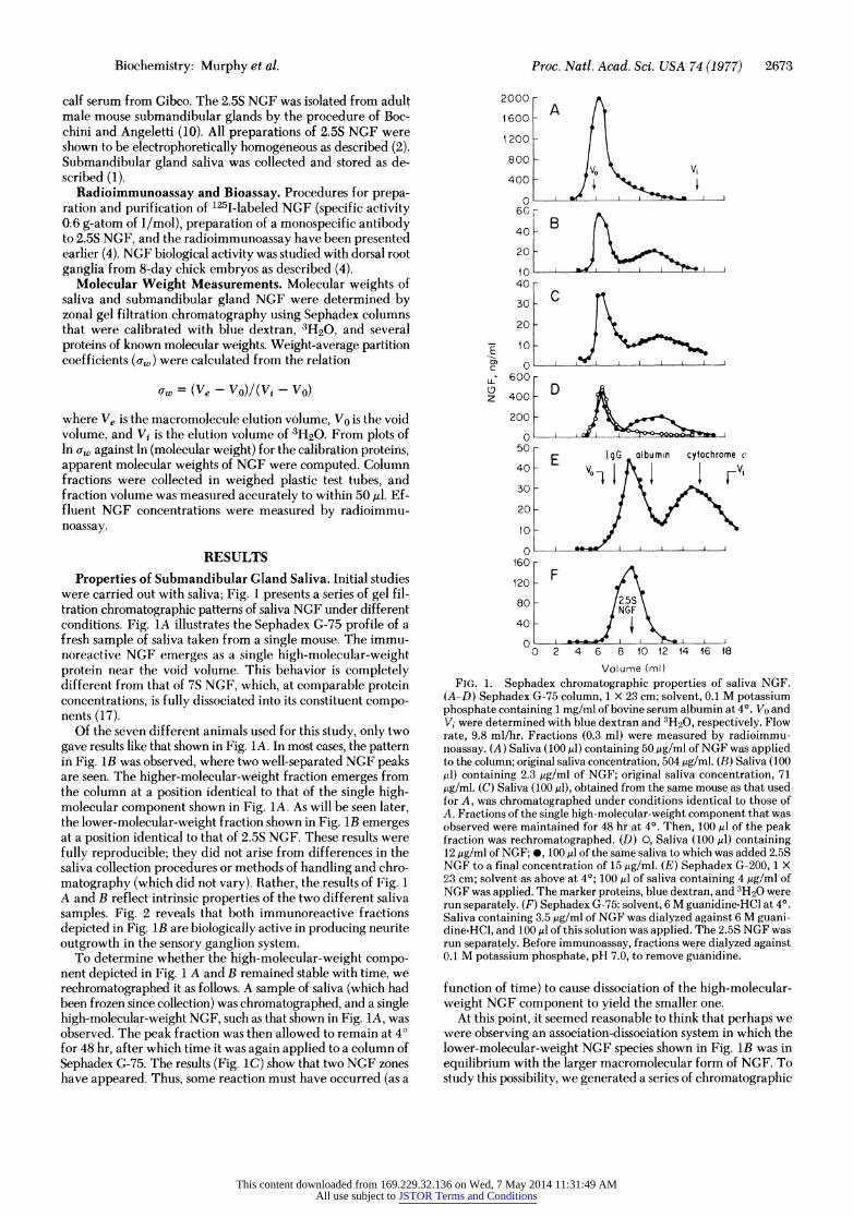
Properties of Submandibular Gland Saliva. Initial studies were carried out with saliva; Fig. 1 presents a series of gel fil- tration chromatographic patterns of saliva NGF under different conditions. Fig. 1A illustrates the Sephadex G-75 profile of a fresh sample of saliva taken from a single mouse. The immu- noreactive NGF emerges as a single high-molecular-weight protein near the void volume. This behavior is completely different from that of 7S NGF, which, at comparable protein concentrations, is fully dissociated into its constituent compo- nents (17).
Of the seven different animals used for this study, only two gave results like that shown in Fig. 1A. In most cases, the pattern in Fig. 1B was observed, where two well-separated NGF peaks are seen. The higher-molecular-weight fraction emerges from the column at a position identical to that of the single high- molecular component shown in Fig. 1A. As will be seen later, the lower-molecular-weight fraction shown in Fig. 1B emerges at a position identical to that of 2.5S NGF. These results were fully reproducible; they did not arise from differences in the saliva collection procedures or methods of handling and chro- matography (which did not vary). Rather, the results of Fig. 1 A and B reflect intrinsic properties of the two different saliva samples. Fig. 2 reveals that both immunoreactive fractions depicted in Fig. lB are biologically active in producing neurite outgrowth in the sensory ganglion system.
To determine whether the high-molecular-weight compo- nent depicted in Fig. 1 A and B remained stable with time, we rechromatographed it as follows. A sample of saliva (which had been frozen since collection) was chromatographed, and a single high-molecular-weight NGF, such as that shown in Fig. 1A, was observed. The peak fraction was then allowed to remain at 40 for 48 hr, after which time it was again applied to a column of Sephadex G-75. The results (Fig. 1C) show that two NGF zones have appeared. Thus, some reaction must have occurred (as a
2000 -
1600- A
1 200 -
800 - V0 ~~~~~~~Vi
400 -
O I v I 1 1 I 60r
40 B
20 X-_ _ _ _
10 40 -
30- C
20 -
6 00 L-
600 -
r 400 -
200 j w
50- 50 E IgG albumin cytochrome c 40- V-1
E V
30 -
20 -
lo I
60
120 - FA 80 2.5S
/NGF\ 40 -
O I I I I I L_ 0 2 4 6 8 10 12 14 16 18
Volume (ml) FIG. 1. Sephadex chromatographic properties of saliva NGF.
(A-D) Sephadex G-75 column, 1 X 23 cm; solvent, 0.1 M potassium phosphate containing 1 mg/rnl of bovine serum albumin at 4?. Vo and Vi were determined with blue dextran and 3H20, respectively. Flow rate, 9.8 ml/hr. Fractions (0.3 ml) were measured by radioimmu- noassay. (A) Saliva (100 ,u) containing 50 ,ug/ml of NGF was applied to the column; original saliva concentration, 504 ,ug/ml. (B) Saliva (100 ktl) containing 2.3 ,ug/ml of NGF; original saliva concentration, 71 ,ug/ml. (C) Saliva (100 ,l), obtained from the same mouse as that used for A, was chromatographed under conditions identical to those of A. Fractions of the single high-molecular-weight component that was observed were maintained for 48 hr at 4?. Then, 100 ,ul of the peak fraction was rechromatographed. (D) 0, Saliva (100,ul) containing 12 ,ug/ml of NGF; *, 100 ,ul of the same saliva to which was added 2.5S NGF to a final concentration of 15 ,ug/ml. (E) Sephadex G-200, 1 X 23 cm; solvent as above at 40; 100 ,ul of saliva containing 4 ,ug/ml of NqGF was applied. The marker proteins, blue dextran, and 3H20 were run separately. (F) Sephadex G-75: solvent, 6 M guanidine-HCl at 4?. Saliva containing 3.5 ,ug/ml of NGF was dialyzed against 6 M guani- cLine-HCl, and 100 ,ul of this solution was applied. The 2.5S NGF was run separately. Before immunoassay, fractions were dialyzed against 0.1 M potassium phosphate, pH 7.0, to remove guanidine.
function of time) to cause dissociation of the high-molecular- vveight NGF component to yield the smaller one.
At this point, it seemed reasonable to think that perhaps we vvere observing an association-dissociation system in which the lower-molecular-weight NGF species shown in Fig. lB was in equilibrium with the larger macromolecular form of NGF. To study this possibility, we generated a series of chromatographic
This content downloaded from 169.229.32.136 on Wed, 7 May 2014 11:31:49 AMAll use subject to JSTOR Terms and Conditions
2674 Biochemistry: Murphy et al. Proc. Natl. Acad. Sci. USA 74 (1977)
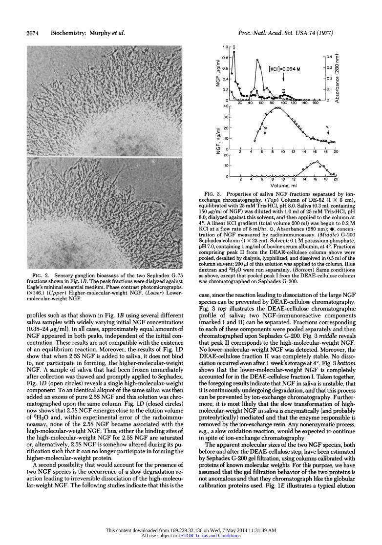
FIG. 2. Sensory ganglion bioassays of the two Sephadex G-75 fractions shown in Fig. 1B. The peak fractions were dialyzed against Eagle's minimal essential medium. Phase contrast photomicrographs. ( X 146.) ( Upper) Higher-molecular-weight NGF. (Lower) Lower- molecular-weight NGF.
profiles such as that shown in Fig. 1B using several different saliva samples with widely varying initial NGF concentrations (0.38-24 ,ug/ml). In all cases, approximately equal amounts of NGF appeared in both peaks, independent of the initial con- centration. These results are not compatible with the existence of an equilibrium reaction. Moreover, the results of Fig. 1D show that when 2.5S NGF is added to saliva, it does not bind to, nor participate in forming, the higher-molecular-weight NGF. A sample of saliva that had been frozen immediately after collection was thawed and promptly applied to Sephadex. Fig. 1D (open circles) reveals a single high-molecular-weight component. To an identical aliquot of the same saliva was then added an excess of pure 2.5S NGF and this solution was chro- matographed upon the same column. Fig. 1D (closed circles) now shows that 2.5S NGF emerges close to the elution volume of 3H20 and, within experimental error of the radioimmu- noassay, none of the 2.5S NGF became associated with the high-molecular-weight NGF. Thus, either the binding sites of the high-molecular-weight NGF for 2.5S NGF are saturated or, alternatively, 2.5S NGF is somehow altered during its pu- rif ication such that it can no longer participate in forming the higher-molecular-weight protein.
A second possibility that would account for the presence of two NGF species is the occurrence of a slow degradation re-
acio eain o revrsbe isoiain f h hghmleu
lar-weight ~ ~~~~ NG.Teflown- tde'idct ht hsi h
1.0 - I
0.8 - 0.4 E
cm 0.6 [KC I]0.094 M -0.3 00 E0 -,02 X
2 Co
20 40 60 80 100 120 140 160
z 2 0424 82
20 _
40 -
30-
-~20 - VO V1
PCY c 10 -
LL: UD 0- L L
2 40 628 10 12 14 16 18 2
10F 2 4 6 8 10 12 14 16 18 20
Volume, ml
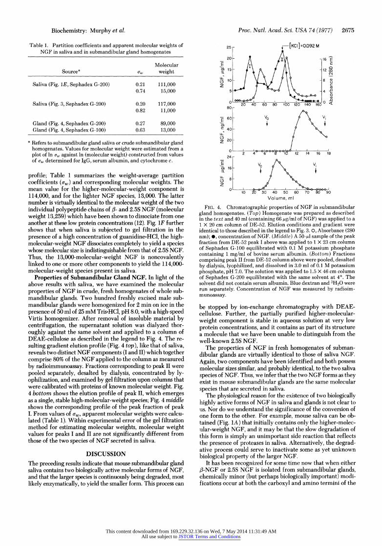
FIG. 3. Properties of saliva NGF fractions separated by ion- exchange chromatography. (Top) Column of DE-52 (1 X 6 cm), equilibrated with 25 mM Tris-HCl, pH 8.0. Saliva (0.3 ml, containing 150 gg/ml of NGF) was diluted with 1.0 ml of 25 mM Tris-HCl, pH 8.0, dialyzed against this solvent, and then applied to the column at 4?. A linear KCl gradient (total volume 200 ml) was begun to 0.2 M KC1 at a flow rate of 8 ml/hr. 0, Absorbance (280 nm); 0, concen- tration of NGF measured by radioimmunoassay. (Middle) G-200 Sephadex column (1 X 23 cm). Solvent: 0.1 M potassium phosphate, pH 7.0, containing 1 mg/ml of bovine serum albumin, at 4?. Fractions comprising peak II from the DEAE-cellulose column above were pooled, desalted by dialysis, lyophilized, and dissolved in 0.5 ml of the column solvent; 200 Al of this solution was applied to the column. Blue dextran and 3H20 were run separately. (Bottom) Same conditions as above, except that pooled peak I from the DEAE-cellulose column was chromatographed on Sephadex G-200.
case, since the reaction leading to dissociation of the large NGF species can be prevented by DEAE-cellulose chromatography. Fig. 3 top illustrates the DEAE-cellulose chromatographic profile of saliva; two NGF-immunoreactive components (marked I and II) can be separated. Fractions corresponding to each of these components were pooled separately and then chromatographed upon Sephadex G-200. Fig. 3 middle reveals that peak II corresponds to the high-molecular-weight NGF. No lower-molecular-weight NGF was detected. Moreover, the DEAE-cellulose fraction II was completely stable. No disso- ciation occurred even after 1 week's storage at 4?. Fig. 3 bottom shows that the lower-molecular-weight NGF is completely accounted for in the DEAE-cellulose fraction I. Taken together, the foregoing results indicate that NGF in saliva is unstable, that it is continuously undergoing degradation, and that this process can be prevented by ion-exchange chromatography. Further- more, it is most likely that the slow transformation of high- molecular-weight NGF in saliva is enzymatically (and probably proteolytically) mediated and that the enzyme responsible is removed by the ion-exchange resin. Any nonenzymatic process, e.g., a slow oxidation reaction, would be expected to continue in spite of ion-exchange chromatography.
The apparent molecular sizes of the two NGF species, both before and after the DEAE-cellulose step, have been estimated by Sephadex G-200 gel filtration, using columns calibrated with proteins of known molecular weights. For this purpose, we have assumed that the gel filtration behavior of the two proteins is not anomalous and that they chromatograph like the globular calibration proteins used. Fig. iE illustrates a typical elution
This content downloaded from 169.229.32.136 on Wed, 7 May 2014 11:31:49 AMAll use subject to JSTOR Terms and Conditions
Biochemistry: Murphy et al. Proc. Natl. Acad. Sci. USA 74 (1977) 2675
Table 1. Partition coefficients and apparent molecular weights of NGF in saliva and in submandibular gland homogenates
Molecular Source* uw weight
Saliva (Fig. 1E, Sephadex G-200) 0.21 111,000 0.74 15,000
Saliva (Fig. 3, Sephadex G-200) 0.20 117,000 0.82 11,000
Gland (Fig. 4, Sephadex G-200) 0.27 89,000 Gland (Fig. 4, Sephadex G-100) 0.63 13,000
* Refers to submandibular gland saliva or crude submandibular gland homogenates. Values for molecular weight were estimated from a plot of ln ow against ln (molecular weight) constructed from values of aw determined for IgG, serum albumin, and cytochrome c.
profile; Table 1 summarizes the weight-average partition coefficients (cw) and corresponding molecular weights. The mean value for the higher-molecular-weight component is 114,000, and for the lighter NGF species, 13,000. The latter number is virtually identical to the molecular weight of the two individual polypeptide chains of fi- and 2.5S NGF (molecular weight 13,259) which have been shown to dissociate from one another at these low protein concentrations (12). Fig. 1F further shows that when saliva is subjected to gel filtration in the presence of a high concentration of guanidine-HCl, the high- molecular-weight NGF dissociates completely to yield a species whose molecular size is indistinguishable from that of 2.5S NGF. Thus, the 13,000-molecular-weight NGF is noncovalently linked to one or more other components to yield the 114,000- molecular-weight species present in saliva.
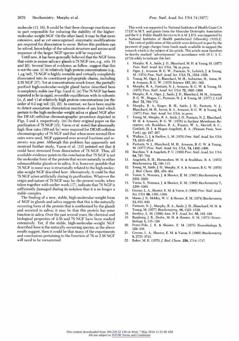
Properties of Submandibular Gland NGF. In light of the above results with saliva, we have examined the molecular properties of NGF in crude, fresh homogenates of whole sub- mandibular glands. Two hundred freshly excised male sub- mandibular glands were homogenized for 2 min on ice in the presence of 50 ml of 25 mM Tris.HCl, pH 8.0, with a high-speed Virtis homogenizer. After removal of insoluble material by centrifugation, the supernatant solution was dialyzed thor- oughly against the same solvent and applied to a column of DEAE-cellulose as described in the legend to Fig. 4. The re- sulting gradient elution profile (Fig. 4 top), like that of saliva, reveals two distinct NGF components (I and II) which together comprise 80% of the NGF applied to the column as measured by radioimmunoassay. Fractions corresponding to peak II were pooled separately, desalted by dialysis, concentrated by ly- ophilization, and examined by gel filtration upon columns that were calibrated with proteins of known molecular weight. Fig. 4 bottom shows the elution profile of peak II, which emerges as a single, stable high-molecular-weight species; Fig. 4 middle shows the corresponding profile of the peak fraction of peak I. From values of cw, apparent molecular weights were calcu- lated (Table 1). Within experimental error of the gel filtration method for estimating molecular weights, molecular weight values for peaks I and II are not significantly different from those of the two species of NGF secreted in saliva.
DISCUSSION The preceding results indicate that mouse submandibular gland saliva contains two biologically active molecular forms of NGF, and that the larger species is continuously being degraded, most likely enzymatically, to yield the smaller form. This process can
25 [KC ]=0.092 M
20 - -1i6 E
LL 10 - - 8
4 2
z X
20 40 60 80 100 120 140 160 80-
~60 - VO V.
C: 40 -
20 -
2 12 14 ~~~~~~16 18
i6-
10 20 30 40 50 60 70 80 90
Volume, ml
FIG. 4. Chromatographic properties of NGF in submandibular gland homogenates. (Top) Homogenate was prepared as described in the text and 40 ml (containing 66 pAg/ml of NGF) was applied to a 1 X 20 cm column of DE-52. Elution conditions and gradient were identical to those described in the legend to Fig. 3. 0, Absorbance (280 nm); *, concentration of NGF. (Middle) A 50-il sample of the peak fraction from DE-52 peak I above was applied to 1 X 23 cm column of Sephadex G-100 equilibrated with 0.1 M potassium phosphate containing 1 mg/ml of bovine serum albumin. (Bottom) Fractions comprising peak II from DE-52 column above were pooled, desalted by dialysis, lyophilized, and dissolved in 3.0 ml of 0.1 M potassium phosphate, pH 7.0. The solution was applied to 1.5 X 46 cm column of Sephadex G-200 equilibrated with the same solvent at 4?. The solvent did not contain serum albumin. Blue dextran and 3H20 were run separately. Concentration of NGF was measured by radioim- munoassay.
be stopped by ion-exchange chromatography with DEAE- cellulose. Further, the partially purified higher-molecular- weight component is stable in aqueous solution at very low protein concentrations, and it contains as part of its structure a molecule that we have been unable to distinguish from the well-known 2.5S NGF.
The properties of NGF in fresh homogenates of subman- dibular glands are virtually identical to those of saliva NGF. Again, two components have been identified and both possess molecular sizes similar, and probably identical, to the two saliva species of NGF. Thus, we infer that the two NGF forms as they exist in mouse submandibular glands are the same molecular species that are secreted in saliva.
The physiological reason for the existence of two biologically ihighly active forms of NGF in saliva and glands is not clear to us. Nor do we understand the significance of the conversion of one form to the other. For example, mouse saliva can be ob- lained (Fig. 1A) that initially contains only the higher-molec- ular-weight NGF, and it may be that the slow degradation of this form is simply an unimportant side reaction that reflects the presence of proteases in saliva. Alternatively, the degrad- ative process could serve to inactivate some as yet unknown biological property of the larger NGF.
It has been recognized for some time now that when either f-NGF or 2.5S NGF is isolated from submandibular glands, chemically minor (but perhaps biologically important) modi- f ications occur at both the carboxyl and amino termini of the
This content downloaded from 169.229.32.136 on Wed, 7 May 2014 11:31:49 AMAll use subject to JSTOR Terms and Conditions
2676 Biochemistry: Murphy et al. Proc. Natl. Acad. Sci. USA 74 (1977)
molecule (11, 16). It could be that these cleavage reactions are in part responsible for reducing the stability of the higher- molecular-weight NGF. On the other hand, it may be that more extensive, and as yet unrecognized, enzymatic modifications are required for dissociation to occur. Before this problem can be solved, knowledge of the subunit structure and amino acid sequence of the larger NGF species will be required.
Until now, it has been generally believed that the NGF form that exists in mouse salivary glands is 7S NGF (see, e.g., refs. 19 and 20). Several lines of evidence, as follow, suggest that this is not the case. (i) At relatively high-protein concentrations (e.g., 1 ,g/ml), 7S NGF is highly unstable and virtually completely dissociated into its constituent polypeptide chains, including 2.5S NGF (17). Yet at concentrations much lower, the partially purified high-molecular-weight gland factor described here is completely stable (see Figs. 3 and 4). (ii) The 7S NGF has been reported to be in rapid, reversible equilibrium with its subunits (a, f, and y) at relatively high protein concentrations (on the order of 0.2 mg/ml) (21, 22). In contrast, we have been unable to detect association-dissociation reactions of any kind which pertain to the high-molecular-weight saliva or gland NGF after the DEAE-cellulose chromatographic procedure depicted in Figs. 3 and 4, respectively. (iii) In their original paper on the purification of 7S NGF (13), Varon et al. noted that abnormally high flow rates (350 ml/hr) were required for DEAE-cellulose chromatography of 7S NGF and that when more normal flow rates were used, NGF appeared over several fractions and re- covery was poor. Although this problem has apparently not received further study, Varon et al. (13) pointed out that it could have stemmed from dissociation of 7S NGF. Thus, all available evidence points to the conclusion that 7S NGF is not the molecular form of the protein that occurs naturally in either submandibular glands or in saliva. It is, however, possible that 7S NGF in some way is structurally related to the high-molec- ular-weight NGF described here. Alternatively, it could be that 7S NGF arises artificially during its purification. Whatever the origin and nature of 7S NGF may be, the present results, when taken together with earlier work (17), indicate that 7S NGF is sufficiently damaged during its isolation that it is no longer a stable complex.
The finding of a new, stable, high-molecular-weight form of NGF in glands and saliva suggests that this is the naturally occurring form of the protein that is synthesized by the glands and secreted in saliva; it may be that this protein has some function in saliva. Over the past several years, the chemical and biological properties of 2.5S and 7S NGF have been studied extensively. Yet, if the stable, high-molecular-weight NGF described here is the naturally occurring species, as the above results suggest, then it could be that many of the experiments and conclusions pertaining to the function of 7S or 2.5S NGF will need to be reexamined.
This work was supported by National Institutes of Health Grant CA 17137 to M.Y. and grants from the Muscular Dystrophy Association and the U.S. Public Health Service to R.A.M. J.D.S. was supported by a National Institutes of Health postdoctoral fellowship (NHLI).
The costs of publication of this article were defrayed in part by the payment of page charges from funds made available to support the research which is the subject of the article. This article must therefore be hereby marked "advertisement" in accordance with 18 U. S. C. ?1734 solely to indicate this fact.
1. Murphy, R. A., Saide, J. D., Blanchard, M. H. & Young, M. (1977) Proc. Natl. Acad. Sci. USA, 74, in press.
2. Oger, J., Arnason, B. G. W., Pantazis, N., Lehrich, J. & Young, M. (1974) Proc. Natl. Acad. Sci. USA 71, 1554-1558.
3. Young, M., Oger, J., Blanchard, M. H., Asdourian, H., Amos, H. & Arnason, B. G. W. (1975) Science 187,361-362.
4. Murphy, R. A., Pantazis, N. J., Arnason, B. G. W. & Young, M. (1975) Proc. Natl. Acad. Sci. USA 72, 1895-1898.
5. Murphy, R. A., Oger, J., Saide, J. D., Blanchard, M. H., Arnason, B. G. W., Hogan, C., Pantazis, N. J. & Young, M. (1977) J. Cell Biol. 72, 769-773.
6. Murphy, R. A., Singer, R. H., Saide, J. D., Pantazis, N. J., Blanchard, M. H., byron, K. S., Arnason, B. G. W. & Young, M. (1977) Proc. Nat. Acad. Sci. USA, in press.
7. Young, M., Murphy, R. A., Saide, J. D., Pantazis, N. J., Blanchard, M. H. & Arnason, B. G. W. (1976) in Surface Membrane Re- ceptors, eds. Bradshaw, R. A., Frazier, W. A., Merrell, R. C., Gottlieb, D. I. & Hogue-Angeletti, R. A. (Plenum Press, New York), pp. 247-267.
8. Wallace, L. J. & Partlow, L. M. (1976) Proc. Natl. Acad. Sci. USA 73, 4210-4214.
9. Pantazis, N. J., Blanchard, M. H., Arnason, B. G. W. & Young, M. (1977) Proc. Natl. Acad. Sci. USA, 74, 1492-1496.
10. Bocchini, V. & Angeletti, P. U. (1969) Proc. Natl. Acad. Sci. USA 64, 787-794.
11. Angeletti, R. H., Hermodson, M. H. & Bradshaw, R. A. (1973) Biochemistry 12, 100-115.
12. Young, M., Saide, J. D., Murphy, R. A. & Arnason, B. G. W. (1976) J. Biol. Chem. 251, 459-464.
13. Varon, S., Nomura, J. & Shooter, E. M. (1967) Biochemistry 6, 2202-2209.
14. Varon, S., Nomura, J. & Shooter, E. M. (1968) Biochemistry 7, 1296-1303.
15. Greene, L. A., Shooter, E. M. & Varon, S. (1968) Proc. Natl. Acad. Sci. USA 60, 1383-1388.
16. Moore, J. B., Mobley, W. C. & Shooter, E. M. (1974) Biochemistry 13, 833-840.
17. Pantazis, N. J., Murphy, R. A., Saide, J. D., Blanchard, M. H. & Young, M. (1977) Biochemistry, 16, 1525-1530.
18. Sreebny, L. M. (1960) Ann. N.Y. Acad. Sci. 85, 182-188. 19. Bamburg, J. R., Derby, M. H. & Shooter, E. M. (1971) Neuro-
biology 1, 115-120. 20. Perez-Polo, J. R. & Shooter, E. M. (1975) Neurobiology 5,
329-338. 21. Greene, L. A., Shooter, E. M. & Varon, S. (1969) Biochemistry
8, 3735-3741. 22. Baker, M. E. (1975) J. Biol. Chem. 250, 1714-1717.
This content downloaded from 169.229.32.136 on Wed, 7 May 2014 11:31:49 AMAll use subject to JSTOR Terms and Conditions