Embed Size (px)
Citation preview
BioMed CentralMolecular Pain
ss
Open AcceResearchModerate hypoxia influences excitability and blocks dendrotoxin sensitive K+ currents in rat primary sensory neuronesMarco Gruss1,2, Giovanni Ettorre1, Annette Jana Stehr1,3, Michael Henrich2,4, Gunter Hempelmann2 and Andreas Scholz*1Address: 1Physiologisches Institut, Justus-Liebig-Universität, 35385 Giessen, Germany, 2Abteilung Anaesthesiologie, Intensivmedizin, Schmerztherapie, Universitätsklinikum Gießen und Marburg, Standort Gießen, 35385 Giessen, Germany, 3Zentrum für Anaesthesiologie, Rettungs- und Intensivmedizin, Robert-Koch-Str.40, 37075 Göttingen, Germany and 4University Laboratory of Physiology, Parks Road, Oxford OX1 3PT, UK
Email: Marco Gruss - [email protected]; Giovanni Ettorre - [email protected]; Annette Jana Stehr - [email protected]; Michael Henrich - [email protected]; Gunter Hempelmann - [email protected]; Andreas Scholz* - [email protected]
* Corresponding author
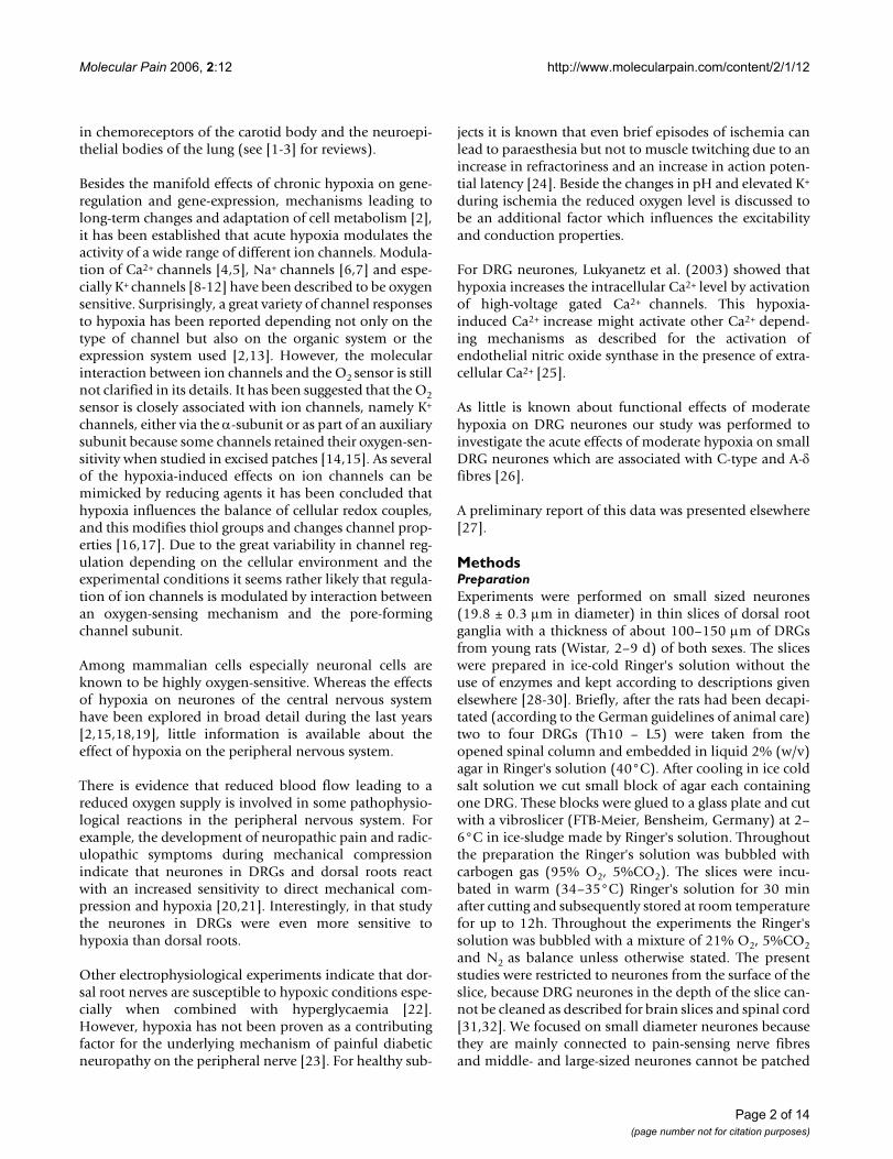
AbstractHypoxia alters neuronal function and can lead to neuronal injury or death especially in the centralnervous system. But little is known about the effects of hypoxia in neurones of the peripheralnervous system (PNS), which survive longer hypoxic periods. Additionally, people haveexperienced unpleasant sensations during ischemia which are dedicated to changes in conductionproperties or changes in excitability in the PNS. However, the underlying ionic conductances indorsal root ganglion (DRG) neurones have not been investigated in detail.
Therefore we investigated the influence of moderate hypoxia (27.0 ± 1.5 mmHg) on actionpotentials, excitability and ionic conductances of small neurones in a slice preparation of DRGs ofyoung rats. The neurones responded within a few minutes non-uniformly to moderate hypoxia:changes of excitability could be assigned to decreased outward currents in most of the neurones(77%) whereas a smaller group (23%) displayed increased outward currents in Ringer solution. Wewere able to attribute most of the reduction in outward-current to a voltage-gated K+ currentwhich activated at potentials positive to -50 mV and was sensitive to 50 nM α-dendrotoxin (DTX).Other toxins that inhibit subtypes of voltage gated K+ channels, such as margatoxin (MgTX),dendrotoxin-K (DTX-K), r-tityustoxin Kα (TsTX-K) and r-agitoxin (AgTX-2) failed to prevent thehypoxia induced reduction. Therefore we could not assign the hypoxia sensitive K+ current to onehomomeric KV channel type in sensory neurones. Functionally this K+ current blockade mightunderlie the increased action potential (AP) duration in these neurones. Altogether these results,might explain the functional impairment of peripheral neurones under moderate hypoxia.
BackgroundIn the last years the knowledge about the effects ofhypoxia on different organ systems showed a remarkable
increase. Most efforts were made to understand theactions of hypoxia in the central nervous system, in theregulation of blood flow, in the sensing of oxygen levels
Published: 31 March 2006
Molecular Pain 2006, 2:12 doi:10.1186/1744-8069-2-12
Received: 27 July 2005Accepted: 31 March 2006
This article is available from: http://www.molecularpain.com/content/2/1/12
© 2006 Gruss et al; licensee BioMed Central Ltd.This is an Open Access article distributed under the terms of the Creative Commons Attribution License (http://creativecommons.org/licenses/by/2.0), which permits unrestricted use, distribution, and reproduction in any medium, provided the original work is properly cited.
Page 1 of 14(page number not for citation purposes)
Molecular Pain 2006, 2:12 http://www.molecularpain.com/content/2/1/12
in chemoreceptors of the carotid body and the neuroepi-thelial bodies of the lung (see [1-3] for reviews).
Besides the manifold effects of chronic hypoxia on gene-regulation and gene-expression, mechanisms leading tolong-term changes and adaptation of cell metabolism [2],it has been established that acute hypoxia modulates theactivity of a wide range of different ion channels. Modula-tion of Ca2+ channels [4,5], Na+ channels [6,7] and espe-cially K+ channels [8-12] have been described to be oxygensensitive. Surprisingly, a great variety of channel responsesto hypoxia has been reported depending not only on thetype of channel but also on the organic system or theexpression system used [2,13]. However, the molecularinteraction between ion channels and the O2 sensor is stillnot clarified in its details. It has been suggested that the O2sensor is closely associated with ion channels, namely K+
channels, either via the α-subunit or as part of an auxiliarysubunit because some channels retained their oxygen-sen-sitivity when studied in excised patches [14,15]. As severalof the hypoxia-induced effects on ion channels can bemimicked by reducing agents it has been concluded thathypoxia influences the balance of cellular redox couples,and this modifies thiol groups and changes channel prop-erties [16,17]. Due to the great variability in channel reg-ulation depending on the cellular environment and theexperimental conditions it seems rather likely that regula-tion of ion channels is modulated by interaction betweenan oxygen-sensing mechanism and the pore-formingchannel subunit.
Among mammalian cells especially neuronal cells areknown to be highly oxygen-sensitive. Whereas the effectsof hypoxia on neurones of the central nervous systemhave been explored in broad detail during the last years[2,15,18,19], little information is available about theeffect of hypoxia on the peripheral nervous system.
There is evidence that reduced blood flow leading to areduced oxygen supply is involved in some pathophysio-logical reactions in the peripheral nervous system. Forexample, the development of neuropathic pain and radic-ulopathic symptoms during mechanical compressionindicate that neurones in DRGs and dorsal roots reactwith an increased sensitivity to direct mechanical com-pression and hypoxia [20,21]. Interestingly, in that studythe neurones in DRGs were even more sensitive tohypoxia than dorsal roots.
Other electrophysiological experiments indicate that dor-sal root nerves are susceptible to hypoxic conditions espe-cially when combined with hyperglycaemia [22].However, hypoxia has not been proven as a contributingfactor for the underlying mechanism of painful diabeticneuropathy on the peripheral nerve [23]. For healthy sub-
jects it is known that even brief episodes of ischemia canlead to paraesthesia but not to muscle twitching due to anincrease in refractoriness and an increase in action poten-tial latency [24]. Beside the changes in pH and elevated K+
during ischemia the reduced oxygen level is discussed tobe an additional factor which influences the excitabilityand conduction properties.
For DRG neurones, Lukyanetz et al. (2003) showed thathypoxia increases the intracellular Ca2+ level by activationof high-voltage gated Ca2+ channels. This hypoxia-induced Ca2+ increase might activate other Ca2+ depend-ing mechanisms as described for the activation ofendothelial nitric oxide synthase in the presence of extra-cellular Ca2+ [25].
As little is known about functional effects of moderatehypoxia on DRG neurones our study was performed toinvestigate the acute effects of moderate hypoxia on smallDRG neurones which are associated with C-type and A-δfibres [26].
A preliminary report of this data was presented elsewhere[27].
MethodsPreparationExperiments were performed on small sized neurones(19.8 ± 0.3 µm in diameter) in thin slices of dorsal rootganglia with a thickness of about 100–150 µm of DRGsfrom young rats (Wistar, 2–9 d) of both sexes. The sliceswere prepared in ice-cold Ringer's solution without theuse of enzymes and kept according to descriptions givenelsewhere [28-30]. Briefly, after the rats had been decapi-tated (according to the German guidelines of animal care)two to four DRGs (Th10 – L5) were taken from theopened spinal column and embedded in liquid 2% (w/v)agar in Ringer's solution (40°C). After cooling in ice coldsalt solution we cut small block of agar each containingone DRG. These blocks were glued to a glass plate and cutwith a vibroslicer (FTB-Meier, Bensheim, Germany) at 2–6°C in ice-sludge made by Ringer's solution. Throughoutthe preparation the Ringer's solution was bubbled withcarbogen gas (95% O2, 5%CO2). The slices were incu-bated in warm (34–35°C) Ringer's solution for 30 minafter cutting and subsequently stored at room temperaturefor up to 12h. Throughout the experiments the Ringer'ssolution was bubbled with a mixture of 21% O2, 5%CO2and N2 as balance unless otherwise stated. The presentstudies were restricted to neurones from the surface of theslice, because DRG neurones in the depth of the slice can-not be cleaned as described for brain slices and spinal cord[31,32]. We focused on small diameter neurones becausethey are mainly connected to pain-sensing nerve fibresand middle- and large-sized neurones cannot be patched
Page 2 of 14(page number not for citation purposes)
Molecular Pain 2006, 2:12 http://www.molecularpain.com/content/2/1/12
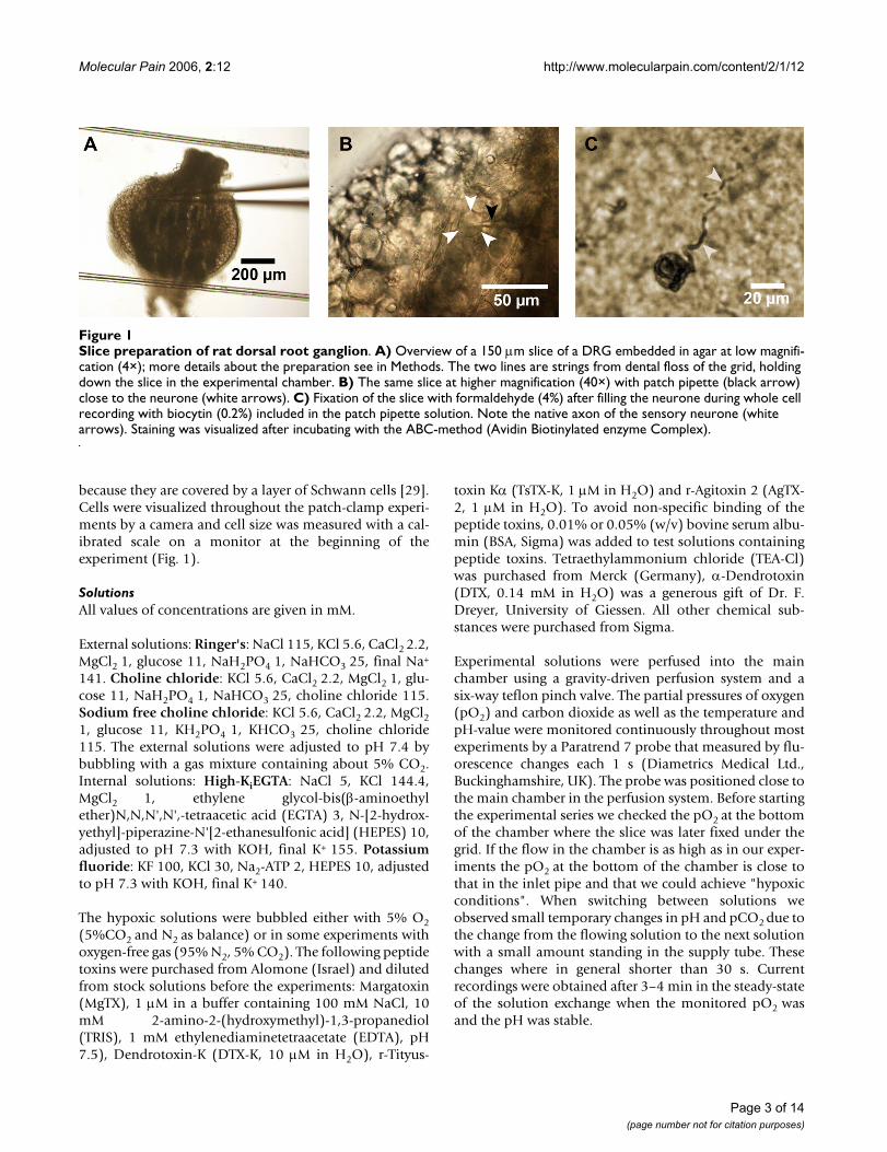
because they are covered by a layer of Schwann cells [29].Cells were visualized throughout the patch-clamp experi-ments by a camera and cell size was measured with a cal-ibrated scale on a monitor at the beginning of theexperiment (Fig. 1).
SolutionsAll values of concentrations are given in mM.
External solutions: Ringer's: NaCl 115, KCl 5.6, CaCl2 2.2,MgCl2 1, glucose 11, NaH2PO4 1, NaHCO3 25, final Na+
141. Choline chloride: KCl 5.6, CaCl2 2.2, MgCl2 1, glu-cose 11, NaH2PO4 1, NaHCO3 25, choline chloride 115.Sodium free choline chloride: KCl 5.6, CaCl2 2.2, MgCl21, glucose 11, KH2PO4 1, KHCO3 25, choline chloride115. The external solutions were adjusted to pH 7.4 bybubbling with a gas mixture containing about 5% CO2.Internal solutions: High-KiEGTA: NaCl 5, KCl 144.4,MgCl2 1, ethylene glycol-bis(β-aminoethylether)N,N,N',N',-tetraacetic acid (EGTA) 3, N-[2-hydrox-yethyl]-piperazine-N'[2-ethanesulfonic acid] (HEPES) 10,adjusted to pH 7.3 with KOH, final K+ 155. Potassiumfluoride: KF 100, KCl 30, Na2-ATP 2, HEPES 10, adjustedto pH 7.3 with KOH, final K+ 140.
The hypoxic solutions were bubbled either with 5% O2(5%CO2 and N2 as balance) or in some experiments withoxygen-free gas (95% N2, 5% CO2). The following peptidetoxins were purchased from Alomone (Israel) and dilutedfrom stock solutions before the experiments: Margatoxin(MgTX), 1 µM in a buffer containing 100 mM NaCl, 10mM 2-amino-2-(hydroxymethyl)-1,3-propanediol(TRIS), 1 mM ethylenediaminetetraacetate (EDTA), pH7.5), Dendrotoxin-K (DTX-K, 10 µM in H2O), r-Tityus-
toxin Kα (TsTX-K, 1 µM in H2O) and r-Agitoxin 2 (AgTX-2, 1 µM in H2O). To avoid non-specific binding of thepeptide toxins, 0.01% or 0.05% (w/v) bovine serum albu-min (BSA, Sigma) was added to test solutions containingpeptide toxins. Tetraethylammonium chloride (TEA-Cl)was purchased from Merck (Germany), α-Dendrotoxin(DTX, 0.14 mM in H2O) was a generous gift of Dr. F.Dreyer, University of Giessen. All other chemical sub-stances were purchased from Sigma.
Experimental solutions were perfused into the mainchamber using a gravity-driven perfusion system and asix-way teflon pinch valve. The partial pressures of oxygen(pO2) and carbon dioxide as well as the temperature andpH-value were monitored continuously throughout mostexperiments by a Paratrend 7 probe that measured by flu-orescence changes each 1 s (Diametrics Medical Ltd.,Buckinghamshire, UK). The probe was positioned close tothe main chamber in the perfusion system. Before startingthe experimental series we checked the pO2 at the bottomof the chamber where the slice was later fixed under thegrid. If the flow in the chamber is as high as in our exper-iments the pO2 at the bottom of the chamber is close tothat in the inlet pipe and that we could achieve "hypoxicconditions". When switching between solutions weobserved small temporary changes in pH and pCO2 due tothe change from the flowing solution to the next solutionwith a small amount standing in the supply tube. Thesechanges where in general shorter than 30 s. Currentrecordings were obtained after 3–4 min in the steady-stateof the solution exchange when the monitored pO2 wasand the pH was stable.
Slice preparation of rat dorsal root ganglionFigure 1Slice preparation of rat dorsal root ganglion. A) Overview of a 150 µm slice of a DRG embedded in agar at low magnifi-cation (4×); more details about the preparation see in Methods. The two lines are strings from dental floss of the grid, holding down the slice in the experimental chamber. B) The same slice at higher magnification (40×) with patch pipette (black arrow) close to the neurone (white arrows). C) Fixation of the slice with formaldehyde (4%) after filling the neurone during whole cell recording with biocytin (0.2%) included in the patch pipette solution. Note the native axon of the sensory neurone (white arrows). Staining was visualized after incubating with the ABC-method (Avidin Biotinylated enzyme Complex).
Page 3 of 14(page number not for citation purposes)

Molecular Pain 2006, 2:12 http://www.molecularpain.com/content/2/1/12
Current and voltage recordingsPatch-clamp pipettes were fabricated from borosilicateglass tubing with internal filament (GC 150F, HarvardApparatus, UK) using a horizontal puller (Sutter, USA)and were heat-polished to final resistances of 2.2 – 11 MΩ(median 4.0 MΩ) measured with internal solution.Recordings were obtained using an Axopatch 200A patch-clamp amplifier (Axon Instruments, USA) and data weredigitized with a Digidata 1200A 12bit AD/DA converter(Axon Instruments, USA) at a sampling frequency of 2kHz and filtered at 1 kHz with a 10-pole low-pass Besselfilter if not otherwise stated. Voltage and current steps anddata acquisition were controlled online by a PC/AT com-puter with pCLAMP8 software (Axon Instruments, USA).
Whole-cell recordings were performed as described byHamill et al. [33], and in most experiments additionalseries resistance compensation (55–80%) was appliedafter adjustment of series resistance and compensation ofwhole-cell capacity. Currents evoked by voltage steps weredigitally corrected off-line for leakage and transients usingappropriately scaled averaged episodes of recordingswhich were evoked by hyperpolarizing pulses.
Voltages are expressed as transmembrane potentials (V).Resting potentials of the neurones are shown next to thecurrent-clamp recordings and are indicated by dottedlines in the figure. Deviations in action potential duration(measured at 0 mV) as well as in current measurements at
Whole-cell recordings in the current-clamp modusFigure 2Whole-cell recordings in the current-clamp modus. A) A neuron which was in carbogen-bubbled control solution (pO2 600 mmHg, left) not spontaneous active, responded in hypoxic solution (pO2 27 mmHg, middle) spontaneously (1 pA). After reoxygenation the effect was reversible and the neurones did not generate an AP. B) The same neuron as shown in A. The neuron generates a second AP in the hypoxic solution compared to control (middle vs. left) but the AP-amplitude was reduced from 99 mV to 79 mV and the duration of the AP was prolonged in hypoxic solution from 2.3 ms to 2.6 ms (measured at half amplitude). C) Same neuron as shown in A and B. Trains of APs elicited in a DRG-neuron by a long lasting depolarizing current injection (750 ms, left). After reduction of pO2 (27 mmHg, middle) the neuron generate few APs and adapted. After reoxygen-ation the effect was reversible (right). 23°C; Pipette solution: High-Ki EGTA; Bath: Ringer's solution.
C
pO2 [mmHg] 48727600
200 ms20 mV
A
0 mV
0 mV
0 mV
20 mV
200 ms
B
20 ms
-70 mV
-70 mV
-72 mV
35 pA
1 pA
101 pA
20 mV
Page 4 of 14(page number not for citation purposes)
Molecular Pain 2006, 2:12 http://www.molecularpain.com/content/2/1/12
the end of the voltage step at +40 mV from control to testconditions were taken as different if larger or smaller than3% from control value. Those cells showing an increasedoutward-current after toxin application were excludedfrom further analysis (n = 3).
Evaluations, fits and statistical analysis were performedwith the programs pCLAMP (6 and 8, Axon Instruments,USA), Origin 6.0 (Microcal Software, USA) and Excel 7(Microsoft, USA). Data are expressed as mean ± standarderror of the mean (S.E.M.). Statistical significance wasassessed using the paired or unpaired Student's t-testwhere appropiate. Experiments were performed at roomtemperature (21 – 23°C).
ResultsEffects of hypoxia on action potentials and thresholdIn patch-clamp experiments with neuronal slices (Fig. 1)most investigators use carbogen (95%O2, 5% CO2)-bub-bled solutions with relatively high glucose concentrations(mostly about 10 mmol/l) to keep their cells healthy[30,34]. During some pilot experiments we tested theeffect of a dramatic change in oxygen level on excitabilityof DRG neurones. We reduced the oxygen level in theRinger's solution from about 600 mmHg to about 30mmHg ("hypoxic") and measured the APs of DRG neu-rones. The large decrease in oxygen level resulted in achange of firing pattern as shown exemplary in figure 2. Aneurone that did not show any spontaneous activityunder carbogen (Fig. 2A) became spontaneously activeunder hypoxia and displayed an increased excitabilty
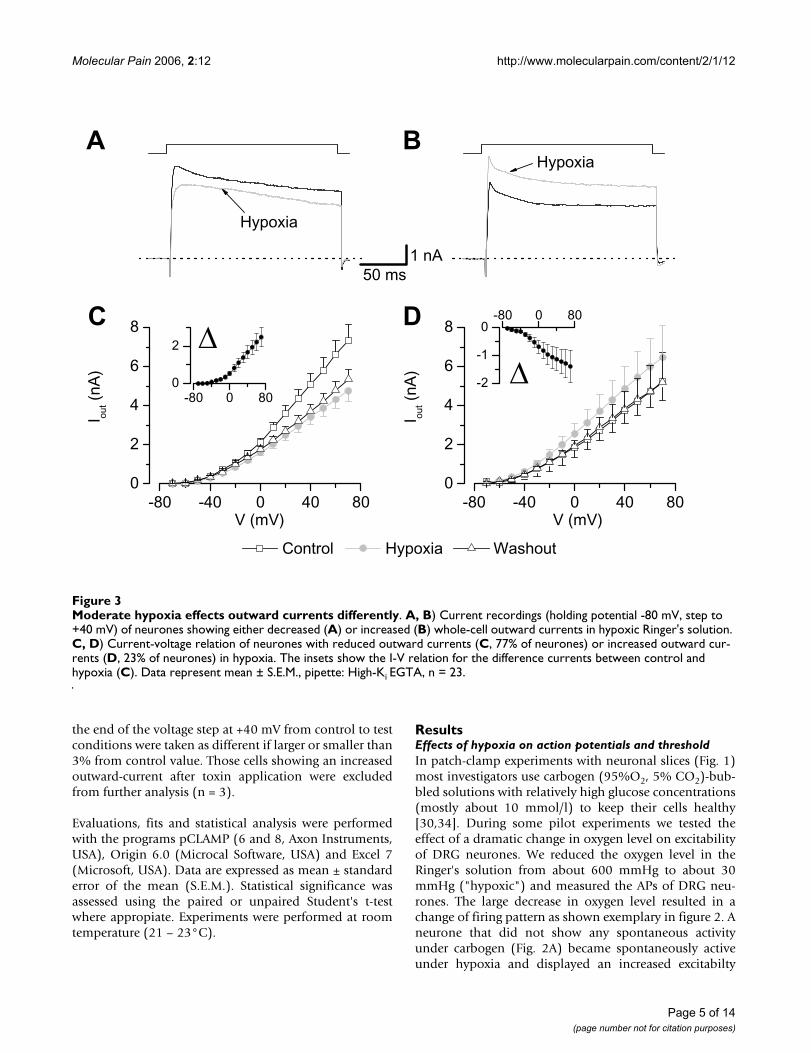
Moderate hypoxia effects outward currents differentlyFigure 3Moderate hypoxia effects outward currents differently. A, B) Current recordings (holding potential -80 mV, step to +40 mV) of neurones showing either decreased (A) or increased (B) whole-cell outward currents in hypoxic Ringer's solution. C, D) Current-voltage relation of neurones with reduced outward currents (C, 77% of neurones) or increased outward cur-rents (D, 23% of neurones) in hypoxia. The insets show the I-V relation for the difference currents between control and hypoxia (C). Data represent mean ± S.E.M., pipette: High-Ki EGTA, n = 23.
-80 -40 0 40 800
2
4
6
8
-80 -40 0 40 800
2
4
6
8
Washout Hypoxia Control
V (mV)V (mV)
I out (
nA)
I out (
nA)
Hypoxia
C D
BAHypoxia
-80 0 80
-2
-1
0
-80 0 800
2
50 ms1 nA
Page 5 of 14(page number not for citation purposes)

Molecular Pain 2006, 2:12 http://www.molecularpain.com/content/2/1/12
when stimulated with small depolarising currents (Fig.2B). Interestingly, when stimulated with stronger currentsthe neuron responded with a train of APs in carbogen-bubbled solution but showed an adapting firing behav-iour in hypoxic solution in parallel we observed a decreasein outward currents. These effects were reversible afterwashout of hypoxic solution to carbogen-bubbledRinger's and demonstrate the complexity of neuronalresponses to hypoxia. Because only a small fraction of sen-sory neurones responded with repetitive firing, wefocused on the effects on single action potentials.
To investigate the oxygen sensitivity of small DRG-neu-rones in more detail we replaced the carbogen-bubbledsolution by a control bubbled with 21% O2 (5% CO2,
74% N2) and tested the effects of moderate hypoxia onaction potentials (APs) and currents of DRG neurones.
Overall, we investigated in 137 cells the effects of moder-ate hypoxia on currents and actions potentials (APs) ofsmall DRG neurones. In 98 experiments the oxygen level(pO2) was measured continuously throughout the experi-ment in control solutions (144.2 ± 0.9 mmHg), inhypoxic solutions (27.0 ± 1.5 mmHg) and in control afterhypoxia (142.9 ± 1.1 mmHg; washout). In the voltage-clamp configuration we tested 127 neurones, 29 weretested with external Ringer's solution and internal High-Ki, 33 in external choline chloride and 65 in sodium freecholine chloride solution with internal potassium fluo-ride to investigate voltage-gated potassium currents sepa-rately.
Functional consequences of hypoxia-induced K+ current blockFigure 4Functional consequences of hypoxia-induced K+ current block. A) Outward-currents (measured at +40 mV) of neu-rones which showed hypoxic sensitive outward-currents (reduction by 43 %, p < 0.01) and in which we measured action potentials (n = 11). B) Corresponding to changes in K+ currents these neurones displayed a 49% prolongation in mean AP duration in hypoxia (p < 0.01, n = 11). C) Hypoxia reduced the threshold to evoke an AP in some neurones. Injection of 200 pA depolarising currents could evoke an AP in hypoxia but not in normoxia and injection of 500 pA could elicit 2 APs in hypoxia compared to one in normoxia. Bath: Ringer's solution, pipette: High-Ki EGTA.
0 mV
0 mV
n.s.p<0.01
200 pA 200 pA
500 pA
F
HypoxiaNormoxia
500 pA
p<0.01
-66 mV
-63 mV
Control Hypoxie Washout0
1
2
3
4
5
6
7
C
B
A
20 mV20 ms
0
1
2
3
4
5
6
n.s.
I out (
nA)
AP
-dur
atio
n (m
s)
Page 6 of 14(page number not for citation purposes)

Molecular Pain 2006, 2:12 http://www.molecularpain.com/content/2/1/12
Different effects of moderate hypoxia on whole-cell outward currentsWhen we measured the effect of hypoxia (11.9 ± 4.7mmHg) on whole-cell currents of small DRG neuroneswith external Ringer's solution and internal High Ki, themajority of the neurones (22 out of 29) showed adecreased steady-state whole-cell outward current inhypoxia (3.40 ± 0.36 nA, measured at +40 mV) withintens of seconds compared to control (4.82 ± 0.43 nA, p <0.001, Fig. 3A, C) whereas seven neurones showed anincrease in outward current in hypoxia (4.87 ± 1.68 nA vs.3.75 ± 1.35 nA in control, p < 0.05, Fig. 3B, D). Theincrease in outward current was reversible (p < 0.05) inwashout (3.83 ± 0.83 nA, n = 6, Fig. 3D). However, the sit-uation in the group with reduced currents was more het-erogeneous; in eight neurones we observed a substantialincrease in current during 3 to 6 min after washout of
hypoxia whereas in other neurones the reduction was irre-versible. Therefore the mean current in washout was onlyslightly greater than in hypoxia (3.00 ± 0.38 nA, n.s., n =20, Fig. 3C).
Effect of hypoxia on action potentialsNext, we tried to look into the functional consequences ofthis inhibition of K+ currents by hypoxia. Neurones whichshowed a decreased whole-cell outward-current inhypoxic Ringer's solution (2.71 ± 0.62 nA) compared tonormoxia (4.97 ± 1.18 nA; p < 0.01, n = 10, washout 2.61± 0.73 nA, n.s., n = 8, Fig. 4A, B) were tested in current-clamp. Currents were measured at +40 mV, which corre-lates to the membrane potential of the peak of an AP inmany neurones. The AP duration in these neurones wasprolonged (measured at 0 mV) from 3.08 ± 0.31 ms incontrol conditions to 5.01 ± 0.77 ms in hypoxia (p < 0.05,
Voltage-dependent K+ currents in hypoxiaFigure 5Voltage-dependent K+ currents in hypoxia. Current recordings (holding potential -80 mV, ∆V 10 mV) in control, hypoxia and washout revealing different reactions of K+ currents to hypoxia. A) Most neurones (63%) showed a decreased K+ current during hypoxia which was irreversible in some cells (n = 17). B) Another group of neurones (26%, n = 7) displayed a reversible increase in K+ current. C) Some neurones showed no (11%) or only small differences in K+ currents during hypoxia (n = 3). Current-voltage relations shown right. Bath: sodium-free choline chloride, pipette: potassium fluoride.
-80 -40 0 40 800
1
2
3
4
5
-80 -40 0 40 800
1
2
3
4
5
I K (
nA)
I K (
nA)
I K (
nA)
V (mV)
WashoutHypoxiaControl
C
B
A
-80 -40 0 40 800
1
2
3
4
5
Washout Hypoxia Control
50 ms1 nA
Page 7 of 14(page number not for citation purposes)
Molecular Pain 2006, 2:12 http://www.molecularpain.com/content/2/1/12
n = 10) and corresponding to partial recovery of the K+
currents in subsequent washout (4.14 ± 0.59 ms, n = 9,n.s., Fig. 4C). The mean threshold, to evoke an AP aboveV>0 mV, did not change in hypoxia (390 ± 62 pA in con-trol vs 360 ± 63 pA, n.s.). The resting membrane potentialremained unchanged in hypoxia (-66.3 ± 2.6 mV) com-pared to normoxia (-68.0 ± 1.8 mV, n.s., washout 65.9 ±2.9 mV, n.s.).
Voltage-gated K+ currentsThe changes in steady-state whole-cell currents could beeither due to a block or an activation of outward-currentsor an increased or decreased inward-current, respectively.Several potassium channels have been shown as a possi-ble target for hypoxia and thus we focused on the effectsof moderate hypoxia on K+ currents in small DRG neu-rones. To investigate isolated voltage-gated K+ currents we
used extracellular choline chloride and intracellularpotassium fluoride.
Again the largest group of neurones (17 out of 32)revealed a reduction of voltage-gated K+ currents inhypoxia (2.37 ± 0.23 nA, at V = +40 mV, sodium freecholine chloride, Fig. 5A) compared to control conditions(3.15 ± 0.19 nA, p < 0.001) which did not recover in mostneurones within 3 – 6 minutes (washout 2.40 ± 0.24 nA,p > 0,05, n = 16). In contrast, eight neurones displayed areversible increase of the K+ current amplitude underhypoxia (Fig. 5B, 2.91 ± 0.15 nA compared to 2.48 ± 0.19nA in normoxic solution, p < 0.05, washout 2.58 ± 0.17nA, p < 0.05) whereas in three neurones the K+ currentremained unchanged (Fig. 5C, 1.81 ± 0.14 nA in hypoxiacompared to 1.81 ± 0.16 nA in normoxia, n.s.). Theseexperiments regarding voltage-gated K+ currents support
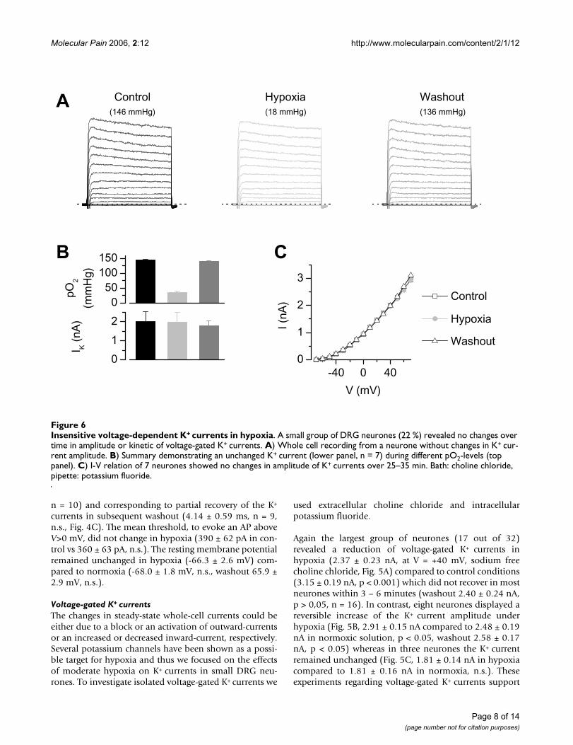
Insensitive voltage-dependent K+ currents in hypoxiaFigure 6Insensitive voltage-dependent K+ currents in hypoxia. A small group of DRG neurones (22 %) revealed no changes over time in amplitude or kinetic of voltage-gated K+ currents. A) Whole cell recording from a neurone without changes in K+ cur-rent amplitude. B) Summary demonstrating an unchanged K+ current (lower panel, n = 7) during different pO2-levels (top panel). C) I-V relation of 7 neurones showed no changes in amplitude of K+ currents over 25–35 min. Bath: choline chloride, pipette: potassium fluoride.
-40 0 400
1
2
3
Washout
Hypoxia
Control
V (mV)
I (nA
)
0
1
2
I K (
nA)
050
100150 CB
A Washout(136 mmHg)
Hypoxia(18 mmHg)
Control(146 mmHg)
pO2
(mm
Hg)
Page 8 of 14(page number not for citation purposes)

Molecular Pain 2006, 2:12 http://www.molecularpain.com/content/2/1/12
the assumption that the observed changes in whole-cellcurrents in Ringer's solution can be assigned mostly toeffects on K+ currents. Noteworthy, we observed in sevenneurones (out of 32 tested) no changes in current ampli-tude or in the current kinetics over 25–35 min eventhough hypoxic solution (pO2 33 ± 5 mmHg) was appliedmeanwhile (Fig. 6). Therefore it seems unlikely that a sim-ple "rundown" phenomenon is responsible for thedecreased outward- or K+-currents in hypoxic solutions. Infurther experiments, we focused on the reduction ofhypoxia-sensitive K+ current during moderate hypoxiawhich was main group of responding neurones.
There are several different K+ currents described in rat'sDRG neurones [29,32,35,36] which can be partially dis-tinguished by their physiological and pharmacologicalproperties.
The hypoxia-sensitive K+ current is TEA-insensitiveTEA is a widely used K+ channel inhibitor which is knownas a relatively unspecific blocker of many voltage-gated K+
channels [29,37]. Eight out of twelve neurones showed adecrease of current amplitude in hypoxic solutions from4.12 ± 0.62 nA in choline chloride solution to 3.65 ± 0.52nA in hypoxia (p < 0.01), whereas four neurones revealedan increase of outward current in hypoxia. In neuroneswith reduced current in hypoxic solution, preapplicationof 5 mM TEA reduced the steady-state amplitude to 2.02 ±0.35 nA (p < 0.001 compared to washout after hypoxia:3.34 ± 0.48 nA). TEA at 5 mM could not abolish a furtherreduction to 1.79 ± 0.32 nA in TEA and hypoxia (p <0.05).
The hypoxia-sensitive K+ current is sensitive to α-DTXFigure 7The hypoxia-sensitive K+ current is sensitive to α-DTX. A) Current recordings (holding potential -80 mV, ∆V 10 mV) of a neurone showing reversibly decreased K+ currents in hypoxic solution. Application of 50 nM α-DTX in normoxic solution in the same neurone prevented the reduction of K+ currents in subsequently applied hypoxia. B) Current-voltage relation of six neurones with reduced K+ currents. After application of 50 nM α-DTX the amplitude of K+ currents did not further decrease in hypoxia. Bath: choline chloride, pipette: potassium fluoride.
I K (
nA)
DTX + HypoxiaDTX
WashoutHypoxiaControl
-80 -40 0 40 800
1
2
3
4
5
DTX DTX + Hypoxia
Control Hypoxia Washout V (mV)
B
A
50 ms
1 nA
Page 9 of 14(page number not for citation purposes)
Molecular Pain 2006, 2:12 http://www.molecularpain.com/content/2/1/12
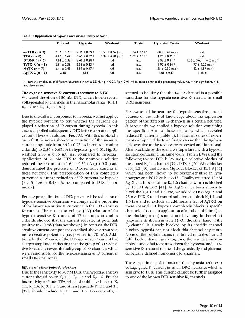
The hypoxic sensitive K+ current is sensitive to DTXWe tested the effect of 50 nM DTX, which blocks severalvoltage-gated K+ channels in the nanomolar range (KV1.1,KV1.2 and KV1.6; [37,38]).
Due to the different responses to hypoxia, we first appliedthe hypoxic solution to test whether the neurone dis-played a reduction of K+ current during hypoxia. In thiscase we applied subsequently DTX before a second appli-cation of hypoxic solution (Fig. 7A). With this protocol 7out of 10 neurones showed a reduction of the mean K+
current amplitude from 2.92 ± 0.73 nA in control (cholinechloride) to 2.36 ± 0.69 nA in hypoxia (p < 0.01, Fig. 5B,washout 2.53 ± 0.66 nA, n.s. compared to hypoxia).Application of 50 nM DTX to the normoxic solutionreduced the K+ current to 1.64 ± 0.51 nA (p < 0.01) anddemonstrated the presence of DTX-sensitive currents inthese neurones. This preapplication of DTX completelyprevented a further reduction of K+ currents by hypoxia(Fig. 5, 1.60 ± 0.48 nA, n.s. compared to DTX in nor-moxia).
Because preapplication of DTX prevented the reduction ofhypoxia-sensitive K+currents we compared the propertiesof the hypoxia-sensitive K+ current with the DTX-sensitiveK+ current. The current to voltage (I-V) relation of thehypoxia-sensitive K+ current of 17 neurones in cholinechloride showed that the current activated at potentialspositive to -50 mV (data not shown). In contrast, the DTX-sensitive current component described above activated atmore negative potentials (i.e. positive to -70 mV). Addi-tionally, the I-V curve of the DTX-sensitive K+ current hada larger amplitude indicating that the group of DTX-sensi-tive K+ current covers the subgroup of K+ channels whichwere responsible for the hypoxia-sensitive K+ current insmall DRG neurones.
Effects of other peptide blockersDue to the sensitivity to 50 nM DTX, the hypoxia-sensitivecurrent should cover KV 1.1, KV 1.2 and KV 1.6. But theinsensitivity to 5 mM TEA, which should have blocked KV1.1, KV 1.6, KV 3.1–3.4 and at least partially KV 2.1 and 2.2[37], should exclude KV 1.1 and 1.6. Consequently, it
seemed to be likely that the KV 1.2 channel is a possiblecandidate for the hypoxia-sensitive K+ current in smallDRG neurones.
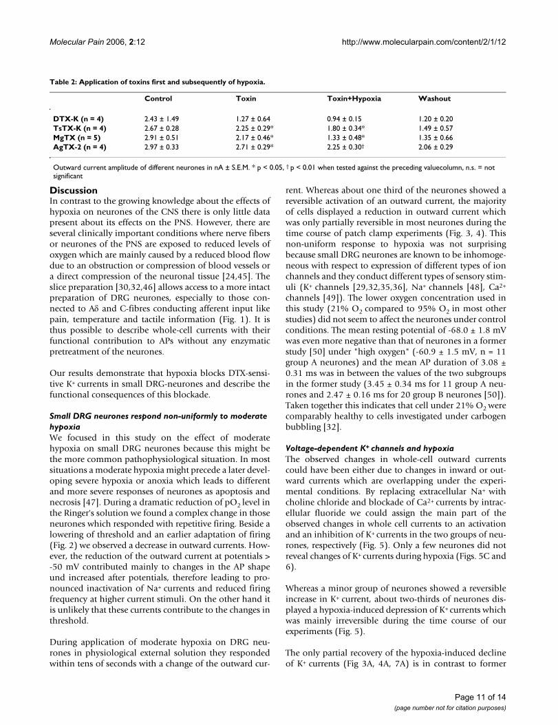
First, we tested the neurones for hypoxia-sensitive currentsbecause of the lack of knowledge about the expressionpattern of the different KV channels in a certain neurone.Subsequently, we applied a hypoxic solution containingthe specific toxin to those neurones which revealedreduced K+ currents (Table 1). In another series of experi-ments we applied the toxin first to ensure that the KVchan-nels sensitive to the toxin were expressed and functional.After blockade by the toxin, we superfused with a hypoxicsolution containing the same toxin (Table 2). We used thefollowing toxins: DTX-k (25 nM), a selective blocker ofthe cloned KV 1.1 channel [39], TsTX-K (20 nM) a blockerof KV 1.2 [40] and 20 nM MgTX as blocker of KV 1.3 [41]which has been shown to be oxygen-sensitive in lym-phozytes and PC12-cells [42,43]. Finally, we tested 10 nMAgTX-2 as blocker of the KV 1.6 channel which is blockedby 10 nM AgTX-2 [44]. As AgTX-2 has been shown toblock the KV1.1 and 1.3, too, we added 20 nM MgTX and25 nM DTX-K to all control solutions to block KV1.1 and1.3 first and to exclude an additional effect of AgTX-2 onthese channels. If hypoxia completely blocks a specificchannel, subsequent application of another inhibitor (i.e.the blocking toxin) should not have any further effect(experiments shown in table 1). On the other hand, if theKV channel is already blocked by its specific channelblocker, hypoxia can not block this channel any more.None of the peptide toxins mentioned in tables 1 and 2fulfil both criteria. Taken together, the results shown intables 1 and 2 fail to narrow down the hypoxia- and DTX-sensitive K+-channel to one of the genetically and pharma-cologically defined homomeric KV channels.
These experiments demonstrate that hypoxia reduces avoltage-gated K+ current in small DRG neurones which issensitive to DTX. This current cannot be further assignedto one of the known DTX sensitive KV channels.
Table 1: Application of hypoxia and subsequently of toxin.
Control Hypoxia Washout Toxin Hypoxia+ Toxin Washout
α-DTX (n = 7) 2.92 ± 0.73 2.36 ± 0.69 † 2.53 ± 0.66 (n.s.) 1.64 ± 0.51 † 1.60 ± 0.48 (n.s.) n.d.TEA (n = 8) 4.12 ± 0.62 3.65 ± 0.52 † 3.34 ± 0.48 (n.s.) 2.02 ± 0.35 † 1.79 ± 0.32 * n.d.DTX-K (n = 6) 3.14 ± 0.32 2.46 ± 0.28 † n.d. n.d. 2.08 ± 0.31 * 1.56 ± 0.63 (n = 2, n.d.)TsTX-K (n = 5) 2.91 ± 0.38 2.53 ± 0.43 * n.d. n.d. 1.92 ± 0.34 † 1.77 ± 0.20 (n.s.)MgTX (n = 7) 2.41 ± 0.48 1.89 ± 0.37 * n.d. n.d. 1.55 ± 0.30 (n.s.) 1.82 ± 0.59 (n.s.)AgTX-2 (n = 2) 2.48 2.15 n.d. n.d. 1.61 ± 0.17 1.25 ±
K+ current amplitude of different neurones in nA ± S.E.M. * p < 0.05, † p < 0.01 when tested against the preceding value, n.s. = not significant, n.d. not determined
Page 10 of 14(page number not for citation purposes)
Molecular Pain 2006, 2:12 http://www.molecularpain.com/content/2/1/12
DiscussionIn contrast to the growing knowledge about the effects ofhypoxia on neurones of the CNS there is only little datapresent about its effects on the PNS. However, there areseveral clinically important conditions where nerve fibersor neurones of the PNS are exposed to reduced levels ofoxygen which are mainly caused by a reduced blood flowdue to an obstruction or compression of blood vessels ora direct compression of the neuronal tissue [24,45]. Theslice preparation [30,32,46] allows access to a more intactpreparation of DRG neurones, especially to those con-nected to Aδ and C-fibres conducting afferent input likepain, temperature and tactile information (Fig. 1). It isthus possible to describe whole-cell currents with theirfunctional contribution to APs without any enzymaticpretreatment of the neurones.
Our results demonstrate that hypoxia blocks DTX-sensi-tive K+ currents in small DRG-neurones and describe thefunctional consequences of this blockade.
Small DRG neurones respond non-uniformly to moderate hypoxiaWe focused in this study on the effect of moderatehypoxia on small DRG neurones because this might bethe more common pathophysiological situation. In mostsituations a moderate hypoxia might precede a later devel-oping severe hypoxia or anoxia which leads to differentand more severe responses of neurones as apoptosis andnecrosis [47]. During a dramatic reduction of pO2 level inthe Ringer's solution we found a complex change in thoseneurones which responded with repetitive firing. Beside alowering of threshold and an earlier adaptation of firing(Fig. 2) we observed a decrease in outward currents. How-ever, the reduction of the outward current at potentials >-50 mV contributed mainly to changes in the AP shapeund increased after potentials, therefore leading to pro-nounced inactivation of Na+ currents and reduced firingfrequency at higher current stimuli. On the other hand itis unlikely that these currents contribute to the changes inthreshold.
During application of moderate hypoxia on DRG neu-rones in physiological external solution they respondedwithin tens of seconds with a change of the outward cur-
rent. Whereas about one third of the neurones showed areversible activation of an outward current, the majorityof cells displayed a reduction in outward current whichwas only partially reversible in most neurones during thetime course of patch clamp experiments (Fig. 3, 4). Thisnon-uniform response to hypoxia was not surprisingbecause small DRG neurones are known to be inhomoge-neous with respect to expression of different types of ionchannels and they conduct different types of sensory stim-uli (K+ channels [29,32,35,36], Na+ channels [48], Ca2+
channels [49]). The lower oxygen concentration used inthis study (21% O2 compared to 95% O2 in most otherstudies) did not seem to affect the neurones under controlconditions. The mean resting potential of -68.0 ± 1.8 mVwas even more negative than that of neurones in a formerstudy [50] under "high oxygen" (-60.9 ± 1.5 mV, n = 11group A neurones) and the mean AP duration of 3.08 ±0.31 ms was in between the values of the two subgroupsin the former study (3.45 ± 0.34 ms for 11 group A neu-rones and 2.47 ± 0.16 ms for 20 group B neurones [50]).Taken together this indicates that cell under 21% O2 werecomparably healthy to cells investigated under carbogenbubbling [32].
Voltage-dependent K+ channels and hypoxiaThe observed changes in whole-cell outward currentscould have been either due to changes in inward or out-ward currents which are overlapping under the experi-mental conditions. By replacing extracellular Na+ withcholine chloride and blockade of Ca2+ currents by intrac-ellular fluoride we could assign the main part of theobserved changes in whole cell currents to an activationand an inhibition of K+ currents in the two groups of neu-rones, respectively (Fig. 5). Only a few neurones did notreveal changes of K+ currents during hypoxia (Figs. 5C and6).
Whereas a minor group of neurones showed a reversibleincrease in K+ current, about two-thirds of neurones dis-played a hypoxia-induced depression of K+ currents whichwas mainly irreversible during the time course of ourexperiments (Fig. 5).
The only partial recovery of the hypoxia-induced declineof K+ currents (Fig 3A, 4A, 7A) is in contrast to former
Table 2: Application of toxins first and subsequently of hypoxia.
Control Toxin Toxin+Hypoxia Washout
DTX-K (n = 4) 2.43 ± 1.49 1.27 ± 0.64 0.94 ± 0.15 1.20 ± 0.20TsTX-K (n = 4) 2.67 ± 0.28 2.25 ± 0.29* 1.80 ± 0.34* 1.49 ± 0.57MgTX (n = 5) 2.91 ± 0.51 2.17 ± 0.46* 1.33 ± 0.48* 1.35 ± 0.66AgTX-2 (n = 4) 2.97 ± 0.33 2.71 ± 0.29* 2.25 ± 0.30† 2.06 ± 0.29
Outward current amplitude of different neurones in nA ± S.E.M. * p < 0.05, † p < 0.01 when tested against the preceding valuecolumn, n.s. = not significant
Page 11 of 14(page number not for citation purposes)
Molecular Pain 2006, 2:12 http://www.molecularpain.com/content/2/1/12
observations of mainly reversible actions of hypoxia on KVchannels [11,12]. It might be possible that some factorsprolong the blockade of potassium channels which mightnot be present in expression systems. Additionally, inabout 25% of experiments either in Ringer's solution orcholinchlorid the neurones showed a reversible increaseof currents under hypoxia. We think that the reversibilityof these experiments together with the stable currents inthe "unchanged" group are clear hints for a specific effectof hypoxia. Furthermore, we cannot exclude that theobserved partial recovery is just a slow or delayed reversi-bility that might have be seen at longer time courses of thewashout period [20]. Differences in recovery from ischae-mia-induced effects have been reported for sensory andmotor axons of human median nerve where the sensoryneurones need a longer time period to return to pre-ischaemic excitability [51].
Beside KV channels other channels might contribute to thehypoxia induced responses under physiological condi-tions. Hypoxia-induced increase of cytoplasmatic Ca2+ hasbeen described for carotid body type I cells [52] and par-ticularly for DRG neurones where it has been shown thatthe hypoxia-induced increase in intracellular Ca2+ concen-tration is due to an influx through L-type Ca2+ channels[5]. Beside stimulating the production and release of NOby endothelial NO synthase in DRG neurones [25] thismight influence Ca2+-dependent channels and other Ca2+-dependent mechanisms (e.g. kinases). However, changesof internal Ca2+ in those experiments with KV channels areunlikely due to the internal fluoride which binds any freeinternal Ca2+ most effectively [53]. Because the relativereduction in presence of choline chloride was similar tothat in Ringer's solution (25% vs. 29%) it supports theidea that the major component is a voltage-dependent K+
current. The hypoxia sensitive current activated mainly atpotentials positive to -50 mV (Figs. 3 and 5A) with volt-age-dependent characteristic which makes a contributionof one the two-pore-domain K+ channels rather unlikely.
The hypoxia-sensitive current is DTX sensitive but insensitive to other specific blockersα-DTX has been described to block KV 1.1, 1.2 and 1.6 and5 mM TEA should have blocked KV 1.1, KV 1.6, KV 3.1–3.4and at least partially KV 2.1 and 2.2, too [37]. It has beenshown previously in ganglia nodosa that the α-DTX-sensi-tive group of KV channels is a subcomponent of the 4-APsensitive K+ current, therefore we have not used 4-AP [54].Consequently, it seemed to be likely that the KV 1.2 chan-nel is responsible or contributes to the hypoxia-sensitiveK+ current in small DRG neurones.
Due to the problem of inhomogeneous distribution ofdifferent KV channels in a specific neurone [35,36], wetried two different sequences of hypoxia and toxin appli-
cation (tables 1 and 2). The hypoxia-sensitive KV channelshould neither show a further reduction in toxin afterapplication of hypoxia (table 1) nor in subsequenthypoxia after application of the toxin (table 2). If therewas no further change in hypoxia after application of thetoxin, the toxin-sensitive current could be the demandedKV channel type already blocked by hypoxia (table 1). Incase of a further reduction there had to be another K+
channel, which is not blocked by this toxin, contributingto the hypoxia-sensitive K+ current in that specific neu-rone.
Even though α-DTX could completely prevent the hypoxiaeffect, none of the other toxins had a similar effect onhypoxia-induced reduction of the K+ current. According tothe literature the toxin concentrations used in this studyshould have been high enough to get a sufficient completeinhibition of the sensitive KV channel. Therefore we can-not assign the hypoxia-sensitive K+ current to one singleKV α-subunit as described in expression systems. Thiscould be due to different KV α-subunits which areexpressed in the same neurone and have assembled toheteromultimeric channels. Compared to homomulti-mera these heteromultimeric channels can have alteredchannel properties and toxin sensitivity. This has beendemonstrated for a heteromultimer of KV1.2/KV1.5 [12]and other heteromultimera of voltage-gated K+ channelswhere an enhanced sensitivity to dendrotoxin has beendemonstrated for the heteromultimer compared to thehomomultimer [55-57].
Therefore we suggest that the hypoxia-sensitive and α-DTX sensitive K+ channel in our preparation is the corre-late to a heteromultimeric channel assembled from differ-ent KV channel α-subunits present in DRG neurones.Alternatively it is possible that a co-assembled β-subunithas altered some electrophysiological properties of anexpressed KV channel α-subunit [58,59] and can even con-fer oxygen-sensitivity to an expressed Kv α-subunit [60].However, it is nearly impossible with whole-cell record-ings to differentiate between "simple" blockade and alter-ations in gating of the channels.
Effects of hypoxia on action potentialsAlthough it is unlikely, that voltage-gated K+ channels areactive around the resting membrane potential of smallDRG neurones (-60 mV to -70 mV; [50]), the block of K+
channels which becomes active at potentials close to thethreshold of APs might influence the excitability of neu-rones under hypoxic conditions. It has been demon-strated that DTX-sensitive currents modulate actionpotentials and regulate excitability in rat visceral sensoryneurones [61]. Application of DTX had only little effect onthe resting membrane potential in that study but influ-enced the shape of APs, increased the excitability and the
Page 12 of 14(page number not for citation purposes)

Molecular Pain 2006, 2:12 http://www.molecularpain.com/content/2/1/12
firing frequency of neurones from nodose ganglia. DTX-sensitive KV channels have been described as fast-activat-ing and slow-inactivating and activate at membranepotentials slightly positive to the resting membrane andclose to the threshold for APs under normoxic conditions[61]. These effects on sensory neurones from nodose gan-glia were observed even though the DTX-sensitive currentfraction was only about 12 – 25 % of the total K+ current.
In our preparation about 42 % (at 0 mV) of the K+ currentwas DTX sensitive (Fig. 4), therefore the effect of this com-ponent on cell excitability might be even higher. Therewas no correlation with changes of the resting membranepotential but in some neurones we observed an increasein excitability (Figs. 2 and 4). This is unlikely to be causedby the changes at +40 mV but might be explained by min-imal changes of the total outward currents active aroundthe threshold potential (see I/V relation of blocked cur-rent in Fig 3C). We could observe a prolongation of actionpotential duration corresponding to the decrease in out-ward-current in 11 neurones (Fig. 4).
Other factors influencing the effects of hypoxia on small DRG neuronesIt cannot be excluded that part of the effects of hypoxia inslices are due to secondary effects after a local release ofneuropeptides under hypoxia. Other investigators do notuse synaptic blockers in their experiments with DRG neu-rones because these neurones normally do not possesssynapses. Further experiments in the presence of synapticblockers might help to clarify if an indirect effect influ-ences the hypoxic modulation of K+ channels.
On the other hand it is reasonable to assume that theobserved effects of hypoxia might have been even biggerwhen tested at a temperature closer to the physiologicallevel (i.e. 37°C) with a higher metabolic rate and oxygenconsumption rate of the tissue. Some ion channels havebeen described to be directly temperature-modulated (e.g.TRPV1, TRPV2, TRPM8, TRAAK, TREK-1 and TREK-2; [62-64]) and might further influence functional responses tohypoxia. Further experiments at body temperature mighthelp to evaluate the effects in more details.
We used young rats (2–9 d) because the slice-technique ofdorsal root ganglia is well established for animals of thisage [29,32]. The possible changes in ion channel distribu-tion during development of the PNS and CNS make itpreferable to test effects on mature animals. However,older animals cannot be investigated easily in the slicetechnique due to their more compact connecting tissue.
Our findings in small DRG neurones to moderate hypoxiamight account for some of the neurological phenomenaas paraesthesia, numbness or pain observed under condi-
tions with a reduced oxygen level in the PNS. In fact, theblockade of a part of voltage-gated KV channels in smallDRG neurones might be one part of the pathophysiologi-cal mechanisms which aggravate the situation after theinitial response of the sensory neurone to hypoxia.
Competing interestsThe authors declare herewith that they have no financialas well as non-financial competitions with any other peo-ple or organizations.
AcknowledgementsThe authors are grateful to Prof. G. Reid, Dr. T. Weiser and Chr. Malik for their helpful comments on the manuscript. We thank B. Agari and O. Becker for excellent technical support. This study was supported by the DFG (VO 20–2 and 3) to A.S and the VW-Stiftung (I/77 794) to A.S.
References1. Haddad GG, Jiang C: O2-sensing mechanisms in excitable cells:
role of plasma membrane K+ channels. Annu Rev Physiol 1997,59:23-42.
2. Lopez-Barneo J, Pardal R, Ortega-Saenz P: Cellular mechanism ofoxygen sensing. Annu Rev Physiol 2001, 63:259-287.
3. Patel AJ, Honoré E: Molecular physiology of oxygen-sensitivepotassium channels. Eur Respir J 2001, 18:221-227.
4. Franco-Obregon A, Urena J, Lopez-Barneo J: Oxygen-sensitive cal-cium channels in vascular smooth muscle and their possiblerole in hypoxic arterial relaxation. Proc Natl Acad Sci USA 1995,92:4715-4719.
5. Lukyanetz EA, Stanika RI, Koval LM, Kostyuk PG: Intracellularmechanisms of hypoxia-induced calcium increase in rat sen-sory neurons. Arch Biochem Biophys 2003, 410:212-221.
6. O'Reilly JP, Cummins TR, Haddad GG: Oxygen deprivation inhib-its Na+ current in rat hippocampal neurones via proteinkinase C. J Physiol 1997, 503 ( Pt 3):479-488.
7. Hammarstrom AKM, Gage PW: Oxygen-sensing persistentsodium channels in rat hippocampus. J Physiol 2000,529:107-118.
8. Liu H, Moczydlowski E, Haddad GG: O2 deprivation inhibitsCa2+-activated K+ channels via cytosolic factors in mice neo-cortical neurons. J Clin Invest 1999, 104:577-588.
9. Buckler KJ, Williams BA, Honore E: An oxygen-, acid- and anaes-thetic-sensitive TASK-like background potassium channel inrat arterial chemoreceptor cells. J Physiol 2000, 525:135-142.
10. Fearon IM, Thompson RJ, Samjoo I, Vollmer C, Doering LC, NurseCA: O2-sensitive K+ channels in immortalised rat chromaf-fin-cell-derived MAH cells. J Physiol 2002, 545:807-818.
11. Osipenko ON, Tate RJ, Gurney AM: Potential role for Kv3.1bchannels as oxygen sensors. Circ Res 2000, 86:534-540.
12. Hulme JT, Coppock EA, Felipe A, Martens JR, Tamkun MM: Oxygensensitivity of cloned voltage-gated K+ channels expressed inthe pulmonary vasculature. Circ Res 1999, 85:489-497.
13. Coppock EA, Martens JR, Tamkun MM: Molecular basis ofhypoxia-induced pulmonary vasoconstriction: role of volt-age-gated K+ channels. Am J Physiol Lung Cell Mol Physiol 2001,281:L1-L12.
14. Ganfornina MD, Lopez-Barneo J: Single K+ channels in mem-brane patches of arterial chemoreceptor cells are modu-lated by O2 tension. Proc Natl Acad Sci USA 1991, 88:2927-2930.
15. Jiang C, Haddad GG: Oxygen deprivation inhibits a K+ channelindependently of cytosolic factors in rat central neurons. JPhysiol 1994, 481:15-26.
16. Archer SL, Huang J, Henry T, Peterson D, Weir EK: A redox-basedO2 sensor in rat pulmonary vasculature. Circ Res 1993,73:1100-1112.
17. Yuan XJ, Tod ML, Rubin LJ, Blaustein MP: Deoxyglucose andreduced glutathione mimic effects of hypoxia on K+ andCa2+ conductances in pulmonary artery cells. Am J Physiol1994, 267:L52-L63.
Page 13 of 14(page number not for citation purposes)

Molecular Pain 2006, 2:12 http://www.molecularpain.com/content/2/1/12
18. Haddad GG, Jiang C: O2 deprivation in the central nervous sys-tem: on mechanisms of neuronal response, differential sen-sitivity and injury. Prog Neurobiol 1993, 40:277-318.
19. Bickler PE, Donohoe PH: Adaptive responses of vertebrate neu-rons to hypoxia. J Exp Biol 2002, 205:3579-3586.
20. Howe JF, Loeser JD, Calvin WH: Mechanosensitivity of dorsalroot ganglia and chronically injured axons: a physiologicalbasis for the radicular pain of nerve root compression. Pain1977, 3:25-41.
21. Sugawara O, Atsuta Y, Iwahara T, Muramoto T, Watakabe M, Take-mitsu Y: The effects of mechanical compression and hypoxiaon nerve root and dorsal root ganglia. An analysis of ectopicfiring using an in vitro model. Spine 1996, 21:2089-2094.
22. Schneider U, Niedermeier W, Grafe P: The paradox betweenresistance to hypoxia and liability to hypoxic damage inhyperglycemic peripheral nerves. Evidence for glycolysisinvolvement. Diabetes 1993, 42:981-987.
23. Obrosova IG: How does glucose generate oxidative stress inperipheral nerve? Int Rev Neurobiol 2002, 50:3-35.
24. Mogyoros I, Kiernan MC, Burke D, Bostock H: Excitability changesin human sensory and motor axons during hyperventilationand ischaemia. Brain 1997, 120 Part 2:317-325.
25. Henrich M, Hoffmann K, Konig P, Gruss M, Fischbach T, Godecke A,Hempelmann G, Kummer W: Sensory neurons respond tohypoxia with NO production associated with mitochondria.Mol Cell Neurosci 2002, 20:307-322.
26. Greffrath W, Binzen U, Schwarz ST, Saaler-Reinhardt S, Treede RD:Co-expression of heat sensitive vanilloid receptor subtypesin rat dorsal root ganglion neurons. Neuroreport 2003,14:2251-2255.
27. Scholz A, Gruß M, Stehr J, Beuche J, Rizzo N, Ettorre G: Moderatehypoxia blocks dendrotoxin sensitive channels in dorsal rootganglion neurones of rat. Pflügers Archiv 2003, 445:S20.
28. Takahashi T: Membrane currents in visually identifiedmotoneurones of neonatal rat spinal cord. J Physiol 1990,423:27-46.
29. Safronov BV, Bischoff U, Vogel W: Single voltage gated K+ chan-nels and their functions in small dorsal root ganglion neu-rones of rat. J Physiol 1996, 493:393-408.
30. Scholz A, Vogel W: Tetrodotoxin-resistant action potentials indorsal root ganglion neurons are blocked by local anesthet-ics. Pain 2000, 89:47-52.
31. Edwards FA, Konnerth A, Sakmann B, Takahashi T: A thin slicepreparation for patch clamp recordings from neurones ofthe mammalian central nervous system. Pflügers Arch 1989,414:600-612.
32. Scholz A, Gruß M, Vogel W: Properties and functions of cal-cium-activated K+ channels in small neurones of rat dorsalroot ganglion studied in a thin slice preparation. J Physiol 1998,513:55-69.
33. Hamill OP, Marty A, Neher E, Sakmann B, Sigworth FJ: Improvedpatch-clamp techniques for high-resolution current record-ing from cells and cell-free membrane patches. Pflügers Arch1981, 391:85-100.
34. Gold MS, Shuster MJ, Levine JD: Characterization of six voltagegated K+ currents in adult rat sensory neurons. J Neurophysiol1996, 75:2629-2646.
35. Ishikawa K, Tanaka M, Black JA, Waxman SG: Changes in expres-sion of voltage-gated potassium channels in dorsal root gan-glion neurons following axotomy. Muscle Nerve 1999,22:502-507.
36. Mathie A, Wooltorton JR, Watkins CS: Voltage-activated potas-sium channels in mammalian neurons and their block bynovel pharmacological agents. Gen Pharmacol 1998, 30:13-24.
37. Harvey AL: Twenty years of dendrotoxins. Toxicon 2001,39:15-26.
38. Robertson B, Owen D, Stow J, Butler C, Newland C: Novel effectsof dendrotoxin homologues on subtypes of mammalian Kv1potassium channels expressed in Xenopus oocytes. FEBS Lett1996, 383:26-30.
39. Rogowski RS, Krueger BK, Collins JH, Blaustein MP: Tityustoxin Kablocks voltage-gated noninactivating K+ channels andunblocks inactivating K+ channels blocked by a-dendrotoxinin synaptosomes. Proc Natl Acad Sci USA 1994, 91:1475-1479.
40. Grissmer S, Dethlefs B, Wasmuth JJ, Goldin AL, Gutman GA, CahalanMD, Chandy KG: Expression and chromosomal localization of
a lymphocyte K+ channel gene. Proc Natl Acad Sci USA 1990,87:9411-9415.
41. Conforti L, Petrovic M, Mohammad D, Lee S, Ma Q, Barone S, Filipov-ich AH: Hypoxia regulates expression and activity of Kv1.3channels in T lymphocytes: a possible role in T cell prolifer-ation. J Immunol 2003, 170:695-702.
42. Conforti L, Millhorn DE: Selective inhibition of a slow-inactivat-ing voltage-dependent K+ channel in rat PC12 cells byhypoxia. J Physiol 1997, 502:293-305.
43. Garcia ML, Garcia-Calvo M, Hidalgo P, Lee A, MacKinnon R: Purifi-cation and characterization of three inhibitors of voltage-dependent K+ channels from Leiurus quinquestriatus var.hebraeus venom. Biochemistry 1994, 33:6834-6839.
44. Brunelle JK, Chandel NS: Oxygen deprivation induced celldeath: an update. Apoptosis 2002, 7:475-482.
45. Scholz A, Reid G: Properties and localisation of ionic channelsin myelinated and unmyelinated nerve fibres. In Ion channelsand physiopathologies of nerve conduction and cell proliferation Volume 1.1st edition. Edited by: Rouzaire-Dubois B, Benoit E and Dubois JM.Fort P.O. Trivandrum, Research Signpost; 2002:7-48.
46. Yusaf SP, Goodman J, Pinnock RD, Dixon AK, Lee K: Expression ofvoltage-gated calcium channel subunits in rat dorsal rootganglion neurons. Neurosci Lett 2001, 311:137-141.
47. Gruß M, Henrich M, König P, Hempelmann G, Vogel W, Scholz A:Ethanol reduces excitability in a subgroup of primary sen-sory neurones by activation of BKCa channels. Eur J Neurosci2001:1246-1256.
48. Lin CS, Kuwabara S, Cappelen-Smith C, Burke D: Responses ofhuman sensory and motor axons to the release of ischaemiaand to hyperpolarizing currents. J Physiol 2002, 541:1025-1039.
49. Buckler KJ, Vaughan-Jones RD: Effects of hypoxia on membranepotential and intracellular calcium in rat neonatal carotidbody type I cells. J Physiol 1994, 476:423-428.
50. Kostyuk PG, Krishtal OA, Pidoplichko VI: Asymmetrical displace-ment currents in nerve cell membrane and effect of internalfluoride. Nature 1977, 267:70-72.
51. Glazebrook PA, Ramirez AN, Schild JH, Shieh CC, Doan T, Wible BA,Kunze DL: Potassium channels Kv1.1, Kv1.2 and Kv1.6 influ-ence excitability of rat visceral sensory neurons. J Physiol 2002,541:467-482.
52. Ruppersberg JP, Schroter KH, Sakmann B, Stocker M, Sewing S, PongsO: Heteromultimeric channels formed by rat brain potas-sium-channel proteins. Nature 1990, 345:535-537.
53. Scott VE, Muniz ZM, Sewing S, Lichtinghagen R, Parcej DN, Pongs O,Dolly JO: Antibodies specific for distinct Kv subunits unveil aheterooligomeric basis for subtypes of alpha-dendrotoxin-sensitive K+ channels in bovine brain. Biochemistry 1994,33:1617-1623.
54. Shamotienko OG, Parcej DN, Dolly JO: Subunit combinationsdefined for K+ channel Kv1 subtypes in synaptic membranesfrom bovine brain. Biochemistry 1997, 36:8195-8201.
55. Heinemann S, Rettig J, Scott V, Parcej DN, Lorra C, Dolly J, Pongs O:The inactivation behaviour of voltage-gated K-channels maybe determined by association of alpha- and beta-subunits. JPhysiol Paris 1994, 88:173-180.
56. Rettig J, Heinemann SH, Wunder F, Lorra C, Parcej DN, Dolly JO,Pongs O: Inactivation properties of voltage-gated K+ channelsaltered by presence of beta-subunit. Nature 1994, 369:289-294.
57. Perez-Garcia MT, Lopez-Lopez JR, Gonzalez C: Kvbeta1.2 subunitcoexpression in HEK293 cells confers O2 sensitivity to kv4.2but not to Shaker channels. Journal of General Physiology 1999,113:897-907.
58. Patapoutian A, Peier AM, Story GM, Viswanath V: Thermotrpchannels and beyond: Mechanisms of temperature sensa-tion. Nat Rev Neurosci 2003, 4:529-539.
59. Maingret F, Lauritzen I, Patel AJ, Heurteaux C, Reyes R, Lesage F, Laz-dunski M, Honore E: TREK-1 is a heat-activated background K+channel. EMBO J 2000, 19:2483-2491.
60. Kang D, Choe C, Kim D: Thermosensitivity of the two-poredomain K+ channels TREK-2 and TRAAK. J Physiol 2005,564:103-116.
Page 14 of 14(page number not for citation purposes)