Embed Size (px)
Citation preview

APPLIED AND ENVIRONMENTAL MICROBIOLOGY, Dec. 2008, p. 7694–7708 Vol. 74, No. 240099-2240/08/$08.00�0 doi:10.1128/AEM.00878-08Copyright © 2008, American Society for Microbiology. All Rights Reserved.
Molecular Microbial Diversity Survey of Sponge Reproductive Stagesand Mechanistic Insights into Vertical Transmission of
Microbial Symbionts�
Susanne Schmitt,1* Hilde Angermeier,1 Roswitha Schiller,1 Niels Lindquist,2 and Ute Hentschel1
University of Wuerzburg, Research Center for Infectious Diseases, Roentgenring 11, D-97070 Wuerzburg, Germany,1 and University ofNorth Carolina at Chapel Hill, Institute of Marine Sciences, 3431 Arendell Street, Morehead City, North Carolina 285572
Received 17 April 2008/Accepted 22 September 2008
Many marine sponges, hereafter termed high-microbial-abundance (HMA) sponges, harbor large andcomplex microbial consortia, including bacteria and archaea, within their mesohyl matrices. To investigatevertical microbial transmission as a strategy to maintain these complex associations, an extensive phylogeneticanalysis was carried out with the 16S rRNA gene sequences of reproductive (n � 136) and adult (n � 88)material from five different Caribbean species, as well as all published 16S rRNA gene sequences from spongeoffspring (n � 116). The overall microbial diversity, including members of at least 13 bacterial phyla and onearchaeal phylum, in sponge reproductive stages is high. In total, 28 vertical-transmission clusters, defined asclusters of phylotypes that are found both in adult sponges and their offspring, were identified. They aredistributed among at least 10 bacterial phyla and one archaeal phylum, demonstrating that the complex adultmicrobial community is collectively transmitted through reproductive stages. Indications of host-speciesspecificity and cospeciation were not observed. Mechanistic insights were provided using a combined electronmicroscopy and fluorescence in situ hybridization analysis, and an indirect mechanism of vertical transmissionvia nurse cells is proposed for the oviparous sponge Ectyoplasia ferox. Based on these phylogenetic andmechanistic results, we suggest the following symbiont transmission model: entire microbial consortia arevertically transmitted in sponges. While vertical transmission is clearly present, additional environmentaltransfer between adult individuals of the same and even different species might obscure possible signals ofcospeciation. We propose that associations of HMA sponges with highly sponge-specific microbial communitiesare maintained by this combination of vertical and horizontal symbiont transmission.
Sponges (phylum Porifera) are evolutionarily ancient Meta-zoa whose origin dates back about 600 million years to thePrecambrian (26). With 7,000 formally described species andan estimated 15,000 extant species, sponges are among themost diverse marine invertebrate groups and are importantcomponents of all aquatic habitats, including freshwater en-vironments, tropical reefs, and even the deep sea (19). De-spite an enormous range of shapes, colors, and sizes, allsponges possess a relatively simple body plan which isadapted to a filter-feeding lifestyle (4). Large volumes ofseawater are pumped through canals embedded in an extra-cellular matrix (55), termed the mesohyl, and microorgan-isms and small unicellular eukaryotes are taken up from theseawater with high efficiency, leaving the expelled wateressentially sterile (37, 54).
Numerous sponges live in permanent and close associationswith microorganisms, and many of them host phylogeneticallydiverse populations of microbes (15, 17, 49). These microor-ganisms are located mainly extracellularly in the sponge me-sohyl in high concentrations, contributing up to 40% of thesponge’s biomass and exceeding microbial seawater concentra-tions by 2 to 4 orders of magnitude (13, 53). Such host species
are therefore termed bacteriosponges or high-microbial-abun-dance (HMA) sponges, in contrast to low-microbial-abun-dance (LMA) sponges that lack large and complex microbialconsortia within their mesohyl (15, 38). The total phylogeneticmicrobial diversity of HMA sponges is extremely high andincludes representatives of up to 16 bacterial phyla and bothmajor archaeal lineages (49). Despite this microbial complex-ity, there exists a high degree of uniformity among the mi-crobial communities from taxonomically and geographicallydisparate HMA sponges. Many microbial phylotypes were re-peatedly detected in different sponges but not in seawater. Therespective microbes were therefore defined as sponge specific(16). Collectively, these microbes possess diverse metabolicpathways (e.g., for photosynthesis, sulfate reduction, and nitri-fication) and their involvement in sponge metabolism suggestsa beneficial interaction (3, 18, 56). Additionally, those associ-ations were shown to be permanent and stable, with low tem-poral and spatial variability in the microbial communities (17).
To maintain stable and specific symbioses, the presence ofsymbionts in each generation must be assured. In the symbio-ses of many insects, including carpenter ants, tsetse flies, andaphids, as well as of certain marine clams, the microbial sym-bionts are strictly vertically transmitted through reproductivestages, resulting in cospeciation (20, 31, 36). Similarly, hori-zontal symbiont transmission between single adult individualsor environmental acquisition can also be highly specific andmaintain obligate symbioses, sometimes also resulting in par-allel evolution. Examples are hydrothermal vent tubeworms,
* Corresponding author. Present address: University of Auckland,School of Biological Sciences, Private Bag 92019, Auckland 1142, NewZealand. Phone: 64-9-3737599, ext. 87785. Fax: 64-9-3737416. E-mail:[email protected].
� Published ahead of print on 26 September 2008.
7694
on June 16, 2019 by guesthttp://aem
.asm.org/
Dow
nloaded from

which rely on their chemoautotrophic symbionts (34), and thesepiolid squid-vibrio symbiosis (33).
The first evidence for vertical transmission in sponges camefrom electron microscopy studies (references 6 and 9 and ref-erences cited therein; 25, 44, 52). To date, microbes have beendetected in oocytes, embryos, and larvae of more than 20oviparous sponges (which release gametes) and viviparoussponges (which release fully developed larvae) (9, 42). Re-cently, a culture-based study successfully isolated the samealphaproteobacterial strain from adult and embryo samples ofthe Caribbean sponge Mycale laxissima, and vertical transmis-sion of this bacterium was suggested by the authors (8). Addi-tional evidence for vertical transmission comes from molecularstudies that allow the phylogenetic identification of microbeswithin sponge reproductive stages and comparison with themicrobial community of the adult. Using Cyanobacteria-spe-cific primers, Oren et al. (35) found a Synechococcus-relatedsequence type in Diacarnus erythraenus adult sponges and theirembryos. However, so far only three molecular studies inves-tigated the total microbial diversity in sponge offspring. Com-plex bacterial consortia were described for each of the speciesM. laxissima, Ircinia felix, and Corticium sp. (8, 45, 46, respec-tively). Sharp et al. (46) also described archaea within Corti-cium sp. embryos and verified the presence of microbes byfluorescence in situ hybridization (FISH). Additionally, a com-plex microbial community was described for the first time in I.felix juvenile sponges which were obtained after larval settlementexperiments (45).
To provide insights into the phylogenetic diversity of bacte-ria and archaea in sponge reproductive stages, a comprehen-sive 16S rRNA gene survey was performed with �250 se-quences obtained from eight sponges representing six orders,two geographic regions, and both oviparous and viviparousmodes of reproduction. Furthermore, the mechanistic aspects
of vertical transmission were investigated in the oviparous Ca-ribbean sponge Ectyoplasia ferox using a combination of elec-tron and fluorescence microscopy. E. ferox was chosen becauseits early reproductive stages are obtainable, allowing mecha-nistic investigations from an early developmental stage on-ward. Based on the phylogenetic and mechanistic results, wepropose a symbiont transmission model for HMA sponge sym-bioses, including a combination of vertical and horizontalsymbiont transmission to maintain the association with sponge-specific, though not species-specific, microbial consortia.
MATERIALS AND METHODS
Sponge collection. The sponges Agelas conifera, Agelas wiedenmayeri, Ectyo-plasia ferox, Smenospongia aurea, and Xestospongia muta were collected by scubadiving off the coast of Key Largo, Florida, in June 2002, June and August 2004,and May 2005 (Table 1). Additional material from E. ferox was collected by scubadiving offshore of French Wells, Bahamas, in July 2003. The samples were takenat depths of 5 to 30 m, transferred in ziplock bags containing seawater to thesurface, and kept on ice in a cooler until further processing (within 4 to 5 h).Offspring samples (oocytes, embryos, and larvae) were collected in larval trapsusing the methodology of Lindquist et al. (27). Additionally, tissue samples of thecorresponding adult individuals were taken. After the adult samples were cutinto small pieces, the adult and offspring materials were washed in sterile filteredseawater, fixed either in 96% ethanol or in 2.5% glutaraldehyde–double-distilledwater (ddH2O), and stored at either �20 or 4°C. The field settlement experi-ments were performed with a portion of the captured S. aurea larvae as describedby Schmitt et al. (45). Briefly, larvae were enclosed in plastic containers (�75 mlin volume), which were placed at a 9-m-deep reef site. These containers consistedof a plastic frame supporting a covering of 200-�m nylon mesh to allow waterexchange. Juveniles were recovered 1 to 3 days postsettlement and preserved asdescribed above.
Extraction of DNA and 16S rRNA gene library construction. Adult samplesthat had been fixed in 96% ethanol were air dried, ground with a mortar andpestle in liquid nitrogen, and DNA extracted using the Fast DNA Spin kit for soil(Q-Biogene, Heidelberg, Germany) in accordance with the manufacturer’s in-structions. Reproductive material containing oocytes and embryos was also airdried and transferred into 150 �l ddH2O, and DNA was extracted by heating thesamples in a water bath for 10 min at 100°C. This same methodology was used to
TABLE 1. Compilation of all available 16S rRNA gene sequences from sponge reproductive stages until December 2007
Host sponge (order)a Collection site(s) (latitude/longitude) Mode ofreproduction Sequence source(s)c
No. ofoffspring
sequencesReference
HMA spongesAgelas conifera
(Agelasida)Key Largo, FL (25°02�N/80°24�W) Oviparous 16S rRNA libraryd 5 This
studyAgelas wiedenmayeri
(Agelasida)Key Largo, FL (25°02�N/80°24�W) Oviparous DGGE 7 This
studyCorticium sp.
(Homosclerophorida)Palau islands (07°23�N/134°38�E) Viviparous 16S rRNA library 18 46
Ectyoplasia ferox(Poecilosclerida)
Key Largo, FL (25°02�N/80°24�W) Oviparous DGGE 28 ThisstudyBahamas islands (24°32�N/75°55�W)
Ircinia felix(Dictyoceratida)
Key Largo, FL (25°02�N/80°24�W) Viviparous DGGE 71 4516S rRNA libraryd 1 This
studySmenospongia aurea(Verongidab)
Key Largo, FL (25°02�N/80°24�W) Viviparous DGGE 19 Thisstudy
Xestospongia muta(Haplosclerida)
Key Largo, FL (25°02�N/80°24�W) Oviparous 16S rRNA library 76 Thisstudy
LMA spongesMycale laxissima
(Poecilosclerida)Key Largo, FL (25°02�N/80°24�W) Viviparous Isolates, 16S rRNA
library27 8
a All sponges belong to the class Demospongiae (classification after Systema Porifera by Hooper and van Soest �19�).b Reclassified after the paper by Schmitt et al. (43).c Sequences were obtained using universal 16S rRNA primers either by the construction of 16S rRNA libraries or by the excision of DGGE bands.d The 16S rRNA library was constructed using group-specific primers.
VOL. 74, 2008 VERTICAL MICROBIAL TRANSMISSION IN MARINE SPONGES 7695
on June 16, 2019 by guesthttp://aem
.asm.org/
Dow
nloaded from
obtain DNA from collections of larvae and juveniles (three, five, or seven indi-viduals pooled immediately after collection). The DNA solutions were then useddirectly as templates (0.01 to 1.0 �g) in the following PCR.
The universal primers 27f and 1492r (23) were used for the PCR amplificationof bacterial 16S rRNA genes. The primer pairs POR389f/POR1130r (12), 11f/Ntspa865r (47), and Arch21f/Arch958r (7) were used for the specific PCR am-plification of the candidate phylum “Poribacteria,” Nitrospira, and Archaea 16SrRNA genes, respectively. Negative controls (PCRs without DNA templates)were included for each 16S rRNA gene amplification. Cycling conditions on aMastercycler gradient (Eppendorf, Hamburg, Germany) were as follows: an initialdenaturing step at 96°C for 5 min; 30 cycles of denaturing at 96°C for 1 min; primerannealing at 54, 64, 60, and 56°C for 1 min; and elongation at 72°C for 90, 50, 90, and50 s for universal, Poribacteria, Nitrospira, and Archaea PCRs, respectively. The PCRprogram ended with a final extension step at 72°C for 5 min.
DGGE. DNA was extracted as described above. The universal 16S rRNA geneprimers 341f with GC-clamp and 907r (32) were used for the PCR amplificationof bacterial 16S rRNA genes. The cycling conditions were as follows: initialdenaturing step at 95°C for 5 min, 30 cycles of denaturing at 95°C for 1 min,primer annealing at 54°C for 1 min and elongation at 72°C for 45 s, followed bya final extension step at 72°C for 10 min. Denaturing gradient gel electrophoresis(DGGE) was performed with a Bio-Rad DCode universal mutation detectionsystem (Bio-Rad, Munchen, Germany) on a 10% (wt/vol) polyacrylamide gel in1 Tris-acetate-EDTA using a 0 to 90% denaturing gradient; 100% denaturantcorresponded to 7 M urea and 40% (vol/vol) formamide. Electrophoresis wasperformed for 6 h at 150 V and 60°C. The gels were stained for 30 min in Sybrgold (Molecular Probes) and scanned on a Typhoon 8600 scanner (AmershamBiosciences). Selected bands were excised with an ethanol-sterilized scalpel andincubated in 25 �l ddH2O overnight at 4°C. A total of 4 �l of eluted DNA wassubsequently used for reamplification with primers 341f and 907r using the PCRconditions described above.
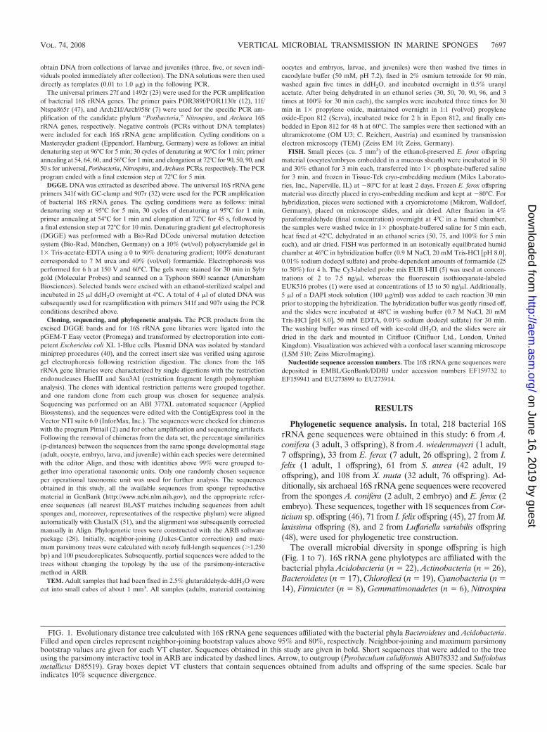
Cloning, sequencing, and phylogenetic analysis. The PCR products from theexcised DGGE bands and for 16S rRNA gene libraries were ligated into thepGEM-T Easy vector (Promega) and transformed by electroporation into com-petent Escherichia coli XL 1-Blue cells. Plasmid DNA was isolated by standardminiprep procedures (40), and the correct insert size was verified using agarosegel electrophoresis following restriction digestion. The clones from the 16SrRNA gene libraries were characterized by single digestions with the restrictionendonucleases HaeIII and Sau3AI (restriction fragment length polymorphismanalysis). The clones with identical restriction patterns were grouped together,and one random clone from each group was chosen for sequence analysis.Sequencing was performed on an ABI 377XL automated sequencer (AppliedBiosystems), and the sequences were edited with the ContigExpress tool in theVector NTI suite 6.0 (InforMax, Inc.). The sequences were checked for chimeraswith the program Pintail (2) and for other amplification and sequencing artifacts.Following the removal of chimeras from the data set, the percentage similarities(p-distances) between the sequences from the same sponge developmental stage(adult, oocyte, embryo, larva, and juvenile) within each species were determinedwith the editor Align, and those with identities above 99% were grouped to-gether into operational taxonomic units. Only one randomly chosen sequenceper operational taxonomic unit was used for further analysis. The sequencesobtained in this study, all the available sequences from sponge reproductivematerial in GenBank (http://www.ncbi.nlm.nih.gov), and the appropriate refer-ence sequences (all nearest BLAST matches including sequences from adultsponges and, moreover, representatives of the respective phylum) were alignedautomatically with ClustalX (51), and the alignment was subsequently correctedmanually in Align. Phylogenetic trees were constructed with the ARB softwarepackage (28). Initially, neighbor-joining (Jukes-Cantor correction) and maxi-mum parsimony trees were calculated with nearly full-length sequences (�1,250bp) and 100 pseudoreplicates. Subsequently, partial sequences were added to thetrees without changing the topology by the use of the parsimony-interactivemethod in ARB.
TEM. Adult samples that had been fixed in 2.5% glutaraldehyde-ddH2O werecut into small cubes of about 1 mm3. All samples (adults, material containing
oocytes and embryos, larvae, and juveniles) were then washed five times incacodylate buffer (50 mM, pH 7.2), fixed in 2% osmium tetroxide for 90 min,washed again five times in ddH2O, and incubated overnight in 0.5% uranylacetate. After being dehydrated in an ethanol series (30, 50, 70, 90, 96, and 3times at 100% for 30 min each), the samples were incubated three times for 30min in 1 propylene oxide, maintained overnight in 1:1 (vol/vol) propyleneoxide-Epon 812 (Serva), incubated twice for 2 h in Epon 812, and finally em-bedded in Epon 812 for 48 h at 60°C. The samples were then sectioned with anultramicrotome (OM U3; C. Reichert, Austria) and examined by transmissionelectron microscopy (TEM) (Zeiss EM 10; Zeiss, Germany).
FISH. Small pieces (ca. 5 mm3) of the ethanol-preserved E. ferox offspringmaterial (oocytes/embryos embedded in a mucous sheath) were incubated in 50and 30% ethanol for 3 min each, transferred into 1 phosphate-buffered salinefor 3 min, and frozen in Tissue-Tek cryo-embedding medium (Miles Laborato-ries, Inc., Naperville, IL) at �80°C for at least 2 days. Frozen E. ferox offspringmaterial was directly placed in cryo-embedding medium and kept at �80°C. Forhybridization, pieces were sectioned with a cryomicrotome (Mikrom, Walldorf,Germany), placed on microscope slides, and air dried. After fixation in 4%paraformaldehyde (final concentration) overnight at 4°C in a humid chamber,the samples were washed twice in 1 phosphate-buffered saline for 5 min each,heat fixed at 42°C, dehydrated in an ethanol series (50, 75, and 100% for 5 mineach), and air dried. FISH was performed in an isotonically equilibrated humidchamber at 46°C in hybridization buffer (0.9 M NaCl, 20 mM Tris-HCl [pH 8.0],0.01% sodium dodecyl sulfate) and probe-dependent amounts of formamide (25to 50%) for 4 h. The Cy3-labeled probe mix EUB I-III (5) was used at concen-trations of 2 to 7.5 ng/�l, whereas the fluorescein isothiocyanate-labeledEUK516 probes (1) were used at concentrations of 15 to 50 ng/�l. Additionally,5 �l of a DAPI stock solution (100 �g/ml) was added to each reaction 30 minprior to stopping the hybridization. The hybridization buffer was gently rinsed off,and the slides were incubated at 48°C in washing buffer (0.7 M NaCl, 20 mMTris-HCl [pH 8.0], 50 mM EDTA, 0.01% sodium dodecyl sulfate) for 30 min.The washing buffer was rinsed off with ice-cold dH2O, and the slides were airdried in the dark and mounted in Citifluor (Citifluor Ltd., London, UnitedKingdom). Visualization was achieved with a confocal laser scanning microscope(LSM 510; Zeiss MicroImaging).
Nucleotide sequence accession numbers. The 16S rRNA gene sequences weredeposited in EMBL/GenBank/DDBJ under accession numbers EF159732 toEF159941 and EU273899 to EU273914.
RESULTS
Phylogenetic sequence analysis. In total, 218 bacterial 16SrRNA gene sequences were obtained in this study: 6 from A.conifera (3 adult, 3 offspring), 8 from A. wiedenmayeri (1 adult,7 offspring), 33 from E. ferox (7 adult, 26 offspring), 2 from I.felix (1 adult, 1 offspring), 61 from S. aurea (42 adult, 19offspring), and 108 from X. muta (32 adult, 76 offspring). Ad-ditionally, six archaeal 16S rRNA gene sequences were recoveredfrom the sponges A. conifera (2 adult, 2 embryo) and E. ferox (2embryo). These sequences, together with 18 sequences from Cor-ticium sp. offspring (46), 71 from I. felix offspring (45), 27 from M.laxissima offspring (8), and 2 from Luffariella variabilis offspring(48), were used for phylogenetic tree construction.
The overall microbial diversity in sponge offspring is high(Fig. 1 to 7). 16S rRNA gene phylotypes are affiliated with thebacterial phyla Acidobacteria (n 22), Actinobacteria (n 26),Bacteroidetes (n 17), Chloroflexi (n 19), Cyanobacteria (n 14), Firmicutes (n 8), Gemmatimonadetes (n 6), Nitrospira
FIG. 1. Evolutionary distance tree calculated with 16S rRNA gene sequences affiliated with the bacterial phyla Bacteroidetes and Acidobacteria.Filled and open circles represent neighbor-joining bootstrap values above 95% and 80%, respectively. Neighbor-joining and maximum parsimonybootstrap values are given for each VT cluster. Sequences obtained in this study are given in bold. Short sequences that were added to the treeusing the parsimony interactive tool in ARB are indicated by dashed lines. Arrow, to outgroup (Pyrobaculum calidiformis AB078332 and Sulfolobusmetallicus D85519). Gray boxes depict VT clusters that contain sequences obtained from adults and offspring of the same species. Scale barindicates 10% sequence divergence.
VOL. 74, 2008 VERTICAL MICROBIAL TRANSMISSION IN MARINE SPONGES 7697
on June 16, 2019 by guesthttp://aem
.asm.org/
Dow
nloaded from

FIG. 2. Evolutionary distance tree calculated with 16S rRNA gene sequences affiliated with the bacterial phyla Nitrospira, Gemmatimonadetes,and Actinobacteria. Details are the same as those provided for Fig. 1.
7698
on June 16, 2019 by guesthttp://aem
.asm.org/
Dow
nloaded from
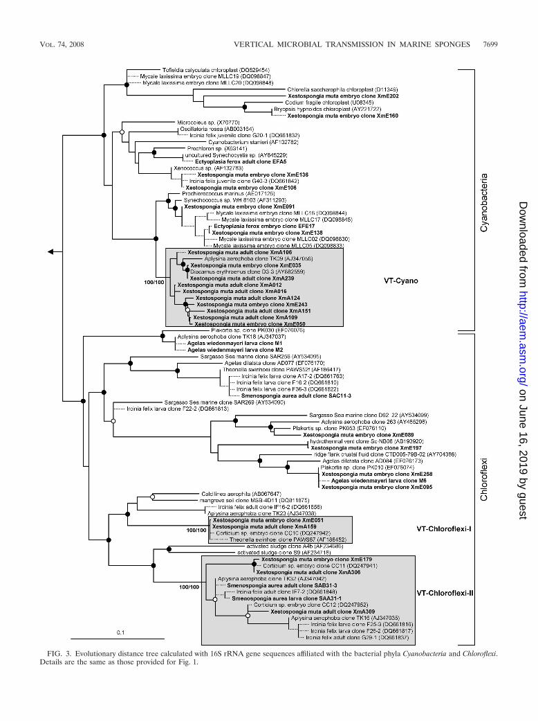
FIG. 3. Evolutionary distance tree calculated with 16S rRNA gene sequences affiliated with the bacterial phyla Cyanobacteria and Chloroflexi.Details are the same as those provided for Fig. 1.
VOL. 74, 2008 VERTICAL MICROBIAL TRANSMISSION IN MARINE SPONGES 7699
on June 16, 2019 by guesthttp://aem
.asm.org/
Dow
nloaded from

FIG. 4. Evolutionary distance tree calculated with 16S rRNA gene sequences affiliated with the bacterial class Alphaproteobacteria. Details arethe same as those provided for Fig. 1. The cluster containing the isolate from M. laxissima adults and embryos is indicated by an unshaded box.
7700 SCHMITT ET AL. APPL. ENVIRON. MICROBIOL.
on June 16, 2019 by guesthttp://aem
.asm.org/
Dow
nloaded from
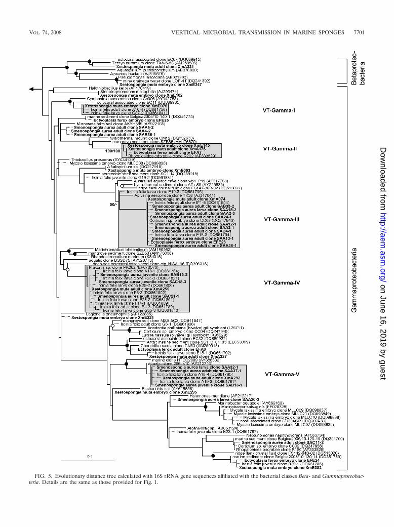
FIG. 5. Evolutionary distance tree calculated with 16S rRNA gene sequences affiliated with the bacterial classes Beta- and Gammaproteobac-teria. Details are the same as those provided for Fig. 1.
VOL. 74, 2008 VERTICAL MICROBIAL TRANSMISSION IN MARINE SPONGES 7701
on June 16, 2019 by guesthttp://aem
.asm.org/
Dow
nloaded from

FIG. 6. Evolutionary distance tree calculated with 16S rRNA gene sequences affiliated with the bacterial phyla Planctomycetes, Verrucomicrobia,and Firmicutes; with the class Deltaproteobacteria; and with the candidate phyla TM7 and Poribacteria. One group of sequences could not beunambiguously affiliated within the Bacteria and is marked as “uncertain affiliation.” Details are the same as those provided for Fig. 1.
7702 SCHMITT ET AL. APPL. ENVIRON. MICROBIOL.
on June 16, 2019 by guesthttp://aem
.asm.org/
Dow
nloaded from
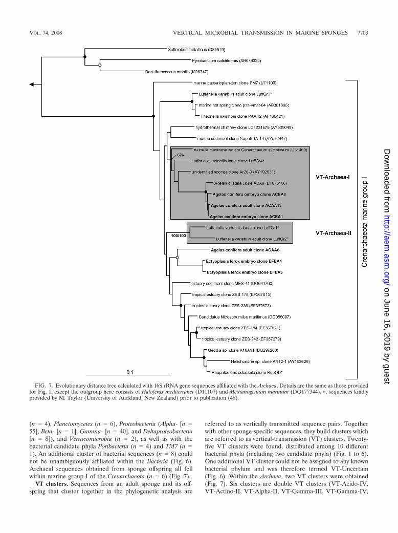
(n 4), Planctomycetes (n 6), Proteobacteria (Alpha- [n 55], Beta- [n 1], Gamma- [n 40], and Deltaproteobacteria[n 8]), and Verrucomicrobia (n 2), as well as with thebacterial candidate phyla Poribacteria (n 4) and TM7 (n 1). An additional cluster of bacterial sequences (n 8) couldnot be unambiguously affiliated within the Bacteria (Fig. 6).Archaeal sequences obtained from sponge offspring all fellwithin marine group I of the Crenarchaeota (n 6) (Fig. 7).
VT clusters. Sequences from an adult sponge and its off-spring that cluster together in the phylogenetic analysis are
referred to as vertically transmitted sequence pairs. Togetherwith other sponge-specific sequences, they build clusters whichare referred to as vertical-transmission (VT) clusters. Twenty-five VT clusters were found, distributed among 10 differentbacterial phyla (including two candidate phyla) (Fig. 1 to 6).One additional VT cluster could not be assigned to any knownbacterial phylum and was therefore termed VT-Uncertain(Fig. 6). Within the Archaea, two VT clusters were obtained(Fig. 7). Six clusters are double VT clusters (VT-Acido-IV,VT-Actino-II, VT-Alpha-II, VT-Gamma-III, VT-Gamma-IV,
FIG. 7. Evolutionary distance tree calculated with 16S rRNA gene sequences affiliated with the Archaea. Details are the same as those providedfor Fig. 1, except the outgroup here consists of Haloferax mediterranei (D11107) and Methanogenium marinum (DQ177344). �, sequences kindlyprovided by M. Taylor (University of Auckland, New Zealand) prior to publication (48).
VOL. 74, 2008 VERTICAL MICROBIAL TRANSMISSION IN MARINE SPONGES 7703
on June 16, 2019 by guesthttp://aem
.asm.org/
Dow
nloaded from

and VT-Nitrospira), and one is a triple VT cluster (VT-Chlo-roflexi-II), as they contain vertically transmitted sequence pairsof two or three sponge species, respectively. Twenty-nine per-cent of A. wiedenmayeri, 53% of Corticium sp., 23% of E. ferox,75% of I. felix, 0% of M. laxissima, 68% of S. aurea, and 23%of X. muta offspring sequences belong to VT clusters.
TEM. E. ferox released orange-colored oocytes which wereembedded in a mucous sheath. Electron micrographs of theoocytes revealed the presence of many inclusions equally scat-tered within the cytoplasm (Fig. 8A) These inclusions vary inelectron density and in size and represent, e.g., lipid droplets (1to 1.5 �m in diameter) and yolk granules (2 to 3 �m in diam-eter). Microorganisms could not be detected in the oocytecytoplasm. The oocyte is surrounded by a layer of amoeboid-shaped nurse cells which are 5 to 8 �m in size (Fig. 8A and B).Each nurse cell contains one 1- to 2-�m large nucleus, severalsmaller vacuoles, and usually one large vesicle with 15 to 20(sometimes more) microorganisms of different sizes of up to2.5 �m and various shapes including rod-like and coccus-likecells (Fig. 8B). At this stage of development, there is a clearspace between nurse cells and the oocyte. However, the oocytealready shows signs of invagination (Fig. 8B). After 2 days, theoocytes developed into early-stage embryos, which were still
embedded in the gelatinous sheath. The embryo consists ofmany small, densely packed amoeboid-shaped cells that con-tain one 2- to 3-�m large nucleus each and again diverseinclusions, including yolk granules (Fig. 8C and D). A thinlamella of collagen fibrils surrounds the embryo (Fig. 8C), andnurse cells are no longer present at this stage of development.Clusters of morphologically diverse microorganisms can befound extracellularly within the embryo, mostly located in theperiphery.
FISH. A cohybridization with the bacterial probe mix EUBI-III and the eukaryotic probe EUK516, in addition to theDNA dye DAPI, was performed on cross-sections of E. feroxoocytes (Fig. 9). The 150- by 100-�m-large oocyte showed nobackground autofluorescence (Fig. 9A). The interior appearedheterogeneous (Fig. 9B). Eukaryotic ribosomes are present inlarge numbers as indicated by many green signals. The 4- to5-�m-large nucleolus is especially prominent in the center ofthe oocyte within the nucleus, which has a size of 20 �m.Additionally, dark blue, circular signals (about 3 �m in diam-eter), as well as red signals of various sizes, are present. Theoocyte is surrounded by nurse cells which can be identified bytheir nuclei due to blue DAPI signals and contain clusters ofmicroorganisms represented by red signals (Fig. 9C).
FIG. 8. TEM of the E. ferox oocyte (A), a layer of nurse cells surrounding the oocyte (B), the surface area (C), and the peripheral part (D) ofan embryo. Arrows indicate clusters of microorganisms. Abbreviations: In, invaginations of oocyte; L, lamella of collagen fibrils; N, nucleus; NC,nurse cells; Oo, oocyte; YG, yolk granules. Scale bars, 5 �m (A and C) and 3 �m (B and D).
7704 SCHMITT ET AL. APPL. ENVIRON. MICROBIOL.
on June 16, 2019 by guesthttp://aem
.asm.org/
Dow
nloaded from

DISCUSSION
Molecular microbial diversity survey of sponge reproductivestages. The first evidence for the vertical transmission of mi-croorganisms in sponges came from electron microscopy stud-ies that documented the presence of microbes in reproductivestages (references 6 and 9 and references cited therein; 25, 44,52). The application of molecular tools allowed the phyloge-netic identification of microorganisms in sponges and theirreproductive stages. Prior to this study, the total microbialdiversity in sponge offspring was analyzed for only three spe-cies in separate studies (8, 45, 46). Here we provide new 16SrRNA gene sequences from five additional sponges and reportthe first comprehensive phylogenetic analysis including morethan 250 16S rRNA gene sequences from sponge reproductivestages.
The observed total phylogenetic diversity of microbes insponge reproductive stages is high and comprises phylotypeswithin one archaeal phylum and 13 bacterial phyla (two ofthem are candidate phyla) including the four classes Alpha-,Beta-, Gamma-, and Deltaproteobacteria (Fig. 1 to 7). Thesehigh numbers almost match the known microbial diversity ofadult sponges, which comprises two archaeal and 16 bacterialphyla and is predicted to be close to the total microbial diver-sity in sponges at the phylum level as assessed by the currentapproaches used (49). Most sponge offspring 16S rRNA genesequences were derived from X. muta and I. felix, representing11 and 8 phyla, respectively. However, just 7 sequences from A.wiedenmayeri embryos are affiliated with 4 phyla and 19 se-quences from Corticium sp. already fall into 8 phyla. Thesenumbers suggest that the presented data largely reflect theoverall microbial diversity in sponge offspring, at least at thephylum level, and they show that equally complex microbialconsortia are present in adult sponges and reproductive stages.
Microbes which are vertically transmitted were determinedby VT clusters which are defined as phylotypes that are presentin adult and offspring samples of the same species. More thantwo-thirds of the offspring sequences from I. felix and S. aureaand more than half from Corticium sp. fall into VT clustersimplying that a large part of the adult microbial community isvertically transmitted in these three species. In A. wiedenmay-eri, E. ferox, and X. muta, about one-third of the offspring-derived sequences are within VT clusters. Phylotypes that donot fall within VT clusters may represent environmental con-
tamination, e.g., seawater microbes. However, it is noteworthythat many of the offspring sequences outside of the VT clustersstill cluster with sequences obtained from other sponge species.For example, within the Gemmatimonadetes phylum (Fig. 2),three embryo sequences from Corticium sp. (clones CC08, -18,and -19) and one from E. ferox (clone EFE5) cluster withclones from the adult sponges I. felix (clone B3-1), Aplysinaaerophoba (clone TK19), and Agelas dilatata (clone AD004).This cluster was not defined as a VT cluster because sequencesfrom adult Corticium sp. and E. ferox are missing. Additionally,all VT clusters except clusters VT-TM7 (Fig. 6) and VT-Ar-chaea-II (Fig. 7) contain additional sequences derived fromother sponge species. The general overlap between offspringand adult microbial communities is striking. It is well recog-nized that HMA sponges share a uniform, sponge-specific mi-crobial community (references 16 and 49 and references citedtherein), and the presented data now demonstrate that thisuniform microbial community is collectively transmittedthrough the reproductive stages.
In contrast to the results for the HMA sponges discussedabove, sequences from M. laxissima embryos tend to clustermuch less with other sponge-derived sequences, and none fallwithin a VT cluster. A recent analysis of the adult microbialcommunity of M. laxissima also showed little overlap with themicrobial diversity of HMA sponges but revealed a specificcommunity in M. laxissima that is different from seawater bac-terioplankton (30). Interestingly, M. laxissima was recentlyclassified as an LMA sponge based on microscopic observa-tions (44). The phylogenetic microbial signature of both theadult sponge and embryos that differ from the signature ofHMA sponges supports this classification. M. laxissima mightcontain its own specific microbial community that is verticallytransmitted. Notable is a bacterial isolate that was cultivatedfrom the adults and embryos of M. laxissima. The respectivesequence was also recovered from a 16S rRNA gene library ofM. laxissima embryos and clusters with the marine alphapro-teobacterium Pseudovibrio denitrificans (Fig. 4). The same phy-lotype was previously detected by cultivation-dependent and-independent approaches in many other sponges but also incorals and marine environments (41, 49). It cannot be dis-counted that the Pseudovibrio-like bacterium was actuallytaken up by sponges from the seawater. However, because it iswidespread among sponges and appears to be vertically trans-
FIG. 9. FISH of E. ferox oocytes. (A) Negative control; (B) whole oocyte; (C) nurse cell layer. Cohybridization with the Cy3-labeled universalbacterial probe mix EUB I-III (red signals) and the fluorescein isothiocyanate-labeled eukaryotic probe EUK516 (green signals). Additionally, thefluorescent dye DAPI (blue signals) was applied. Arrows indicate clusters of microorganisms. Abbreviations: N, nucleus; Nu, nucleolus; YG, yolkgranules. Scale bars, 50 �m (A and B) and 10 �m (C).
VOL. 74, 2008 VERTICAL MICROBIAL TRANSMISSION IN MARINE SPONGES 7705
on June 16, 2019 by guesthttp://aem
.asm.org/
Dow
nloaded from

mitted in M. laxissima, it probably represents a true spongesymbiont.
Mechanism of vertical transmission in E. ferox. The phylo-genetic results demonstrated vertical transmission in eight spe-cies representing six different orders and two modes of repro-duction (ovipary and vivipary) (Table 1). The passage ofsymbionts through reproductive stages appears to be commonand widespread among sponges. However, differences in themechanism of symbiont transmission, e.g., between oviparousand viviparous species, are almost unknown (but see refer-ences 9 and 29). In this study, we conducted a combined elec-tron and fluorescence microscopic examination of microbeswithin oocytes and embryos of the Caribbean sponge E. ferox.This oviparous species was chosen because oocytes can becollected, allowing the examination of vertical transmissionfrom the early reproductive stages onward.
The oocyte is large and represents a late stage of gametedevelopment. As fertilization was not observed, this unicellularstage could possibly also be a zygote. Typically, the oocytecontains many inclusions, including yolk and lipids, whichmostly serve as nutrient reserves during the gamete and em-bryo growth periods (Fig. 8). The oocyte is metabolically highlyactive, as demonstrated by the large number of eukaryoticribosomes throughout the cytoplasm (Fig. 9B). The nucleus islocated in the center of the oocyte and contains several nucle-oli (data not shown) of which one is visible in Fig. 9B as abright green signal due to the synthesis of new rRNA. Theoocyte is encircled by a layer of nurse cells (Fig. 8A and B).Nurse cells play a major role in the nutrition, and consequentlythe growth, of oocytes. The transfer of substances from nurse
cells to oocytes via cytoplasmic bridges and/or direct phagocy-tosis of the whole nurse cell by oocytes has been described forseveral viviparous species (e.g., see references 11 and 22) aswell as oviparous species (e.g., see references 14 and 24). Inthis developmental stage of E. ferox, the nurse cells have notyet made contact with the oocyte, but the phagocytotic activityof the gamete is already visible (Fig. 8B). The presence ofmicroorganisms within nurse cells was shown in electron mi-crographs (Fig. 8A and B) and further confirmed by clusters ofbacterial-specific red signals encircling the oocyte in fluores-cence micrographs (Fig. 9B and C). Single red signals were alsoobserved within the oocyte in FISH analyses but were absent inthe negative control (Fig. 9A). These signals might correspondto microbes. However, as microbes within the oocyte werenever detected in the electron micrographs, the identity ofthese red signals remains unclear.
The embryonic stage of development that is represented byFig. 8C and D is characterized by many densely packed spongecells that include lipids, yolk reserves, and other inclusions.The embryo is surrounded by a lamella of collagen fibrils. Suchfilamentous structures were previously described, e.g., the en-veloping embryos of Spongia sp. and Hippospongia lachne (22)as well as Latrunculia magnifica (21). Ilan (21) suggested thatthe fibrils are secreted by a layer of sponge cells. However, theorigin as well as the function of this lamella in E. ferox remainsunknown. Clusters of microorganisms are present extracellu-larly within the embryo (Fig. 8D).
E. ferox, like all other sponges, lacks reproductive organs,and gamete production occurs within the mesohyl of adultsponges. Based on the microscopic observations, the following
FIG. 10. Proposed symbiont transmission model in HMA sponges. Adult mesohyl microbial consortia are vertically transmitted throughnonfeeding reproductive stages, resulting in symbiont separation (solid line). During the feeding life stage of sponges, occasional horizontalsymbiont transfer occurs, leading to symbiont dispersal among sponge individuals and even species (dashed line). Uniformly shared, sponge-specific microbial consortia are maintained by this combination of vertical and horizontal transmission.
7706 SCHMITT ET AL. APPL. ENVIRON. MICROBIOL.
on June 16, 2019 by guesthttp://aem
.asm.org/
Dow
nloaded from

mechanism of microbial transmission in E. ferox is proposed:during the establishment of oocytes, nurse cells take up adultmicroorganisms from the mesohyl via phagocytosis. Presum-ably, this uptake occurs mainly at random. However, nurse cellphagocytosis of cyanobacteria in the cortex region and subse-quent transport to the developing eggs deeper within thesponge were observed in Chondrilla australiensis (52), suggest-ing a possible control of symbiont uptake by these cells. After-wards, the reproductive propagules, along with associatednurse cells that now form a cell layer around the oocytes,become embedded in a mucous sheath that is produced andthen released by the sponge. The same mode of reproductionwas observed to occur in other sponges, including X. muta, A.conifera, and A. wiedenmayeri (N. Lindquist, personal observa-tion). Microorganisms are then either transferred from nursecells to oocytes or, at a later stage, to the developing embryo.In the majority of oviparous species as well as in at least oneviviparous sponge, the transfer of microbes appears to occurdirectly as the microbes are taken up by the oocyte itself fromthe adult mesohyl (reference 9 and references cited therein).In E. ferox and some other sponges, such as Chondrosia reni-formis (24), the transfer takes place indirectly because themicrobes are translocated through nurse cells to the progeny.
Transmission model for symbioses of HMA sponges andcomplex microbial consortia. Cospeciation between spongesand microbes is often difficult to prove due to the complexity ofthe microbial consortia and/or the generally conservative char-acter of some phylogenetic markers such as the 16S rDNAgene. Based on largely congruent phylogenies, cospeciationwas demonstrated for the filamentous cyanobacterium Oscil-latoria spongeliae and dictyoceratid sponges (39, 50) and for analphaproteobacterial symbiont and halichondrid sponges (10).However, in both cases, at least one host-switching event oc-curred. Our data demonstrate the transmission of a highlysponge-specific microbial community via reproductive stages.However, the phylogenetic trees show no indication of host-species specificity or cospeciation.
Based on our phylogenetic results and the mechanistic de-tails described for E. ferox, we therefore propose the followingsymbiont transmission model for associations of HMA spongesand their microbial consortia (Fig. 10): during sponge repro-duction, gametes or later reproductive stages take up microbesfrom the adult mesohyl microbial community. Thereby, at leastin some species, including E. ferox, nurse cells are involved inmicrobial incorporation and might actually represent the im-portant factor fostering selection of this association. Spongelarvae are unable to feed and might therefore be regarded as aclosed system with no exchange with the environment. Verticaltransmission therefore causes the separation of symbionts indifferent sponges which might eventually result in genetic dif-ferentiation and would ultimately lead to cospeciation. Afterthe settlement of sponge larvae and their subsequent meta-morphosis into juveniles, the filter-feeding life phase begins,allowing the uptake of seawater microbes. In this phase, thehorizontal transfer of symbionts between host individuals orenvironmental acquisition from seawater appears to be possi-ble, at least to some degree, and evidence from sponges wasrecently discussed in detail by Taylor and colleagues (49).Symbionts might be released by sponges due to tissue damageby predators and environmental factors (currents and storms)
or during spawning periods. The uptake of symbionts couldpossibly occur during filter-feeding periods of the host. It mightalso be conceivable that sponge larvae acquire microbial sym-bionts that colonize the outer surface of the larvae, after therelease of larvae from the adult sponge. Such symbiont uptakeand release mechanisms would result in symbiont dispersalamong host individuals and even species, thus obscuring thepattern of cospeciation in phylogenetic trees (Fig. 10).
The importance of vertical transmission lies in the efficientpassage of symbionts to the next generation. In HMA sponges,vertical transmission represents a bottleneck in the sense thatonly members of the specific adult microbial community, butapparently no seawater microorganisms, are transferred throughreproductive stages. Horizontal transmission acts against symbi-ont separation and specialization. Instead of leading to cospecia-tion, this mechanism might maintain a uniformly shared microbialcommunity in HMA sponges. We propose that a combination ofvertical and horizontal symbiont transmission maintains the asso-ciations of HMA sponges with highly sponge-specific, complexmicrobial communities.
ACKNOWLEDGMENTS
We gratefully acknowledge the staff of the University of NorthCarolina at Wilmington’s NOAA National Undersea Research Centerin Key Largo, FL, for their exceptional assistance during the field work.We especially thank M. Taylor (University of Auckland, New Zealand)for kindly providing 16S rRNA gene archaeal sequences prior to pub-lication and for valuable comments on the manuscript. We also thankJ. Cowart (University of North Carolina at Wilmington) for spongesampling and M. Meinhold and C. Gernert (University of Wuerzburg,Germany) for technical assistance in the lab.
This work was supported by Deutsche Forschungsgemeinschaftgrants HE3299/1-1 and 1-2 to U.H. and UNCW/NURC and NSFChemical Oceanography Program grants (NA03OAR4300088 andOCE 0351893/OCE 0531422, respectively) to N.L. and C. S. Martens.
REFERENCES
1. Amann, R. I., B. J. Binder, R. J. Olson, S. W. Chisholm, R. Devereux, andD. A. Stahl. 1990. Combination of 16S rRNA-targeted oligonucleotideprobes with flow cytometry for analyzing microbial populations. Appl. En-viron. Microbiol. 56:1919–1925.
2. Ashelford, K. E., N. A. Chuzhanova, J. C. Fry, A. J. Jones, and A. J.Weightman. 2005. At least 1 in 20 16S rRNA sequence records currently heldin public repositories is estimated to contain substantial anomalies. Appl.Environ. Microbiol. 71:7724–7736.
3. Bayer, K., S. Schmitt, and U. Hentschel. 2008. Physiology, phylogeny and insitu evidence for bacterial and archaeal nitrifiers in the marine sponge Apl-ysina aerophoba. Environ. Microbiol. 10:2942–2955.
4. Brusca, R. C., and G. J. Brusca. 1990. Phylum Porifera: the sponges, p. 181–210.In A. D. Sinauer (ed.), Invertebrates. Sinauer Press, Sunderland, MA.
5. Daims, H., A. Bruhl, R. Amann, K. H. Schleifer, and M. Wagner. 1999. Thedomain-specific probe EUB338 is insufficient for the detection of all Bacte-ria: development and evaluation of a more comprehensive probe set. Syst.Appl. Microbiol. 22:434–444.
6. deCaralt, S., M. J. Uriz, and R. H. Wijffels. 2007. Vertical transmission andsuccessive location of symbiotic bacteria during embryo development andlarva formation in Corticium candelabrum (Porifera: Demospongiae). J. Mar.Biol. Assoc. UK 87:1693–1699.
7. DeLong, E. F. 1992. Archaea in coastal marine environments. Proc. Natl.Acad. Sci. USA 89:5685–5689.
8. Enticknap, J. J., M. Kelly, O. Peraud, and R. T. Hill. 2006. Characterizationof a culturable alphaproteobacterial symbiont common to many marinesponges and evidence for vertical transmission via sponge larvae. Appl.Environ. Microbiol. 72:3724–3732.
9. Ereskovsky, A. V., E. Gonobobleva, and A. Vishnyakov. 2005. Morphologicalevidence for vertical transmission of symbiotic bacteria in the viviparoussponge Halisarca dujardini Johnston (Porifera, Demospongiae, Halisarca).Mar. Biol. 146:869–875.
10. Erpenbeck, D., J. A. J. Breeuwer, H. C. van der Velde, and R. W. M. vanSoest. 2002. Unravelling host and symbiont phylogenies of halichondridsponges (Demospongiae, Porifera) using a mitochondrial marker. Mar. Biol.141:377–386.
VOL. 74, 2008 VERTICAL MICROBIAL TRANSMISSION IN MARINE SPONGES 7707
on June 16, 2019 by guesthttp://aem
.asm.org/
Dow
nloaded from
11. Fell, P. E. 1969. The involvement of nurse cells in oogenesis and embryonicdevelopment in the marine sponge, Haliclona ecbasis. J. Morphol. 127:133–150.
12. Fieseler, L., M. Horn, M. Wagner, and U. Hentschel. 2004. Discovery of thenovel candidate phylum “Poribacteria” in marine sponges. Appl. Environ.Microbiol. 70:3724–3732.
13. Friedrich, A. B., I. Fischer, P. Proksch, J. Hacker, and U. Hentschel. 2001.Temporal variation of the microbial community associated with the Medi-terranean sponge Aplysina aerophoba. FEMS Microbiol. Ecol. 38:105–113.
14. Gallissian, M. F., and J. Vacelet. 1976. Ultrastructure de quelques stades del’ovogenese des spongiaires du genre Verongia (Dictyoceratida). Ann. Sci.Nat. Zool. 18:381–404.
15. Hentschel, U., L. Fieseler, M. Wehrl, C. Gernert, M. Steinert, M. Horn, andJ. Hacker. 2003. Microbial diversity of marine sponges, p. 59–88. In W. E. G.Muller (ed.), Molecular marine biology of sponges. Springer, Heidelberg,Germany.
16. Hentschel, U., J. Hopke, M. Horn, A. B. Friedrich, M. Wagner, J. Hacker,and B. S. Moore. 2002. Molecular evidence for a uniform microbial commu-nity in sponges from different oceans. Appl. Environ. Microbiol. 68:4431–4440.
17. Hentschel, U., K. M. Usher, and M. W. Taylor. 2006. Marine sponges asmicrobial fermenters. FEMS Microbiol. Ecol. 55:167–177.
18. Hoffmann, F., O. Larsen, V. Thiel, H. T. Rapp, T. Rape, W. Michaelis, andJ. Reitner. 2005. An anaerobic world in sponges. Geomicrobiol. J. 22:1–10.
19. Hooper, J. N. A., and R. W. M. van Soest. 2002. Systema Porifera. A guideto the classification of sponges. Plenum Publishers, New York, NY.
20. Hurtado, L. A., M. Mateos, R. A. Lutz, and R. C. Vrijenhoek. 2003. Couplingof bacterial endosymbionts and host mitochondrial genomes in the hydro-thermal vent clam Calyptogena magnifica. Appl. Environ. Microbiol. 69:2058–2064.
21. Ilan, M. 1995. Reproductive biology, taxonomy, and aspects of chemicalecology of Latrunculiidae (Porifera). Biol. Bull. 188:306–312.
22. Kaye, H. 1991. Sexual reproduction in four Caribbean commercial sponges.II. Oogenesis and transfer of bacterial symbionts. Invertebr. Reprod. Dev.19:13–24.
23. Lane, D. J. 1991. 16S/23S rRNA sequencing, p. 115–175. In E. Stackebrandtand M. Goodfellow (ed.), Nucleic acid techniques in bacterial systematics.Wiley, London, United Kingdom.
24. Levi, C., and P. Levi. 1976. Embryogenese de Chondrosia reniformis (Nardo),demosponge ovipare, et transmission des bacteries symbiotiques. Ann. Sci.Nat. Zool. 18:367–380.
25. Levi, C., and A. Porte. 1962. Etude au microscope electronique de l’epongeOscarella lobularis Schmidt et de sa larve amphiblastula. Cah. Biol. Mar.3:307–315.
26. Li, C. W., J. Y. Chen, and T. E. Hua. 1998. Precambrian sponges with cellularstructures. Science 279:879–882.
27. Lindquist, N., R. Bolser, and K. Laing. 1997. Timing of larval release by twoCaribbean demosponges. Mar. Ecol. Prog. Ser. 155:309–313.
28. Ludwig, W., O. Strunk, R. Westram, L. Richter, H. Meier, Yadhukumar, A.Buchner, T. Lai, S. Steppi, G. Jobb, W. Forster, I. Brettske, S. Gerber, A. W.Ginhart, O. Gross, S. Grumann, S. Hermann, R. Jost, A. Konig, T. Liss, R.Lussmann, M. May, B. Nonhoff, B. Reichel, R. Strehlow, A. Stamatakis, N.Stuckmann, A. Vilbig, M. Lenke, T. Ludwig, A. Bode, and K. H. Schleifer.2004. ARB: a software environment for sequence data. Nucleic Acids Res.32:1363–1371.
29. Maldonado, M. 2007. Intergenerational transmission of symbiotic bacteria inoviparous and viviparous demosponges, with emphasis on intracytoplasmi-cally-compartmented bacterial types. J. Mar. Biol. Assoc. UK 87:1701–1713.
30. Mohamed, N. M., J. J. Enticknap, J. E. Lohr, S. M. McIntosh, and R. T. Hill.2008. Changes in bacterial communities of the marine sponges Mycale lax-issima on transfer into aquaculture. Appl. Environ. Microbiol. 74:1209–1222.
31. Moran, N. A., and P. Baumann. 2000. Bacterial endosymbionts in animals.Curr. Opin. Microbiol. 3:270–275.
32. Muyzer, G., T. Brinkhoff, U. Nubel, C. Santegoeds, H. Schafer, and C.Wawer. 1998. Denaturing gradient gel electrophoresis (DGGE) in microbialecology, p. 1–27. In A. D. L. Akkermans, J. D. van Elsas, and F. J. de Bruijn(ed.), Molecular microbial ecology, manual 3.4.4. Kluwer, Dordrecht, TheNetherlands.
33. Nishiguchi, M. K., E. G. Ruby, and M. J. McFall-Ngai. 1998. Competitivedominance among strains of luminous bacteria provides an unusual form ofevidence for parallel evolution in sepiolid squid-vibrio symbioses. Appl.Environ. Microbiol. 64:3209–3213.
34. Nussbaumer, A. D., C. R. Fisher, and M. Bright. 2006. Horizontal endosym-biont transmission in hydrothermal vent tubeworms. Nature 441:345–348.
35. Oren, M., L. Steindler, and M. Ilan. 2005. Transmission, plasticity and themolecular identification of cyanobacterial symbionts in the Red Sea spongeDiacarnus erythraenus. Mar. Biol. 148:35–41.
36. Peek, A. S., R. A. Feldman, R. A. Lutz, and R. C. Vrijenhoek. 1998. Cospe-ciation of chemoautotrophic bacteria and deep sea clams. Proc. Natl. Acad.Sci. USA 95:9962–9966.
37. Reiswig, H. 1974. Water transport, respiration and energetics of three tropicalmarine sponges. J. Exp. Mar. Biol. Ecol. 14:231–249.
38. Reiswig, H. 1981. Partial carbon and energy budgets of the bacteriospongeVerongia fistularis (Porifera: Demospongiae) in Barbados. Mar. Ecol. 2:273–293.
39. Ridley, C. P., P. R. Bergquist, M. K. Harper, D. J. Faulkner, J. N. A. Hooper,and M. G. Haygood. 2005. Speciation and biosynthetic variation in fourdictyoceratid sponges and their cyanobacterial symbiont, Oscillatoria spongeliae.Chem. Biol. 12:397–406.
40. Sambrook, J., and D. Russell. 2001. Molecular cloning: a laboratory manual,3rd ed. Cold Spring Harbor Laboratory Press, Cold Spring Harbor, NY.
41. Scheuermayer, M., S. Pimentel-Elardo, L. Fieseler, L. Grozdanov, and U.Hentschel. 2006. Microorganisms of sponges: phylogenetic diversity andbiotechnological potential, p. 289–312. In P. Proksch and W. E. G. Muller(ed.), Frontiers in marine biotechnology. Horizon Bioscience, Norfolk,United Kingdom.
42. Schmitt, S. 2007. Vertical microbial transmission in Caribbean bacterio-sponges. Ph.D. thesis. University of Wuerzburg, Wuerzburg, Germany.
43. Schmitt, S., U. Hentschel, S. Zea, T. Dandekar, and M. Wolf. 2005. ITS-2and 18S rRNA gene phylogeny of Aplysinidae (Verongida, Demospongiae).J. Mol. Evol. 60:327–336. (Erratum, 61:148–150.)
44. Schmitt, S., M. Wehrl, N. Lindquist, J. B. Weisz, and U. Hentschel. 2007.Morphological and molecular analyses of microorganisms in Caribbean reefadult sponges and in corresponding reproductive material, p. 561–568. In M.Custodio, G. Lobo-Hajdu, E. Hajdu, and G. Muricy (ed.), Porifera research:biodiversity, innovation & sustainability. Museo National, Rio de Janeiro,Brazil.
45. Schmitt, S., J. B. Weisz, N. Lindquist, and U. Hentschel. 2007. Verticaltransmission of a phylogenetically complex microbial consortium in the vi-viparous sponge Ircinia felix. Appl. Environ. Microbiol. 73:2067–2078.
46. Sharp, K. H., B. Eam, D. J. Faulkner, and M. G. Haygood. 2007. Verticaltransmission of diverse microbes in the tropical sponge Corticium sp. Appl.Environ. Microbiol. 73:622–629.
47. Siripong, S., J. J. Kelly, D. A. Stahl, and B. E. Rittmann. 2006. Impact ofprehybridization PCR amplification on microarray detection of nitrifyingbacteria in wastewater treatment plant samples. Environ. Microbiol. 8:1564–1574.
48. Steger, D., P. Ettinger-Epstein, S. Whalan, U. Hentschel, R. DeNys, M.Wagner, and M. W. Taylor. 2008. Diversity and mode of transmission ofammonia-oxidizing archaea in marine sponges. Environ. Microbiol. 10:1087–1094.
49. Taylor, M. W., R. Radax, D. Steger, and M. Wagner. 2007. Sponge-associ-ated microorganisms: evolution, ecology, and biotechnological potential. Mi-crobiol. Mol. Biol. Rev. 71:295–347.
50. Thacker, R. W., and S. Starnes. 2003. Host specificity of the symbioticcyanobacterium Oscillatoria spongeliae in marine sponges, Dysidea spp. Mar.Biol. 142:643–648.
51. Thompson, J. D., T. J. Gibson, F. Plewniak, F. Jeanmougin, and D. G.Higgins. 1997. The CLUSTAL_X windows interface: flexible strategies formultiple sequence alignment aided by quality analysis tools. Nucleic AcidsRes. 25:4876–4882.
52. Usher, K. M., J. Kuo, J. Fromont, and D. C. Sutton. 2001. Vertical trans-mission of cyanobacterial symbionts in the marine sponge Chondrilla aus-traliensis (Demospongiae). Hydrobiologia 461:15–23.
53. Webster, N. S., and R. T. Hill. 2001. The culturable microbial community ofthe Great Barrier Reef sponge Rhopaloeides odorabile is dominated by an�-Proteobacterium. Mar. Biol. 138:843–851.
54. Wehrl, M., M. Steinert, and U. Hentschel. 2007. Bacterial uptake by themarine sponge Aplysina aerophoba. Microb. Ecol. 53:355–365.
55. Weisz, J. B., N. Lindquist, and C. S. Martens. 2008. Do associated microbialabundances impact marine demosponge pumping rates and tissue densities?Oecologia 155:367–376.
56. Wilkinson, C. R. 1983. Net primary productivity in coral reef sponges. Sci-ence 219:410–412.
7708 SCHMITT ET AL. APPL. ENVIRON. MICROBIOL.
on June 16, 2019 by guesthttp://aem
.asm.org/
Dow
nloaded from


![Biological Characterisation of Haliclona (?gellius sp ... · the sponge species Chondrilla nucula [56], Ircinia felix, Aplysina cauliformis, Niphates erecta ... Biological Characterisation](https://img.pdfslide.us/doc/110x75/5d06124388c993ea578ce8e5/biological-characterisation-of-haliclona-gellius-sp-the-sponge-species.jpg)

![DOES THE ODOR FROM SPONGES OF THE GENUS Ircinia …people.uncw.edu/pawlikj/2002JCEPaw.pdf · 2007. 3. 10. · P1: GCO Journal of Chemical Ecology [joec] pp482-joec-372103 June 28,](https://img.pdfslide.us/doc/110x75/60b8a0e054d3447a187dc103/does-the-odor-from-sponges-of-the-genus-ircinia-2007-3-10-p1-gco-journal-of.jpg)