Embed Size (px)
Citation preview

lsevier.com/locate/ydbio
Developmental Biology 2
Molecular mechanisms underlying inner ear patterning
defects in kreisler mutants
Daniel Choo a,*, Jaye Ward a, Alisa Reece a, Hongwei Dou a, Zhengshi Lin b, John Greinwald a
a Center for Hearing and Deafness Research, Department of Otolaryngology Head and Neck Surgery, University of Cincinnati College of Medicine,
Cincinnati Children’s Hospital Medical Center, 3333 Burnet Avenue, Cincinnati, OH 45229-3039, USAb Laboratory of Molecular Biology, National Institute on Deafness and Other Communication Disorders,
5 Research Court-Rm 2B34, Rockville, MD 20850, USA
Received for publication 24 May 2005, revised 4 October 2005, accepted 6 October 2005
Available online 1 December 2005
Abstract
Prior studies have shown that kreisler mutants display early inner ear defects that are related to abnormal hindbrain development and signaling.
These defects in kreisler mice have been linked to mutation of the kr/mafB gene. To investigate potential relevance of kr/mafB and abnormal
hindbrain development in inner ear patterning, we analyzed the ear morphogenesis in kreisler mice using a paint-fill technique. We also examined
the expression patterns of a battery of genes important for normal inner ear patterning and development. Our results indicate that the loss of dorsal
otic structures such as the endolymphatic duct and sac is attributable to the downregulation of Gbx2, Dlx5 and Wnt2b in the dorsal region of the
otocyst. In contrast, the expanded expression domain of Otx2 in the ventral otic region likely contributes to the cochlear phenotype seen in kreisler
mutants. Sensory organ development is also markedly disrupted in kreisler mutants. This pattern of defects and gene expression changes is
remarkably similar to that observed in Gbx2 mutants. Taken together, the data show an important role for hindbrain cues, and indirectly, kr/mafB,
in guiding inner ear morphogenesis. The data also identify Gbx2, Dlx5, Wnt2b and Otx2 as key otic genes ultimately affected by perturbation of
the kr/mafB –hindbrain pathway.
D 2005 Elsevier Inc. All rights reserved.
Keywords: Inner ear; Kreisler; MafB; Endolymphatic sac; Wnt2b; Otx2; Dlx5; Gbx2; Mouse
Introduction
In the United States, congenital permanent hearing loss has
been estimated to affect as many as 1–3 infants per 1000 births
(1991; 2000; Mehl and Thomson, 1998). Reports have also
estimated that upwards of 50–60% of cases of congenital
hearing loss have a genetic etiology (Brookhouser, 1993). The
presence of a hearing loss at birth suggests that many cases may
result from a perturbation of inner ear development that makes
normal hearing impossible. Accordingly, it is important to study
the genetic processes involved in inner ear development that
may have relevance to such a common congenital disorder.
First described in the early 1940s by Hertwig and then later
by Deol and Ruben, the kreisler mouse was generated by X-ray
mutagenesis and identified by its circling behavior and
0012-1606/$ - see front matter D 2005 Elsevier Inc. All rights reserved.
doi:10.1016/j.ydbio.2005.10.007
* Corresponding author.
E-mail address: [email protected] (D. Choo).
deafness noted in offspring of the irradiated founders (Hertwig,
1944). Morphological studies described the earliest defect in
kreisler mutant inner ears as a failure of the endolymphatic
duct to differentiate from the otocyst (Deol, 1964; Ruben,
1973). Subsequently, the inner ears developed into cystic,
poorly differentiated structures in mutant mice. Roughly 50
years after the original description of the kreisler strain by
Hertwig, Cordes and Barsch (1994) identified the genetic
defect as a chromosomal microinversion affecting a zinc finger,
leucine zipper transcription factor referred to as the kr/mafB
gene (kreisler/musculoaponeurotic fibrosarcoma B). kreisler
mutants fail to develop rhombomeres 5 and 6 of the embryonic
hindbrain and this neural tube defect is associated with a loss of
normal kr/mafB expression in these rhombomeres. Studies also
suggest that those regions of the neural tube that fail to
differentiate into rhombomeres 5 and 6 appear to adopt a
rhombomeres 4 identity and are subsequently eliminated by
programmed cell death (McKay et al., 1994). Intriguingly,
89 (2006) 308 – 317
www.e

D. Choo et al. / Developmental Biology 289 (2006) 308–317 309
expression studies have confirmed that kr/mafB is not normally
expressed in the early developing ear itself but is instead
normally restricted to those rhombomeres (5 and 6) immedi-
ately adjacent to the otic region between embryonic day 8.5–
10. As a result, investigators have proposed that loss of
hindbrain kr/mafB expression in mutant mice results in an
altered inductive signal from the hindbrain to the developing
ear that ultimately leads to the inner ear malformations.
However, the potential molecular targets of this hindbrain
signaling pathway have yet to be identified. We focused on otic
gene expressions patterns to examine the molecular processes
in the developing inner ear that are ultimately affected by
perturbation of the kr/mafB –hindbrain pathway. While a
complex cascade of genetic events in the hindbrain is clearly
involved prior to effects observed in the inner ear, we focused
our attention on identification of early otocyst-specific genes.
Our data demonstrate that the absence of rhombomeres 5 and 6
and loss of normal hindbrain kr/mafB activity ultimately results
in a loss of Gbx2, Dlx5 andWnt2b expression in the dorsal part
of the otic epithelium that eventually gives rise to the
endolymphatic duct. Otx2, a gene important for cochlear
patterning, shows a medially-expanded expression pattern
suggesting an inhibitory role of kr/mafB in its regulation.
Later expression of otic markers (such as lunatic fringe, Lfng,
Neurofilament 68, NF68, Pendred syndrome gene, Pds and
Tyrosinase-related protein 2, Trp2) is also altered in kreisler
mutants suggesting that global ear patterning is perturbed by
abnormal hindbrain patterning and kr/mafB mutation.
Materials and methods
Mice
The kreisler mouse colony described in this study was maintained and
handled according to an IACUC-approved protocol.
Homozygous mutants were generated by mating male homozygotes with
female heterozygotes. The morning a vaginal plug was observed was
designated as embryonic day 0.5. A PCR-based genotyping method was
performed as previously described (Frohman et al., 1993).
Paint filling
Mouse embryos were harvested at each embryonic day between E10 and
E17 and processed for paint filling as previously described (Choo et al., 1998;
Martin and Swanson, 1993; Morsli et al., 1998).
Whole mount and serial section situ hybridization
Embryos between E8.5 and E11.5 were processed for wholemount in situ
hybridization with older embryos processed for cryosections as previously
described (Choo et al., 1998; Morsli et al., 1998, 1999). Sense and antisense
riboprobes were generated from plasmids for: Pax2 (Torres et al., 1996), Gbx2
(Liu and Joyner, 2001; Millet et al., 1999), Otx2 (Morsli et al., 1999), Dlx5
(Depew et al., 1999), Pds (Everett et al., 1999), Trp2 (Zhao and Overbeek,
1999), Bmp4, Nf68 and Lfng (Morsli et al., 1998), Gata3 (Karis et al., 2001)
and Wnt2b (Ng et al., 2002).
Assay for programmed cell death and cell proliferation
Terminal dUTP Nick End Labeling (TUNEL) was performed to assay for
cells undergoing programmed cell death using an ApoptagR Plus kit (Oncor,
Inc) as previously described (Choo et al., 1998). At least 5 kreisler and control
embryos at each embryonic day between E9.5 and E13.5 were examined by
TUNEL assay (n = 5 at E9.5, n = 6 at E10.5, n = 5 at E11.5, n = 5 at E12.5 and
n = 5 at E13.5).
An immunohistochemical technique using a biotinylated mouse monoclo-
nal antibody against Proliferating Cell Nuclear Antigen (PCNA, Zymed Labs,
Inc.) was utilized as an assay of cell proliferation (Takahashi et al., 1994; Tsue
et al., 1994; Umemoto et al., 1995). Cryosections from kreisler mutant and
control embryos between E9.5 and E13.5 were incubated with the anti-PCNA-
antiserum for 30–60 min after preblocking for 10 min according to
manufacturer’s recommended guidelines. A manufacturer-supplied secondary
antibody and diaminobenzidine (DAB)–peroxidase reaction was then used to
detect the antibody along with a hematoxylin counterstain.
Total counts of TUNEL- and PCNA-labeled cells were taken from serial
sections of the inner ear from an equal number of kreisler and control embryos
between E9.5 and E13.5 (n = 5 at E9.5, n = 6 at E10.5, n = 4 at E11.5, n = 4 at
E12.5 and n = 4 at E13.5).
Results
The kreisler adult ear phenotype
The kreisler strain used in these studies was a congenic line
derived in a C3H background (Cordes and Barsch, 1994). This
differed from the CBA strain previously described by Ruben
(Ruben, 1973) as well as the B6CBACa strain at Jackson
Laboratories (Bar Harbor, ME). Accordingly, we confirmed
that the adult behavioral, hearing loss and inner ear phenotype
in our C3H strain was identical compared to that reported in
earlier studies. Hyperactive circling behaviors and the absence
of Preyer’s reflex, auditory brainstem responses and an
endocochlear potential were all consistent with previously
reported kreisler phenotypes. Serial semithin plastic sections of
4 adult mutant mice also showed the same poorly differentiated
inner ear structures and dilated cochlear morphologies consis-
tent with earlier reports of kreisler mice. These functional and
morphologic data confirm previous reports of the kreisler
phenotype and are not shown here.
Developmental morphology of the kreisler inner ear
Because of the intricate anatomy of the mouse inner ear
and the potential for missing subtle anomalies even with
meticulous serial sectioning, we analyzed developing kreisler
inner ears using a paint-fill technique from the otocyst stage
through embryonic day 17 (E17). Prior morphologic studies
of kr/mafB�/� mutants in different background strains relied
on histologic sections without any 3-dimensional reconstruc-
tion to define the phenotype (Deol, 1964; Ruben, 1973). The
paint-fill technique provided a rapid and sensitive method for
demonstrating the kr/mafB�/� phenotype. More than 60
kreisler mutant embryos from E10 to E17 were paint-filled
to evaluate the developmental morphology. Seven to 10
homozygous kreisler embryos (along with an equal number
of heterozygous control littermates) were analyzed at each
embryonic day.
The earliest difference between kr/mafB�/� and control
littermates could be demonstrated at E9–E9.5 when the
position of the early otic cup could be noted in a more laterally

D. Choo et al. / Developmental Biology 289 (2006) 308–317310
displaced position relative to the neural tube (data not shown).
Fig. 1A shows an E10.5 kr/mafB�/� paint-injected specimen
along with a control littermate (Fig. 1B). The arrowed brackets
in Fig. 1A indicate the abnormal separation between the
otocyst and the neural tube edge (nt). At this stage, 9 of 10 kr/
mafB�/� embryos showed an absence of the early endolym-
phatic duct (ed, endolymphatic duct in Fig. 1B) at the dorsal
surface of the otocyst. By E10.75, the kr/mafB�/� otocysts
show a ventral elongation indicating initial outgrowth of a
cochlear projection (Fig. 1C). This rudimentary cochlea
continues to expand ventrally through E11 and E12 with a
circumferentially distended cochlear duct noted in most speci-
mens at E11.5 and E12.5 (arrows in Figs. 1D, E and F; n = 6/8 for
E11.5; n = 7/9 for E12.5) compared to the E12.5 control (Fig. 1G,
arrows). In some kr/mafB�/� E12.5 specimens, an initial coiling
of the distal cochlear projection could be noted (see Fig. 1E).
The vertical canal plate at E11.5 is frequently smaller
compared to controls (Fig. 1D, n = 6/8). Semicircular canal
morphology showed greater variability than the early
cochlear projection at E11 or E12. As demonstrated in Figs.
1E and F, canal plates showed varying degrees of central
resorption (arrowheads in Fig. 1E) or even absence of
Fig. 1. Paint fill data from E10 to E15 embryos. The kreisler mutant in panel A fails
demonstrated in a control littermate (ed in B). Also apparent in the kreisler mutant
bracketed arrow in panel A. (C) E10.75 kreisler mutant shows an absence of the en
can be noted along with hypoplastic canal plates. At E12.5 (E and F), kreisler mutant
of canal plate resorption are marked by arrowheads. In panel F, the vertical canal p
Cochlear duct outgrowth and coiling is somewhat variable at this stage (arrows in E a
inner ear phenotypes fall into 3 morphological categories of increasing severity (Typ
and sac (eds) and common crus (cc) (arrowheads). The canals, saccule (sac) and coc
and posterior semicircular canals are affected in addition to the absence of the eds a
severe phenotype (Type III) shown in (J) shows all three semicircular canals being
shown in (K) for comparison. Orientation axes in panel C apply to all panels. D,
semicircular canal; psc, posterior semicircular canal; asc, anterior semicircular cana
recognizable vertical canal plates (Fig. 1F). The lateral canal
showed less tendency for perturbation with 7/9 E12 mutant
specimens showing at least a rudimentary lateral canal plate
(lcp in Fig. 1F).
By E15.5, the kr/mafB�/� specimens typically showed an
obvious cochlear duct with a minimal amount of coiling (Figs.
1H–J). Note that a poorly differentiated saccule-like structure
can be seen in some specimens at E15 (see Fig. 1H; sac,
saccule). Although canal malformation is variable, the lateral
canal is the least commonly affected and is identifiable in 18/22
specimens between E15 and E17. Notably, ears that did not
develop a lateral canal displayed a more severe overall ear
phenotype with grossly malformed cochlear ducts and no
evidence of a common crus, other semicircular canals or the
endolymphatic duct and sac (EDS). It is noteworthy that 2
embryos showed interaural differences indicating the potential
for variability of the ear phenotype within a given specimen.
This intra-specimen variability could also be observed in terms
of the degree of cochlear coiling. Among all paint-filled
embryos (between E13 and E17, n = 37), differences in
semicircular canal and/or cochlear morphology from one ear to
the other (within one embryo) were noted in 4 specimens. A
to develop an early endolymphatic duct (ed) at the dorsal end of the otocyst as
is its laterally displaced otocyst relative to the neural tube (nt) marked by the
dolymphatic duct (arrow). (D) By E11.5, a ventral cochlear projection (arrows)
s display varying degrees of canal plate abnormalities. In panel E, atypical areas
late is largely absent although a lateral canal plate (lcp) can still be observed.
nd F). A normal E12.5 paint-filled inner ear is shown in panel G. By E15.5, the
e I, II and III). Type I inner ears (H) show an absence of the endolymphatic duct
hlea are not completely normal, but are present. In Type II ears (I), the anterior
nd cc. The lsc persists as does a poorly differentiated cochlear duct. The most
affected/absent along with the missing eds and cc. A control E15.5 inner ear is
dorsal; A, anterior. Scale bars = 100 Am. lcp, lateral canal plate; lsc, lateral
l; coch, cochlea.

D. Choo et al. / Developmental Biology 289 (2006) 308–317 311
heterozygous (kr/mafB+/�) E15.5 paint-filled inner ear is
shown in Fig. 1K.
Based upon the paint-fill data of embryos between E15 and
E17, we grouped the 22 kreisler specimens into 3 phenotypic
categories (I, II and III) based upon the pattern of inner ear
structures affected (for summary, see Table 1). The predomi-
nant defect in kreisler mutant ears is an absence of the EDS and
common crus (CC) with dilated cochleas and variable defects
of the anterior and posterior semicircular canals. Type I inner
ears showed the specific absence of the EDS and CC (n = 7).
The Type II phenotype included the EDS and CC defects along
with abnormalities of the anterior and posterior canals (n = 10).
The Type III phenotype was the most severe with no EDS, CC
and all semicircular canals affected (n = 5). In all phenotypes,
the cochlear ducts were abnormal and showed globular and
nominally-coiled appearances. In one Type III specimen, a
ventral cochlear projection could not be identified.
A loss of early endolymphatic duct and sac markers in kreisler
mutants
One of the earliest developmental aberrations of kreisler
mice is the failure of the endolymphatic duct to evaginate from
the dorsal tip of the otocyst. Based upon this morphologic
observation, we examined genes expressed in the dorsal and
medial aspects of E9–11 otocysts that could serve as molecular
markers of the developing endolymphatic duct (such as
Wnt2b). We also studied the expression patterns of genes such
as Dlx5 and Gbx2, whose null mutant phenotype included an
EDS phenotype (Depew et al., 1999; Lin et al., 2005).
At E9 (¨20 somites), the otic epithelium diffusely expresses
Dlx5 and is comparable in kreisler mutants and controls (data
not shown). However, at E9.5, Dlx5 signal in the dorsal–
medial otocyst is down-regulated (compare arrows in Figs. 2A
and E) while the dorsal–lateral signal is unchanged (arrow-
Table 1
Classification of the morphological defects in the inner ears of kreisler mice
Number of specimens
with defects of specific
inner ear structures
% of total mutants
(n = 22) with an
absence of specific
ear structuresType I,
n = 7
Type II,
n = 10
Type III,
n = 5
EDS 7 (6) 10 (10) 5 (5) 95 (21/22)
CC 7 (6) 10 (10) 5 (5) 95 (21/22)
ASC 1 (0) 10 (6) 5 (5) 50 (11/22)
PSC 1 (0) 10 (4) 5 (5) 41 (9/22)
LSC 1 (0) 3 (0) 5 (5) 23 (5/22)
AA 1 (0) 10 (4) 5 (5) 41 (9/22)
PA 1 (0) 10 (3) 5 (4) 32 (7/22)
LA 0 (0) 8 (0) 5 (3) 14 (3/22)
Utricle 7 (0) 10 (0) 5 (0) 0 (0/22)
Saccule 7 (0) 10 (2) 5 (3) 23 (5/22)
Cochlea 7 (0) 10 (0) 5 (1) 5 (1/22)
EDS, endolymphatic duct and sac; CC, common crus; ASC, anterior
semicircular canal; PSC, posterior semicircular canal; LSC, lateral semicircular
canal; AA, anterior ampulla; PA, posterior ampulla; LA, lateral ampulla;
numbers in parentheses indicate number of specimens completely lacking the
structure.
heads in Figs. 2A and E). Similarly, Gbx2 expression is absent
in E9 kr/mafB�/� embryos as evidenced by the loss of signal at
the dorsal rim of the otocyst (arrows in Fig. 2C and arrowheads
outlining the otocyst in Fig. 2G). Acknowledging that Gbx2 is
also expressed in the mid–hindbrain junction (in addition to
the otocyst), we carefully examined the neural tube expression
of Gbx2 in kreisler mutants. Gbx2 expression in the mid–
hindbrain region (arrows in Figs. 2D and H), is unchanged in
kreisler mutants. The only change in kreisler mutants is the loss
of Gbx2 signal in the dorsal otocyst (arrowheads in Figs. 2D
and H).
At E11 (Figs. 2B, I, K), control specimens show a well-
defined ED and substantial elongation along the dorsal–ventral
axis. Dlx5 expression persists in the kr/mafB+/� inner ears and
can be observed along the dorso-medial otic epithelium and
continues laterally in the epithelium that eventually gives rise
to the semicircular pouch (arrows, Fig. 2B). In kr/mafB�/�
specimens (Figs. 2F, M and O), the otocyst is noticeably
smaller than control inner ears and no ED outpouching is
observed. However, Dlx5 expression in the dorsal half of the
mutant otocyst appears comparable to the expression seen in
controls (arrows, Fig. 2F). In contrast, Gbx2 expression is
absent in the kreisler E11 ears (Fig. 2M) with no Gbx2 mRNA
detected in other regions of the otocyst (n = 10). Arrows in Fig.
2M mark the approximate regions of the medial otocyst where
the endolymphatic duct should be forming and where Gbx2
expression is noted in the control ear (compare to arrows in
Fig. 2I). Gbx2 expression continues to be absent at E12 and
E13 in kreisler mutants (data not shown).
Similar to Gbx2, Wnt2b expression is absent at E9.5 in
kreisler mutants (n = 7, Fig. 2N). The earliest Wnt2b
expression demonstrable by in situ hybridization was E9.5
(20 somites, Fig. 2J). Loss of Wnt2b signal was also noted at
E10 (n = 5) and E11 (n = 4) as well as later stages in kr/
mafB�/� otocysts that lack an endolymphatic duct (compare
arrows in Figs. 2K and O). In E11 controls, Wnt2b is normally
expressed in the epithelium marking the endolymphatic duct
(n = 4, arrows in Fig. 2K).
An expansion of early Otx2 expression in kreisler mutants
In the developing mouse central nervous system, Otx2 and
Gbx2 interactions have been implicated in refining and/or
positioning the midbrain–hindbrain boundary by means of a
mutually antagonistic mechanism (Li and Joyner, 2001; Millet
et al., 1999). Given that our data indicated a down-regulation of
Gbx2 in the developing kreisler otocyst, we studied Otx2 in
kreisler mutants to determine whether analogous Otx2–Gbx2
interactions were involved in inner ear patterning and if these
were potentially perturbed by kr/mafB mutation and the
hindbrain abnormalities in kreisler mice. At E11 (Figs. 2L
and P), in situ data show a medial expansion of the Otx2
expression domain in the kreisler cochlear ducts (n = 4).
Normally restricted to the lateral epithelium of the early
cochlea (arrows in Fig. 2L), Otx2 transcripts were noted to
extend along the medial cochlear duct (arrowheads in Fig. 2P).
A similar expression pattern for Otx2 persists at E12.5 (n = 2)
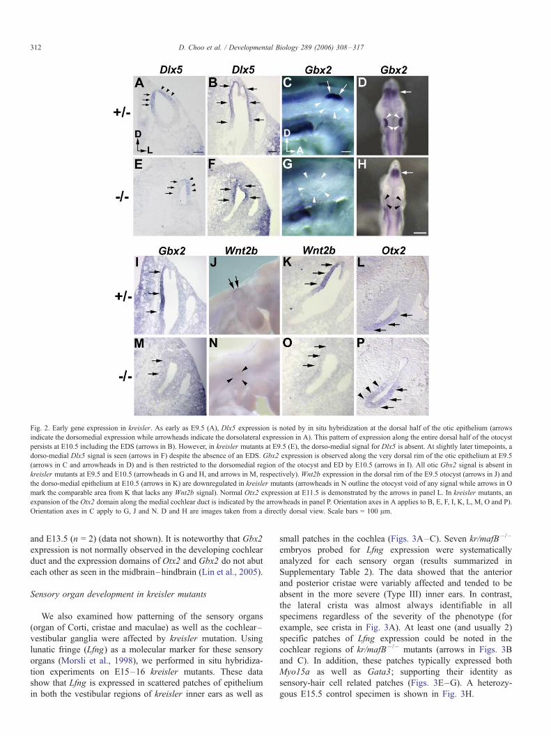
Fig. 2. Early gene expression in kreisler. As early as E9.5 (A), Dlx5 expression is noted by in situ hybridization at the dorsal half of the otic epithelium (arrows
indicate the dorsomedial expression while arrowheads indicate the dorsolateral expression in A). This pattern of expression along the entire dorsal half of the otocyst
persists at E10.5 including the EDS (arrows in B). However, in kreisler mutants at E9.5 (E), the dorso-medial signal for Dlx5 is absent. At slightly later timepoints, a
dorso-medial Dlx5 signal is seen (arrows in F) despite the absence of an EDS. Gbx2 expression is observed along the very dorsal rim of the otic epithelium at E9.5
(arrows in C and arrowheads in D) and is then restricted to the dorsomedial region of the otocyst and ED by E10.5 (arrows in I). All otic Gbx2 signal is absent in
kreisler mutants at E9.5 and E10.5 (arrowheads in G and H, and arrows in M, respectively). Wnt2b expression in the dorsal rim of the E9.5 otocyst (arrows in J) and
the dorso-medial epithelium at E10.5 (arrows in K) are downregulated in kreisler mutants (arrowheads in N outline the otocyst void of any signal while arrows in O
mark the comparable area from K that lacks any Wnt2b signal). Normal Otx2 expression at E11.5 is demonstrated by the arrows in panel L. In kreisler mutants, an
expansion of the Otx2 domain along the medial cochlear duct is indicated by the arrowheads in panel P. Orientation axes in A applies to B, E, F, I, K, L, M, O and P).
Orientation axes in C apply to G, J and N. D and H are images taken from a directly dorsal view. Scale bars = 100 Am.
D. Choo et al. / Developmental Biology 289 (2006) 308–317312
and E13.5 (n = 2) (data not shown). It is noteworthy that Gbx2
expression is not normally observed in the developing cochlear
duct and the expression domains of Otx2 and Gbx2 do not abut
each other as seen in the midbrain–hindbrain (Lin et al., 2005).
Sensory organ development in kreisler mutants
We also examined how patterning of the sensory organs
(organ of Corti, cristae and maculae) as well as the cochlear–
vestibular ganglia were affected by kreisler mutation. Using
lunatic fringe (Lfng) as a molecular marker for these sensory
organs (Morsli et al., 1998), we performed in situ hybridiza-
tion experiments on E15–16 kreisler mutants. These data
show that Lfng is expressed in scattered patches of epithelium
in both the vestibular regions of kreisler inner ears as well as
small patches in the cochlea (Figs. 3A–C). Seven kr/mafB�/�
embryos probed for Lfng expression were systematically
analyzed for each sensory organ (results summarized in
Supplementary Table 2). The data showed that the anterior
and posterior cristae were variably affected and tended to be
absent in the more severe (Type III) inner ears. In contrast,
the lateral crista was almost always identifiable in all
specimens regardless of the severity of the phenotype (for
example, see crista in Fig. 3A). At least one (and usually 2)
specific patches of Lfng expression could be noted in the
cochlear regions of kr/mafB�/� mutants (arrows in Figs. 3B
and C). In addition, these patches typically expressed both
Myo15a as well as Gata3; supporting their identity as
sensory-hair cell related patches (Figs. 3E–G). A heterozy-
gous E15.5 control specimen is shown in Fig. 3H.

Fig. 3. In situ hybridization experiments performed on E15.5 embryos show that the patterning of inner ear sensory organs is disrupted by kr/mafB mutation and
abnormal hindbrain development. Using Lfng as a marker for the developing sensory organs, in situ data show that Lfng can be observed in cristae-like structures
(crista in A) and scattered epithelial patches in the cochleas of kreisler mutants (arrows in B and C). A control specimen showing the normal Lfng expression in the
E15.5 cochlea is shown in panel D and demonstrates the signal in the supporting cells of the hair cell regions of the cochlea. Similarly, sensory organ generation
was assessed by examining Myo15a (E), GATA3 (F, G and H) and Nf68 (I and J) in E15.5 inner ears. Myo15a transcripts were noted in scattered patches of the
kreisler cochleas (arrows in E). GATA3 expression could be demonstrated in scattered hair cell-like regions of the cochlea (arrows in F) as well as the spiral
ganglion (gangl in G). Normal GATA3 signal is shown in panel H along the entire floor of the cochlear duct. Nf68 expression was clearly observed in the
developing vestibular ganglion of kreisler embryos (gangl in I) although morphologically, the ganglion appeared smaller than that seen in controls (J). Orientation
axes in panel F apply to all panels. D, dorsal; M, Medial; Lfng, lunatic fringe; Myo15a, myosin 15a; Nf68, neurofilament 68; gangl, ganglion; coch, cochlea; canal,
semicircular canal. Scale bars = 100 Am.
D. Choo et al. / Developmental Biology 289 (2006) 308–317 313
Four kreisler embryos at E15.5 were studied by in situ
hybridization using a probe for Gata3 as a means of assessing
spiral ganglion development. All 4 specimens were either
Type II or III based upon semicircular canal defects, the
absence of an EDS and the typical uncoiled cochlear duct. In 3
mutant ears, a small spiral ganglion could be demonstrated by
Gata3 in situ hybridization (Fig. 3G). In the 4th specimen
(Type III), a definitive spiral ganglion could not be identified.
Analogously, alternate sections from the above E15.5 speci-
mens were probed with Nf68 as a means of examining
vestibular ganglion development. Distinct Nf68 signal in
proximity to the vestibular structures in kreisler mutants was
observed in all specimens (gangl, Fig. 3I). Fig. 3J shows a
control specimen with the anticipated Nf68 expression in the
vestibular ganglion.
Cell proliferation and programmed cell death in kreisler inner
ears
An abnormality in regulated cell proliferation and/or
programmed cell death during inner ear morphogenesis were
considered potentially relevant to the generation of inner ear
malformations in kreisler mice. Accordingly, we immunohis-
tochemically labelled proliferating cells (using a monoclonal
mouse anti-PCNA-antibody) and TUNEL-labelled those cells
undergoing apoptosis using alternate serial sections from
embryos between E9.5 and E13.5.
At E9.5, proliferating cells are observed diffusely through-
out the otic epithelium of both control and kreisler mutant
embryos (Figs. 4A and E). No statistically significant
difference in the number of proliferating cells could be
demonstrated between kreisler mutants (n = 5) and controls
(n = 5) at this stage (a � 0.10). However, a significantly
increased number of TUNEL profiles (a � 0.05) was noted in
the kreisler otocysts (n = 6, Fig. 4B) compared to controls (n =
6, Fig. 4F) at E9.5. A particularly obvious increase in TUNEL-
labelled profiles was observed in the lateral otocyst (bracketed
arrows in Fig. 4B) as well as the geniculate ganglion region of
kreisler mutants (arrowheads in Fig. 4B) compared to control
specimens.
Between E10.5 and E13.5, no statistically significant
changes in the number of PCNA-labelled or TUNEL-labelled
cells could be demonstrated in the otic epithelium (n = 6 at
E10.5; n = 4 at E11.5; n = 3 at E12.5; n = 3 at E13.5). Despite
the size and shape of kreisler inner ears being different and
smaller than control ears (see Fig. 1), the patterns of both
PCNA-labelled and TUNEL-labelled cells in the inner ears
were similar between kreisler and control specimens (E10.5
PCNA and TUNEL data shown in Figs. 4C, D, G and H).
Specific attention was directed at areas such as the fusion plate

Fig. 4. Immunolabeling of proliferating cells using an anti-PCNA-antiserum shows diffuse labeling of otic epithelial cells at E9.5 that is comparable in kreisler
mutants and controls (A and E, respectively). TUNEL labeling of apoptotic cells at E9.5 show an obvious and significant increase in apoptotic cells in kreisler
otocysts (B) compared to controls (F). Bracketed arrow in B marks the lateral otocyst wall where an increased number of TUNEL profiles can be noted. Arrowheads
in B mark the geniculate ganglion area also demonstrating an increase in apoptotic cells. At E10.5, no statistically significant changes in either proliferating cells (C
and G) or apoptotic cells can be demonstrated (D and H). PCNA-immunostaining shows labelled proliferating cells throughout the otic epithelium of kreisler (C) and
control (G) inner ears. TUNEL experiments show scattered patches of labelled cells at the ventromedial region of the otocyst as well as the more dorsal regions of the
otocyst (short arrows in D). In the area of the geniculate ganglion, robust TUNEL labeling is again noted (long arrow in D). In E10.5 controls, a similar pattern of
TUNEL staining can be observed in the ventromedial and dorsal regions of the otocyst (arrows in H). Orientation axis in A applies to all panels. D, Dorsal; L,
Lateral; PCNA, proliferating cell nuclear antigen; TUNEL, terminal dUTP nick end-labeling. Scale bar in E applies to all panels. Scale bar = 100 Am.
D. Choo et al. / Developmental Biology 289 (2006) 308–317314
region in the canal pouches at E11.5–12.5, which have
previously been demonstrated as ‘‘hotspots’’ of PCD in
normally developing inner ears (Fekete et al., 1997; Nishikori
et al., 1999). No changes in TUNEL labeling could be
observed in those areas between 11.5 and E13.5 (data not
shown).
Discussion
Morphogenesis of the kreisler inner ear
This report provides new morphologic data on the devel-
opmental inner ear phenotype in kreisler generated by paint
filling of the membranous labyrinth. We drew several conclu-
sions regarding the effects of kr/mafB mutation and the loss of
rhombomeres 5 and 6 on inner ear morphogenesis. First, the
hindbrain perturbations in kreisler mutants globally affect inner
ear development. The cochlea, vestibular structures, ganglia
and EDS are all perturbed to some extent. Our observation that
the EDS and common crus are almost always affected, while
the lateral canal is least often affected, suggest that a more
dorsal and medial (vs. lateral) defect is incurred by loss of
normal kr/mafB function. The progressive dilation of the
membranous labyrinth in kreisler mutants led us to consider
that the early failure to form an EDS may contribute to this
distended phenotype. Taking into consideration the proposed
endolymph-regulating role of the EDS in the inner ear, it seems
reasonable to speculate that absence of the EDS contributes to a
failure of kreisler ears to develop or maintain a proper
endolymph composition and volume. Correlative evidence of
the EDS being relevant to inner ear morphogenesis can be
drawn from Mansour’s report on Fgf3 null mutants (Mansour
et al., 1993) and Foxi1 mutants (Hulander et al., 2003). In the
Fgf3 mutant mice, a similar developmental failure to form an
EDS results in a comparable (although notably less severe)
distended inner ear phenotype. In the case of Foxi1 mice, a
failure to form a properly functioning EDS results in a much
milder, but still dilated overall inner ear phenotype. Accord-
ingly, one possibility is that kr/mafB mutation results in a
failure of the EDS to develop which then disrupts endolymph
homeostasis and this abnormal endolymph volume/composi-
tion then disrupts normal morphogenesis of the entire inner ear.
Given the dramatically abnormal molecular patterning in
kreisler inner ears (elaborated on below), we find it difficult
to attribute all of these changes to the absence of an EDS.
Semicircular canal defects in kreisler
Paint-fill data from kreisler mutants suggested that canal
defects could potentially be due to altered cell proliferation
during canal pouch formation and/or dysregulated apoptosis.
The majority of kreisler E11.5 embryos (Fig. 2D) demonstrated
smaller canal pouch compared to controls. This observation
raised the possibility that an adequate canal pouch never
developed in these kreisler mutants and subsequent defects of
the canals are due to this failure to generate a requisite mass of
canal epithelium. Based upon tritiated-thymidine experiments,
Ruben reported that kreisler otocysts demonstrated a shortened
cell cycle and, somewhat contradictorily, implicated a higher
mitotic rate (Ruben, 1973). However, our PCNA experiments
looking at cell proliferation in kreisler ears between E9.5 and
E13.5 have not shown any obvious differences in cell
proliferation compared to controls.
Similarly, TUNEL labelling of cells undergoing prog-
rammed cell death did not show any marked changes except
at E9.5 when kreisler mutant otocysts showed a statistically
significant increase in TUNEL profiles. The number of
apoptotic cells also seemed to be particularly increased along

Fig. 5. Genes likely involved in the kr/mafB –hindbrain pathway and early
inner ear patterning. In the posterior hindbrain, a complex interaction of Fgfs
and Hox genes likely regulates kr/mafB activity. Binding sites for MyoD and
kr/mafB have been identified in the promoter of kr/mafB suggesting a role in kr/
mafB (auto)regulation. Data from Lin et al. also suggest that Gbx2 in the
hindbrain negatively regulates kr/mafB expression. Our data focusing on the
early otocyst show that normal cues from the kr/mafB –hindbrain pathway are
required for Gbx2 expression in the ear. Dorsomedial Dlx5 and Wnt2b
expression at E9.5 is also dependent upon normal hindbrain cues while ventral
expression of Otx2 in the otocyst is negatively regulated by the kr/mafB –
hindbrain pathway. Arrows from the hindbrain to the otocyst do not imply
direct interactions or relationships.
D. Choo et al. / Developmental Biology 289 (2006) 308–317 315
the lateral otocyst epithelium (Fig. 4B). Fate-mapping the rim
of the otic cup in chicken has shown that a large region of the
lateral otic epithelium gives rise to the vertical and lateral canal
pouches (Brigande et al., 2000). Assuming mice have a similar
fate map as that of chicken, the increased cell death in the
lateral wall of kreisler otocysts at E9.5 could have contributed
to the smaller size of the vertical canal pouch observed at E11.
Whether the initial increase in cell death and the size reduction
of the canal pouch contributed to the expanded and seemingly
disorganized areas of resorption in the canal plates at later
stages is not clear.
A loss of endolymphatic duct markers in kreisler mutants
We initially selected candidate otic targets of the kr/mafB –
hindbrain pathway based upon their expression pattern in the
dorsal and/or medial otocyst (which gives rise to the EDS) or
based upon a similar EDS phenotype in null mutant mice. As
an example, Dlx5 and Gbx2 were studied given the
comparable absence of an EDS in some Dlx5 mutant mice
(Depew et al., 1999) and a consistent lack of an EDS in Gbx2
mice (Lin et al., 2005). Our in situ data demonstrate that Dlx5
expression at E9.5 along the dorsomedial otocyst is dependent
upon normal cues from the hindbrain, and in turn, normal kr/
mafB activity (see Fig. 2E). Interestingly, Dlx5 expression at
E9.5 and later in the developing canal pouches is not affected
in kreisler mutants. Since kreisler mutants display both EDS
and semicircular canal defects, these data suggest that the
specific Dlx5 expression in the dorsomedial otocyst is
required for EDS differentiation while Dlx5 expression alone
is not sufficient for normal canal differentiation. It seems
reasonable to speculate that combined signaling by multiple
Dlx paralogs might be necessary for proper semicircular canal
differentiation as seen in other animal models and organ
systems (Solomon and Fritz, 2002).
Gbx2 expression was consistently absent in the dorsomedial
region of the otocyst in kreisler mutants (see Figs. 2G and H).
Later Gbx2 expression in the ear was also absent (Fig. 2M).
These data suggest that otic expression of Gbx2 is important
for proper EDS development and is impacted by hindbrain kr/
mafB expression and a normally developed posterior hindbrain.
Since Gbx2 is also expressed in the developing midbrain/
hindbrain during this developmental stage (Figs. 2D and H) (Li
et al., 2002), it is necessary to acknowledge that hindbrain kr/
mafB –Gbx2 interactions could also be relevant. However, in
kreisler mutants, Gbx2 expression in the mid–hindbrain is
unchanged (arrows in Figs. 2D and H) whereas its expression
in the inner ear is absent (compare arrowheads in Figs. 2D and
H). Accordingly, we interpret these data as indicating that the
otic expression of Gbx2 (rather than the mid–hindbrain Gbx2
expression) is important for EDS development. Pertinent to this
discussion, Lin et al.’s report on the inner ear phenotypes of
Gbx2 mutants discusses the phenotypic similarities between
Gbx2 and kreisler mutants (Lin et al., 2005). In examining the
molecular mechanisms underlying the EDS phenotype ob-
served in Gbx2 mutants, Lin et al. similarly concluded that the
loss of otic Gbx2 expression was particularly relevant to EDS
development and proposed that Gbx2 represented a key
downstream target of hindbrain signaling. Considering our
kreisler data along with Lin’s Gbx2 data, we propose that the
data are consistent in identifying otic Gbx2 as a key target of
hindbrain signaling and that otic Gbx2 is particularly relevant
for EDS development.
Our in situ data also show that Wnt2b expression is absent
in kreisler inner ears between E9.5 and E13. The role of Wnt2b
in inner ear development has not been reported. Our earliest
signal for Wnt2b in heterozygous embryos was detected at
E9.5. Temporally, this places Wnt2b as potentially downstream
of Gbx2 and Dlx5. Assimilating these data together, we
conclude that loss of normal kr/mafB activity and disrupted
hindbrain patterning initiates a cascade of molecular events in
the otocyst that are mediated by Gbx2 and Dlx5, and
subsequently, Wnt2b.
Cochlear patterning in kreisler mutants
The expression domain of Otx2 in the otocyst is expanded
in kreisler mutants, similar to the observations in Gbx2
knockout mice (Lin et al., 2005). Taken together with earlier
reports on the complimentary expression boundaries of Otx2
and Gbx2 in the developing chick inner ear (Hidalgo-Sanchez
et al., 2000; Sanchez-Calderon et al., 2002) and the antago-
nistic relationship between Gbx2 and Otx2 in the midbrain–
hindbrain region, we propose that loss of kr/mafB expression
and hindbrain signaling results in the downregulation of Gbx2
expression in the inner ear which then leads to an expansion of
the Otx2 domain in the mutant cochleas. We further propose
that such an altered Otx2 domain might then perturb the normal
patterning of the cochlea and result in altered cell populations
in the kreisler cochleas. The unusual and scattered hair cell
patches in kreisler ears (as shown by Lfng, Myo15a and
GATA3 in situ hybridization data, Figs. 3A–I) are all consistent
with such a process.

D. Choo et al. / Developmental Biology 289 (2006) 308–317316
kr/mafB and hindbrain cues for patterning the inner ear
The putative pathways involved in early otocyst differenti-
ation are schematically presented in Fig. 5. Fully acknowledg-
ing gaps in our understanding of the hindbrain-to-ear signaling
pathways, this model provided a template into which certain
components in the distal pathway could be inserted.
At the level of the hindbrain, expression of kr/mafB is likely
autoregulated by its own product as well as MyoD. Both kr/
mafB and MyoD have been shown to activate Maf recognition
elements (MAREs) in the kr/mafB promoter region (Huang,
1999). Various Hox genes have also been demonstrated to
affect hindbrain segmentation and kr/mafB expression in
rhombomere 5 (Barrow et al., 2000) while Fgfs have been
shown in chick embryos to regulate kr/mafB expression (Marin
and Charnay, 2000). Hox proteins also interact with kr/mafB to
inhibit its DNA binding, transactivation and transforming
activities (Kataoka et al., 2001). And as reported by Lin et
al., Gbx2 likely plays a negative inhibitory role in hindbrain kr/
mafB expression (Lin et al., 2005). As a result, a myriad of
molecular interactions involving kr/mafB are ongoing in the
posterior hindbrain in a manner that is temporally and spatially
relevant to early inner ear patterning.
At the otocyst level, our data as well as the data from Lin et
al. suggest that Gbx2 is one of the key otic targets of hindbrain
signaling. Furthermore, in the dorsal and medial regions of the
otocyst, disruption of normal hindbrain development and cues
by kr/mafB mutation perturbs Dlx5 and Wnt2b which appears
to be associated with failure of the EDS to form. In the more
ventral portion of the early otocyst, loss of normal hindbrain
signals in kreisler mutants results in an expanded Otx2
expression domain. Given the negative regulatory effect of
Gbx2 on Otx2 in the CNS, it seems plausible to consider
similar mechanisms might be utilized in the ear and that loss of
otic Gbx2 activity in kreisler mutants would then allow for the
expansion of the Otx2 expression domain in kreisler cochleas.
With regards to the actual signals that convey the cues from
the hindbrain to the inner ear (the schematic red arrows in Fig.
5), several molecules (Wnts and Fgfs, for example) have been
implicated as either inducing formation of the inner ear or
providing cues for its differentiation (Anniko and Schacht,
1984; Ladher et al., 2000; Represa et al., 1991; Wright and
Mansour, 2003). However, the exact signals from the hindbrain
and the targets of these hindbrain signals in the inner ear have
remained elusive.
It is also critical to acknowledge the additional signaling
cues from ectoderm and mesoderm, as proposed by Ladher and
Wright, (Ladher et al., 2000; Wright et al., 2004) that together
with neural signals comprise the overall developmental
program that guides inner ear morphogenesis. The loss of
normal hindbrain cues in kreisler mutants may perturb not only
the neural cues for inner ear development, but also the balance
of signals from surrounding tissues that guide proper ear
development. This area requires further investigation.
Gaps clearly exist in identifying additional components of
the kr/mafB –hindbrain pathway that ultimately guides ear
development. However, by focusing on the early otocyst, we
have begun identifying differentially expressed genes in this
pathway that may help us develop a more refined hypothesis of
the ‘‘upstream’’ molecular pathways. Continuing experiments
include array-based comparisons of early (E9–E10.5) kreisler
and control transcripts in an attempt to identify other
differentially expressed otic genes that are regulated by the
hindbrain–kreisler pathway.
Acknowledgments
We are indebted to S. Cordes for providing us with the
kreisler mice used in this study. We wish to thank P. Gruss, L.
Everett, J. Rubenstein, S. Vainio, C. Ragsdale and K.
Campbell for sharing probes with us. We also gratefully
acknowledge very helpful discussions with D. Wu and J.
Whitsett and assistance with statistical analysis provided by
Dr. Meinzen-derr.
Appendix A. Supplementary data
Supplementary data associated with this article can be found
in the online version at doi:10.1016/j.ydbio.2005.10.007.
References
1991. Joint Committee on Infant Hearing 1990 position statement. ASHA, 3–6R
(Suppl.).
2000. Year 2000 position statement: principles and guidelines for early hearing
detection and intervention programs. Joint Committee on Infant Hearing,
American Academy of Audiology, American Academy of Pediatrics,
American Speech–Language–Hearing Association, and Directors of
Speech and Hearing Programs in State Health and Welfare Agencies.
Pediatrics 106, 798–817.
Anniko, M., Schacht, J., 1984. Inductive tissue interactions during inner ear
development. Arch. Oto-Rhino-Laryngol. 240, 17–33.
Barrow, J.R., Stadler, H.S., Capecchi, M.R., 2000. Roles of Hoxa1 and Hoxa2
in patterning the early hindbrain of the mouse. Development 127, 933–944.
Brigande, J.V., Iten, L.E., Fekete, D.M., 2000. A fate map of chick otic cup
closure reveals lineage boundaries in the dorsal otocyst. Dev. Biol. 227,
256–270.
Brookhouser, P., 1993. Sensorineural hearing loss in children. In:
Cummings, C. (Ed.), Otolaryngol. Head Neck Surg., vol. 4. Mosby-Year
Book, St. Louis, pp. 3080–3102.
Choo, D., Sanne, J., Wu, D., 1998. The differential sensitivities of inner ear
structures to retinoic acid during development. Dev. Biol. 204, 136–150.
Cordes, S., Barsch, G., 1994. The mouse segmentation gene Kr encodes a novel
basic domain-leucine zipper transcription factor. Cell 79, 1025–1034.
Deol, M., 1964. The abnormalities of the inner ear in kreisler mice. J. Embryol.
Exp. Morphol. 12, 475–490.
Depew, M., Liu, J., Long, J., Presley, R., Meneses, J., Pedersen, R., Rubenstein,
J., 1999. Dlx5 regulates regional development of the branchial arches and
sensory capsules. Development 126, 3831–3846.
Everett, L.A., Morsli, H., Wu, D.K., Green, E.D., 1999. Expression pattern
of the mouse ortholog of the Pendred’s syndrome gene (Pds) suggests a
key role for pendrin in the inner ear. Proc. Natl. Acad. Sci. U. S. A. 96,
9727–9732.
Fekete, D.M., Homburger, S.A., Waring, M.T., Riedl, A.E., Garcia, L.F., 1997.
Involvement of programmed cell death in morphogenesis of the vertebrate
inner ear. Development 124, 2451–2461.
Frohman, M.A., Martin, G.R., Cordes, S.P., Halamek, L.P., Barsh, G.S., 1993.
Altered rhombomere-specific gene expression and hyoid bone differentia-
tion in the mouse segmentation mutant, kreisler (kr). Development 117,
925–936.

D. Choo et al. / Developmental Biology 289 (2006) 308–317 317
Hertwig, P., 1944. Die Genese der Hirn-und GehorgangsmiBbildungen bei
Rontgen-mutierten Kreisler-Mausen. Z. Menschl. Vererb. Konstitutionsl.
28, 327.
Hidalgo-Sanchez, M., Alvarado-Mallart, R., Alvarez, I.S., 2000. Pax2, Otx2,
Gbx2 and Fgf8 expression in early otic vesicle development. Mech. Dev.
95, 225–229.
Huang, K., 1999. Cloning and characterization of promoter of the mouse mafB
gene. Hokkaido Igaku Zasshi 74, 419–430.
Hulander, M., Kiernan, A.E., Blomqvist, S.R., Carlsson, P., Samuelsson, E.J.,
Johansson, B.R., Steel, K.P., Enerback, S., 2003. Lack of pendrin
expression leads to deafness and expansion of the endolymphatic
compartment in inner ears of Foxi1 null mutant mice. Development 130,
2013–2025.
Karis, A., Pata, I., van Doorninck, J., Grosveld, F., de Zeeuw, C., de Caprona,
D., Fritzsch, B., 2001. Transcription factor GATA-3 alters pathway
selection of olivocochlear neurons and affects morphogenesis of the ear.
J. Comp. Neurol. 429, 615–630.
Kataoka, K., Yoshitomo-Nakagawa, K., Shioda, S., Nishizawa, M., 2001. A set
of Hox proteins interact with the Maf oncoprotein to inhibit its DNA
binding, transactivation, and transforming activities. J. Biol. Chem. 276,
819–826.
Ladher, R.K., Anakwe, K.U., Gurney, A.L., Schoenwolf, G.C., Francis-West,
P.H., 2000. Identification of synergistic signals initiating inner ear
development. Science 290, 1965–1967.
Li, J.Y., Joyner, A.L., 2001. Otx2 and Gbx2 are required for refinement and
not induction of mid–hindbrain gene expression. Development 128,
4979–4991.
Li, J.Y., Lao, Z., Joyner, A.L., 2002. Changing requirements for Gbx2 in
development of the cerebellum and maintenance of the mid/hindbrain
organizer. Neuron 36, 31–43.
Lin, Z., Cantos, R., Patente, M., Wu, D.K., 2005. Gbx2 is required for the
morphogenesis of the mouse inner ear: a downstream candidate of
hindbrain signaling. Development 132, 2309–2318.
Liu, A., Joyner, A.L., 2001. EN and GBX2 play essential roles downstream of
FGF8 in patterning the mouse mid/hindbrain region. Development 128,
181–191.
Mansour, S., Goddard, J., Capecchi, M., 1993. Mice homozygous for a targeted
disruption of the proto-oncogene int-2 have developmental defects in the
tail and inner ear. Development 117, 13–28.
Marin, F., Charnay, P., 2000. Hindbrain patterning: FGFs regulate Krox20
and mafB/kr expression in the otic/preotic region. Development 127,
4925–4935.
Martin, P., Swanson, G., 1993. Descriptive and experimental analysis of the
epithelial remodelling that controls semicircular canal formation in the
developing mouse inner ear. Dev. Biol. 159, 549–558.
McKay, I., Muchamore, I., Krumlauf, R., Maden, M., Lumsden, A., Lewis, J.,
1994. The Kreisler mouse: a hindbrain segmentation mutant that lacks two
rhombomeres. Development 120, 2199–2211.
Mehl, A.L., Thomson, V., 1998. Newborn hearing screening: the great
omission. Pediatrics 101, E4.
Millet, S., Campbell, K., Epstein, D.J., Losos, K., Harris, E., Joyner, A.L.,
1999. A role for Gbx2 in repression of Otx2 and positioning the
mid/hindbrain organizer. Nature 401, 161–164.
Morsli, H., Choo, D., Ryan, A., Johnson, R., Wu, D., 1998. Development of the
mouse inner ear and its sensory organs. J. Neurosci. 18, 3327–3335.
Morsli, H., Tuorto, F., Choo, D., Postiglione, M., Simeone, A., Wu, D., 1999.
Otx1 and Otx 2 activities are required for the normal development of the
mouse inner ear. Development 126, 2335–2343.
Ng, J.K., Kawakami, Y., Buscher, D., Raya, A., Itoh, T., Koth, C.M.,
Rodriguez Esteban, C., Rodriguez-Leon, J., Garrity, D.M., Fishman,
M.C., Izpisua Belmonte, J.C., 2002. The limb identity gene Tbx5
promotes limb initiation by interacting with Wnt2b and Fgf10. Develop-
ment 129, 5161–5170.
Nishikori, T., Hatta, T., Kawauchi, H., Otani, H., 1999. Apoptosis during
inner ear development in human and mouse embryos: an analysis by
computer-assisted three-dimensional reconstruction. Anat. Embryol. (Berl)
200, 19–26.
Represa, J., Leon, Y., Miner, C., Giraldez, F., 1991. The int-2 proto-oncogene is
responsible for induction of the inner ear. Nature 353, 561–563.
Ruben, R., 1973. Development and cell kinetics of the kreisler (kr/kr) mouse.
Laryngoscope 83, 1440–1468.
Sanchez-Calderon, H., Martin-Partido, G., Hidalgo-Sanchez, M., 2002.
Differential expression of Otx2, Gbx2, Pax2, and Fgf8 in the developing
vestibular and auditory sensory organs. Brain Res. Bull. 57, 321–323.
Solomon, K.S., Fritz, A., 2002. Concerted action of two dlx paralogs in sensory
placode formation. Development 129, 3127–3136.
Takahashi, M., Hokunan, K., Unno, T., 1994. S-phase cells in the guinea pig
endolymphatic sac. Acta Oto-Laryngol. 114, 259–263.
Torres, M., Gomez-Pardo, E., Gruss, P., 1996. Pax2 contributes to inner ear
patterning and optic nerve trajectory. Development 122, 3381–3391.
Tsue, T.T., Watling, D.L., Weisleder, P., Coltrera, M.D., Rubel, E.W., 1994.
Identification of hair cell progenitors and intermitotic migration of their
nuclei in the normal and regenerating avian inner ear. J. Neurosci. 14,
140–152.
Umemoto, M., Sakagami, M., Fukazawa, K., Ashida, K., Kubo, T., Senda, T.,
Yoneda, Y., 1995. Hair cell regeneration in the chick inner ear following
acoustic trauma: ultrastructural and immunohistochemical studies. Cell
Tissue Res. 281, 435–443.
Wright, T.J., Mansour, S.L., 2003. Fgf3 and Fgf10 are required for mouse otic
placode induction. Development 130, 3379–3390.
Wright, T.J., Ladher, R., McWhirter, J., Murre, C., Schoenwolf, G.C., Mansour,
S.L., 2004. Mouse FGF15 is the ortholog of human and chick FGF19, but is
not uniquely required for otic induction. Dev. Biol. 269, 264–275.
Zhao, S., Overbeek, P.A., 1999. Tyrosinase-related protein 2 promoter targets
transgene expression to ocular and neural crest-derived tissues. Dev. Biol.
216, 154–163.


![[Kreisler] Cadenzas to Beethoven Violin Concerto](https://img.pdfslide.us/doc/110x75/577cc5361a28aba7119bb045/kreisler-cadenzas-to-beethoven-violin-concerto.jpg)