Embed Size (px)
Citation preview
Molecular Mechanisms of Preeclampsia
Tammy Hod1, Ana Sofia Cerdeira1,2, and S. Ananth Karumanchi1,3
1Department of Medicine, Obstetrics & Gynecology, Beth Israel Deaconess Medical Center and HarvardMedical School, Boston, Massachusetts 02214
2Gulbenkian Program for Advanced Medical Education, 1067-001 Lisbon, Portugal3Howard Hughes Medical Institute, Chevy Chase, Maryland 20815
Correspondence: [email protected]
Preeclampsia is a pregnancy-specific disease characterized by new onset hypertension andproteinuria after 20 wk of gestation. It is a leading cause of maternal and fetal morbidity andmortality worldwide. Exciting discoveries in the last decade have contributed to a betterunderstanding of the molecular basis of this disease. Epidemiological, experimental, andtherapeutic studies from several laboratories have provided compelling evidence that anantiangiogenic state owing to alterations in circulating angiogenic factors leads to pre-eclampsia. In this review, we highlight the role of key circulating antiangiogenic factors aspathogenic biomarkers and in the development of novel therapies for preeclampsia.
Preeclampsia is a leading cause of maternalmorbidity and mortality worldwide, com-
plicating 2%–8% of pregnancies (Duley 2009).It is a multisystemic disorder with both mater-nal and fetal manifestations. In the mother,alongside with hypertension and proteinuria,the disease can progress to widespread micro-angiopathy that mainly affects the kidney, liver,and brain. Thrombocytopenia, liver dysfunc-tion, microangiopathic hemolytic anemia, acuterenal failure, placental abruption, visual distur-bances, stroke, seizures, and maternal death areserious consequences of preeclampsia. In thefetus, preeclampsia is associated with intrauter-ine growth restriction and prematurity. Besidesadverse pregnancy outcomes, women with pre-eclampsia have an increased risk of future healthcomplications (Irgens et al. 2001; Bellamy et al.
2007; Vikse et al. 2008; Wang et al. 2013b; Chenet al. 2014).
Recent data suggest that alterations in cir-culating angiogenic factors play a pathogenicrole in preeclampsia. Angiogenesis, the processof new blood vessel formation from preexistingones, is tightly regulated by angiogenic factors.Importantly, angiogenic factors are also essen-tial for maintenance of normal vessel health;they provide important cues for organ develop-ment. Increased levels of the antiangiogenic fac-tors, soluble fms-like tyrosine kinase 1 (sFlt-1)and soluble Endoglin (sEng) trap circulatingvascular endothelial growth factor (VEGF), pla-cental growth factor (PlGF) and transforminggrowth factor b (TGFb) respectively, decreas-ing their free levels, leading to endothelial dys-function and the clinical manifestations of the
Editors: Diana W. Bianchi and Errol R. Norwitz
Additional Perspectives on Molecular Approaches to Reproductive and Newborn Medicine available at
www.perspectivesinmedicine.org
Copyright # 2015 Cold Spring Harbor Laboratory Press; all rights reserved; doi: 10.1101/cshperspect.a023473
Cite this article as Cold Spring Harb Perspect Med 2015;5:a023473
1
ww
w.p
ersp
ecti
vesi
nm
edic
ine.
org
on July 22, 2018 - Published by Cold Spring Harbor Laboratory Press http://perspectivesinmedicine.cshlp.org/Downloaded from
disease (Maynard et al. 2003; Levine et al. 2004,2006b; Venkatesha et al. 2006; Romero andChaiworapongsa 2013). Interestingly, the pre-eclampsia-like signs and symptoms are alsoseen in cancer patients receiving antiangiogenicchemotherapy (Hurwitz et al. 2004; Patel et al.2008; Vigneau et al. 2014).
The search for the ability to better diagnose,predict, and prevent preeclampsia as well as themechanisms of its pathogenesis to develop atherapy that safely prolongs gestation has beenextensive. Exciting data on angiogenic factors ascentral contributors to the pathogenesis of pre-eclampsia, candidate biomarkers, and thera-peutic targets open clinically meaningful per-spectives for the near future.
PATHOGENESIS
Angiogenic Factors as Mediators of theMaternal Syndrome
Taylor and Roberts posited that a dysfunctionalplacenta releases “toxic” factors into the mater-nal circulation that trigger the clinical syndromeof preeclampsia (Roberts et al. 1989). The pla-centa is both necessary and sufficient to causethe disease, and delivery of the placenta is theonly treatment (Powe et al. 2011). Over the lastdecade, preeclampsia has been recognized as anantiangiogenic state (Romero and Chaiwora-pongsa 2013). Excessive placental productionof antiangiogenic factors sFlt-1 and sEng areliberated into the maternal circulation inducingthe clinical syndrome. Preeclamptic placentasshow overexpression of sFlt-1 and sEng. Theirlevels are increased in the serum of preeclampticwomen weeks before the appearance of overtclinical manifestations of the disease and theycorrelate with disease severity (Levine et al.2006b)
Importantly, sFlt-1 overexpression in preg-nant rodents is sufficient to induce the hall-marks of the disease: hypertension, proteinuria,and the characteristic kidney lesion, glomerularendotheliosis (Maynard et al. 2003). In addition,we showed that alterations in circulating sFlt-1may explain a number of risk factors for pre-eclampsia such as multiple gestation, trisomy 13,
nulliparity, and molar pregnancies (Powe et al.2011). Addition of sEng to this model induces amore severe phenotype including thrombo-cytopenia, cerebral edema, and fetal growth re-striction (Venkatesha et al. 2006; Maharaj et al.2008).
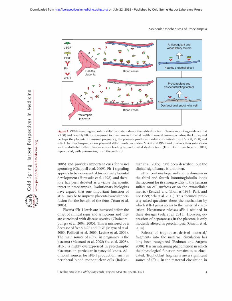
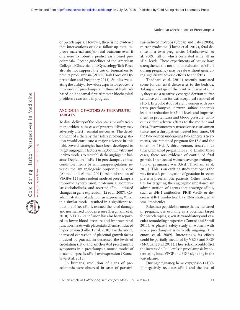
Fms-like tyrosine kinase 1 (Flt-1), and solu-ble Flt-1 (sFlt-1) are the products of FLT-1 gen-erated by differential mRNA processing. Flt-1,also known as vascular endothelial growth fac-tor receptor 1 (VEGFR-1), is a membrane span-ning receptor for VEGF and PlGF. It comprisesan extracellular domain with seven immuno-globulin-like domains (ligand-binding), amembrane domain, and a cytoplasmic tyrosinekinase domain. Soluble Flt-1 is a shorter iso-form that retains the ligand-binding domainbut lacks the cytoplasmic and transmembranedomains and is therefore secreted into the cir-culation (Kendall and Thomas 1993; Huckleand Roche 2004; Thomas et al. 2007). sFlt-1can also be generated by cleavage of the mem-brane receptor by proteases, although the phys-iologic significance of this process is not known(Rahimi et al. 2009; Zhao et al. 2010). SolubleFlt-1 binds VEGF and PlGF in circulation andlocally in tissues acting as a scavenger that pre-vents them from interacting with their mem-brane receptors on the endothelium (Fig. 1).Therefore, an increase in the concentration ofsFlt-1 decreases free, and thus bioactive, PlGFand VEGF. Another splicing variant of VEGFR-1, sFlt1-14, generated primarily in nonendothe-lial cells, functions similarly to sFlt-1 as a potentVEGF inhibitor. Interestingly, conversion ofVEGFR-1 mRNA to sFlt1-14 prevents VEGFR-1-mediated signal transmission, in contrast tosFlt-1, whose production in endothelial cellsis accompanied by a large excess of the trans-membrane receptor. Expression of sFlt1-14 hasbeen shown to be significantly increased in pre-eclampsia. Specifically, syncytial knots wereidentified as the major source of local and circu-lating sFlt1-14 (Sela et al. 2008). The exact role ofthe different splicing variants of VEGFR-1 inantiangiogenic state remains to be elucidated.
The physiologic function of sFlt-1 is notclearly understood. sFlt-1 is crucial for mainte-nance of cornea avascularity (Ambati et al.
T. Hod et al.
2 Cite this article as Cold Spring Harb Perspect Med 2015;5:a023473
ww
w.p
ersp
ecti
vesi
nm
edic
ine.
org
on July 22, 2018 - Published by Cold Spring Harbor Laboratory Press http://perspectivesinmedicine.cshlp.org/Downloaded from
2006) and provides important cues for vesselsprouting (Chappell et al. 2009). Flt-1 signalingappears to be nonessential for normal placentaldevelopment (Hiratsuka et al. 1998), and there-fore has been debated as a viable therapeutictarget in preeclampsia. Evolutionary biologistshave argued that one important function ofsFlt-1 may be to improve placental vascular per-fusion for the benefit of the fetus (Yuan et al.2005).
Plasma sFlt-1 levels are increased before theonset of clinical signs and symptoms and theyare correlated with disease severity (Chaiwora-pongsa et al. 2004, 2005). This is mirrored by adecrease of free VEGF and PlGF (Maynard et al.2003; Polliotti et al. 2003; Levine et al. 2004).The main source of sFlt-1 in pregnancy is theplacenta (Maynard et al. 2003; Gu et al. 2008).sFlt-1 is highly overexpressed in preeclampticplacentas, in particular in syncytial knots. Ad-ditional sources for sFlt-1 production, such asperipheral blood mononuclear cells (Rajaku-
mar et al. 2005), have been described, but theclinical significance is unknown.
sFlt-1 contains heparin-binding domains inthe third and fourth immunoglobulin loopsthat account for its strong avidity to the heparansulfate on cell surfaces or on the extracellularmatrix (Kendall and Thomas 1993; Park andLee 1999; Sela et al. 2011). This chemical prop-erty raised questions about the mechanism bywhich sFlt-1 gains access to the maternal circu-lation. Heparanase releases sFlt-1 retained inthese storages (Sela et al. 2011). However, ex-pression of heparanases in the placenta is onlymodestly altered in preeclampsia (Ginath et al.2014).
Release of trophoblast-derived material/fragments into the maternal circulation haslong been recognized (Redman and Sargent2000). It is an intriguing phenomenon in whichthe physiological function remains to be eluci-dated. Trophoblast fragments are a significantsource of sFlt-1 in the maternal circulation in
Preclampsiaplacenta
Blood vessel
Dysfunctional endothelial cell
Procoagulant andvasoconstricting factors
Healthy endothelial cellBlood vesselHealthy
placentasFlt-1
Flt-1
PIGF
VEGFAnticoagulant and
vasodilatory factors
Figure 1. VEGF signaling and role of sFlt-1 in maternal endothelial dysfunction. There is mounting evidence thatVEGF, and possibly PlGF, are required to maintain endothelial health in several tissues including the kidney andperhaps the placenta. In normal pregnancy, the placenta produces modest concentrations of VEGF, PlGF, andsFlt-1. In preeclampsia, excess placental sFlt-1 binds circulating VEGF and PlGF and prevents their interactionwith endothelial cell-surface receptors leading to endothelial dysfunction. (From Karumanchi et al. 2005;reproduced, with permission, from the author.)
Molecular Mechanisms of Preeclampsia
Cite this article as Cold Spring Harb Perspect Med 2015;5:a023473 3
ww
w.p
ersp
ecti
vesi
nm
edic
ine.
org
on July 22, 2018 - Published by Cold Spring Harbor Laboratory Press http://perspectivesinmedicine.cshlp.org/Downloaded from
preeclampsia. The first report of trophoblastdeportation comes from Schmorl who showedtrophoblast material in lungs of patients thatdied from eclampsia (Lapaire et al. 2007). It isnow recognized that the detachment of syncy-tial knots from the placenta results in free, mul-tinucleated trophoblast microparticles that aremetabolically active and capable of de novogene translation, including the ability to synthe-size sFlt-1 protein from endogenous stores ofmRNA (Rajakumar et al. 2012). This phenom-enon is enhanced in preeclampsia, suggestingthat an accelerated shedding of syncytial aggre-gates is a proximal event in the pathogenesis ofpreeclampsia (Buurma et al. 2013).
VEGF signaling is not only important forangiogenesis but also for maintaining endothe-lial health in certain specialized vascular bedssuch as liver, kidney, brain as well as endocrineorgans such as thyroid tissue, where it is con-stitutively expressed to maintain endothelialfenestrations (Kamba et al. 2006). There is com-pelling evidence that the growth of some tumorsis dependent on VEGF-induced angiogenesis(Sitohy et al. 2011). VEGF inhibitor therapycaused early striking vascular changes in isletcell tumors. Within 24 hours, endothelial fenes-trations and sprouts disappeared, patency waslost, and flow ceased in some vessels. Tumorsvessels lacking fenestrations were less responsiveto the inhibitors, suggesting that vessel pheno-type may be predictive of responsiveness toVEGF inhibitors (Inai et al. 2004). Cessationof anti-VEGF therapy led to complete revascu-larization within the first week after the treat-ment stopped which fully regressed during a sec-ond treatment (Mancuso et al. 2006). VEGF,which is strongly expressed by podocytes, al-though its binding sites are localized on glomer-ular endothelial cells, plays a paramount role inmaintaining glomerular integrity under normalphysiologic conditions, such that when intra-glomerular VEGF levels decrease capillary en-dothelial cells swell, capillary loops collapse,glomerular filtration is impaired, and protein-uria develops (Mattot et al. 2002; Eremina andQuaggin 2004). Moreover, after renal injury,there is a transient increase in VEGF expressionfollowed by a decrease in its expression resulting
in glomerular and peritubular capillary loss,which correlates with the severity of glomerulo-sclerosis, interstitial fibrosis, and tubular atro-phy. This shows that lack of persistence of VEGFexpression may be a primary factor in the pro-gression of renal insufficiency (Kang et al.2001a,b).
VEGF treatment promoted glomerulo-genesis and vascularization of implanted or-ganoids and enabled the generation of vascular-ized nephrons from single cell suspensions(Xinaris et al. 2012). Genetic studies in micehave provided compelling evidence that VEGFis involved in the induction and maintenance ofthe fenestrae in glomerular capillary endothelialcells (Eremina et al. 2003). Moreover, slit-dia-phragm proteins such as nephrin are signifi-cantly reduced in podocytes in animals receivingVEGF inhibitors (Sugimoto et al. 2003) as wellas in renal biopsy studies in preeclamptic sub-jects (Garovic et al. 2007). Podocyte VEGFknockdown disrupts integrin activity via de-creased VEGFR2 signaling, thereby damagingthe glomerular filtration barrier, causing pro-teinuria and acute renal failure (Veron et al.2012). VEGF also mediates endothelium-de-pendent vasodilatation and is thus importantfor blood pressure regulation (He et al. 1999;Facemire et al. 2009). Hypertension and pro-teinuria with glomerular damage resemblingpreeclampsia are adverse effects of VEGF inhib-itors therapy given to nonpregnant cancer pa-tients (Eremina et al. 2008; Patel et al. 2008; Vi-gneau et al. 2014). Furthermore, the treatmentof pregnant rats or mice with sFlt-1 results inhypertension, proteinuria, and glomerular en-dotheliosis, the classic lesions of preeclampsia(Maynard et al. 2003; Lu et al. 2007). Taken to-gether, these studies and others suggest thatVEGF inhibition prompts endothelial dysfunc-tion, hypertension, and proteinuria.
Other factors released by the placenta actsynergistically with sFlt-1 to induce an antian-giogenic environment. Placental endoglin is up-regulated in preeclampsia, releasing sEng to thematernal circulation. sEng interferes with TGF-b signaling and eNOS activation and therebycauses endothelial dysfunction (Venkateshaet al. 2006). Overexpression of sEng and sFlt-1
T. Hod et al.
4 Cite this article as Cold Spring Harb Perspect Med 2015;5:a023473
ww
w.p
ersp
ecti
vesi
nm
edic
ine.
org
on July 22, 2018 - Published by Cold Spring Harbor Laboratory Press http://perspectivesinmedicine.cshlp.org/Downloaded from
in mice also induces cerebral edema that resem-bles eclampsia and severe preeclampsia (Maha-raj et al. 2008). sEng is increased in the sera ofpreeclamptic women 2–3 mo before clinicalsigns of preeclampsia develop, and its bloodlevels correlate with disease severity (Levine etal. 2006a; Noori et al. 2010). Semaphorin 3B, anovel trophoblastic secreted antiangiogenicprotein may also synergize with sFlt1 by inter-fering with VEGF signaling and contributing tothe pathogenesis of preeclampsia (Zhou et al.2013).
Recently, a pathophysiological explanationwas provided for the epidemiological associa-tion of peripartum cardiomyopathy (PPCM)with preeclampsia (Bello et al. 2013). ExogenoussFlt-1 alone caused diastolic dysfunction inwild type mice, and profound systolic dysfunc-tion in mice lacking cardiac PGC-1a, a powerfulregulator of angiogenesis. Furthermore, plasmasamples from women with PPCM containedabnormally high levels of sFlt-1. These data in-dicate that PPCM is a two-hit combinationof, first, systemic antiangiogenesis during latepregnancy which is substantially worsened inpreeclampsia and/or in multiple gestation andsecond, a host susceptibility attributed to insuf-ficient local proangiogenic defenses in the heart(Patten et al. 2012).
Mechanisms of Placentation Defectsin Preeclampsia
Although high levels of sFlt-1 and sEng arewidely recognized as contributors to the path-ogenesis of the disease, the upstream mecha-nisms of placental damage remain to be eluci-dated. Several mechanisms have been proposedsuch as placental hypoxia, oxidative stress, en-doplasmic stress, inflammation, altered NK cellsignaling, autoantibodies against the angioten-sin receptor, and others (Chaiworapongsa et al.2014).
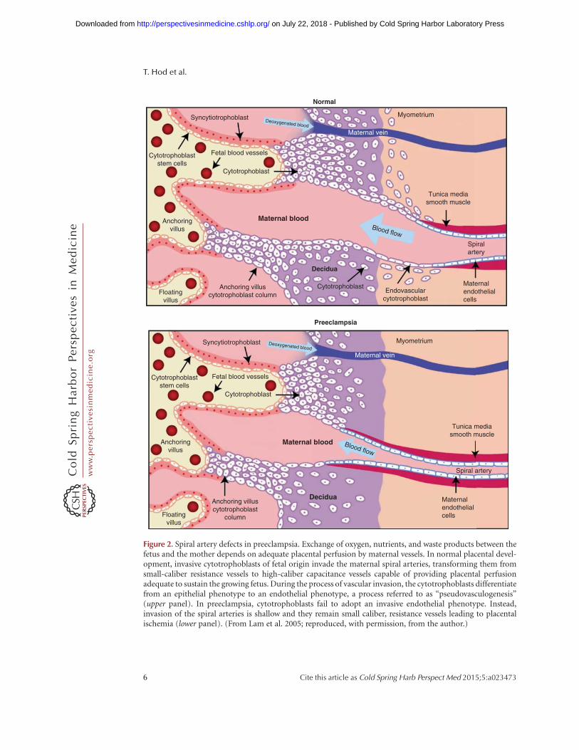
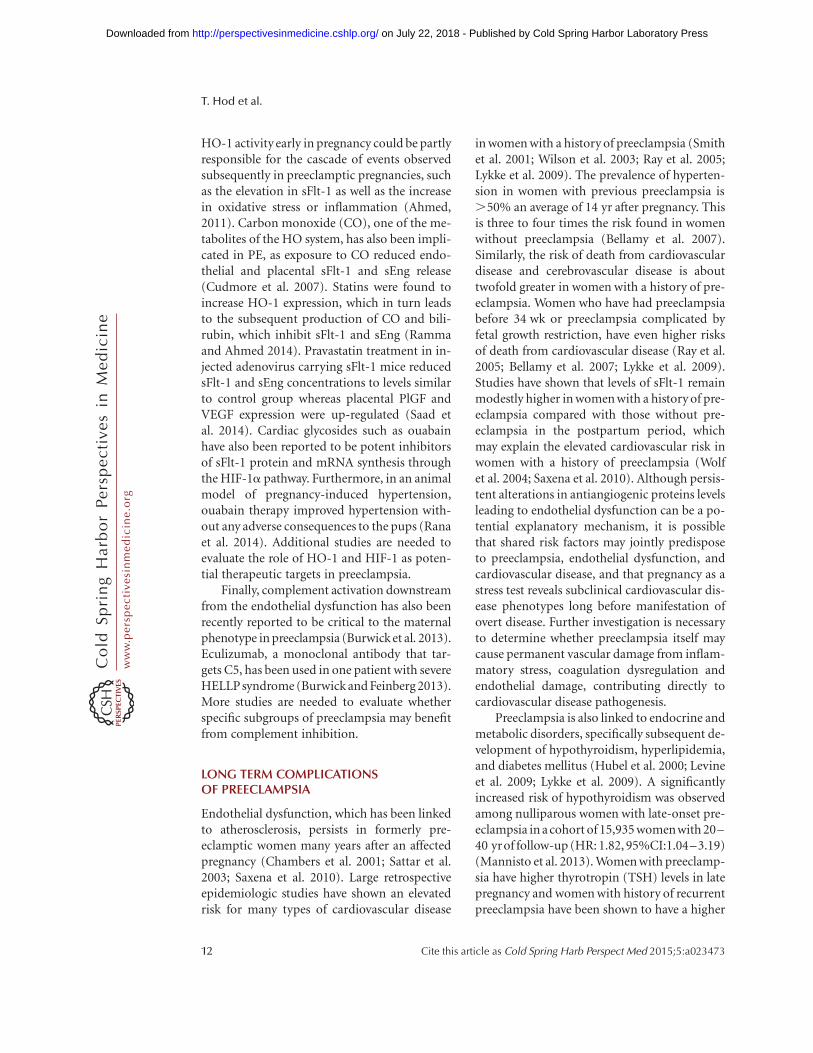
During normal placental development, thematernal uterine spiral arteries undergo an ex-tensive remodeling, transforming them into lowresistance, high bore blood vessels adequate toprovide nutrients to the placenta and fetus(Brosens et al. 1967). In preeclampsia, cytotro-
phoblasts (CTBs) fail to invade the myome-trium and the physiological changes of the spiralarteries are restricted to the decidua (Fig. 2).Invading CTBs up-regulate expression of mole-cules that are central to uterine invasion andpseudovasculogenesis (the process by whichCTB switch their adhesion molecules to mimicthat of vascular cells) (Damsky and Fisher 1998).Expression of molecules from the VEGF familychanges as invasion of these cells through theuterine wall takes place. Invasive CTBs in earlygestation express VEGF-A, VEGF-C, PlGF,VEGFR-1 and VEGFR-3 and at term, VEGF-A,PlGF, and VEGFR-1. The interactions betweenthese molecules are critical for invasion andpseudovasculogenesis because interference withligand binding altered integrin a1, an adhesionmolecule highlyexpressed byendovascular CTB,resulting in increased apoptosis of these cells.Interestingly, in severe preeclampsia, expressionof VEGF-A and VEGFR-1 in the cytotropho-blasts is down-regulated but sFlt-1 release is in-creased (Zhou et al. 2002). These findings sug-gest that dysregulation of angiogenic factors inthe maternal–fetal interface is associated withfailed pseudovasculogenesis and impaired pla-centation (Zhou et al. 2002; Fisher 2004).
Epidemiological data suggest that the im-mune system may play a role in the pathogenesisof preeclampsia (Robillard et al. 2011). Uterinenatural killer (uNK) cells, which are abundantin decidualized endometrium, surround spi-ral arteries and secrete a range of angiogenicgrowth factors play that a major role in immu-nology-related placentation, vascular remodel-ing of uterine arteries, and angiogenesis (Croyet al. 2000). When chorionic plate arteries andspiral arteries are cultured with conditionedmedium from uNK cells, disruption of vascularsmooth muscle cells and breakdown of extracel-lular matrix components is induced. These re-sults show that uNK cells contribute to the earlystages of spiral artery remodeling (Robson et al.2012). Genetic studies have associated naturalkiller (NK) cells with preeclampsia. Specificcombinations of killer immunoglobulin-like re-ceptors (NK cell receptors for receptors forMHC class I ligands) haplotypes in the motherand trophoblast HLA-C types in the fetus are
Molecular Mechanisms of Preeclampsia
Cite this article as Cold Spring Harb Perspect Med 2015;5:a023473 5
ww
w.p
ersp
ecti
vesi
nm
edic
ine.
org
on July 22, 2018 - Published by Cold Spring Harbor Laboratory Press http://perspectivesinmedicine.cshlp.org/Downloaded from
Floatingvillus
Anchoring villuscytotrophoblast column
Preeclampsia
Anchoringvillus
Maternal blood
Decidua
Blood flow
Tunica mediasmooth muscle
Spiralartery
Maternalendothelialcells
Endovascularcytotrophoblast
Cytotrophoblast
Cytotrophoblaststem cells
Cytotrophoblast
Syncytiotrophoblast
Normal
Maternal vein
MyometriumDeoxygenated blood
Fetal blood vessels
Floatingvillus
Anchoring villuscytotrophoblast
column
Anchoringvillus
Maternal blood
Decidua
Blood flow
Tunica mediasmooth muscle
Spiral artery
Cytotrophoblaststem cells
Cytotrophoblast
Syncytiotrophoblast
Maternal vein
MyometriumDeoxygenated blood
Fetal blood vessels
Maternalendothelialcells
Figure 2. Spiral artery defects in preeclampsia. Exchange of oxygen, nutrients, and waste products between thefetus and the mother depends on adequate placental perfusion by maternal vessels. In normal placental devel-opment, invasive cytotrophoblasts of fetal origin invade the maternal spiral arteries, transforming them fromsmall-caliber resistance vessels to high-caliber capacitance vessels capable of providing placental perfusionadequate to sustain the growing fetus. During the process of vascular invasion, the cytotrophoblasts differentiatefrom an epithelial phenotype to an endothelial phenotype, a process referred to as “pseudovasculogenesis”(upper panel). In preeclampsia, cytotrophoblasts fail to adopt an invasive endothelial phenotype. Instead,invasion of the spiral arteries is shallow and they remain small caliber, resistance vessels leading to placentalischemia (lower panel). (From Lam et al. 2005; reproduced, with permission, from the author.)
T. Hod et al.
6 Cite this article as Cold Spring Harb Perspect Med 2015;5:a023473
ww
w.p
ersp
ecti
vesi
nm
edic
ine.
org
on July 22, 2018 - Published by Cold Spring Harbor Laboratory Press http://perspectivesinmedicine.cshlp.org/Downloaded from
strongly associated with preeclampsia (Hiby etal. 2004). Moreover, uNK cells have a distinctphenotype, as well as a differential gene ex-pression, compared with circulating NK cells(Koopman et al. 2003). Recently it has beenshown that exposure to a combination of hyp-oxia, TGF-b1, and a demethylating agent resultsin transformation of peripheral NK cells todecidual NK-like cells, including expression ofdecidual NK cell markers, the ability to secreteVEGF, reduced cytotoxicity, and promotionof invasion of human trophoblast cell lines.These striking findings have potential therapeu-tic applications for placental disorders asso-ciated with altered NK cell biology (Cerdeiraet al. 2013).
Impaired Corin expression or function inthe pregnant uterus has been suggested as an-other mechanism for failed spiral artery remod-eling. Local atrial natriuretic peptide (ANP)production by Corin, a cardiac protease thatactivates ANP, promoted trophoblast invasionand spiral artery remodeling to prevent hyper-tension in pregnancy (Cui et al. 2012). PregnantCorin- or ANP-deficient mice develop hyper-tension and proteinuria. Moreover, in pre-eclamptic women, uterine, Corin mRNA andprotein levels are significantly lower than in nor-mal pregnancies. Of note, Corin levels in plas-ma, probably derived from the heart, are higherin preeclamptic women and did not reflect thelevels in tissues (Cui et al. 2012).
Inadequate placentation, owing to deficienttrophoblast invasion of uterine spiral arteries, acharacteristic of preeclampsia, can lead to pla-cental hypoxia that can lead to abnormal ex-pression of angiogenic factors. Persistent pla-cental hypoxia promotes hypoxia-induciblefactor-1 alpha (HIF-1a) release, which fostersa proliferative noninvasive trophoblast pheno-type (Rajakumar et al. 2005), further aggravat-ing hypoxia. HIF-1a promotes transforminggrowth factor-b3 (TGF-b3) expression, an in-hibitor of trophoblast differentiation (Caniggiaet al. 2000). Endoglin (Eng), related to a de-crease in invasive functions, is a coreceptor forTGF-b, which is expressed on cytotrophoblastsduring the first trimester and is promoted byTGF-b1 and TGF-b3. TGF-b3 also promotes
the production of sEng (Mano et al. 2011).In addition, VEGF and PlGF are induced underhypoxic conditions (Tissot van Patot et al. 2004;Nishimoto et al. 2009) and are inhibited by sFlt-1. Interestingly, sEng and sFlt-1 expression areboth stimulated under ischemic conditions,thus contributing to the systemic endothelialdysfunction (Gilbert et al. 2007, 2009).
The renin–angiotensin system (RAS) maybe involved in the pathogenesis of preeclampsia.Decidual expression of AT1 receptor, the geneencoding the angiotensin (Ang II) type 1 recep-tor is higher in preeclampsia than in normalpregnancies (Herse et al. 2007). Ang concentra-tion, which is also increased in the chorionicvilli in preeclampsia, may promote uteroplacen-tal dysfunction via vasoconstriction (Anton etal. 2008). Levels of stimulatory Ang type 1 re-ceptor autoantibodies (AT1-AAs) are elevatedin �70%–95% of preeclamptic women (Siddi-qui et al. 2010). Injection of AT1-AAs frompregnant women to pregnant mice induced hy-pertension, proteinuria, placental abnormali-ties, and glomerular endotheliosis, which arethe key features of preeclampsia (Zhou et al.2008). The binding of AT1-AAs to AT1-R in-duces sFlt-1 and sEng production by humanvillous explants through tumor necrosis factor(TNF)-a pathways (Irani et al. 2010). TNF-a isalso increased in preeclampsia owing to placen-tal ischemia and it stimulates placental produc-tion of sEng and sFlt-1 (Parrish et al. 2010).Interestingly, high levels of AT1-AAs correlatewith the disease severity because of their asso-ciation with the presence of hypertension, pro-teinuria, and sFlt-1 (Siddiqui et al. 2010).
A pathogenic role of heme-oxygenase-1(HO-1) in preeclampsia has been identified(Ahmed and Cudmore 2009). Heme break-down metabolites generated by the enzymaticactivity of HO-1 account for its angiogenic andvasodilatory properties. Placental HO-1 expres-sion and exhaled CO levels were both found tobe reduced in severe preeclamptic women. In-terestingly, in vitro experiments showed thatHO-1 induction increased CO production anddown-regulated secretion of sFlt-1 (Levytskaet al. 2013). More recently, endometrial VEGFup-regulation has also been shown in a mouse
Molecular Mechanisms of Preeclampsia
Cite this article as Cold Spring Harb Perspect Med 2015;5:a023473 7
ww
w.p
ersp
ecti
vesi
nm
edic
ine.
org
on July 22, 2018 - Published by Cold Spring Harbor Laboratory Press http://perspectivesinmedicine.cshlp.org/Downloaded from
model to induce placental sFlt1 production andcontribute to preeclampsia (Fan et al. 2014).
Placental underperfusion, secondary to de-ficient conversion of the spiral arteries was re-cently linked to endoplasmic reticulum (ER)stress, which can lead to activation of proin-flammatory pathways, contributing to maternalendothelial cell activation (Burton and Yung2011). The ER is a hub for proper folding andexport of peptides, guided by ER-specific chap-erones. ER stress can dysregulate the function ofchaperones, resulting in export of misfoldedproteins into the circulation (Redman 2008).Administration of aberrant transthyretin, im-munoprecipitated from PE serum to pregnantmice induces a full spectrum of preeclampsia-like features, whereas immunodepletion of thetransthyretin from preeclamptic serum amelio-rates most of the disease features and reducessFlt-1 and sEng. Moreover, aggregation of trans-thyretin protein is found in serum and in pla-cental tissue from preeclamptic pregnancies.It is possible that hypoxia, which has beenshown to control transthyretin expression anduptake, and ER stress destabilize transthyretininto a misfolded conformation and aggregationacquiring the potential to induce antiangio-genic factors (Kalkunte et al. 2013). The pres-ence of b-amyloid aggregates in placentas ofwomen with PE and fetal growth restriction(Buhimschi et al. 2014) may further supportthe pathogenic role of ER stress and misfoldedproteins in preeclampsia.
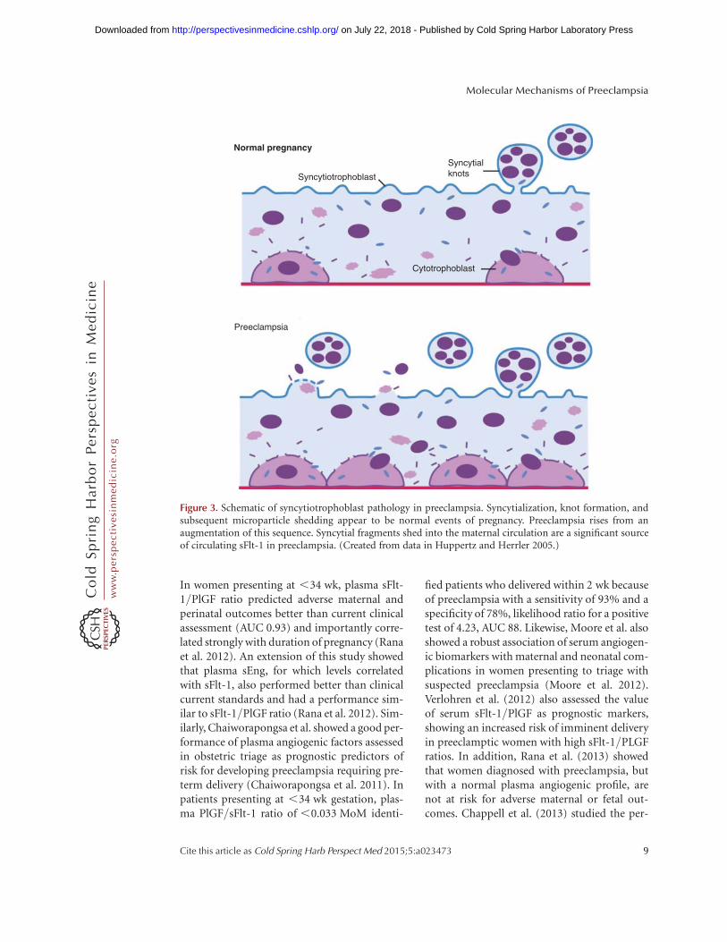
Placental pathological lesions similar to pre-eclampsia also occur in cases of intrauterinegrowth restriction (IUGR), but without mater-nal disease (Khong et al. 1986). Increased syn-cytiotrophoblast microparticles are found inwomen with early onset preeclampsia, but notin cases of normotensive IUGR suggesting thatsyncytial pathology may be unique to pre-eclampsia (Goswami et al. 2006). These biolog-ically active syncytial l microparticles can trans-port toxic proteins, such as sFlt1 and sEng intothe maternal circulation, where they mediatethe major manifestations of preeclampsia (Fig.3) (Rajakumar et al. 2012). We and others havehypothesized that the absence of a second insultof syncytial debris shedding, crucial for the ap-
pearance of the systemic disease in PE, protectswomen with fetal IUGR from the full blownmaternal syndrome observed in preeclampticwomen. Changes not only in quantity, butalso in the size of the circulating microparticles,might be important in the pathogenesis of pre-eclampsia (Redman et al. 2012). Moreover, dif-ferences in physical and antigenic characteristicsof normal and preeclamptic placental micro-and nanovesicles produced by placental perfu-sion or mechanical disruption have been ob-served (Tannetta et al. 2013). It is therefore pos-sible that specific differences in microparticlesof preeclampsia versus normal or IUGR preg-nancies are the basis for their distinct pathology.
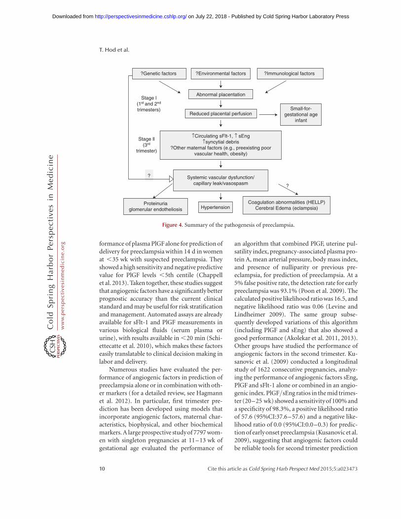
The mechanisms by which angiogenic fac-tor expression in the placenta contribute to thepathophysiology of preeclampsia are yet to bedefined. However, most models described aboveand others show increased sFlt-1 and sEng se-cretion, which further strengthens its role in thestage II or the effector pathway that leads to theclinical syndrome of preeclampsia (Fig. 4).
ANGIOGENIC FACTORS AS BIOMARKERS
Measurement of angiogenic factors using auto-mated assays (sFlt-1 and PlGF) has proven to beclinically useful for routine diagnosis of pre-eclampsia (Ohkuchi et al. 2010; Schiettecatte etal. 2010; Sunderji et al. 2010; Verlohren et al.2010; Benton et al. 2011; Knudsen et al. 2012).Several groups have shown that sFlt-1 and freePlGF levels can be used to differentiate pre-eclampsia from diseases that mimic preeclamp-sia, such as chronic hypertension, gestationalhypertension, kidney disease, and gestationalthrombocytopenia (Salahuddin et al. 2007;Sunderji et al. 2010; Perni et al. 2012; Rolfo etal. 2012; Verdonk et al. 2012).
Similar to the field of cardiovascular medi-cine with the introduction of troponins, riskstratification of women presenting at obstetricaltriage with suspected preeclampsia may be animportant advance in obstetrical clinical prac-tice. Rana et al. tested the performance ofangiogenic factors as predictors of adverse out-comes in patients presenting at obstetrical triagewith suspected preeclampsia (Rana et al. 2012).
T. Hod et al.
8 Cite this article as Cold Spring Harb Perspect Med 2015;5:a023473
ww
w.p
ersp
ecti
vesi
nm
edic
ine.
org
on July 22, 2018 - Published by Cold Spring Harbor Laboratory Press http://perspectivesinmedicine.cshlp.org/Downloaded from
In women presenting at ,34 wk, plasma sFlt-1/PlGF ratio predicted adverse maternal andperinatal outcomes better than current clinicalassessment (AUC 0.93) and importantly corre-lated strongly with duration of pregnancy (Ranaet al. 2012). An extension of this study showedthat plasma sEng, for which levels correlatedwith sFlt-1, also performed better than clinicalcurrent standards and had a performance sim-ilar to sFlt-1/PlGF ratio (Rana et al. 2012). Sim-ilarly, Chaiworapongsa et al. showed a good per-formance of plasma angiogenic factors assessedin obstetric triage as prognostic predictors ofrisk for developing preeclampsia requiring pre-term delivery (Chaiworapongsa et al. 2011). Inpatients presenting at ,34 wk gestation, plas-ma PlGF/sFlt-1 ratio of ,0.033 MoM identi-
fied patients who delivered within 2 wk becauseof preeclampsia with a sensitivity of 93% and aspecificity of 78%, likelihood ratio for a positivetest of 4.23, AUC 88. Likewise, Moore et al. alsoshowed a robust association of serum angiogen-ic biomarkers with maternal and neonatal com-plications in women presenting to triage withsuspected preeclampsia (Moore et al. 2012).Verlohren et al. (2012) also assessed the valueof serum sFlt-1/PlGF as prognostic markers,showing an increased risk of imminent deliveryin preeclamptic women with high sFlt-1/PLGFratios. In addition, Rana et al. (2013) showedthat women diagnosed with preeclampsia, butwith a normal plasma angiogenic profile, arenot at risk for adverse maternal or fetal out-comes. Chappell et al. (2013) studied the per-
Cytotrophoblast
Syncytiotrophoblast
Normal pregnancy
Syncytialknots
Preeclampsia
Figure 3. Schematic of syncytiotrophoblast pathology in preeclampsia. Syncytialization, knot formation, andsubsequent microparticle shedding appear to be normal events of pregnancy. Preeclampsia rises from anaugmentation of this sequence. Syncytial fragments shed into the maternal circulation are a significant sourceof circulating sFlt-1 in preeclampsia. (Created from data in Huppertz and Herrler 2005.)
Molecular Mechanisms of Preeclampsia
Cite this article as Cold Spring Harb Perspect Med 2015;5:a023473 9
ww
w.p
ersp
ecti
vesi
nm
edic
ine.
org
on July 22, 2018 - Published by Cold Spring Harbor Laboratory Press http://perspectivesinmedicine.cshlp.org/Downloaded from
formance of plasma PlGF alone for prediction ofdelivery for preeclampsia within 14 d in womenat ,35 wk with suspected preeclampsia. Theyshowed a high sensitivity and negative predictivevalue for PlGF levels ,5th centile (Chappellet al. 2013). Taken together, these studies suggestthat angiogenic factors have a significantly betterprognostic accuracy than the current clinicalstandard and may be useful for risk stratificationand management. Automated assays are alreadyavailable for sFlt-1 and PlGF measurements invarious biological fluids (serum plasma orurine), with results available in ,20 min (Schi-ettecatte et al. 2010), which makes these factorseasily translatable to clinical decision making inlabor and delivery.
Numerous studies have evaluated the per-formance of angiogenic factors in prediction ofpreeclampsia alone or in combination with oth-er markers (for a detailed review, see Hagmannet al. 2012). In particular, first trimester pre-diction has been developed using models thatincorporate angiogenic factors, maternal char-acteristics, biophysical, and other biochemicalmarkers. A large prospective studyof 7797 wom-en with singleton pregnancies at 11–13 wk ofgestational age evaluated the performance of
an algorithm that combined PlGF, uterine pul-satility index, pregnancy-associated plasma pro-tein A, mean arterial pressure, body mass index,and presence of nulliparity or previous pre-eclampsia, for prediction of preeclampsia. At a5% false positive rate, the detection rate for earlypreeclampsia was 93.1% (Poon et al. 2009). Thecalculated positive likelihood ratio was 16.5, andnegative likelihood ratio was 0.06 (Levine andLindheimer 2009). The same group subse-quently developed variations of this algorithm(including PlGF and sEng) that also showed agood performance (Akolekar et al. 2011, 2013).Other groups have studied the performance ofangiogenic factors in the second trimester. Ku-sanovic et al. (2009) conducted a longitudinalstudy of 1622 consecutive pregnancies, analyz-ing the performance of angiogenic factors sEng,PlGF and sFlt-1 alone or combined in an angio-genic index. PlGF/sEng ratios in the mid trimes-ter (20–25 wk) showed a sensitivityof 100% anda specificity of 98.3%, a positive likelihood ratioof 57.6 (95%CI:37.6–57.6) and a negative like-lihood ratio of 0.0 (95%CI:0.0–0.3) for predic-tion of earlyonset preeclampsia (Kusanovic et al.2009), suggesting that angiogenic factors couldbe reliable tools for second trimester prediction
?Genetic factors ?Environmental factors ?Immunological factors
Small-for-gestational age
infant
↑Circulating sFlt-1, ↑ sEng↑syncytial debris
?Other maternal factors (e.g., preexisting poorvascular health, obesity)
Proteinuriaglomerular endotheliosis Hypertension
Coagulation abnormalities (HELLP)Cerebral Edema (eclampsia)
Systemic vascular dysfunction/capillary leak/vasospasm
?
?
Reduced placental perfusion
Stage ll(3rd
trimester)
Stage l(1st and 2nd
trimesters)
Abnormal placentation
Figure 4. Summary of the pathogenesis of preeclampsia.
T. Hod et al.
10 Cite this article as Cold Spring Harb Perspect Med 2015;5:a023473
ww
w.p
ersp
ecti
vesi
nm
edic
ine.
org
on July 22, 2018 - Published by Cold Spring Harbor Laboratory Press http://perspectivesinmedicine.cshlp.org/Downloaded from
of preeclampsia. However, there is no evidencethat interventions or close follow up may im-prove maternal and/or fetal outcome even ifone were to robustly predict early onset pre-eclampsia. Recent guidelines of the AmericanCollege of Obstetrics and Gynecology Task Forcealso do not support the use of biomarkers topredict preeclampsia (ACOG Task Force on Hy-pertension and Pregnancy 2013). Studies evalu-ating the utility of low-dose aspirin to reduce theincidence of preeclampsia in those at high riskbased on abnormal first trimester biochemicalprofile are currently in progress.
ANGIOGENIC FACTORS AS THERAPEUTICTARGETS
To date, delivery of the placenta is the only treat-ment, which in the case of preterm delivery mayadversely affect neonatal outcomes. The devel-opment of a therapy that safely prolongs gesta-tion would constitute a major advance in thisfield. Several strategies have been developed totarget angiogenic factors using both in vitro andin vivo models to reestablish the angiogenic bal-ance. Depletion of sFlt-1 in preeclamptic villouscondition media by immunoprecipitation re-verses the antiangiogenic properties in vitro(Ahmad and Ahmed 2004). Administration ofVEGFA-121 intoa rodent modelof preeclampsiaimproved hypertension, proteinuria, glomeru-lar endotheliosis, and reverted sFlt-1 inducedchanges in gene expression (Li et al. 2007). Co-administration of adenovirus expressing VEGFin a similar model, resulted in a significant re-duction of free sFlt-1, rescued the renal damageand normalized blood pressure (Bergmann et al.2010). VEGF-121 infusion has also been report-ed to lower blood pressure and improve renalfunction in rats with placental ischemia-inducedhypertension (Gilbert et al. 2010). Furthermore,increased expression of placental growth factorinduced by pravastatin decreased the levels ofcirculating sFlt-1 and ameliorated preeclampticsymptoms in a preeclampsia mouse model ofplacental-specific sFlt-1 overexpression (Kuma-sawa et al. 2011).
In humans, resolution of signs of pre-eclampsia were observed in cases of parvovi-
rus-induced hydrops (Stepan and Faber 2006),mirror syndrome (Llurba et al. 2012), fetal de-mise in a twin pregnancies (Hladunewich etal. 2009), all of which correlated with fall insFlt1 levels. These experiments of nature havestrengthened the notion that reduction of sFlt-1during pregnancy may be safe without generat-ing significant adverse effects to the fetus.
Thadhani et al. (2011) recently translatedsome fundamental discoveries to the bedside.Taking advantage of the positive charge of sFlt-1, they used a negatively charged dextran sulfatecellulose column for extracorporeal removal ofsFlt-1. In a pilot study of eight women with pre-term preeclampsia, dextran sulfate apheresislead to a reduction in sFlt-1 levels and improve-ment in proteinuria and blood pressure, with-out evident adverse effects to the mother andfetus. Five womenwere treated once, twowomentwice, and a third patient treated four times. Ofthe two women undergoing two apheresis treat-ments, one remained pregnant for 15 d and theother for 19 d. A third woman, treated fourtimes, remained pregnant for 23 d. In all of thesecases, there was evidence of continued fetalgrowth. In untreated women, average prolonga-tion of pregnancy was 3.6 d (Thadhani et al.2011). This is an exciting study that opens theway for a safe prolongation of gestation in severepreterm preeclamptic patients. Other modali-ties for targeting the angiogenic imbalance areadministration of agents that scavenge sFlt-1,such as sFlt-1 antibodies, PlGF, VEGF, or de-crease sFlt-1 production by siRNA strategies orsmall molecules.
Relaxin, a peptide hormone that is increasedin pregnancy, is evolving as a potential targetfor preeclampsia, given its vasodilatory and vas-cular remodeling properties (Conrad and Shroff2011). A phase I safety study in women withsevere preeclampsia is currently ongoing (Un-emori et al. 2009). Interestingly, its effectscould be partially mediated by VEGF and PlGF(McGuane et al. 2011). Thus, relaxin could offsetthe increased sFlt-1 levels in preeclampsia by po-tentiating local VEGF and PlGF signaling in thevasculature.
During pregnancy, heme oxygenase-1 (HO-1) negatively regulates sFlt-1 and the loss of
Molecular Mechanisms of Preeclampsia
Cite this article as Cold Spring Harb Perspect Med 2015;5:a023473 11
ww
w.p
ersp
ecti
vesi
nm
edic
ine.
org
on July 22, 2018 - Published by Cold Spring Harbor Laboratory Press http://perspectivesinmedicine.cshlp.org/Downloaded from
HO-1 activity early in pregnancy could be partlyresponsible for the cascade of events observedsubsequently in preeclamptic pregnancies, suchas the elevation in sFlt-1 as well as the increasein oxidative stress or inflammation (Ahmed,2011). Carbon monoxide (CO), one of the me-tabolites of the HO system, has also been impli-cated in PE, as exposure to CO reduced endo-thelial and placental sFlt-1 and sEng release(Cudmore et al. 2007). Statins were found toincrease HO-1 expression, which in turn leadsto the subsequent production of CO and bili-rubin, which inhibit sFlt-1 and sEng (Rammaand Ahmed 2014). Pravastatin treatment in in-jected adenovirus carrying sFlt-1 mice reducedsFlt-1 and sEng concentrations to levels similarto control group whereas placental PlGF andVEGF expression were up-regulated (Saad etal. 2014). Cardiac glycosides such as ouabainhave also been reported to be potent inhibitorsof sFlt-1 protein and mRNA synthesis throughthe HIF-1a pathway. Furthermore, in an animalmodel of pregnancy-induced hypertension,ouabain therapy improved hypertension with-out any adverse consequences to the pups (Ranaet al. 2014). Additional studies are needed toevaluate the role of HO-1 and HIF-1 as poten-tial therapeutic targets in preeclampsia.
Finally, complement activation downstreamfrom the endothelial dysfunction has also beenrecently reported to be critical to the maternalphenotype in preeclampsia (Burwick et al. 2013).Eculizumab, a monoclonal antibody that tar-gets C5, has been used in one patient with severeHELLP syndrome (Burwick and Feinberg 2013).More studies are needed to evaluate whetherspecific subgroups of preeclampsia may benefitfrom complement inhibition.
LONG TERM COMPLICATIONSOF PREECLAMPSIA
Endothelial dysfunction, which has been linkedto atherosclerosis, persists in formerly pre-eclamptic women many years after an affectedpregnancy (Chambers et al. 2001; Sattar et al.2003; Saxena et al. 2010). Large retrospectiveepidemiologic studies have shown an elevatedrisk for many types of cardiovascular disease
in women with a history of preeclampsia (Smithet al. 2001; Wilson et al. 2003; Ray et al. 2005;Lykke et al. 2009). The prevalence of hyperten-sion in women with previous preeclampsia is.50% an average of 14 yr after pregnancy. Thisis three to four times the risk found in womenwithout preeclampsia (Bellamy et al. 2007).Similarly, the risk of death from cardiovasculardisease and cerebrovascular disease is abouttwofold greater in women with a history of pre-eclampsia. Women who have had preeclampsiabefore 34 wk or preeclampsia complicated byfetal growth restriction, have even higher risksof death from cardiovascular disease (Ray et al.2005; Bellamy et al. 2007; Lykke et al. 2009).Studies have shown that levels of sFlt-1 remainmodestly higher in women with a history of pre-eclampsia compared with those without pre-eclampsia in the postpartum period, whichmay explain the elevated cardiovascular risk inwomen with a history of preeclampsia (Wolfet al. 2004; Saxena et al. 2010). Although persis-tent alterations in antiangiogenic proteins levelsleading to endothelial dysfunction can be a po-tential explanatory mechanism, it is possiblethat shared risk factors may jointly predisposeto preeclampsia, endothelial dysfunction, andcardiovascular disease, and that pregnancy as astress test reveals subclinical cardiovascular dis-ease phenotypes long before manifestation ofovert disease. Further investigation is necessaryto determine whether preeclampsia itself maycause permanent vascular damage from inflam-matory stress, coagulation dysregulation andendothelial damage, contributing directly tocardiovascular disease pathogenesis.
Preeclampsia is also linked to endocrine andmetabolic disorders, specifically subsequent de-velopment of hypothyroidism, hyperlipidemia,and diabetes mellitus (Hubel et al. 2000; Levineet al. 2009; Lykke et al. 2009). A significantlyincreased risk of hypothyroidism was observedamong nulliparous women with late-onset pre-eclampsia in a cohort of 15,935 womenwith 20–40 yrof follow-up (HR: 1.82, 95%CI:1.04–3.19)(Mannisto et al. 2013). Women with preeclamp-sia have higher thyrotropin (TSH) levels in latepregnancy and women with history of recurrentpreeclampsia have been shown to have a higher
T. Hod et al.
12 Cite this article as Cold Spring Harb Perspect Med 2015;5:a023473
ww
w.p
ersp
ecti
vesi
nm
edic
ine.
org
on July 22, 2018 - Published by Cold Spring Harbor Laboratory Press http://perspectivesinmedicine.cshlp.org/Downloaded from
risk of developing subsequently reduced thyroidfunction many years after preeclampsia. The as-sociation was stronger if the high concentrationof TSH was combined with absence of thyroidperoxidase antibodies, and particularly strong ifpreeclampsia had occurred in two pregnancies,suggesting that the hypothyroid function in pre-eclampsia may occur independent of the auto-immune mechanisms accepted as the most likelycause of hypothyroidism in iodine replete wom-en (Levine et al. 2009). The increased risk forthyroid dysfunction among women with pre-eclampsia is thought to be mediated throughantiangiogenic proteins, which act as antago-nists to VEGF and PlGF, causing endothelialdysfunction and capillary regression in severaltissues, including the thyroid (Maynard et al.2005; Levine et al. 2006b). This theory is furtherstrengthened by studies in mice using VEGFinhibitors such as sFlt-1, which have shownsubstantial thyroid capillary regression and in-creased TSH concentrations (Kamba et al. 2006;Kamba and McDonald 2007).
Hypertensive diseases are also associatedwith worsened renal outcomes, mainly in-creased albuminuria and an increased risk offuture chronic kidney disease (CKD) and end-stage renal disease (ESRD). A meta-analysis con-firms the association between preeclampsia andalbuminuria in the long term. Thirty-one per-cent of women who had preeclampsia developedmicroalbuminuria at an average of 7.1 yr post-partum, in contrast to only 7% of women whohad uncomplicated pregnancies (McDonaldet al. 2010). An increased risk of renal diseasewas shown in a large retrospective cohort studyof 570,433 women and an average follow-up of17 yr after initial pregnancy. The relative risk ofESRD in women who had preeclampsia in thefirst pregnancy was close to five. Women withtwo or three episodes of preeclampsia had a15-fold increase in the risk of developing ESRD(Vikse et al. 2008). Moreover, the severity of hy-pertensive disease during pregnancy correlatedwith progression to ESRD (Wang et al. 2013a).
In addition to the maternal complicationsof preeclampsia detailed above, preeclampsiasignificantly increases perinatal and neonatalmorbidities. Maternal preeclampsia is a fre-
quent cause of preterm birth (Basso et al.2006) and serum levels of sFlt-1 in the motherare inversely related to gestational age and birthweight of the newborn (Veas et al. 2011). sFlt-1,which is markedly increased in amniotic fluidduring the second and third trimesters of preg-nancies complicated by preeclampsia (Vuorelaet al. 2000; Wang et al. 2010), may directly reachthe fetal lung via fetal breathing or through anintramembranous pathway and fetal swallowingto enter the fetal lung. Studies have shown thatVEGF inhibition during early neonatal periodresults in persistent abnormalities of alveolarand pulmonary vascular structures into and be-yond infancy, which are characteristic of path-ological changes in human bronchopulmonarydysplasia (BPD) (Le Cras et al. 2002; Tang etal. 2004). Furthermore, excess intra-amnioticsFlt-1, causing transient impairment of VEGFsignaling in the fetus, was sufficient to causesustained abnormalities of lung structure dur-ing infancy as well as biventricular hypertrophymost probably secondary to pulmonary andsystemic hypertension in 14-d-old rats (Tanget al. 2012). These findings are interesting be-cause offspring of mothers with preeclampsiaare at risk for pulmonary vascular dysfunctionlater in life (Jayet et al. 2010). Preeclampsia-in-duced vascular dysfunction may have otherclinical consequences in offspring, such as anincreased risk of hypertension (Seidman et al.1991; Vatten et al. 2003) and stroke (Kajantieet al. 2009). Interestingly, the antiangiogenicstate of gestational hypertension/preeclampsiaprotects the infant from retinopathy of prema-turity (Yu et al. 2012), a disorder characterizedby the overproduction of VEGF (Pierce et al.1995; Robbins et al. 1997).
Taken together, all of the maternal and fetalcomplications mentioned above suggest a cen-tral role of antiangiogenic factors, not only inthe pathogenesis of preeclampsia but also asleading contributing factors to unfavorable ma-ternal as well as fetal long term consequences.
CONCLUSION
Preeclampsia is a pregnancy-specific diseasecharacterized by an antiangiogenic state (see
Molecular Mechanisms of Preeclampsia
Cite this article as Cold Spring Harb Perspect Med 2015;5:a023473 13
ww
w.p
ersp
ecti
vesi
nm
edic
ine.
org
on July 22, 2018 - Published by Cold Spring Harbor Laboratory Press http://perspectivesinmedicine.cshlp.org/Downloaded from
Fig. 4, for summary). Many questions remain tobe answered, namely the upstream mechanismsof the deregulation of angiogenic factors. Theangiogenic imbalance can be quantified in plas-ma or serum by automated assays and usedfor clinical decision making and therapeuticmonitoring in clinical trials. Administration ofproangiogenic factors or removal of antian-giogenic factors is a promising approach forpreeclampsia treatment. Preeclampsia is also as-sociated with long-term health risks to bothmother and child. More studies are needed toevaluate the underlying mechanisms leading tolong-term cardiovascular disease in women ex-posed to preeclampsia.
COMPETING INTEREST STATEMENT
S.A.K has financial interest in Aggamin Thera-peutics, a consultant to Siemens and has Grantfunding from Thermofisher. S.A.K is a co-in-ventor on patents related to angiogenic bio-markers in preeclampsia that are held by theBeth Israel Deaconess Medical Center.
REFERENCES
Ahmed A. 2011. New insights into the etiology of pre-eclampsia: Identification of key elusive factors for thevascular complications. Thromb Res 127: S72–S75.
Ahmad S, Ahmed A. 2004. Elevated placental soluble vas-cular endothelial growth factor receptor-1 inhibits angio-genesis in preeclampsia. Circ Res 95: 884–891.
Ahmed A, Cudmore MJ. 2009. Can the biology of VEGF andhaem oxygenases help solve pre-eclampsia? Biochem SocTrans 37: 1237–1242.
Akolekar R, Syngelaki A, Sarquis R, Zvanca M, NicolaidesKH. 2011. Prediction of early, intermediate and late pre-eclampsia from maternal factors, biophysical and bio-chemical markers at 11–13 weeks. Prenat Diagn 31:66–74.
Akolekar R, Syngelaki A, Poon L, Wright D, Nicolaides KH.2013. Competing risks model in early screening for pre-eclampsia by biophysical and biochemical markers. FetalDiagn Ther 33: 8–15.
Ambati BK, Nozaki M, Singh N, Takeda A, Jani PD, Suthar T,Albuquerque RJ, Richter E, Sakurai E, Newcomb MT, etal. 2006. Corneal avascularity is due to soluble VEGFreceptor-1. Nature 443: 993–997.
American College of Obstetricians and Gynecologists; TaskForce on Hypertension in Pregnancy. 2013. Hypertensionin pregnancy. Report of the American College of Obste-tricians and Gynecologists’ Task Force on Hypertensionin Pregnancy. Obstet Gynecol 122: 1122–1131.
Anton L, Merrill DC, Neves LA, Stovall K, Gallagher PE, DizDI, Moorefield C, Gruver C, Ferrario CM, Brosnihan KB.2008. Activation of local chorionic villi angiotensin IIlevels but not angiotensin (1–7) in preeclampsia. Hyper-tension 51: 1066–1072.
Basso O, Rasmussen S, Weinberg CR, Wilcox AJ, Irgens LM,Skjaerven R. 2006. Trends in fetal and infant survivalfollowing preeclampsia. JAMA 296: 1357–1362.
Bellamy L, Casas JP, Hingorani AD, Williams DJ. 2007. Pre-eclampsia and risk of cardiovascular disease and cancer inlater life: Systematic review and meta-analysis. BMJ 335:974.
Bello N, Rendon IS, Arany Z. 2013. The relationship be-tween pre-eclampsia and peripartum cardiomyopathy:A systematic review and meta-analysis. J Am Coll Cardiol62: 1715–1723.
Benton SJ, Hu Y, Xie F, Kupfer K, Lee SW, Magee LA, vonDadelszen P. 2011. Angiogenic factors as diagnostic testsfor preeclampsia: A performance comparison betweentwo commercial immunoassays. Am J Obstet Gynecol205: 469 e461–e468.
Bergmann A, Ahmad S, Cudmore M, Gruber AD, WittschenP, Lindenmaier W, Christofori G, Gross V, Gonzalves A,Grone HJ, et al. 2010. Reduction of circulating solubleFlt-1 alleviates preeclampsia-like symptoms in a mousemodel. J Cell Mol Med 14: 1857–1867.
Brosens I, Robertson WB, Dixon HG. 1967. The physiolog-ical response of the vessels of the placental bed to normalpregnancy. J Pathol Bacteriol 93: 569–579.
Buhimschi IA, Nayeri UA, Zhao G, Shook LL, Pensalfini A,Funai EF, Bernstein IM, Glabe CG, Buhimschi CS. 2014.Protein misfolding, congophilia, oligomerization, anddefective amyloid processing in preeclampsia. Sci TranslMed 6: 245ra292.
Burton GJ, Yung HW. 2011. Endoplasmic reticulum stress inthe pathogenesis of early-onset pre-eclampsia. PregnancyHypertens 1: 72–78.
Burwick RM, Feinberg BB. 2013. Eculizumab for the treat-ment of preeclampsia/HELLP syndrome. Placenta 34:201–203.
Burwick RM, Fichorova RN, Dawood HY, Yamamoto HS,Feinberg BB. 2013. Urinary excretion of C5b-9 in severepreeclampsia: Tipping the balance of complement acti-vation in pregnancy. Hypertension 62: 1040–1045.
Buurma AJ, Penning ME, Prins F, Schutte JM, Bruijn JA,Wilhelmus S, Rajakumar A, Bloemenkamp KW, Karu-manchi SA, Baelde HJ. 2013. Preeclampsia is associatedwith the presence of transcriptionally active placentalfragments in the maternal lung. Hypertension 62: 608–613.
Caniggia I, Mostachfi H, Winter J, Gassmann M, Lye SJ,Kuliszewski M, Post M. 2000. Hypoxia-inducible fac-tor-1 mediates the biological effects of oxygen on humantrophoblast differentiation through TGFb3. J Clin Invest105: 577–587.
Cerdeira AS, Rajakumar A, Royle CM, Lo A, Husain Z,Thadhani RI, Sukhatme VP, Karumanchi SA, KopcowHD. 2013. Conversion of peripheral blood NK cells to adecidual NK-like phenotype by a cocktail of defined fac-tors. J Immunol 190: 3939–3948.
Chaiworapongsa T, Romero R, Espinoza J, Bujold E, MeeKim Y, Goncalves LF, Gomez R, Edwin S. 2004. Evidence
T. Hod et al.
14 Cite this article as Cold Spring Harb Perspect Med 2015;5:a023473
ww
w.p
ersp
ecti
vesi
nm
edic
ine.
org
on July 22, 2018 - Published by Cold Spring Harbor Laboratory Press http://perspectivesinmedicine.cshlp.org/Downloaded from
supporting a role for blockade of the vascular endothelialgrowth factor system in the pathophysiology of pre-eclampsia. Am J Obstet Gynecol 190: 1541–1547; discus-sion 1547–1550.
Chaiworapongsa T, Romero R, Kim YM, Kim GJ, Kim MR,Espinoza J, Bujold E, Goncalves L, Gomez R, Edwin S, etal. 2005. Plasma soluble vascular endothelial growth fac-tor receptor-1 concentration is elevated prior to the clin-ical diagnosis of pre-eclampsia. J Matern Fetal NeonatalMed 17: 3–18.
Chaiworapongsa T, Romero R, Savasan ZA, Kusanovic JP,Ogge G, Soto E, Dong Z, Tarca A, Gaurav B, Hassan SS.2011. Maternal plasma concentrations of angiogenic/anti-angiogenic factors are of prognostic value in patientspresenting to the obstetrical triage area with the suspicionof preeclampsia. J Matern Fetal Neona 24: 1187–1207.
Chaiworapongsa T, Chaemsaithong P, Yeo L, Romero R.2014. Pre-eclampsia part 1: Current understanding ofits pathophysiology. Nat Rev Nephrol 10: 466–480.
Chambers JC, Fusi L, Malik IS, Haskard DO, De Swiet M,Kooner JS. 2001. Association of maternal endothelialdysfunction with preeclampsia. JAMA 285: 1607–1612.
Chappell JC, Taylor SM, Ferrara N, Bautch VL. 2009. Localguidance of emerging vessel sprouts requires soluble Flt-1. Dev Cell 17: 377–386.
Chappell LC, Duckworth S, Seed PT, Griffin M, Myers J,Mackillop L, Simpson N, Waugh J, Anumba D, KennyLC, et al. 2013. Diagnostic accuracy of placental growthfactor in women with suspected preeclampsia: A prospec-tive multicenter study. Circulation 128: 2121–2131.
Chen CW, Jaffe IZ, Karumanchi SA. 2014. Pre-eclampsiaand cardiovascular disease. Cardiovasc Res 101: 579–586.
Conrad KP, Shroff SG. 2011. Effects of relaxin on arterialdilation, remodeling, and mechanical properties. CurrHypertens Rep 13: 409–420.
Croy BA, Ashkar AA, Minhas K, Greenwood JD. 2000. Canmurine uterine natural killer cells give insights into thepathogenesis of preeclampsia? J Soc Gynecol Investig 7:12–20.
Cudmore M, Ahmad S, Al-Ani B, Fujisawa T, Coxall H,Chudasama K, Devey LR, Wigmore SJ, Abbas A, HewettPW, et al. 2007. Negative regulation of soluble Flt-1 andsoluble endoglin release by heme oxygenase-1. Circula-tion 115: 1789–1797.
Cui Y, Wang W, Dong N, Lou J, Srinivasan DK, Cheng W,Huang X, Liu M, Fang C, Peng J, et al. 2012. Role of corinin trophoblast invasion and uterine spiral artery remod-elling in pregnancy. Nature 484: 246–250.
Damsky CH, Fisher SJ. 1998. Trophoblast pseudo-vasculo-genesis: Faking it with endothelial adhesion receptors.Curr Opin Cell Biol 10: 660–666.
Duley L. 2009. The global impact of pre-eclampsia andeclampsia. Semin Perinatol 33: 130–137.
Eremina V, Quaggin SE. 2004. The role of VEGF-A in glo-merular development and function. Curr Opin NephrolHypertens 13: 9–15.
Eremina V, Sood M, Haigh J, Nagy A, Lajoie G, Ferrara N,Gerber HP, Kikkawa Y, Miner JH, Quaggin SE. 2003.Glomerular-specific alterations of VEGF-A expressionlead to distinct congenital and acquired renal diseases. JClin Invest 111: 707–716.
Eremina V, Jefferson JA, Kowalewska J, Hochster H, Haas M,Weisstuch J, Richardson C, Kopp JB, Kabir MG, BackxPH, et al. 2008. VEGF inhibition and renal thromboticmicroangiopathy. N Engl J Med 358: 1129–1136.
Facemire CS, Nixon AB, Griffiths R, Hurwitz H, CoffmanTM. 2009. Vascular endothelial growth factor receptor 2controls blood pressure by regulating nitric oxide syn-thase expression. Hypertension 54: 652–658.
Fan X, Rai A, Kambham N, Sung JF, Singh N, Petitt M, DhalS, Agrawal R, Sutton RE, Druzin ML, et al. 2014. Endo-metrial VEGF induces placental sFLT1 and leads to preg-nancy complications. J Clin Invest 124: 4941–4952.
Fisher SJ. 2004. The placental problem: Linking abnormalcytotrophoblast differentiation to the maternal symp-toms of preeclampsia. Reprod Biol Endocrinol 2: 53.
Garovic VD, Wagner SJ, Petrovic LM, Gray CE, Hall P, Su-gimoto H, Kalluri R, Grande JP. 2007. Glomerular expres-sion of nephrin and synaptopodin, but not podocin, isdecreased in kidney sections from women with pre-eclampsia. Nephrol Dial Transplant 22: 1136–1143.
Gilbert JS, Babcock SA, Granger JP. 2007. Hypertensionproduced by reduced uterine perfusion in pregnant ratsis associated with increased soluble fms-like tyrosine ki-nase-1 expression. Hypertension 50: 1142–1147.
Gilbert JS, Gilbert SA, Arany M, Granger JP. 2009. Hyper-tension produced by placental ischemia in pregnant ratsis associated with increased soluble endoglin expression.Hypertension 53: 399–403.
Gilbert JS, Verzwyvelt J, Colson D, Arany M, KarumanchiSA, Granger JP. 2010. Recombinant vascular endothelialgrowth factor 121 infusion lowers blood pressure andimproves renal function in rats with placentalischemia-induced hypertension. Hypertension 55: 380–385.
Ginath S, Lurie S, Golan A, Amsterdam A, Sandbank J,Sadan O, Kovo M. 2014. The expression of heparanasein normal and preeclamptic placentas. J Matern FetalNeonatal Med: 1–5. doi: 10.3109/14767058.2014.962506.
Goswami D, Tannetta DS, Magee LA, Fuchisawa A, RedmanCW, Sargent IL, von Dadelszen P. 2006. Excess syncytio-trophoblast microparticle shedding is a feature of early-onset pre-eclampsia, but not normotensive intrauterinegrowth restriction. Placenta 27: 56–61.
Gu Y, Lewis DF, Wang Y. 2008. Placental productions andexpressions of soluble endoglin, soluble fms-like tyrosinekinase receptor-1, and placental growth factor in normaland preeclamptic pregnancies. J Clin Endocrinol Metab93: 260–266.
Hagmann H, Thadhani R, Benzing T, Karumanchi SA, Ste-pan H. 2012. The promise of angiogenic markers for theearly diagnosis and prediction of preeclampsia. ClinChem 58: 837–845.
He H, Venema VJ, Gu X, Venema RC, Marrero MB, CaldwellRB. 1999. Vascular endothelial growth factor signals en-dothelial cell production of nitric oxide and prostacyclinthrough flk-1/KDR activation of c-Src. J Biol Chem 274:25130–25135.
Herse F, Dechend R, Harsem NK, Wallukat G, Janke J, QadriF, Hering L, Muller DN, Luft FC, Staff AC. 2007. Dys-regulation of the circulating and tissue-based renin-an-giotensin system in preeclampsia. Hypertension 49: 604–611.
Molecular Mechanisms of Preeclampsia
Cite this article as Cold Spring Harb Perspect Med 2015;5:a023473 15
ww
w.p
ersp
ecti
vesi
nm
edic
ine.
org
on July 22, 2018 - Published by Cold Spring Harbor Laboratory Press http://perspectivesinmedicine.cshlp.org/Downloaded from
Hiby SE, Walker JJ, O’Shaughnessy KM, Redman CW, Car-rington M, Trowsdale J, Moffett A. 2004. Combinationsof maternal KIR and fetal HLA-C genes influence the riskof preeclampsia and reproductive success. J Exp Med 200:957–965.
Hiratsuka S, Minowa O, Kuno J, Noda T, Shibuya M. 1998.Flt-1 lacking the tyrosine kinase domain is sufficient fornormal development and angiogenesis in mice. Proc NatlAcad Sci 95: 9349–9354.
Hladunewich MA, Steinberg G, Karumanchi SA, Levine RJ,Keating S, Kingdom J, Keunen J. 2009. Angiogenic factorabnormalities and fetal demise in a twin pregnancy. NatRev Nephrol 5: 658–662.
Hubel CA, Snaedal S, Ness RB, Weissfeld LA, Geirsson RT,Roberts JM, Arngrimsson R. 2000. Dyslipoproteinaemiain postmenopausal women with a history of eclampsia.BJOG 107: 776–784.
Huckle WR, Roche RI. 2004. Post-transcriptional control ofexpression of sFlt-1, an endogenous inhibitor of vascularendothelial growth factor. J Cell Biochem 93: 120–132.
Huppertz B, Herrler A. 2005. Regulation of proliferationand apoptosis during development of the preimplanta-tion embryo and the placenta. Birth Defects Res C EmbryoToday 75: 249–261.
Hurwitz H, Fehrenbacher L, Novotny W, Cartwright T,Hainsworth J, Heim W, Berlin J, Baron A, Griffing S,Holmgren E, et al. 2004. Bevacizumab plus irinotecan,fluorouracil, and leucovorin for metastatic colorectalcancer. N Engl J Med 350: 2335–2342.
Inai T, Mancuso M, Hashizume H, Baffert F, Haskell A,Baluk P, Hu-Lowe DD, Shalinsky DR, Thurston G, Yan-copoulos GD, et al. 2004. Inhibition of vascular endothe-lial growth factor (VEGF) signaling in cancer causes lossof endothelial fenestrations, regression of tumor vessels,and appearance of basement membrane ghosts. Am JPathol 165: 35–52.
Irani RA, Zhang Y, Zhou CC, Blackwell SC, Hicks MJ, RaminSM, Kellems RE, Xia Y. 2010. Autoantibody-mediatedangiotensin receptor activation contributes to pre-eclampsia through tumor necrosis factor-a signaling.Hypertension 55: 1246–1253.
Irgens HU, Reisaeter L, Irgens LM, Lie RT. 2001. Long termmortality of mothers and fathers after pre-eclampsia:Population based cohort study. BMJ 323: 1213–1217.
Jayet PY, Rimoldi SF, Stuber T, Salmon CS, Hutter D, RexhajE, Thalmann S, Schwab M, Turini P, Sartori-Cucchia C, etal. 2010. Pulmonary and systemic vascular dysfunction inyoung offspring of mothers with preeclampsia. Circula-tion 122: 488–494.
Kajantie E, Eriksson JG, Osmond C, Thornburg K, BarkerDJ. 2009. Pre-eclampsia is associated with increased riskof stroke in the adult offspring: The Helsinki birth cohortstudy. Stroke 40: 1176–1180.
Kalkunte SS, Neubeck S, Norris WE, Cheng SB, KostadinovS, Vu Hoang D, Ahmed A, von Eggeling F, Shaikh Z,Padbury J, et al. 2013. Transthyretin is dysregulated inpreeclampsia, and its native form prevents the onset ofdisease in a preclinical mouse model. Am J Pathol 183:1425–1436.
Kamba T, McDonald DM. 2007. Mechanisms of adverseeffects of anti-VEGF therapy for cancer. Br J Cancer 96:1788–1795.
Kamba T, Tam BY, Hashizume H, Haskell A, Sennino B,Mancuso MR, Norberg SM, O’Brien SM, Davis RB, Go-wen LC, et al. 2006. VEGF-dependent plasticity of fenes-trated capillaries in the normal adult microvasculature.Am J Physiol Heart Circ Physiol 290: H560–H576.
Kang DH, Anderson S, Kim YG, Mazzalli M, Suga S, Jeffer-son JA, Gordon KL, Oyama TT, Hughes J, Hugo C, et al.2001a. Impaired angiogenesis in the aging kidney: Vas-cular endothelial growth factor and thrombospondin-1in renal disease. Am J Kidney Dis 7: 601–611.
Kang DH, Hughes J, Mazzali M, Schreiner GF, Johnson RJ.2001b. Impaired angiogenesis in the remnant kidneymodel. II: Vascular endothelial growth factor administra-tion reduces renal fibrosis and stabilizes renal function. JAm Soc Nephrol 12: 1448–1457.
Karumanchi SA, Maynard SE, Stillman IE, Epstein FH, Su-khatme VP. 2005. Preeclampsia: A renal perspective. Kid-ney Int 67: 2101–2113.
Kendall RL, Thomas KA. 1993. Inhibition of vascular endo-thelial cell growth factor activity by an endogenously en-coded soluble receptor. Proc Natl Acad Sci 90: 10705–10709.
Khong TY, De Wolf F, Robertson WB, Brosens I. 1986. In-adequate maternal vascular response to placentation inpregnancies complicated by pre-eclampsia and by small-for-gestational age infants. Br J Obstet Gynaecol 93:1049–1059.
Knudsen UB, Kronborg CS, von Dadelszen P, Kupfer K, LeeS-W, Vittinghus E, Allen JG, Redman CW. 2012. A singlerapid point-of-care placental growth factor determina-tion as an aid in the diagnosis of preeclampsia. PregnancyHypertension: An International Journal of Women’s Car-diovascular Health 2: 8–15.
Koopman LA, Kopcow HD, Rybalov B, Boyson JE, OrangeJS, Schatz F, Masch R, Lockwood CJ, Schachter AD, ParkPJ, et al. 2003. Human decidual natural killer cells are aunique NK cell subset with immunomodulatory poten-tial. J Exp Med 198: 1201–1212.
Kumasawa K, Ikawa M, Kidoya H, Hasuwa H, Saito-Fujita T,Morioka Y, Takakura N, Kimura T, Okabe M. 2011. Prav-astatin induces placental growth factor (PGF) and ame-liorates preeclampsia in a mouse model. Proc Natl AcadSci 108: 1451–1455.
Kusanovic JP, Romero R, Chaiworapongsa T, Erez O, MittalP, Vaisbuch E, Mazaki-Tovi S, Gotsch F, Edwin SS, GomezR, et al. 2009. A prospective cohort study of the value ofmaternal plasma concentrations of angiogenic and anti-angiogenic factors in early pregnancy and midtrimesterin the identification of patients destined to develop pre-eclampsia. J Matern Fetal Neona 22: 1021–1038.
Lam C, Lim KH, Karumanchi SA. 2005. Circulating angio-genic factors in the pathogenesis and prediction of pre-eclampsia. Hypertension 46: 1077–1085.
Lapaire O, Holzgreve W, Oosterwijk JC, Brinkhaus R, Bian-chi DW. 2007. Georg Schmorl on trophoblasts in thematernal circulation. Placenta 28: 1–5.
Le Cras TD, Markham NE, Tuder RM, Voelkel NF, AbmanSH. 2002. Treatment of newborn rats with a VEGF recep-tor inhibitor causes pulmonary hypertension and abnor-mal lung structure. Am J Physiol Lung Cell Mol Physiol283: L555–L562.
T. Hod et al.
16 Cite this article as Cold Spring Harb Perspect Med 2015;5:a023473
ww
w.p
ersp
ecti
vesi
nm
edic
ine.
org
on July 22, 2018 - Published by Cold Spring Harbor Laboratory Press http://perspectivesinmedicine.cshlp.org/Downloaded from
Levine RJ, Lindheimer MD. 2009. First-trimester predictionof early preeclampsia: A possibility at last! Hypertension53: 747–748.
Levine RJ, Maynard SE, Qian C, Lim KH, England LJ, Yu KF,Schisterman EF, Thadhani R, Sachs BP, Epstein FH, et al.2004. Circulating angiogenic factors and the risk of pre-eclampsia. N Engl J Med 350: 672–683.
Levine RJ, Lam C, Qian C, Yu KF, Maynard SE, Sachs BP,Sibai BM, Epstein FH, Romero R, Thadhani R, et al.2006a. Soluble endoglin and other circulating antiangio-genic factors in preeclampsia. N Engl J Med 355: 992–1005.
Levine RJ, Lam C, Qian C, Yu KF, Maynard SE, Sachs BP,Sibai BM, Epstein FH, Romero R, Thadhani R, et al.2006b. Soluble endoglin and other circulating antiangio-genic factors in preeclampsia. N Engl J Med 355: 992–1005.
Levine RJ, Vatten LJ, Horowitz GL, Qian C, RomundstadPR, Yu KF, Hollenberg AN, Hellevik AI, Asvold BO, Kar-umanchi SA. 2009. Pre-eclampsia, soluble fms-like tyro-sine kinase 1, and the risk of reduced thyroid function:Nested case-control and population based study. BMJ339: b4336.
Levytska K, Kingdom J, Baczyk D, Drewlo S. 2013. Hemeoxygenase-1 in placental development and pathology.Placenta 34: 291–298.
Li Z, Zhang Y, Ying Ma J, Kapoun AM, Shao Q, Kerr I, LamA, O’Young G, Sannajust F, Stathis P, et al. 2007. Recom-binant vascular endothelial growth factor 121 attenuateshypertension and improves kidney damage in a rat modelof preeclampsia. Hypertension 50: 686–692.
Llurba E, Marsal G, Sanchez O, Dominguez C, Alijotas-ReigJ, Carreras E, Cabero L. 2012. Angiogenic and antiangio-genic factors before and after resolution of maternal mir-ror syndrome. Ultrasound Obstet Gynecol 40: 367–369.
Lu F, Longo M, Tamayo E, Maner W, Al-Hendy A, AndersonGD, Hankins GD, Saade GR. 2007. The effect of over-expression of sFlt-1 on blood pressure and the occurrenceof other manifestations of preeclampsia in unrestrainedconscious pregnant mice. Am J Obstet Gynecol 196: 396e391–e397; discussion 396 e397.
Lykke JA, Langhoff-Roos J, Sibai BM, Funai EF, Triche EW,Paidas MJ. 2009. Hypertensive pregnancy disorders andsubsequent cardiovascular morbidity and type 2 diabetesmellitus in the mother. Hypertension 53: 944–951.
Maharaj AS, Walshe TE, Saint-Geniez M, Venkatesha S,Maldonado AE, Himes NC, Matharu KS, KarumanchiSA, D’Amore PA. 2008. VEGF and TGF-b are requiredfor the maintenance of the choroid plexus and epen-dyma. J Exp Med 205: 491–501.
Mancuso MR, Davis R, Norberg SM, O’Brien S, Sennino B,Nakahara T, Yao VJ, Inai T, Brooks P, Freimark B, et al.2006. Rapid vascular regrowth in tumors after reversal ofVEGF inhibition. J Clin Invest 116: 2610–2621.
Mannisto T, Karumanchi SA, Pouta A, Vaarasmaki M, Men-dola P, Miettola S, Surcel HM, Bloigu A, Ruokonen A,Jarvelin MR, et al. 2013. Preeclampsia, gestational hyper-tension and subsequent hypothyroidism. Pregnancy Hy-pertens 3: 21–27.
Mano Y, Kotani T, Shibata K, Matsumura H, Tsuda H, Su-migama S, Yamamoto E, Iwase A, Senga T, Kikkawa F.
2011. The loss of endoglin promotes the invasion of ex-travillous trophoblasts. Endocrinology 152: 4386–4394.
Mattot V, Moons L, Lupu F, Chernavvsky D, Gomez RA,Collen D, Carmeliet P. 2002. Loss of the VEGF(164)and VEGF(188) isoforms impairs postnatal glomerularangiogenesis and renal arteriogenesis in mice. J Am SocNephrol 13: 1548–1560.
Maynard SE, Min JY, Merchan J, Lim KH, Li J, Mondal S,Libermann TA, Morgan JP, Sellke FW, Stillman IE, et al.2003. Excess placental soluble fms-like tyrosine kinase 1(sFlt1) may contribute to endothelial dysfunction, hyper-tension, and proteinuria in preeclampsia. J Clin Invest111: 649–658.
Maynard SE, Venkatesha S, Thadhani R, Karumanchi SA.2005. Soluble fms-like tyrosine kinase 1 and endothelialdysfunction in the pathogenesis of preeclampsia. PediatrRes 57: 1R–7R.
McDonald SD, Han Z, Walsh MW, Gerstein HC, DevereauxPJ. 2010. Kidney disease after preeclampsia: A systematicreview and meta-analysis. Am J Kidney Dis 55: 1026–1039.
McGuane JT, Danielson LA, Debrah JE, Rubin JP, Novak J,Conrad KP. 2011. Angiogenic growth factors are new andessential players in the sustained relaxin vasodilatorypathway in rodents and humans. Hypertension 57:1151–1160.
Moore AG, Young H, Keller JM, Ojo LR, Yan J, Simas TA,Maynard SE. 2012. Angiogenic biomarkers for predictionof maternal and neonatal complications in suspected pre-eclampsia. J Matern Fetal Neona 25: 2651–2657.
Nishimoto F, Sakata M, Minekawa R, Okamoto Y, Miyake A,Isobe A, Yamamoto T, Takeda T, Ishida E, Sawada K, et al.2009. Metal transcription factor-1 is involved in hypoxia-dependent regulation of placenta growth factor in tro-phoblast-derived cells. Endocrinology 150: 1801–1808.
Noori M, Donald AE, Angelakopoulou A, Hingorani AD,Williams DJ. 2010. Prospective study of placental angio-genic factors and maternal vascular function before andafter preeclampsia and gestational hypertension. Circu-lation 122: 478–487.
Ohkuchi A, Hirashima C, Suzuki H, Takahashi K, YoshidaM, Matsubara S, Suzuki M. 2010. Evaluation of a new andautomated electrochemiluminescence immunoassay forplasma sFlt-1 and PlGF levels in women with preeclamp-sia. Hypertens Res 33: 422–427.
Park M, Lee ST. 1999. The fourth immunoglobulin-like loopin the extracellular domain of FLT-1, a VEGF receptor,includes a major heparin-binding site. Biochem BiophysRes Commun 264: 730–734.
Parrish MR, Murphy SR, Rutland S, Wallace K, Wenzel K,Wallukat G, Keiser S, Ray LF, Dechend R, Martin JN, et al.2010. The effect of immune factors, tumor necrosis fac-tor-a, and agonistic autoantibodies to the angiotensin IItype I receptor on soluble fms-like tyrosine-1 and solubleendoglin production in response to hypertension duringpregnancy. Am J Hypertens 23: 911–916.
Patel TV, Morgan JA, Demetri GD, George S, Maki RG,Quigley M, Humphreys BD. 2008. A preeclampsia-likesyndrome characterized by reversible hypertension andproteinuria induced by the multitargeted kinase inhibi-tors sunitinib and sorafenib. J Natl Cancer Inst 100: 282–284.
Molecular Mechanisms of Preeclampsia
Cite this article as Cold Spring Harb Perspect Med 2015;5:a023473 17
ww
w.p
ersp
ecti
vesi
nm
edic
ine.
org
on July 22, 2018 - Published by Cold Spring Harbor Laboratory Press http://perspectivesinmedicine.cshlp.org/Downloaded from
Patten IS, Rana S, Shahul S, Rowe GC, Jang C, Liu L, HackerMR, Rhee JS, Mitchell J, Mahmood F, et al. 2012. Cardiacangiogenic imbalance leads to peripartum cardiomyop-athy. Nature 485: 333–338.
Perni U, Sison C, Sharma V, Helseth G, Hawfield A, Suthan-thiran M, August P. 2012. Angiogenic factors in superim-posed preeclampsia: A longitudinal study of women withchronic hypertension during pregnancy. Hypertension59: 740–746.
Pierce EA, Avery RL, Foley ED, Aiello LP, Smith LE. 1995.Vascular endothelial growth factor/vascular permeabilityfactor expression in a mouse model of retinal neovascu-larization. Proc Natl Acad Sci 92: 905–909.
Polliotti BM, Fry AG, Saller DN, Mooney RA, Cox C, MillerRK. 2003. Second-trimester maternal serum placentalgrowth factor and vascular endothelial growth factorfor predicting severe, early-onset preeclampsia. ObstetGynecol 101: 1266–1274.
Poon LC, Kametas NA, Maiz N, Akolekar R, Nicolaides KH.2009. First-trimester prediction of hypertensive disordersin pregnancy. Hypertension 53: 812–818.
Powe CE, Levine RJ, Karumanchi SA. 2011. Preeclampsia, adisease of the maternal endothelium: The role of antian-giogenic factors and implications for later cardiovasculardisease. Circulation 123: 2856–2869.
Rahimi N, Golde TE, Meyer RD. 2009. Identification ofligand-induced proteolytic cleavage and ectodomainshedding of VEGFR-1/FLT1 in leukemic cancer cells.Cancer Res 69: 2607–2614.
Rajakumar A, Michael HM, Rajakumar PA, Shibata E, Hu-bel CA, Karumanchi SA, Thadhani R, Wolf M, Harger G,Markovic N. 2005. Extra-placental expression of vascularendothelial growth factor receptor-1, (Flt-1) and solubleFlt-1 (sFlt-1), by peripheral blood mononuclear cells(PBMCs) in normotensive and preeclamptic pregnantwomen. Placenta 26: 563–573.
Rajakumar A, Cerdeira AS, Rana S, Zsengeller Z, EdmundsL, Jeyabalan A, Hubel CA, Stillman IE, Parikh SM, Kar-umanchi SA. 2012. Transcriptionally active syncytial ag-gregates in the maternal circulation may contribute tocirculating soluble fms-like tyrosine kinase 1 in pre-eclampsia. Hypertension 59: 256–264.
Ramma W, Ahmed A. 2014. Therapeutic potential of statinsand the induction of heme oxygenase-1 in preeclampsia.J Reprod Immunol 101 – 102: 153–160.
Rana S, Cerdeira AS, Wenger J, Salahuddin S, Lim KH,Ralston SJ, Thadhani RI, Karumanchi SA. 2012. Plasmaconcentrations of soluble endoglin versus standard eval-uation in patients with suspected preeclampsia. PLoSONE 7: e48259.
Rana S, Schnettler WT, Powe C, Wenger J, Salahuddin S,Cerdeira AS, Verlohren S, Perschel FH, Arany Z, LimKH, et al. 2013. Clinical characterization and outcomesof preeclampsia with normal angiogenic profile. Hyper-tens Pregnancy 32: 189–201.
Rana S, Rajakumar A, Geahchan C, Salahuddin S, CerdeiraAS, Burke SD, George EM, Granger JP, Karumanchi SA.2014. Ouabain inhibits placental sFlt1 production by re-pressing HSP27-dependent HIF-1a pathway. FASEB J 28:4324–4334.
Ray JG, Vermeulen MJ, Schull MJ, Redelmeier DA. 2005.Cardiovascular health after maternal placental syn-
dromes (CHAMPS): Population-based retrospective co-hort study. Lancet 366: 1797–1803.
Redman CW. 2008. The endoplasmic reticulum stress ofplacental impoverishment. Am J Pathol 173: 311–314.
Redman CW, Sargent IL. 2000. Placental debris, oxidativestress and pre-eclampsia. Placenta 21: 597–602.
Redman CW, Tannetta DS, Dragovic RA, Gardiner C,Southcombe JH, Collett GP, Sargent IL. 2012. Review:Does size matter? Placental debris and the pathophysiol-ogy of pre-eclampsia. Placenta 33: S48–S54.
Robbins SG, Conaway JR, Ford BL, Roberto KA, Penn JS.1997. Detection of vascular endothelial growth factor(VEGF) protein in vascular and non-vascular cells ofthe normal and oxygen-injured rat retina. Growth Factors14: 229–241.
Roberts JM, Taylor RN, Musci TJ, Rodgers GM, Hubel CA,McLaughlin MK. 1989. Preeclampsia: An endothelial celldisorder. Am J Obstet Gynecol 161: 1200–1204.
Robillard PY, Dekker G, Chaouat G, Hulsey TC, Saftlas A.2011. Epidemiological studies on primipaternity and im-munology in preeclampsia—A statement after twelveyears of workshops. J Reprod Immunol 89: 104–117.
Robson A, Harris LK, Innes BA, Lash GE, Aljunaidy MM,Aplin JD, Baker PN, Robson SC, Bulmer JN. 2012. Uter-ine natural killer cells initiate spiral artery remodeling inhuman pregnancy. FASEB J 26: 4876–4885.
Rolfo A, Attini R, Nuzzo AM, Piazzese A, Parisi S, FerraresiM, Todros T, Piccoli GB. 2012. Chronic kidney diseasemay be differentially diagnosed from preeclampsia byserum biomarkers. Kidney Int 83: 177–181.
Romero R, Chaiworapongsa T. 2013. Preeclampsia: A linkbetween trophoblast dysregulation and an antiangiogenicstate. J Clin Invest 123: 2775–2777.
Saad AF, Kechichian T, Yin H, Sbrana E, Longo M, Wen M,Tamayo E, Hankins GD, Saade GR, Costantine MM.2014. Effects of pravastatin on angiogenic and placentalhypoxic imbalance in a mouse model of preeclampsia.Reprod Sci 21: 138–145.
Salahuddin S, Lee Y, Vadnais M, Sachs BP, Karumanchi SA,Lim KH. 2007. Diagnostic utility of soluble fms-like ty-rosine kinase 1 and soluble endoglin in hypertensive dis-eases of pregnancy. Am J Obstet Gynecol 197: 28 e21–e26.
Sattar N, Ramsay J, Crawford L, Cheyne H, Greer IA. 2003.Classic and novel risk factor parameters in women with ahistory of preeclampsia. Hypertension 42: 39–42.
Saxena AR, Karumanchi SA, Brown NJ, Royle CM, McElrathTF, Seely EW. 2010. Increased sensitivity to angiotensin IIis present postpartum in women with a history of hyper-tensive pregnancy. Hypertension 55: 1239–1245.
Schiettecatte J, Russcher H, Anckaert E, Mees M, Leeser B,Tirelli AS, Fiedler GM, Luthe H, Denk B, Smitz J. 2010.Multicenter evaluation of the first automated ElecsyssFlt-1 and PlGF assays in normal pregnancies and pre-eclampsia. Clin Biochem 43: 768–770.
Seidman DS, Laor A, Gale R, Stevenson DK, Mashiach S,Danon YL. 1991. Pre-eclampsia and offspring’s bloodpressure, cognitive ability and physical development at17-years-of-age. Br J Obstet Gynaecol 98: 1009–1014.
Sela S, Itin A, Natanson-Yaron S, Greenfield C, Goldman-Wohl D, Yagel S, Keshet E. 2008. A novel human-specificsoluble vascular endothelial growth factor receptor 1:
T. Hod et al.
18 Cite this article as Cold Spring Harb Perspect Med 2015;5:a023473
ww
w.p
ersp
ecti
vesi
nm
edic
ine.
org
on July 22, 2018 - Published by Cold Spring Harbor Laboratory Press http://perspectivesinmedicine.cshlp.org/Downloaded from
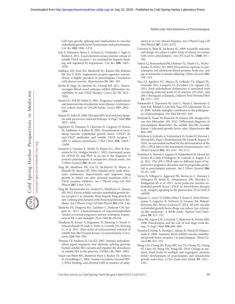
Cell-type-specific splicing and implications to vascularendothelial growth factor homeostasis and preeclampsia.Circ Res 102: 1566–1574.
Sela S, Natanson-Yaron S, Zcharia E, Vlodavsky I, Yagel S,Keshet E. 2011. Local retention versus systemic release ofsoluble VEGF receptor-1 are mediated by heparin-bind-ing and regulated by heparanase. Circ Res 108: 1063–1070.
Siddiqui AH, Irani RA, Blackwell SC, Ramin SM, KellemsRE, Xia Y. 2010. Angiotensin receptor agonistic autoan-tibody is highly prevalent in preeclampsia: Correlationwith disease severity. Hypertension 55: 386–393.
Sitohy B, Nagy JA, Jaminet SC, Dvorak HF. 2011. Tumor-surrogate blood vessel subtypes exhibit differential sus-ceptibility to anti-VEGF therapy. Cancer Res 71: 7021–7028.
Smith GC, Pell JP, Walsh D. 2001. Pregnancy complicationsand maternal risk of ischaemic heart disease: A retrospec-tive cohort study of 129,290 births. Lancet 357: 2002–2006.
Stepan H, Faber R. 2006. Elevated sFlt1 level and preeclamp-sia with parvovirus-induced hydrops. N Engl J Med 354:1857–1858.
Sugimoto H, Hamano Y, Charytan D, Cosgrove D, KieranM, Sudhakar A, Kalluri R. 2003. Neutralization of circu-lating vascular endothelial growth factor (VEGF) byanti-VEGF antibodies and soluble VEGF receptor 1(sFlt-1) induces proteinuria. J Biol Chem 278: 12605–12608.
Sunderji S, Gaziano E, Wothe D, Rogers LC, Sibai B, Kar-umanchi SA, Hodges-Savola C. 2010. Automated assaysfor sVEGF R1 and PlGF as an aid in the diagnosis ofpreterm preeclampsia: A prospective clinical study. AmJ Obstet Gynecol 202: 40 e41–e47.
Tang JR, Markham NE, Lin YJ, McMurtry IF, Maxey A,Kinsella JP, Abman SH. 2004. Inhaled nitric oxide atten-uates pulmonary hypertension and improves lunggrowth in infant rats after neonatal treatment with aVEGF receptor inhibitor. Am J Physiol Lung Cell MolPhysiol 287: L344–L351.
Tang JR, Karumanchi SA, Seedorf G, Markham N, AbmanSH. 2012. Excess soluble vascular endothelial growth fac-tor receptor-1 in amniotic fluid impairs lung growth inrats: Linking preeclampsia with bronchopulmonary dys-plasia. Am J Physiol Lung Cell Mol Physiol 302: L36–L46.
Tannetta DS, Dragovic RA, Gardiner C, Redman CW, Sar-gent IL. 2013. Characterisation of syncytiotrophoblastvesicles in normal pregnancy and pre-eclampsia: Expres-sion of Flt-1 and endoglin. PLoS ONE 8: e56754.
Thadhani R, Kisner T, Hagmann H, Bossung V, Noack S,Schaarschmidt W, Jank A, Kribs A, Cornely OA, KreyssigC, et al. 2011. Pilot study of extracorporeal removal ofsoluble fms-like tyrosine kinase 1 in preeclampsia. Circu-lation 124: 940–950.
Thomas CP, Andrews JI, Liu KZ. 2007. Intronic polyadeny-lation signal sequences and alternate splicing generatehuman soluble Flt1 variants and regulate the abundanceof soluble Flt1 in the placenta. FASEB J 21: 3885–3895.
Tissot van Patot MC, Bendrick-Peart J, Beckey VE, SerkovaN, Zwerdlinger L. 2004. Greater vascularity, lowered HIF-1/DNA binding, and elevated GSH as markers of adap-
tation to in vivo chronic hypoxia. Am J Physiol Lung CellMol Physiol 287: L525–L532.
Unemori E, Sibai B, Teichman SL. 2009. Scientific rationaleand design of a phase I safety study of relaxin in womenwith severe preeclampsia. Ann NY Acad Sci 1160: 381–384.
Vatten LJ, Romundstad PR, Holmen TL, Hsieh CC, Tricho-poulos D, Stuver SO. 2003. Intrauterine exposure to pre-eclampsia and adolescent blood pressure, body size, andage at menarche in female offspring. Obstet Gynecol 101:529–533.
Veas CJ, Aguilera VC, Munoz IJ, Gallardo VI, Miguel PL,Gonzalez MA, Lamperti LI, Escudero CA, Aguayo CR.2011. Fetal endothelium dysfunction is associated withcirculating maternal levels of sE-selectin, sVCAM1, andsFlt-1 during pre-eclampsia. J Matern Fetal Neonatal Med24: 1371–1377.
Venkatesha S, Toporsian M, Lam C, Hanai J, Mammoto T,Kim YM, Bdolah Y, Lim KH, Yuan HT, Libermann TA, etal. 2006. Soluble endoglin contributes to the pathogene-sis of preeclampsia. Nat Med 12: 642–649.
Verdonk K, Visser W, Russcher H, Danser AH, Steegers EA,van den Meiracker AH. 2012. Differential diagnosis ofpreeclampsia: Remember the soluble fms-like tyrosinekinase 1/placental growth factor ratio. Hypertension 60:884–890.
Verlohren S, Galindo A, Schlembach D, Zeisler H, Herraiz I,Moertl MG, Pape J, Dudenhausen JW, Denk B, Stepan H.2010. An automated method for the determination of thesFlt-1/PlGF ratio in the assessment of preeclampsia. Am JObstet Gynecol 202: 161 e161–161 e111.
Verlohren S, Herraiz I, Lapaire O, Schlembach D, Moertl M,Zeisler H, Calda P, Holzgreve W, Galindo A, Engels T, etal. 2012. The sFlt-1/PlGF ratio in different types of hy-pertensive pregnancy disorders and its prognostic poten-tial in preeclamptic patients. Am J Obstet Gynecol 206:58 e51–e58.
Veron D, Villegas G, Aggarwal PK, Bertuccio C, Jimenez J,Velazquez H, Reidy K, Abrahamson DR, Moeckel G,Kashgarian M, et al. 2012. Acute podocyte vascular en-dothelial growth factor (VEGF-A) knockdown disruptsaVb3 integrin signaling in the glomerulus. PLoS ONE 7:e40589.
Vigneau C, Lorcy N, Dolley-Hitze T, Jouan F, Arlot-Bonne-mains Y, Laguerre B, Verhoest G, Goujon JM, Belaud-Rotureau MA, Rioux-Leclercq N. 2014. All anti-vascularendothelial growth factor drugs can induce ‘pre-eclamp-sia-like syndrome’: A RARe study. Nephrol Dial Trans-plant 29: 325–332.
Vikse BE, Irgens LM, Leivestad T, Skjaerven R, Iversen BM.2008. Preeclampsia and the risk of end-stage renal dis-ease. N Engl J Med 359: 800–809.
Vuorela P, Helske S, Hornig C, Alitalo K, Weich H, Halmes-maki E. 2000. Amniotic fluid–soluble vascular endothe-lial growth factor receptor-1 in preeclampsia. Obstet Gy-necol 95: 353–357.
Wang CN, Chang SD, Peng HH, Lee YS, Chang YL, ChengPJ, Chao AS, Wang TH, Wang HS. 2010. Change in am-niotic fluid levels of multiple anti-angiogenic proteinsbefore development of preeclampsia and intrauterinegrowth restriction. J Clin Endocrinol Metab 95: 1431–1441.
Molecular Mechanisms of Preeclampsia
Cite this article as Cold Spring Harb Perspect Med 2015;5:a023473 19
ww
w.p
ersp
ecti
vesi
nm
edic
ine.
org
on July 22, 2018 - Published by Cold Spring Harbor Laboratory Press http://perspectivesinmedicine.cshlp.org/Downloaded from
Wang IK, Muo CH, Chang YC, Liang CC, Chang CT, Lin SY,Yen TH, Chuang FR, Chen PC, Huang CC, et al. 2013a.Association between hypertensive disorders during preg-nancy and end-stage renal disease: A population-basedstudy. CMAJ 185: 207–213.
Wang IK, Muo CH, Chang YC, Liang JC, Chang CT, Lin SY,Yen TH, Chuang FR, Chen PC, Huang CC, et al. 2013b.Association between hypertensive disorders during preg-nancy and end-stage renal disease: A population-basedstudy. CMAJ 185: 207–213.
Wilson BJ, Watson MS, Prescott GJ, Sunderland S, CampbellDM, Hannaford P, Smith WC. 2003. Hypertensive dis-eases of pregnancy and risk of hypertension and stroke inlater life: Results from cohort study. BMJ 326: 845.
Wolf M, Hubel CA, Lam C, Sampson M, Ecker JL, Ness RB,Rajakumar A, Daftary A, Shakir AS, Seely EW, et al. 2004.Preeclampsia and future cardiovascular disease: Potentialrole of altered angiogenesis and insulin resistance. J ClinEndocrinol Metab 89: 6239–6243.
Xinaris C, Benedetti V, Rizzo P, Abbate M, Corna D, Azzol-lini N, Conti S, Unbekandt M, Davies JA, Morigi M, et al.2012. In vivo maturation of functional renal organoidsformed from embryonic cell suspensions. J Am Soc Neph-rol 23: 1857–1868.
Yu XD, Branch DW, Karumanchi SA, Zhang J. 2012. Pre-eclampsia and retinopathy of prematurity in pretermbirths. Pediatrics 130: e101–e107.
Yuan HT, Haig D, Ananth Karumanchi S. 2005. Angiogenicfactors in the pathogenesis of preeclampsia. Curr Top DevBiol 71: 297–312.
Zhao S, Gu Y, Fan R, Groome LJ, Cooper D, Wang Y. 2010.Proteases and sFlt-1 release in the human placenta. Pla-centa 31: 512–518.
Zhou Y, McMaster M, Woo K, Janatpour M, Perry J, Karpa-nen T, Alitalo K, Damsky C, Fisher SJ. 2002. Vascularendothelial growth factor ligands and receptors that reg-ulate human cytotrophoblast survival are dysregulated insevere preeclampsia and hemolysis, elevated liver en-zymes, and low platelets syndrome. Am J Pathol 160:1405–1423.
Zhou CC, Zhang Y, Irani RA, Zhang H, Mi T, Popek EJ,Hicks MJ, Ramin SM, Kellems RE, Xia Y. 2008. Angio-tensin receptor agonistic autoantibodies induce pre-eclampsia in pregnant mice. Nat Med 14: 855–862.
Zhou Y, Gormley MJ, Hunkapiller NM, Kapidzic M, Sto-lyarov Y, Feng V, Nishida M, Drake PM, Bianco K, Wang F,et al. 2013. Reversal of gene dysregulation in culturedcytotrophoblasts reveals possible causes of preeclampsia.J Clin Invest 123: 2862–2872.
T. Hod et al.
20 Cite this article as Cold Spring Harb Perspect Med 2015;5:a023473
ww
w.p
ersp
ecti
vesi
nm
edic
ine.
org
on July 22, 2018 - Published by Cold Spring Harbor Laboratory Press http://perspectivesinmedicine.cshlp.org/Downloaded from
August 20, 20152015; doi: 10.1101/cshperspect.a023473 originally published onlineCold Spring Harb Perspect Med
Tammy Hod, Ana Sofia Cerdeira and S. Ananth Karumanchi Molecular Mechanisms of Preeclampsia
Subject Collection Molecular Approaches to Reproductive and Newborn Medicine
Information in SpermIntergenerational Transfer of Epigenetic
Oliver J. Rando TechnologiesOutcomes with Assisted Reproductive A Molecular Perspective on Procedures and
Butts, et al.Monica A. Mainigi, Carmen Sapienza, Samantha
Programming: Molecular ApproachesEffects of Maternal Obesity on Fetal
Caterina Neri and Andrea G. Edlow Have Single-Gene DisorderstoSequencing in Critically Ill Neonates Suspected
Whole-Exome Sequencing and Whole-Genome
KingsmoreLaurie D. Smith, Laurel K. Willig and Stephen F.
The Neonatal Salivary TranscriptomeJill L. Maron Blood Group Using Next-Generation Sequencing
Noninvasive Antenatal Determination of Fetal
Morten Hanefeld DziegielKlaus Rieneck, Frederik Banch Clausen and
Tract Development, Adult Function, and Fertility Genes in Female ReproductiveHoxThe Role of
Hongling Du and Hugh S. Taylor Newborn ScreeningGenome-Scale Sequencing to Augment Current Potential Uses and Inherent Challenges of Using
Jonathan S. Berg and Cynthia M. Powell
InterfaceMaternal−Molecular Cross-Talk at the Feto
Gendie E. Lashthe Decidual ClockMolecular Regulation of Parturition: The Role of
Snegovskikh, et al.Errol R. Norwitz, Elizabeth A. Bonney, Victoria V.
PerspectiveMolecular Regulation of Parturition: A Myometrial
Montalbano, et al.Nora E. Renthal, Koriand'r C. Williams, Alina P.
Molecular Mechanisms of Preeclampsia
KarumanchiTammy Hod, Ana Sofia Cerdeira and S. Ananth
of Fetal Single-Gene DisordersGenome-Wide Sequencing for Prenatal Detection
Ignatia B. Van den Veyver and Christine M. Eng Maternal Plasma DNADiseases Using Massively Parallel Sequencing of Noninvasive Prenatal Screening for Genetic
Lyn S. Chitty and Y. M. Dennis LoMicroRNA in Ovarian Biology and Disease
ChristensonLynda K. McGinnis, Lacey J. Luense and Lane K. The Oocyte's Perspective on the Incoming Sperm
Confrontation, Consolidation, and Recognition:
David Miller
http://perspectivesinmedicine.cshlp.org/cgi/collection/ For additional articles in this collection, see
Copyright © 2015 Cold Spring Harbor Laboratory Press; all rights reserved
on July 22, 2018 - Published by Cold Spring Harbor Laboratory Press http://perspectivesinmedicine.cshlp.org/Downloaded from