Embed Size (px)
Citation preview
RESEARCH Open Access
Molecular mechanisms of growthdepression in broiler chickens (Gallus Gallusdomesticus) mediated by immune stress: ahepatic proteome studyAijuan Zheng1, Anrong Zhang1, Zhimin Chen1, Shoaib Ahmed Pirzado1, Wenhuan Chang1, Huiyi Cai1,Wayne L. Bryden2 and Guohua Liu1*
Abstract
Background: Immunological stress decreases feed intake, suppresses growth and induces economic losses.However, the underlying molecular mechanism remains unclear. Label-free liquid chromatography and massspectrometry (LC-MS) proteomics techniques were employed to investigate effects of immune stress on the hepaticproteome changes of Arbor Acres broilers (Gallus Gallus domesticus) challenged with Escherichia colilipopolysaccharide (LPS).
Results: Proteomic analysis indicated that 111 proteins were differentially expressed in the liver of broiler chickensfrom the immune stress group. Of these, 28 proteins were down-regulated, and 83 proteins were up-regulated inthe immune stress group. Enrichment analysis showed that immune stress upregulated the expression of hepaticproteins involved in defense function, amino acid catabolism, ion transport, wound healing, and hormonesecretion. Furthermore, immune stress increased valine, leucine and isoleucine degradation pathways.
Conclusion: The data suggests that growth depression of broiler chickens induced by immune stress is triggeredby hepatic proteome alterations, and provides a new insight into the mechanism by which immune challengeimpairs poultry production.
Keywords: Broiler chickens, Hepatic proteome, Immune stress, Lipopolysaccharide
BackgroundIntensive poultry production is conducted in an environ-ment that imposes many stressors on the bird. Thestressed bird instigates an integrated response to main-tain homeostasis through cross-talk between the centralnervous, endocrine and immune systems [1]. Stressorsin the bird’s environment, include feeding management,overcrowding, temperature extremes, dust and litter
condition, pathogen challenges, vaccination, and psycho-logical factors [2, 3]. All of these stressors can have a cu-mulative impact on poultry behavior and physiology,thus affecting the immune response and inducing im-munologically mediated stress or immune stress [4, 5].Immune stress is harmful to the bird and can be miti-gated by improving the bird’s environment. Studies haveshown that stress dysregulates the immune response byincreasing the release of inflammatory cytokines andstress hormones [6, 7], reducing NK cell activity,lymphocyte populations, lymphocyte proliferation, anti-body production and reactivating latent viral infections
© The Author(s). 2021 Open Access This article is licensed under a Creative Commons Attribution 4.0 International License,which permits use, sharing, adaptation, distribution and reproduction in any medium or format, as long as you giveappropriate credit to the original author(s) and the source, provide a link to the Creative Commons licence, and indicate ifchanges were made. The images or other third party material in this article are included in the article's Creative Commonslicence, unless indicated otherwise in a credit line to the material. If material is not included in the article's Creative Commonslicence and your intended use is not permitted by statutory regulation or exceeds the permitted use, you will need to obtainpermission directly from the copyright holder. To view a copy of this licence, visit http://creativecommons.org/licenses/by/4.0/.The Creative Commons Public Domain Dedication waiver (http://creativecommons.org/publicdomain/zero/1.0/) applies to thedata made available in this article, unless otherwise stated in a credit line to the data.
* Correspondence: [email protected] Laboratory of Feed Biotechnology of Ministry of Agriculture and RuralAffairs, Institute of Feed Research, Chinese Academy of Agricultural Sciences,No.12 Zhongguancun south street, Haidian district, Beijing 100081, ChinaFull list of author information is available at the end of the article
Zheng et al. Journal of Animal Science and Biotechnology (2021) 12:90 https://doi.org/10.1186/s40104-021-00591-1
[8, 9]. In response to an immune challenge, the appetiteand growth performance of the bird will decline [10–12]. Immune stress can also disrupt the balance andcomposition of the cecal microflora, impair intestinalmucosal immune function, and reduce ileal protein di-gestibility [13].A bird or animal’s metabolic priorities are rearranged
in response to immune stress, resulting in the redistribu-tion of nutrients away from muscle protein depositionand growth to support upregulation of the immune re-sponse [14, 15]. The liver plays a pivotal role in nutrientmetabolism, and nutrient repartitioning following an im-mune challenge when it is enriched with components ofthe immune system, including macrophages and naturalkiller T-cells; highlighting the vital role of the liver inimmunology [16].Despite extensive research on the effects of immune
stress in broiler chickens, changes in the avian hepaticproteome and the molecular mechanisms induced by animmune insult are not well understood. Proteins as thefunctional carrier of genes can provide both genomicand functional information [17]. Proteomics represents anew strategy to delineate the molecular basis of thephysiological changes in the liver during chicken growth[18, 19]. This approach determines the differential pat-terns of protein abundance and has been used to dem-onstrate their functional relationships to external factors[20, 21]. Lipopolysaccharide (LPS) injection is a classicalmodel for inducing immune stress in broiler chickens [7,10, 22, 23]. In the present experiment, this model wasused to investigate the hypothesis that changes in the ex-pression of the hepatic proteome of broilers occur fol-lowing immune challenge and help explain the responseof the bird. Our findings clarify protein expression andbiological process changes in the liver of immunologic-ally challenged broilers and provides further informationto assist in maintaining the health and productivity ofmeat or broiler chickens.
MethodsMaterials and reagentsAll chemicals were purchased from Sigma-Aldrich (St.Louis., Missouri, USA) except modified sequencinggrade trypsin that was bought from Promega (Madison,WI, USA). LPS from E. coli (O55:B5) was used in thepresent experiment.
Bird managementA total of 144 one-day-old, male, Arbor Acres (AA)broiler chickens were purchased from Huadu ChickenCo. (Beijing, China). The chicks were randomly dividedinto two groups: challenged with saline (control group)or LPS (treatment group). Each group had 6 replicateswith 12 birds in each replicate. The distribution of cages
was arranged to avoid any location effects within thepoultry house. The chickens were reared in two phasesand fed a starter diet during d 0–21 and a grower dietduring d 22–42. The composition of these corn-soybeanbased diets are shown in Table 1. All chickens were in-oculated and subjected to a photoperiod of 16 h lightand 8 h dark in accordance with the AA Broiler Manage-ment Guide. The room temperature was maintained at33–35 °C on d 0–3, at 32–34 °C on d 4–7 and graduallyreduced to the maintenance temperature of 20 °C byd 42. The relative humidity was kept at 70% duringthe first week and thereafter at about 60%.
Experimental treatments and LPS administrationFor the first 5 weeks of the study all birds were main-tained in a similar manner. On d 36, 38, and 40, allchickens (each group had 6 replicates with 12 birds ineach replicate) were injected intravenously with either1 mL sterile saline (control group) or LPS (treatmentgroup) dissolved in saline at an approximate dose of5.0 mg/kg body weight (LPS or immune stress group).The injection protocol is the established method usedwhen inducing an immunological challenge with LPS[4, 7, 10, 22, 23]. The protocol commenced at 5 weeksof age to avoid endocrine and physiological changesthat occur during the starter phase and to permitadditional muscle samples to be collected for meatanalysis; results reported separately.
Performance parametersThe body weight of all birds in each replicate was mea-sured on d 36 (before the first injection, W0), d 38(2 days after the first injection, W38), d 40 (2 days afterthe second injection, W40) and d 42 (2 days after the thirdinjection, W42). The change in body weight caused bysaline or LPS treatment was expressed as body weightgain (W1 =W38- W0, W2 =W40- W0, W3 =W42- W0).W1–3 indicates body weight gain after the first, second orthird injection of LPS. Mortality was recorded daily.
Sample collection and parameters determined in bloodOn d 42, all birds were weighed after a 12 h-fast. Threebirds from each replicate were selected randomly,electrically stunned, and manually slaughtered within5min [24]. Blood was collected using vacutainer tubes.The serum, obtained by centrifugation at 1,500 × gfor 15min, was used for the determination of hormonesand inflammatory factors. The concentrations of adreno-corticotropic hormone (ACTH), corticosterone (CORT),growth hormone (GH) and insulin-like growth factor-1(IGF-1), interleukin-1β (IL-1β), interleukin-6 (IL-6),and tumour necrosis factor-α (TNF-α) were determinedby quantitative sandwich enzyme immunoassay usingcommercial kits (Beijing North Institute of Biological
Zheng et al. Journal of Animal Science and Biotechnology (2021) 12:90 Page 2 of 19
Technology, Beijing, China), according to the manufac-turer’s instructions.The middle section of the major or right lobe of the liver
was sampled and washed with PBS buffer (NaCl 8 g/L,Na2HPO4 1.44 g/L, KH2PO4 0.24 g/L, KCl 0.2 g/L, pH 7.2)to remove any blood and contaminants on the surface. Aliver sample (about 2 g) was taken and put into 5mLultra-low temperature freezing tubes (Free Sterile). Sam-ples were immediately frozen in liquid nitrogen and storedat − 80 °C. Likewise, intestinal and muscle samples werealso collected and the outcome of their analyses willbe published elsewhere.
Protein extraction and digestionThe liver samples of three chickens from each replicate(cage) were combined as a biological replicate, homoge-nized by pestle in liquid nitrogen. Six biological repli-cates of each group were analyzed. Protein extractionwas performed as previously described [18]. In short,after homogenization the samples were then mixed witha lysis buffer containing 8 mol urea, 2 mol thiourea, 4%3-[(3-cholamidopropyl) dimethylammonio]-1-propane-sulfonate, 20 mmol Trisbase, 30 mmol dithiothreitol(DTT), and protease inhibitors in ice for 30 min. Thesample was then centrifuged at 15,000 × g for 20 min at10 °C to remove the insoluble fractions. Three volumesof ice-cold acetone were added to the recovered super-natant and allowed to stand at 20 °C for 4 h to precipi-tate the proteins. Subsequently, the protein pellets werecentrifuged at 8,000 × g at 10 °C for 20 min. The super-natant was discarded, followed by extraction of the pro-tein pellet at room temperature. The recovered proteinswere re-suspended in 100–150 μL of 5 mol urea, andprotein concentration was quantified by the Bradfordassay after diluting 50 times. Of each sample, 200 μg ofproteins were used by adding four volumes of 40 mmolNH4HCO3, mixing with DTT (final concentration 10mmol) for 1 h, and then alkylating with iodoacetamide(final concentration 50 mmol) for 1 h in the dark. Thesurplus iodoacetamide was quenched by DTT (finalconcentration 30 mmol). To digest protein into pep-tides, sequencing grade modified trypsin was used(enzyme/protein ratio of 1:100 (W/W)) at 37 °C for14 h. The enzymatic digestion was stopped by adding1 μL of formic acid to the solution. The digested pep-tide samples were desalted using a C18 column (AgilentTechnologies Inc., Santa Clara, CA, USA). The elutedpeptide solution was collected and extracted using aSpeedVac system (RVC 2–18, Marin Christ, Osterod,Germany) and stored at −80 °C for subsequent LC-MS/MS analysis.
Liquid chromatography and mass spectrometry (LC −MS/MS) analysisThe digested peptide samples were re-dissolved in 50 μLof 0.1% formic acid. Three replicates of each samplewere run using a Q-Exactive mass spectrometer(Thermo Fisher Scientific, USA) and coupled to theEASY-nLC 1000 system using a nano electrospray ionsource (Thermo Fisher Scientific, USA). To enrich thepeptide samples, they were first loaded onto a 2 cm longtrap column (75 μm inner diameter fused silica contain-ing 3 μm Aqua C18 beads, Thermo Fisher Scientific,USA) for 2 min in buffer A (0.1% acetic acid) at a flowrate of 10 μL/min. Secondly, the peptides were separatedby an analytical column (15 cm long, 50 μm inner diam-eter fused silica column filing with 2 μm Aqua C18
Table 1 Ingredient and nutrient composition of experimentalbroiler diets
Starter (1–21 d),g/kg
Grower (22–42 d),g/kg
Ingredient
Corn 593.1 604.2
Soybean meal 298.8 288.7
Cotton seed meal 50.0 30.0
Soybean oil 15.1 39.8
L-Lysine 1.5 0.9
DL-Methionine 1.4 1.6
Limestone 12.7 10.2
CaHPO4 19.4 16.6
NaCl 3.0 3.0
Choline chloride 2.0 2.0
Vitamin premix 0.3 0.3
Mineral premixa 1.0 1.0
Zeolite powder 1.7 1.7
Total 1000 1000
Nutrient concentrationsb
Metabolic energy, MJ/kg 12.35 13.02
Crude protein 211.8 198.4
Calcium 10.1 8.5
Available phosphorus 4.5 4.0
Total phosphorus 6.9 6.3
Lysine 11.4 10.5
Methionine 4.9 4.8
Methionine + Cysteine 8.3 8.1
Threonine 7.7 2.2aThe premix provided the following per kg diet: vitamin A 10, 000 IU, vitaminD3 2,000 IU, vitamin E 10 IU, vitamin K3 2.5 mg, vitamin B1 1 mg, vitamin B2 6mg, vitamin B3 10 mg, vitamin B5 40 mg, vitamin B6 3 mg, vitamin B11 0.3 mg,vitamin B12 0.01 mg, biotin 0.12 mg, Cu (as copper sulfate) 8 mg, Fe (as ferroussulfate) 80 mg, Mn (as manganese sulfate) 60 mg, Zn (as zinc sulfate) 40 mg,Se (as sodium selenite) 0.15 mg, I (as potassium iodide) 0.35 mgbCalculated values
Zheng et al. Journal of Animal Science and Biotechnology (2021) 12:90 Page 3 of 19
beads, Thermo Fisher Scientific, USA) using a 120 mingradient. Peptides were gradient eluted for 110 min witha linear gradient from 8% to 30% acetonitrile at a flowrate of 300 nL/min. The eluting peptides from the ana-lytical column were directly infused into a Q-Exactivemass spectrometer via electrospray ionization. The set-tings for a data-dependent mode to collect the MS andMS/MS data were as follows: one full scan (resolution70,000 at 400 m/z; 350 to 1,600 m/z) followed by top 20MS/MS scans using higher-energy collisional dissoci-ation in the linear ion trap mass spectrometer (reso-lution: 15,000, isolation window: 2 m/z, normalizedcollision energy: 28) using dynamic exclusion (charge ex-clusion: unassigned 1, > 8; peptide match: preferred; ex-clude isotopes: on; dynamic exclusion: 30 s). Foridentification and abundance level quantification of pro-teins, the MS/MS data in RAW were retrieved usingXcalibur (version 3.0, Thermo Fisher Scientific, USA)and searched using in-house PEAKS software (version8.5, Bioinformatics Solutions Inc., CAN).A database containing protein sequences of Gallus
Gallus domesticus including common contaminants wasdownloaded from NCBI and used, totaling to 76,213 en-tries (downloaded 25 June, 2020). The parameters of thesearch database were as follows: trypsin; maximum missedcleavage: 2; precursor ion and MS/MS tolerances: 15 ppmand 0.05 Da; a fixed modification: carbamidomethyl(C, + 57.02); and a variable modification: methionine oxi-dation (M, + 15.99), asparagine and glutamine deamin-ation (+ 0.984 Da). The fusion-decoy database searchstrategy with threshold false discovery rate (FDR ≤ 1%)was used to control the FDR at both the protein and pep-tide levels. A protein was considered as identified only if ithad at least one unique peptide. To quantify the relativeprotein abundance in the livers of broiler chickens bothfrom the control group and immune stress group, threereplications of each sample were performed in the quanti-fication module of PEAKS software (version 8.5) via alabel-free strategy. Feature detection was performed sepa-rately on each sample using the expectation-maximizationalgorithm. Using the high-performance retention timealignment algorithms, the features of the same peptidefrom three replicates of each sample were reliably aligned[25]. Normalization was done by dividing each matrix by afactor of the samples obtained as follows: the total ioncurrent (TIC) of the individual sample / the TIC of thereference sample. Quantification of protein abundance inthe livers in all samples of broiler chickens was done usingthe sum of the three highest ion peak intensities of thetryptic peptides.
GO term enrichment analysisTo understand the biological implications of the identi-fied proteins in the liver of broiler chickens, identifiers
of protein symbol ID numbers were used as an input forGO term enrichment (functional classes and pathway)using ClueGOv2.3.2, a Cytoscape plug-in (http://www.ici.upmc.fr/cluego/) [26]. The number of proteins identi-fied from the samples was compared with the number offunctionally GO annotated proteins in the entire broilerchicken (Gallus Gallus domesticus) genome for enrich-ment analysis. The significantly enriched GO terms inbiological processes and pathways were reported using aright-sided hyper-geometric test and only a P-value < 0.05was considered. Then, Bonferroni step-down procedurewas used to correct the P-value to control FDR. Func-tional grouping of the terms was based on GO hierarchy.The tree level was ranged from 3 to 8, and kappa scorelevel was 0.4. For comparison purpose, sharing 65% of theterms was considered to be merged.
Protein–protein interaction analysisA protein–protein interaction network of differentialproteins was constructed using the STRING 11.0 (http://string-db.org/) [27]. The network nodes represent pro-teins, and the edges represent the predicted functionalassociations.
Statistical analysisMeans of replicate were used as the experimental unitfor statistical analysis. The data of blood parameterswere analyzed by Independent-Samples T-Test moduleusing SPSS 17.0 software (version 17.0, SPSS Inc., Chi-cago, IL, USA). Results are presented as the mean ± SE.Differences between means were considered statisticallysignificant at P < 0.05.Proteins from different samples were considered to be
significantly changed in their abundance only when theyattained the criteria (P-value < 0.05 and a fold change of> 1.5 or < 0.5).
ResultsGrowth and wellbeing of all chicks was normal for thefirst 5 weeks of the study or until the LPS challenge wasintroduced.
Effects of body weight gain of broilers challenged withLPSThe effects of immune stress on body weight gain ofbroilers is shown in Table 2. Body weight gain in broilersinjected with LPS was significantly lower than in the un-challenged broilers.
Changes of serum hormones and cytokines of broilerschallenged with LPSAs shown in Table 3, the serum concentrations ofACTH, CORT, IL-1β, TNF-α and IL-6 in broilersinjected with LPS were significantly higher than in the
Zheng et al. Journal of Animal Science and Biotechnology (2021) 12:90 Page 4 of 19
unchallenged broilers. However, GH and IGF- І concen-trations in serum decreased significantly in the broilersfrom the immune stress group.
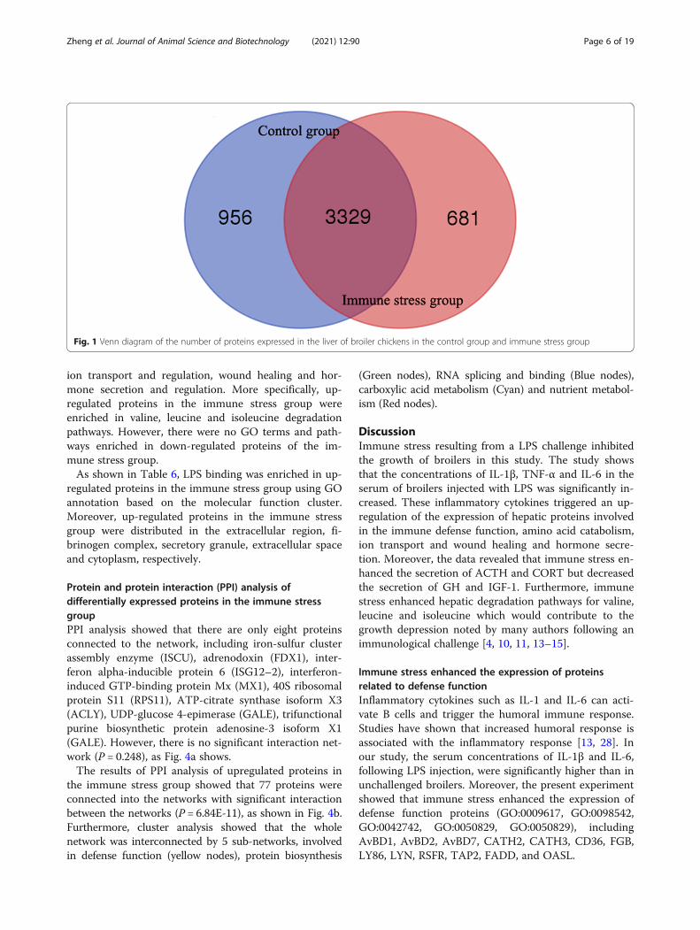
Qualitative differential analysis of hepatic proteome inbroiler chickens between the control and the immunestress groupProtein numbers expressed in the liver of broiler chickensIn the present study, a total of 4,966 proteins were iden-tified in the liver tissues of broiler chickens. In the con-trol group, 4,285 proteins (2,307 groups) were identifiedand 4,010 proteins (2,182 groups) were identified in theLPS group. As shown in Figs. 1, 3,329 proteins wereexpressed in both the control and treatment groups.
GO and KEGG analysis of unique proteins speciallyexpressed in the control groupAs shown in Fig. 2a, KEGG pathway analysis was per-formed on specifically expressed proteins in the controlgroup and demonstrated enrichment of endocytosis, per-oxisome, Golgi vesicle transport, RNA transport, prote-asome, protein processing in endoplasmic reticulum,fatty acid degradation, spliceosome, ribosome and pyru-vate metabolism pathways.Go analysis showed that the following biological pro-
cesses were enriched in the control group, including, re-sponse to toxic substances, oxidation-reduction, aminoacid metabolism, small molecule biosynthesis, transpor-tation (hydrogen or proton transport), proteins biosyn-thesis (organonitrogen compound metabolic andbiosynthetic processes, translation), actin polymerizationor depolymerization and its regulation, nucleic acid bio-synthesis and metabolism (nucleoside phosphate meta-bolic process, nucleoside biosynthetic process,nucleoside monophosphate metabolic process etc.), fattyacid metabolism (fatty acid metabolic process and lipid
catabolic process, etc.), cofactor and coenzyme biosyn-thetic and metabolic process, organic acid metabolism(organic acid catabolic or biosynthetic process, carbox-ylic acid, monocarboxylic acid and glycosyl compoundmetabolic or biosynthetic process).
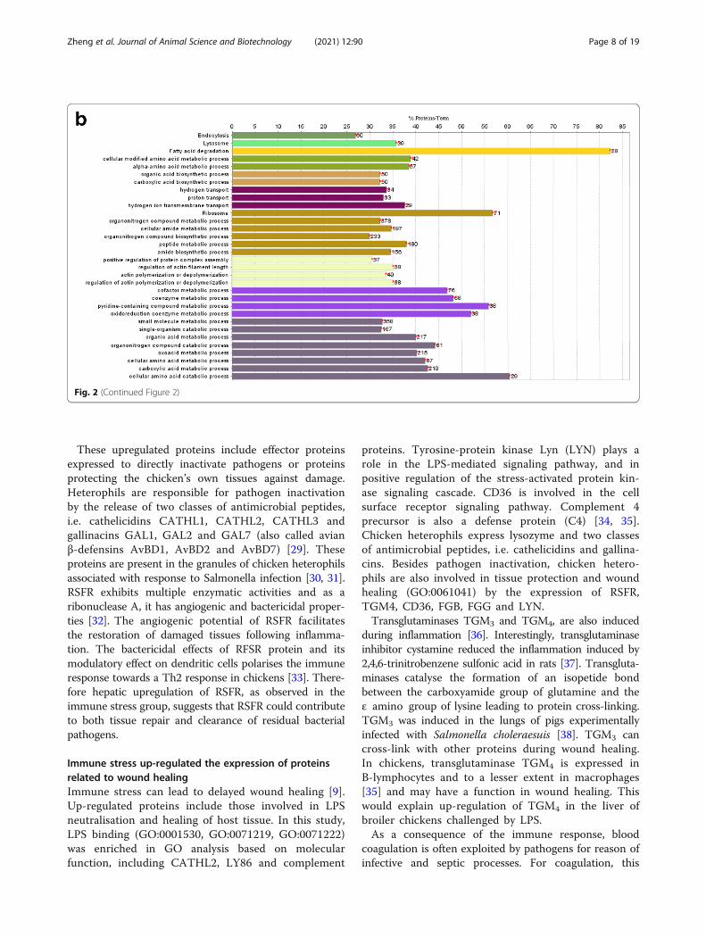
GO and KEGG analysis of unique proteins speciallyexpressed in the immune stress groupAs shown in Fig. 2b, KEGG pathway analysis was per-formed on specifically expressed proteins in the immunestress group. Endocytosis, lysosome, fatty acid degrad-ation, ribosome pathways were enriched.Go analysis showed that the following biological pro-
cesses were enriched in the LPS group, including, aminoacid metabolism, organic acid and carboxylic acid bio-synthesis, transportation (hydrogen or proton transport),organonitrogen compound metabolic and biosyntheticprocesses, positive regulation of protein complex assem-bly, actin polymerization or depolymerization and itsregulation, cofactor and coenzyme metabolism, organicacid metabolism (organic acid, carboxylic acid, oxoacidand amino acid metabolic and catabolic processes).
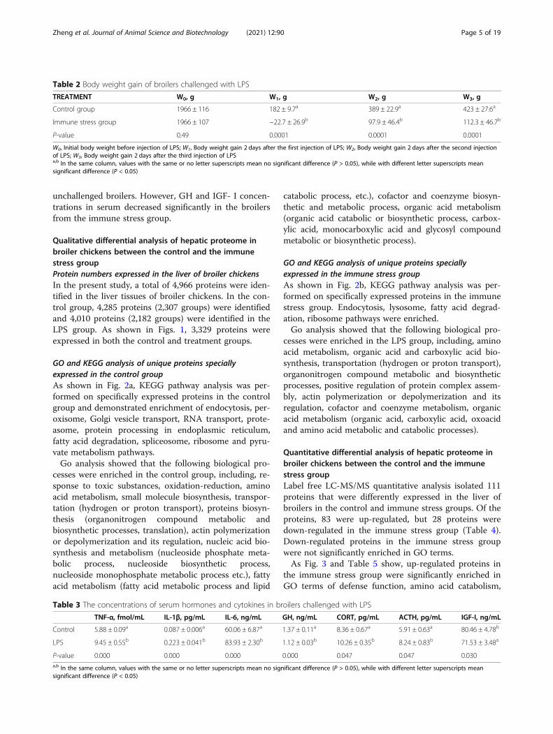
Quantitative differential analysis of hepatic proteome inbroiler chickens between the control and the immunestress groupLabel free LC-MS/MS quantitative analysis isolated 111proteins that were differently expressed in the liver ofbroilers in the control and immune stress groups. Of theproteins, 83 were up-regulated, but 28 proteins weredown-regulated in the immune stress group (Table 4).Down-regulated proteins in the immune stress groupwere not significantly enriched in GO terms.As Fig. 3 and Table 5 show, up-regulated proteins in
the immune stress group were significantly enriched inGO terms of defense function, amino acid catabolism,
Table 2 Body weight gain of broilers challenged with LPS
TREATMENT W0, g W1, g W2, g W3, g
Control group 1966 ± 116 182 ± 9.7a 389 ± 22.9a 423 ± 27.6a
Immune stress group 1966 ± 107 −22.7 ± 26.9b 97.9 ± 46.4b 112.3 ± 46.7b
P-value 0.49 0.0001 0.0001 0.0001
W0, Initial body weight before injection of LPS; W1, Body weight gain 2 days after the first injection of LPS; W2, Body weight gain 2 days after the second injectionof LPS; W3, Body weight gain 2 days after the third injection of LPSa,b In the same column, values with the same or no letter superscripts mean no significant difference (P > 0.05), while with different letter superscripts meansignificant difference (P < 0.05)
Table 3 The concentrations of serum hormones and cytokines in broilers challenged with LPS
TNF-α, fmol/mL IL-1β, pg/mL IL-6, ng/mL GH, ng/mL CORT, pg/mL ACTH, pg/mL IGF-І, ng/mL
Control 5.88 ± 0.09a 0.087 ± 0.006a 60.06 ± 6.87a 1.37 ± 0.11a 8.36 ± 0.67a 5.91 ± 0.63a 80.46 ± 4.78b
LPS 9.45 ± 0.55b 0.223 ± 0.041b 83.93 ± 2.30b 1.12 ± 0.03b 10.26 ± 0.35b 8.24 ± 0.83b 71.53 ± 3.48a
P-value 0.000 0.000 0.000 0.000 0.047 0.047 0.030a,b In the same column, values with the same or no letter superscripts mean no significant difference (P > 0.05), while with different letter superscripts meansignificant difference (P < 0.05)
Zheng et al. Journal of Animal Science and Biotechnology (2021) 12:90 Page 5 of 19
ion transport and regulation, wound healing and hor-mone secretion and regulation. More specifically, up-regulated proteins in the immune stress group wereenriched in valine, leucine and isoleucine degradationpathways. However, there were no GO terms and path-ways enriched in down-regulated proteins of the im-mune stress group.As shown in Table 6, LPS binding was enriched in up-
regulated proteins in the immune stress group using GOannotation based on the molecular function cluster.Moreover, up-regulated proteins in the immune stressgroup were distributed in the extracellular region, fi-brinogen complex, secretory granule, extracellular spaceand cytoplasm, respectively.
Protein and protein interaction (PPI) analysis ofdifferentially expressed proteins in the immune stressgroupPPI analysis showed that there are only eight proteinsconnected to the network, including iron-sulfur clusterassembly enzyme (ISCU), adrenodoxin (FDX1), inter-feron alpha-inducible protein 6 (ISG12–2), interferon-induced GTP-binding protein Mx (MX1), 40S ribosomalprotein S11 (RPS11), ATP-citrate synthase isoform X3(ACLY), UDP-glucose 4-epimerase (GALE), trifunctionalpurine biosynthetic protein adenosine-3 isoform X1(GALE). However, there is no significant interaction net-work (P = 0.248), as Fig. 4a shows.The results of PPI analysis of upregulated proteins in
the immune stress group showed that 77 proteins wereconnected into the networks with significant interactionbetween the networks (P = 6.84E-11), as shown in Fig. 4b.Furthermore, cluster analysis showed that the wholenetwork was interconnected by 5 sub-networks, involvedin defense function (yellow nodes), protein biosynthesis
(Green nodes), RNA splicing and binding (Blue nodes),carboxylic acid metabolism (Cyan) and nutrient metabol-ism (Red nodes).
DiscussionImmune stress resulting from a LPS challenge inhibitedthe growth of broilers in this study. The study showsthat the concentrations of IL-1β, TNF-α and IL-6 in theserum of broilers injected with LPS was significantly in-creased. These inflammatory cytokines triggered an up-regulation of the expression of hepatic proteins involvedin the immune defense function, amino acid catabolism,ion transport and wound healing and hormone secre-tion. Moreover, the data revealed that immune stress en-hanced the secretion of ACTH and CORT but decreasedthe secretion of GH and IGF-1. Furthermore, immunestress enhanced hepatic degradation pathways for valine,leucine and isoleucine which would contribute to thegrowth depression noted by many authors following animmunological challenge [4, 10, 11, 13–15].
Immune stress enhanced the expression of proteinsrelated to defense functionInflammatory cytokines such as IL-1 and IL-6 can acti-vate B cells and trigger the humoral immune response.Studies have shown that increased humoral response isassociated with the inflammatory response [13, 28]. Inour study, the serum concentrations of IL-1β and IL-6,following LPS injection, were significantly higher than inunchallenged broilers. Moreover, the present experimentshowed that immune stress enhanced the expression ofdefense function proteins (GO:0009617, GO:0098542,GO:0042742, GO:0050829, GO:0050829), includingAvBD1, AvBD2, AvBD7, CATH2, CATH3, CD36, FGB,LY86, LYN, RSFR, TAP2, FADD, and OASL.
Fig. 1 Venn diagram of the number of proteins expressed in the liver of broiler chickens in the control group and immune stress group
Zheng et al. Journal of Animal Science and Biotechnology (2021) 12:90 Page 6 of 19
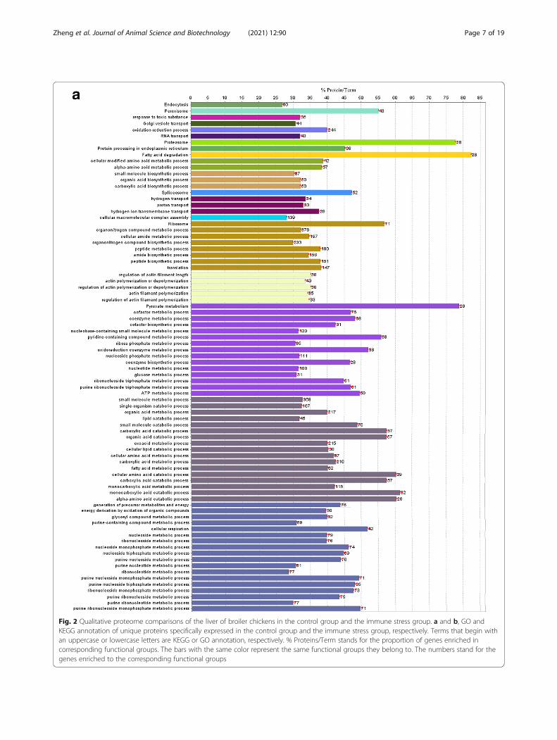
Fig. 2 Qualitative proteome comparisons of the liver of broiler chickens in the control group and the immune stress group. a and b, GO andKEGG annotation of unique proteins specifically expressed in the control group and the immune stress group, respectively. Terms that begin withan uppercase or lowercase letters are KEGG or GO annotation, respectively. % Proteins/Term stands for the proportion of genes enriched incorresponding functional groups. The bars with the same color represent the same functional groups they belong to. The numbers stand for thegenes enriched to the corresponding functional groups
Zheng et al. Journal of Animal Science and Biotechnology (2021) 12:90 Page 7 of 19
These upregulated proteins include effector proteinsexpressed to directly inactivate pathogens or proteinsprotecting the chicken’s own tissues against damage.Heterophils are responsible for pathogen inactivationby the release of two classes of antimicrobial peptides,i.e. cathelicidins CATHL1, CATHL2, CATHL3 andgallinacins GAL1, GAL2 and GAL7 (also called avianβ-defensins AvBD1, AvBD2 and AvBD7) [29]. Theseproteins are present in the granules of chicken heterophilsassociated with response to Salmonella infection [30, 31].RSFR exhibits multiple enzymatic activities and as aribonuclease A, it has angiogenic and bactericidal proper-ties [32]. The angiogenic potential of RSFR facilitatesthe restoration of damaged tissues following inflamma-tion. The bactericidal effects of RFSR protein and itsmodulatory effect on dendritic cells polarises the immuneresponse towards a Th2 response in chickens [33]. There-fore hepatic upregulation of RSFR, as observed in theimmune stress group, suggests that RSFR could contributeto both tissue repair and clearance of residual bacterialpathogens.
Immune stress up-regulated the expression of proteinsrelated to wound healingImmune stress can lead to delayed wound healing [9].Up-regulated proteins include those involved in LPSneutralisation and healing of host tissue. In this study,LPS binding (GO:0001530, GO:0071219, GO:0071222)was enriched in GO analysis based on molecularfunction, including CATHL2, LY86 and complement
proteins. Tyrosine-protein kinase Lyn (LYN) plays arole in the LPS-mediated signaling pathway, and inpositive regulation of the stress-activated protein kin-ase signaling cascade. CD36 is involved in the cellsurface receptor signaling pathway. Complement 4precursor is also a defense protein (C4) [34, 35].Chicken heterophils express lysozyme and two classesof antimicrobial peptides, i.e. cathelicidins and gallina-cins. Besides pathogen inactivation, chicken hetero-phils are also involved in tissue protection and woundhealing (GO:0061041) by the expression of RSFR,TGM4, CD36, FGB, FGG and LYN.Transglutaminases TGM3 and TGM4, are also induced
during inflammation [36]. Interestingly, transglutaminaseinhibitor cystamine reduced the inflammation induced by2,4,6-trinitrobenzene sulfonic acid in rats [37]. Transgluta-minases catalyse the formation of an isopetide bondbetween the carboxyamide group of glutamine and theε amino group of lysine leading to protein cross-linking.TGM3 was induced in the lungs of pigs experimentallyinfected with Salmonella choleraesuis [38]. TGM3 cancross-link with other proteins during wound healing.In chickens, transglutaminase TGM4 is expressed inB-lymphocytes and to a lesser extent in macrophages[35] and may have a function in wound healing. Thiswould explain up-regulation of TGM4 in the liver ofbroiler chickens challenged by LPS.As a consequence of the immune response, blood
coagulation is often exploited by pathogens for reason ofinfective and septic processes. For coagulation, this
Fig. 2 (Continued Figure 2)
Zheng et al. Journal of Animal Science and Biotechnology (2021) 12:90 Page 8 of 19
trigger is usually some form of vascular injury, followedby activation. In the classical waterfall model, each acti-vated protein goes on to activate the next protein in arapidly expanding cascade of reactions which quickly re-sults in the local formation of a fibrin clot to seal the in-jury [39]. For example, FG are targeted by bacteria, thusoffering a straightforward explanation of positive selec-tion. FG is comprised of the α, β, and γ genes of fibrino-gen (FG) (FGA, FGB, and FGG) [40]. In mammals, fibrin(ogen) also serves as a platform for migrating cells, canact as a chemoattractant, and regulates inflammation byactivating immune cells, especially macrophages [41]. Inthe avian thymus, genes encoding fibrinogen subunits(FGA, FGG and FGB) were among the most significantlyexpressed genes in the broiler after exposure to heatstress and LPS treatments [42]. The present studyshowed that the biological processes (GO:0050818, GO:0072376, GO:0007599, GO:1900046, GO:0007596, GO:0030168, GO:0030193 and GO:0070527) were enriched,including FGA, FGG and FGB which were up-regulated in the liver of broilers challenged with LPS.This suggests that chickens stimulated by LPS wereconstantly triggering their body systems to “heal thedamage”.
Immune stress enhanced the expression of proteinsrelated to amino acid catabolismKEGG pathway analysis indicated that the valine, leucineand isoleucine degradation pathway (GO:0000280) wassignificantly enriched, involving AGXT2, HIBCH, IL4I1.IL4I1 that also play a role in the L-phenylalanine cata-bolic process. IL4I1 was up-regulated in the spleen [22],the bursa of Fabricius [43] and the thymus gland whenbirds were exposed to LPS [44]. In this study, IL4I1 wasup-regulated in the liver of broilers challenged with LPS.HIBCH is involved in L-valine degradation. AGXT2plays a role in the glyoxylate catabolic process, L-alaninecatabolic process, glycine biosynthetic process and regu-lation of nitric oxide biosynthesis. Enhancing organicacid catabolism processes (GO:0016054, GO:0009063,GO:0046395, GO:1901606) confirms that body proteinand fat anabolism will be reduced by immune stress,resulting in lower feed utilisation and decreased growthperformance [14].OAT has ornithine-oxo-acid transaminase activity and
is associated with L-proline biosynthesis. SLC27A4 posi-tively regulates serine/threonine kinase activity and par-ticipate in phosphatidylcholine biosynthesis. Up-regulated expression of OAT and SLC27A4 indicatesthat catabolism will be enhanced in order to meet thenutrients required to synthesize immune effector mole-cules. This repartitioning of nutrients away from growthand development will reduce bird productivity [15].
Immune stress upregulated the expression of iontransport proteinsCells of the innate and adaptive immune systems ex-press various ion transporters that allow the influxand efflux of ions across the plasma membrane ortheir release from intracellular organelles such as theendoplasmic reticulum (ER), mitochondria, and lyso-somes [45]. Stimulation of antigen receptors results ina rapid increase in Ca2+ originating from the ER andthe extracellular space through PM Ca2+ channels thatis required for sustained Ca2+ elevations [46]. SRI isinvolved in the regulation of high voltage-gated cal-cium channel activity. CLIC2 is related to the regula-tion and release of sequestered Ca2+ into the cytosolby sarcoplasmic reticulum. Ca2+ signals also mediateT cell motility. In this study, the up-regulation of pro-teins associated with ion transport (GO:0051279, GO:0097553, GO:0032845, GO:0051238, GO:0032387,GO:1903650, GO:0051282, GO:0051208, GO:0051283,GO:0090279, GO:0010522, GO:1903169, GO:0060402,GO:0051209 and GO:1902656) suggests that immunestress could trigger the innate and adaptive immunefunction by inducing the hepatic expression of SRI,CLIC2 and LYN in broilers.
Immune stress increased the expression of proteinsrelated to hormone secretionWhen under immune stress, excessive inflammatory cy-tokines may lead to the activation of the HPA axis, in-creasing the secretion of ACTH and CORT, andreducing the secretion of the growth promoting hor-mones such as GH and IGF-1 [47]. In our experiment,function enrichment analysis of up-regulated proteinsshowed the positive regulation of peptide and hormonesecretion (GO:0090277 and GO:0046887) and positiveregulation of peptide secretion (GO:0002793) wereenriched, including FGB, FGG and SRI.In the lymphocyte life cycle, T and B cells numbers
will be reduced through apoptosis at different stages ofontological development of the immune system to avoidthe accumulation and the potential for autoimmunity.However, apoptosis induced by external factors, such asvaccination-induced stress, would cause adverse re-sponses that affect growth performance. It has beenshown that stress can trigger the apoptosis of pre-B cellsby inducing high concentrations of glucocorticoid,resulting in the reduction of the number of B lympho-cytes and suppressed immunity. It has been determinedthat the infectious bursal disease vaccine can induceapoptotic effects in the bursa of Fabricius [48]. Studieshave shown that serum ACTH and CORT concentra-tions significantly increase due to immune stress in-duced by LPS [7, 49], and the elevated concentrations ofserum ACTH and CORT in these studies are consistent
Zheng et al. Journal of Animal Science and Biotechnology (2021) 12:90 Page 9 of 19
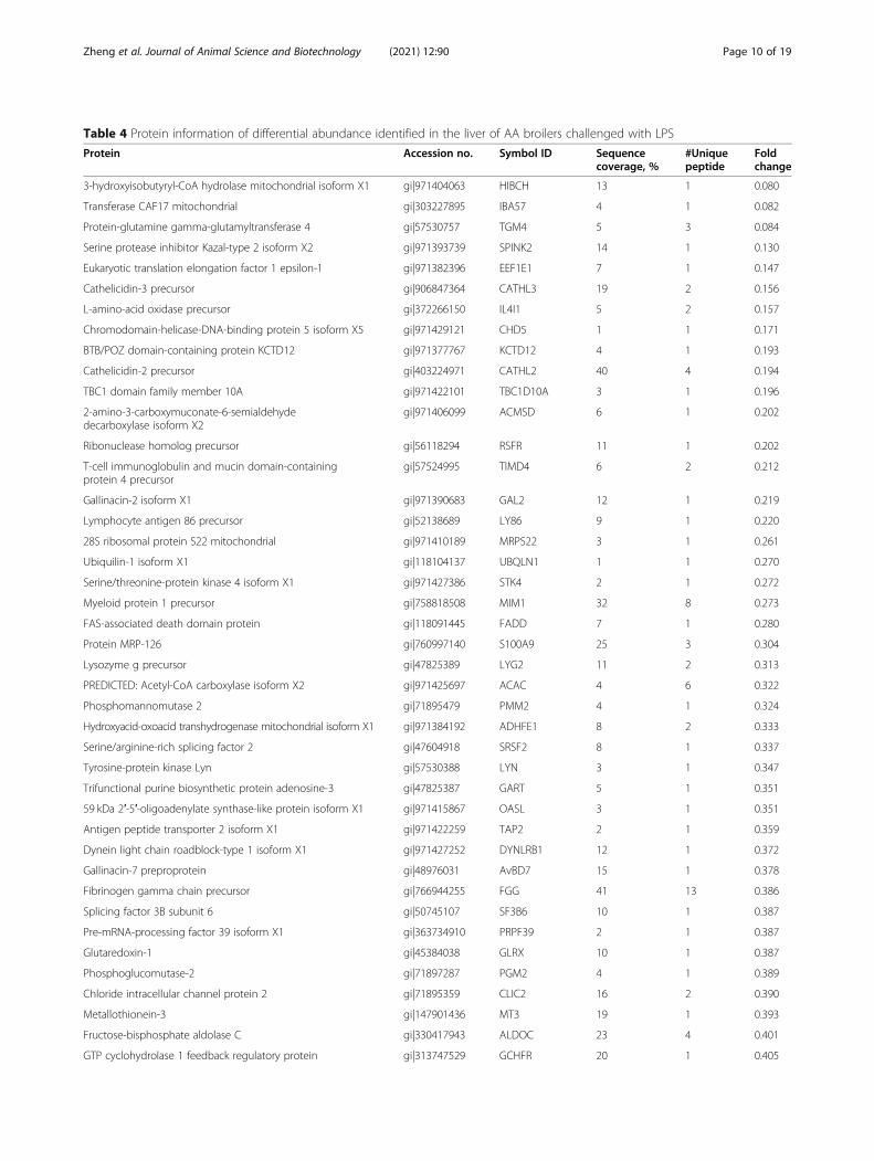
Table 4 Protein information of differential abundance identified in the liver of AA broilers challenged with LPS
Protein Accession no. Symbol ID Sequencecoverage, %
#Uniquepeptide
Foldchange
3-hydroxyisobutyryl-CoA hydrolase mitochondrial isoform X1 gi|971404063 HIBCH 13 1 0.080
Transferase CAF17 mitochondrial gi|303227895 IBA57 4 1 0.082
Protein-glutamine gamma-glutamyltransferase 4 gi|57530757 TGM4 5 3 0.084
Serine protease inhibitor Kazal-type 2 isoform X2 gi|971393739 SPINK2 14 1 0.130
Eukaryotic translation elongation factor 1 epsilon-1 gi|971382396 EEF1E1 7 1 0.147
Cathelicidin-3 precursor gi|906847364 CATHL3 19 2 0.156
L-amino-acid oxidase precursor gi|372266150 IL4I1 5 2 0.157
Chromodomain-helicase-DNA-binding protein 5 isoform X5 gi|971429121 CHD5 1 1 0.171
BTB/POZ domain-containing protein KCTD12 gi|971377767 KCTD12 4 1 0.193
Cathelicidin-2 precursor gi|403224971 CATHL2 40 4 0.194
TBC1 domain family member 10A gi|971422101 TBC1D10A 3 1 0.196
2-amino-3-carboxymuconate-6-semialdehydedecarboxylase isoform X2
gi|971406099 ACMSD 6 1 0.202
Ribonuclease homolog precursor gi|56118294 RSFR 11 1 0.202
T-cell immunoglobulin and mucin domain-containingprotein 4 precursor
gi|57524995 TIMD4 6 2 0.212
Gallinacin-2 isoform X1 gi|971390683 GAL2 12 1 0.219
Lymphocyte antigen 86 precursor gi|52138689 LY86 9 1 0.220
28S ribosomal protein S22 mitochondrial gi|971410189 MRPS22 3 1 0.261
Ubiquilin-1 isoform X1 gi|118104137 UBQLN1 1 1 0.270
Serine/threonine-protein kinase 4 isoform X1 gi|971427386 STK4 2 1 0.272
Myeloid protein 1 precursor gi|758818508 MIM1 32 8 0.273
FAS-associated death domain protein gi|118091445 FADD 7 1 0.280
Protein MRP-126 gi|760997140 S100A9 25 3 0.304
Lysozyme g precursor gi|47825389 LYG2 11 2 0.313
PREDICTED: Acetyl-CoA carboxylase isoform X2 gi|971425697 ACAC 4 6 0.322
Phosphomannomutase 2 gi|71895479 PMM2 4 1 0.324
Hydroxyacid-oxoacid transhydrogenase mitochondrial isoform X1 gi|971384192 ADHFE1 8 2 0.333
Serine/arginine-rich splicing factor 2 gi|47604918 SRSF2 8 1 0.337
Tyrosine-protein kinase Lyn gi|57530388 LYN 3 1 0.347
Trifunctional purine biosynthetic protein adenosine-3 gi|47825387 GART 5 1 0.351
59 kDa 2′-5′-oligoadenylate synthase-like protein isoform X1 gi|971415867 OASL 3 1 0.351
Antigen peptide transporter 2 isoform X1 gi|971422259 TAP2 2 1 0.359
Dynein light chain roadblock-type 1 isoform X1 gi|971427252 DYNLRB1 12 1 0.372
Gallinacin-7 preproprotein gi|48976031 AvBD7 15 1 0.378
Fibrinogen gamma chain precursor gi|766944255 FGG 41 13 0.386
Splicing factor 3B subunit 6 gi|50745107 SF3B6 10 1 0.387
Pre-mRNA-processing factor 39 isoform X1 gi|363734910 PRPF39 2 1 0.387
Glutaredoxin-1 gi|45384038 GLRX 10 1 0.387
Phosphoglucomutase-2 gi|71897287 PGM2 4 1 0.389
Chloride intracellular channel protein 2 gi|71895359 CLIC2 16 2 0.390
Metallothionein-3 gi|147901436 MT3 19 1 0.393
Fructose-bisphosphate aldolase C gi|330417943 ALDOC 23 4 0.401
GTP cyclohydrolase 1 feedback regulatory protein gi|313747529 GCHFR 20 1 0.405
Zheng et al. Journal of Animal Science and Biotechnology (2021) 12:90 Page 10 of 19
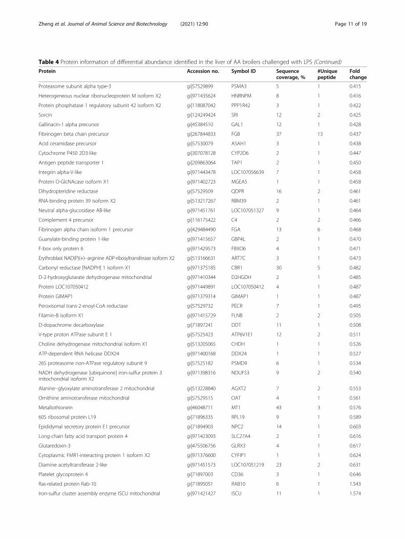
Table 4 Protein information of differential abundance identified in the liver of AA broilers challenged with LPS (Continued)
Protein Accession no. Symbol ID Sequencecoverage, %
#Uniquepeptide
Foldchange
Proteasome subunit alpha type-3 gi|57529899 PSMA3 5 1 0.415
Heterogeneous nuclear ribonucleoprotein M isoform X2 gi|971435624 HNRNPM 8 1 0.416
Protein phosphatase 1 regulatory subunit 42 isoform X2 gi|118087042 PPP1R42 3 1 0.422
Sorcin gi|124249424 SRI 12 2 0.425
Gallinacin-1 alpha precursor gi|45384510 GAL1 12 1 0.428
Fibrinogen beta chain precursor gi|267844833 FGB 37 13 0.437
Acid ceramidase precursor gi|57530079 ASAH1 3 1 0.438
Cytochrome P450 2D3-like gi|307078128 CYP2D6 2 1 0.447
Antigen peptide transporter 1 gi|209863064 TAP1 2 1 0.450
Integrin alpha-V-like gi|971443478 LOC107056639 7 1 0.458
Protein O-GlcNAcase isoform X1 gi|971402723 MGEA5 1 1 0.458
Dihydropteridine reductase gi|57529509 QDPR 16 2 0.461
RNA-binding protein 39 isoform X2 gi|513217267 RBM39 2 1 0.461
Neutral alpha-glucosidase AB-like gi|971451761 LOC107051327 9 1 0.464
Complement 4 precursor gi|116175422 C4 2 2 0.466
Fibrinogen alpha chain isoform 1 precursor gi|429484490 FGA 13 6 0.468
Guanylate-binding protein 1-like gi|971415657 GBP4L 2 1 0.470
F-box only protein 6 gi|971429573 FBXO6 4 1 0.471
Erythroblast NAD(P)(+)--arginine ADP-ribosyltransferase isoform X2 gi|513166631 ART7C 3 1 0.473
Carbonyl reductase [NADPH] 1 isoform X1 gi|971375185 CBR1 30 5 0.482
D-2-hydroxyglutarate dehydrogenase mitochondrial gi|971410344 D2HGDH 2 1 0.485
Protein LOC107050412 gi|971449891 LOC107050412 4 1 0.487
Protein GIMAP1 gi|971379314 GIMAP1 1 1 0.487
Peroxisomal trans-2-enoyl-CoA reductase gi|57529732 PECR 7 1 0.495
Filamin-B isoform X1 gi|971415729 FLNB 2 2 0.505
D-dopachrome decarboxylase gi|71897241 DDT 11 1 0.508
V-type proton ATPase subunit E 1 gi|57525423 ATP6V1E1 12 2 0.511
Choline dehydrogenase mitochondrial isoform X1 gi|513205065 CHDH 1 1 0.526
ATP-dependent RNA helicase DDX24 gi|971400168 DDX24 1 1 0.527
26S proteasome non-ATPase regulatory subunit 9 gi|57525182 PSMD9 6 1 0.534
NADH dehydrogenase [ubiquinone] iron-sulfur protein 3mitochondrial isoform X2
gi|971398316 NDUFS3 9 2 0.540
Alanine--glyoxylate aminotransferase 2 mitochondrial gi|513228840 AGXT2 7 2 0.553
Ornithine aminotransferase mitochondrial gi|57529515 OAT 4 1 0.561
Metallothionein gi|46048711 MT1 43 3 0.576
60S ribosomal protein L19 gi|71896335 RPL19 9 1 0.589
Epididymal secretory protein E1 precursor gi|71894903 NPC2 14 1 0.603
Long-chain fatty acid transport protein 4 gi|971423093 SLC27A4 2 1 0.616
Glutaredoxin-3 gi|475506756 GLRX3 4 1 0.617
Cytoplasmic FMR1-interacting protein 1 isoform X2 gi|971376600 CYFIP1 1 1 0.624
Diamine acetyltransferase 2-like gi|971451573 LOC107051219 23 2 0.631
Platelet glycoprotein 4 gi|71897003 CD36 3 1 0.646
Ras-related protein Rab-10 gi|71895051 RAB10 6 1 1.543
Iron-sulfur cluster assembly enzyme ISCU mitochondrial gi|971421427 ISCU 11 1 1.574
Zheng et al. Journal of Animal Science and Biotechnology (2021) 12:90 Page 11 of 19
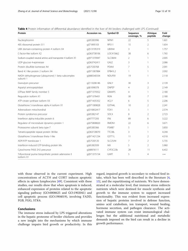
with those observed in the current experiment. Highconcentrations of ACTH and CORT induces apoptoticeffects in spleen lymphocytes [49]. Consistent with thesestudies, our results show that when apoptosis is induced,enhanced expression of proteins related to the apoptoticsignaling pathway (GO:0008625 and GO:1902041) andcell apoptotic process (GO:1904019), involving FADD,FGB, FGG, STK4.
ConclusionsThe immune stress induced by LPS triggered alterationsin the hepatic proteome of broiler chickens and providesa new insight into the mechanisms by which immunechallenge impairs bird growth or productivity. In this
regard, impaired growth is secondary to reduced feed in-take, which has been well described in the literature [4,15], and the repartitioning of nutrients. We have demon-strated at a molecular level, that immune stress redirectsnutrients which were destined for muscle synthesis andgrowth to the immune system to support increasedfunctionality. This was evident from increased expres-sion of hepatic proteins involved in defense function,amino acid catabolism, ion transport, wound healing,hormone secretion, and pathogen clearance. The acti-vated immune system can resist immunological chal-lenges but the additional nutritional and metabolicdemands imposed on the bird can result in a decline ingrowth performance.
Table 4 Protein information of differential abundance identified in the liver of AA broilers challenged with LPS (Continued)
Protein Accession no. Symbol ID Sequencecoverage, %
#Uniquepeptide
Foldchange
Nucleophosmin gi|45383996 NPM1 22 5 1.601
40S ribosomal protein S11 gi|71895103 RPS11 15 2 1.654
UBX domain-containing protein 4 isoform X4 gi|513195319 UBXN4 3 1 1.757
C-factor-like isoform X2 gi|363738106 LOC415662 38 6 1.765
Sodium-coupled neutral amino acid transporter 4 isoform X1 gi|971370947 SLC38A4 2 1 2.005
UDP-glucose 4-epimerase gi|363742411 GALE 3 1 2.086
Protein disulfide-isomerase A4 gi|57530768 PDIA4 7 5 2.091
Band 4.1-like protein 2 isoform X4 gi|971387884 EPB41L2 1 1 2.092
NADH dehydrogenase [ubiquinone] 1 beta subcomplexsubunit 1
gi|480540334 NDUFB1 19 1 2.118
Granulysin precursor gi|113206146 GNLY 30 3 2.139
Aspartyl aminopeptidase gi|61098378 DNPEP 4 2 2.149
GTPase IMAP family member 5 gi|971379352 GIMAP5 4 1 2.180
Regucalcin isoform X1 gi|971376431 RGN 30 7 2.180
ATP-citrate synthase isoform X3 gi|971435352 ACLY 6 5 2.206
Glutathione S-transferase alpha 4 isoform X1 gi|971389828 GSTA4L 10 2 2.241
Adrenodoxin mitochondrial gi|310832417 FDX1 5 1 2.574
Protein syndesmos precursor gi|45382147 SDC4 8 2 2.723
Interferon alpha-inducible protein 6 gi|47777293 IFI6 89 6 3.222
Regulator of microtubule dynamics protein 1 gi|475808820 RMDN1 22 6 3.671
Proteasome subunit beta type-7 gi|45383366 PSMB7 5 1 3.850
Tetratricopeptide repeat protein 38-like gi|363728070 TTC38L 3 1 4.244
Glutathione S-transferase theta-1-like gi|971421234 GSTT1L 11 2 4.318
ADP/ATP translocase 1 gi|57530120 SLC25A4 7 2 5.735
Interferon-induced GTP-binding protein Mx gi|45382939 MX 5 2 5.960
Cytochrome P450 2H2 precursor gi|48976111 CYP2C23b 28 11 6.432
Trifunctional purine biosynthetic protein adenosine-3isoform X1
gi|971375154 GART 5 4 +∞
Zheng et al. Journal of Animal Science and Biotechnology (2021) 12:90 Page 12 of 19
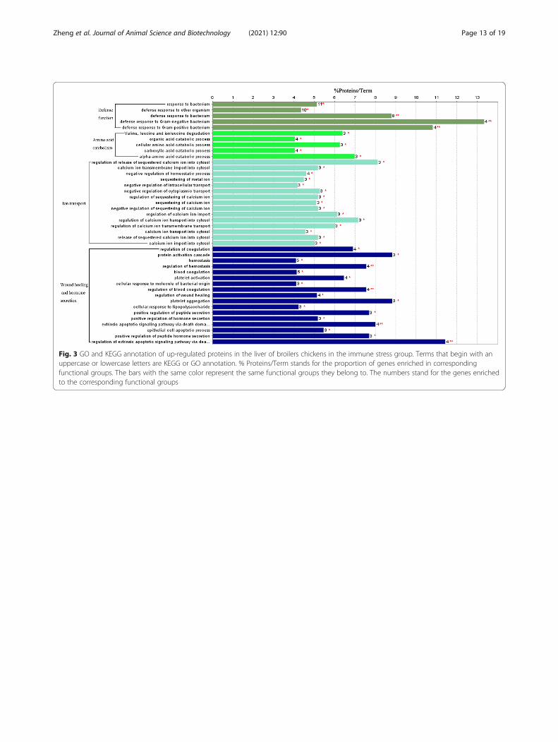
Fig. 3 GO and KEGG annotation of up-regulated proteins in the liver of broilers chickens in the immune stress group. Terms that begin with anuppercase or lowercase letters are KEGG or GO annotation. % Proteins/Term stands for the proportion of genes enriched in correspondingfunctional groups. The bars with the same color represent the same functional groups they belong to. The numbers stand for the genes enrichedto the corresponding functional groups
Zheng et al. Journal of Animal Science and Biotechnology (2021) 12:90 Page 13 of 19
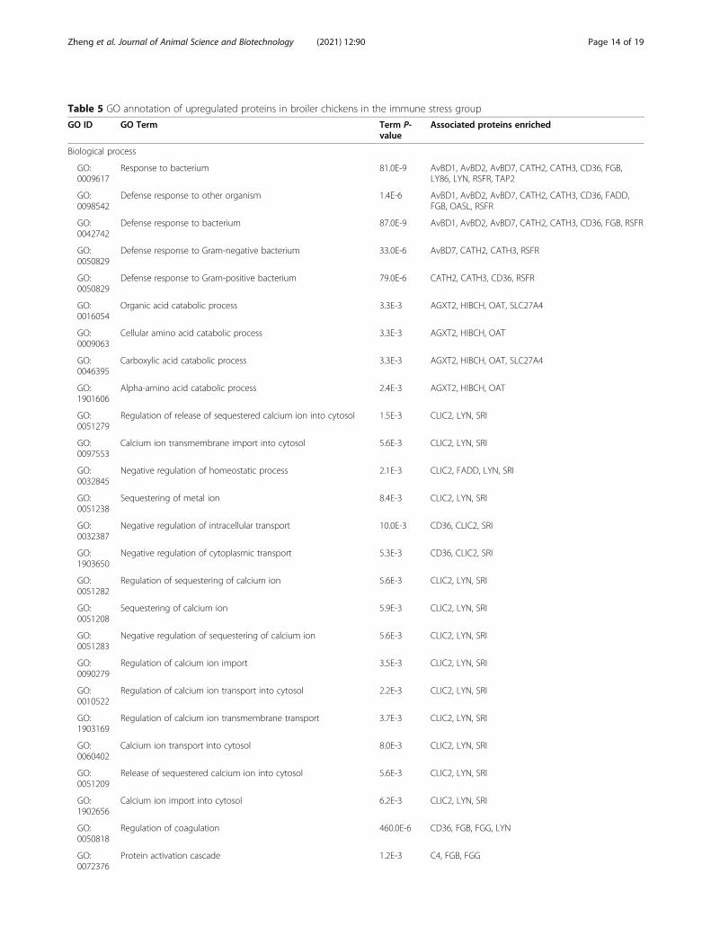
Table 5 GO annotation of upregulated proteins in broiler chickens in the immune stress group
GO ID GO Term Term P-value
Associated proteins enriched
Biological process
GO:0009617
Response to bacterium 81.0E-9 AvBD1, AvBD2, AvBD7, CATH2, CATH3, CD36, FGB,LY86, LYN, RSFR, TAP2
GO:0098542
Defense response to other organism 1.4E-6 AvBD1, AvBD2, AvBD7, CATH2, CATH3, CD36, FADD,FGB, OASL, RSFR
GO:0042742
Defense response to bacterium 87.0E-9 AvBD1, AvBD2, AvBD7, CATH2, CATH3, CD36, FGB, RSFR
GO:0050829
Defense response to Gram-negative bacterium 33.0E-6 AvBD7, CATH2, CATH3, RSFR
GO:0050829
Defense response to Gram-positive bacterium 79.0E-6 CATH2, CATH3, CD36, RSFR
GO:0016054
Organic acid catabolic process 3.3E-3 AGXT2, HIBCH, OAT, SLC27A4
GO:0009063
Cellular amino acid catabolic process 3.3E-3 AGXT2, HIBCH, OAT
GO:0046395
Carboxylic acid catabolic process 3.3E-3 AGXT2, HIBCH, OAT, SLC27A4
GO:1901606
Alpha-amino acid catabolic process 2.4E-3 AGXT2, HIBCH, OAT
GO:0051279
Regulation of release of sequestered calcium ion into cytosol 1.5E-3 CLIC2, LYN, SRI
GO:0097553
Calcium ion transmembrane import into cytosol 5.6E-3 CLIC2, LYN, SRI
GO:0032845
Negative regulation of homeostatic process 2.1E-3 CLIC2, FADD, LYN, SRI
GO:0051238
Sequestering of metal ion 8.4E-3 CLIC2, LYN, SRI
GO:0032387
Negative regulation of intracellular transport 10.0E-3 CD36, CLIC2, SRI
GO:1903650
Negative regulation of cytoplasmic transport 5.3E-3 CD36, CLIC2, SRI
GO:0051282
Regulation of sequestering of calcium ion 5.6E-3 CLIC2, LYN, SRI
GO:0051208
Sequestering of calcium ion 5.9E-3 CLIC2, LYN, SRI
GO:0051283
Negative regulation of sequestering of calcium ion 5.6E-3 CLIC2, LYN, SRI
GO:0090279
Regulation of calcium ion import 3.5E-3 CLIC2, LYN, SRI
GO:0010522
Regulation of calcium ion transport into cytosol 2.2E-3 CLIC2, LYN, SRI
GO:1903169
Regulation of calcium ion transmembrane transport 3.7E-3 CLIC2, LYN, SRI
GO:0060402
Calcium ion transport into cytosol 8.0E-3 CLIC2, LYN, SRI
GO:0051209
Release of sequestered calcium ion into cytosol 5.6E-3 CLIC2, LYN, SRI
GO:1902656
Calcium ion import into cytosol 6.2E-3 CLIC2, LYN, SRI
GO:0050818
Regulation of coagulation 460.0E-6 CD36, FGB, FGG, LYN
GO:0072376
Protein activation cascade 1.2E-3 C4, FGB, FGG
Zheng et al. Journal of Animal Science and Biotechnology (2021) 12:90 Page 14 of 19
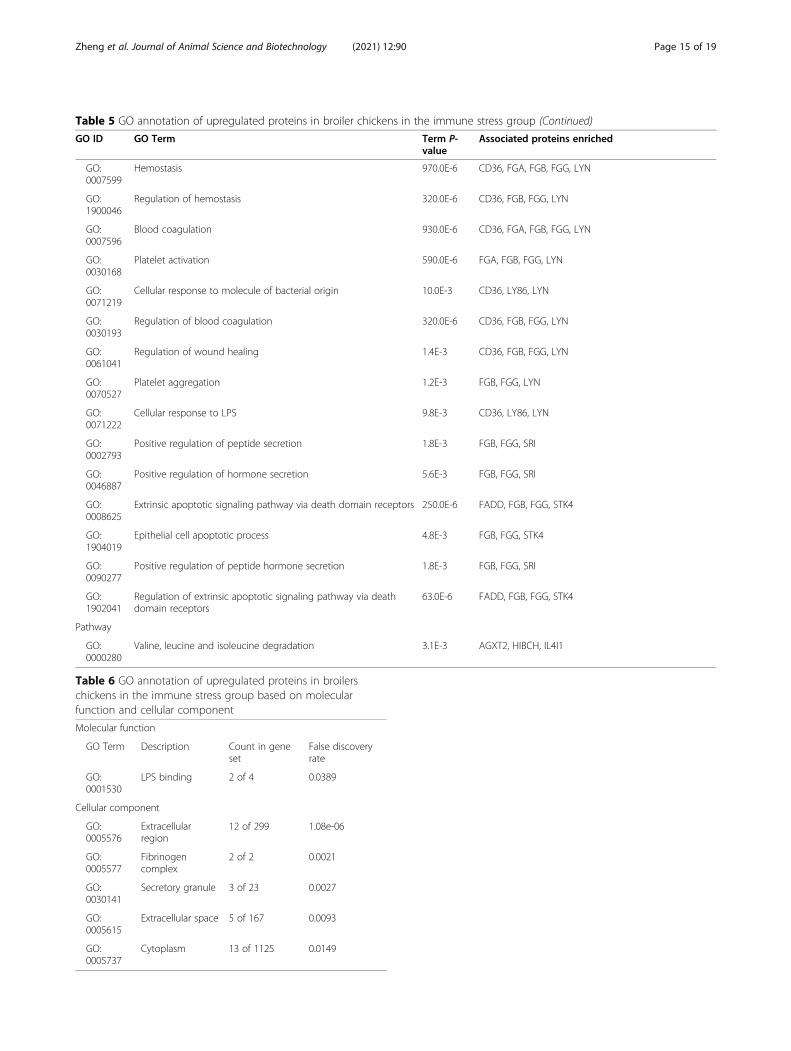
Table 5 GO annotation of upregulated proteins in broiler chickens in the immune stress group (Continued)
GO ID GO Term Term P-value
Associated proteins enriched
GO:0007599
Hemostasis 970.0E-6 CD36, FGA, FGB, FGG, LYN
GO:1900046
Regulation of hemostasis 320.0E-6 CD36, FGB, FGG, LYN
GO:0007596
Blood coagulation 930.0E-6 CD36, FGA, FGB, FGG, LYN
GO:0030168
Platelet activation 590.0E-6 FGA, FGB, FGG, LYN
GO:0071219
Cellular response to molecule of bacterial origin 10.0E-3 CD36, LY86, LYN
GO:0030193
Regulation of blood coagulation 320.0E-6 CD36, FGB, FGG, LYN
GO:0061041
Regulation of wound healing 1.4E-3 CD36, FGB, FGG, LYN
GO:0070527
Platelet aggregation 1.2E-3 FGB, FGG, LYN
GO:0071222
Cellular response to LPS 9.8E-3 CD36, LY86, LYN
GO:0002793
Positive regulation of peptide secretion 1.8E-3 FGB, FGG, SRI
GO:0046887
Positive regulation of hormone secretion 5.6E-3 FGB, FGG, SRI
GO:0008625
Extrinsic apoptotic signaling pathway via death domain receptors 250.0E-6 FADD, FGB, FGG, STK4
GO:1904019
Epithelial cell apoptotic process 4.8E-3 FGB, FGG, STK4
GO:0090277
Positive regulation of peptide hormone secretion 1.8E-3 FGB, FGG, SRI
GO:1902041
Regulation of extrinsic apoptotic signaling pathway via deathdomain receptors
63.0E-6 FADD, FGB, FGG, STK4
Pathway
GO:0000280
Valine, leucine and isoleucine degradation 3.1E-3 AGXT2, HIBCH, IL4I1
Table 6 GO annotation of upregulated proteins in broilerschickens in the immune stress group based on molecularfunction and cellular component
Molecular function
GO Term Description Count in geneset
False discoveryrate
GO:0001530
LPS binding 2 of 4 0.0389
Cellular component
GO:0005576
Extracellularregion
12 of 299 1.08e-06
GO:0005577
Fibrinogencomplex
2 of 2 0.0021
GO:0030141
Secretory granule 3 of 23 0.0027
GO:0005615
Extracellular space 5 of 167 0.0093
GO:0005737
Cytoplasm 13 of 1125 0.0149
Zheng et al. Journal of Animal Science and Biotechnology (2021) 12:90 Page 15 of 19
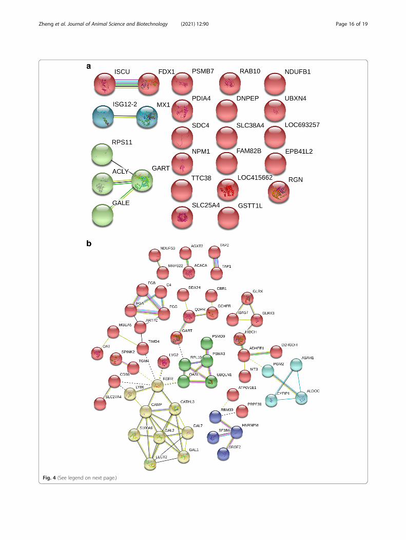
Fig. 4 (See legend on next page.)
Zheng et al. Journal of Animal Science and Biotechnology (2021) 12:90 Page 16 of 19
AbbreviationsLC-MS: Label-free liquid chromatography and mass spectrometry;LPS: Lipopolysaccharide; AA: Arbor Acres; ACTH: Adrenocorticotropichormone; CORT: Corticosterone; GH: Growth hormone; IGF-1: Insulin-likegrowth factor-1; IL-1β: Interleukin-1β; IL-6: Interleukin-6; TNF-α: Tumournecrosis factor-α; DTT: Dithiothreitol; TIC: Total ion current; GO: GeneOntology; KEGG: Kyoto Encyclopedia of Genes and Genomes; HIBCH: 3-hydroxyisobutyryl-CoA hydrolase mitochondrial isoform X1;IBA57: Transferase CAF17 mitochondrial; TGM4: Protein-glutamine gamma-glutamyltransferase 4; SPINK2: Serine protease inhibitor Kazal-type 2 isoformX2; EEF1E1: Eukaryotic translation elongation factor 1 epsilon-1;CATHL3: Cathelicidin-3 precursor; IL4I1: L-amino-acid oxidase precursor;CHD5: Chromodomain-helicase-DNA-binding protein 5 isoform X5;KCTD12: BTB/POZ domain-containing protein KCTD12; CATHL2: Cathelicidin-2precursor; TBC1D10A: TBC1 domain family member 10A; ACMSD: 2-amino-3-carboxymuconate-6-semialdehyde decarboxylase isoform X2;RSFR: Ribonuclease homolog precursor; TIMD4: T-cell immunoglobulin andmucin domain-containing protein 4 precursor; GAL2: Gallinacin-2 isoform X1;LY86: Lymphocyte antigen 86 precursor; MRPS22: 28S ribosomal protein S22mitochondrial; UBQLN1: Ubiquilin-1 isoform X1; STK4: Serine/threonine-protein kinase 4 isoform X1; MIM1: Myeloid protein 1 precursor; FADD: FAS-associated death domain protein; S100A9: Protein MRP-126; LYG2: Lysozymeg precursor; ACAC: PREDICTED: acetyl-CoA carboxylase isoform X2;PMM2: Phosphomannomutase 2; ADHFE1: Hydroxyacid-oxoacidtranshydrogenase mitochondrial isoform X1; SRSF2: Serine/arginine-richsplicing factor 2; LYN: Tyrosine-protein kinase Lyn; GART: Trifunctional purinebiosynthetic protein adenosine-3; OASL: 59 kDa 2'-5'-oligoadenylatesynthase-like protein isoform X1; TAP2: Antigen peptide transporter 2 isoformX1; DYNLRB1: Dynein light chain roadblock-type 1 isoform X1;AvBD7: Gallinacin-7 preproprotein; FGG: Fibrinogen gamma chain precursor;SF3B6: Splicing factor 3B subunit 6; PRPF39: Pre-mRNA-processing factor 39isoform X1; GLRX: Glutaredoxin-1; PGM2: Phosphoglucomutase-2;CLIC2: Chloride intracellular channel protein 2; MT3: Metallothionein-3;ALDOC: Fructose-bisphosphate aldolase C; GCHFR: GTP cyclohydrolase 1feedback regulatory protein; PSMA3: Proteasome subunit alpha type-3;HNRNPM: Heterogeneous nuclear ribonucleoprotein M isoform X2;PPP1R42: Protein phosphatase 1 regulatory subunit 42 isoform X2;SRI: Sorcin; GAL1: Gallinacin-1 alpha precursor; FGB: Fibrinogen beta chainprecursor; ASAH1: Acid ceramidase precursor; CYP2D6: Cytochrome P4502D3-like; TAP1: Antigen peptide transporter 1; LOC107056639: Integrin alpha-V-like; MGEA5: Protein O-GlcNAcase isoform X1; QDPR: Dihydropteridinereductase; RBM39: RNA-binding protein 39 isoform X2;LOC107051327: Neutral alpha-glucosidase AB-like; C4: Complement 4precursor; FGA: Fibrinogen alpha chain isoform 1 precursor;GBP4L: Guanylate-binding protein 1-like; FBXO6: F-box only protein 6;ART7C: Erythroblast NAD(P)(+)--arginine ADP-ribosyltransferase isoform X2;CBR1: Carbonyl reductase [NADPH] 1 isoform X1; D2HGDH: D-2-hydroxyglutarate dehydrogenase mitochondrial; LOC107050412: ProteinLOC107050412; GIMAP1: Protein GIMAP1; PECR: Peroxisomal trans-2-enoyl-CoA reductase; FLNB: Filamin-B isoform X1; DDT: D-dopachromedecarboxylase; ATP6V1E1: V-type proton ATPase subunit E 1; CHDH: Cholinedehydrogenase mitochondrial isoform X1; DDX24: ATP-dependent RNAhelicase DDX24; PSMD9: 26S proteasome non-ATPase regulatory subunit 9;NDUFS3: NADH dehydrogenase [ubiquinone] iron-sulfur protein 3mitochondrial isoform X2; AGXT2: Alanine--glyoxylate aminotransferase 2mitochondrial; OAT: Ornithine aminotransferase mitochondrial;MT1: Metallothionein; RPL19: 60S ribosomal protein L19; NPC2: Epididymalsecretory protein E1 precursor; SLC27A4: Long-chain fatty acid transportprotein 4; GLRX3: Glutaredoxin-3; CYFIP1: Cytoplasmic FMR1-interacting
protein 1 isoform X2; LOC107051219: Diamine acetyltransferase 2-like;CD36: Platelet glycoprotein 4; RAB10: Ras-related protein Rab-10; ISCU: Iron-sulfur cluster assembly enzyme ISCU mitochondrial; NPM1: Nucleophosmin;RPS11: 40S ribosomal protein S11; UBXN4: UBX domain-containing protein 4isoform X4; LOC415662: C-factor-like isoform X2; SLC38A4: Sodium-coupledneutral amino acid transporter 4 isoform X1; GALE: UDP-glucose 4-epimerase;PDIA4: Protein disulfide-isomerase A4; EPB41L2: Band 4.1-like protein 2isoform X4; NDUFB1: NADH dehydrogenase [ubiquinone] 1 beta subcomplexsubunit 1; GNLY: Granulysin precursor; DNPEP: Aspartyl aminopeptidase;GIMAP5: GTPase IMAP family member 5; RGN: Regucalcin isoform X1;ACLY: ATP-citrate synthase isoform X3; GSTA4L: Glutathione S-transferasealpha 4 isoform X1; FDX1: Adrenodoxin mitochondrial; SDC4: Proteinsyndesmos precursor; IFI6: Interferon alpha-inducible protein 6;RMDN1: Regulator of microtubule dynamics protein 1; PSMB7: Proteasomesubunit beta type-7; TTC38L: tetratricopeptide repeat protein 38-like;GSTT1L: GLUTATHIONE S-transferase theta-1-like; SLC25A4: ADP/ATPtranslocase 1; MX: Interferon-induced GTP-binding protein Mx;CYP2C23b: Cytochrome P450 2H2 precursor; GART: Trifunctional purinebiosynthetic protein adenosine-3 isoform X1
AcknowledgementsWe want to show our appreciation to Professor Li Jianke for his help todesign of the present study.
Authors’ contributionsA. Zheng and W.L.B. contributed to the concept and design of the work. H.C.and G.L. contributed to the design of the work. SAP and A. Zhang executedthe experiments. Z.C, W.C and A. Zheng contributed to the analysis andinterpretation of the data. A. Zheng drafted the manuscript. W.L.B. and A.Zheng contributed to the final approval of the version for publication. Allthe authors read and approved the final manuscript.
FundingSponsored by National Natural Science Foundation of China (grant no.31101731) and National Key Research and Development Program of China(No.2018YFD0500600) and The Agricultural Science and TechnologyInnovation Program (ASTIP).
Availability of data and materialsThe datasets used and analyzed during the current study are available fromthe corresponding author on reasonable request.
Declarations
Ethics approval and consent to participateThe feeding trial was conducted according to the guidelines for animalexperiments set out by the National Institute of Animal Health. Allprocedures involving animals such as welfare and ethical issues wereapproved by the Chinese Academy of Agricultural Sciences (statement no.AEC-CAAS-20191106).
Consent for publicationNot applicable.
Competing interestsThe authors declare that they have no competing interests.
(See figure on previous page.)Fig. 4 Protein and protein interaction network of differentially expressed proteins in the liver of broilers chickens in the immune stress group. Aand B represent interaction network of down-regulated and up-regulated proteins in the liver of broilers chickens in the immune stress group,respectively .Each ball represents node protein, the same color balls represent node proteins clustered in the same sub network. The solid lineindicates that the interaction score between the two proteins is more than 0.5 (the dotted line indicates that the score is less than 0.5). Differentcolor solid lines between proteins represent evidence of association. Red lines indicate fusion evidence, green lines indicate neighborhoodevidence, blue lines indicate co-occurrence evidence, purple lines indicate experimental evidence, yellow lines indicate text mining evidence,light blue lines indicate database evidence, and black lines indicate co-expression evidence
Zheng et al. Journal of Animal Science and Biotechnology (2021) 12:90 Page 17 of 19
Author details1Key Laboratory of Feed Biotechnology of Ministry of Agriculture and RuralAffairs, Institute of Feed Research, Chinese Academy of Agricultural Sciences,No.12 Zhongguancun south street, Haidian district, Beijing 100081, China.2School of Agriculture and Food Sciences, University of Queensland, Gatton,QLD 4343, Australia.
Received: 27 November 2020 Accepted: 6 April 2021
References1. Husband AJ. The immune system and integrated homeostasis. Immunol
Cell Biol. 1995;73(4):377–82. https://doi.org/10.1038/icb.1995.58.2. Lai HT, Nieuwland MG, Kemp B, Aarnink AJ, Parmentier HK. Effects of dust
and airborne dust components on antibody responses, body weight gain,and heart morphology of broilers. Poult Sci. 2009;88(9):1838–49. https://doi.org/10.3382/ps.2009-00129.
3. Star L, Decuypere E, Parmentier HK, Kemp B. Effect of single or combinedclimatic and hygienic stress in four layer lines: 2. Endocrine and oxidativestress responses. Poult Sci. 2008;87(6):1031–8. https://doi.org/10.3382/ps.2007-00143.
4. Klasing KC, Laurin DE, Peng RK, Fry DM. Immunologically mediated growthdepression in chicks: influence of feed intake, corticosterone andinterleukin-1. J Nutr. 1987;117:1629–37. https://doi.org/10.1093/jn/117.9.1629.
5. Husband AJ. Role of central nervous system and behaviour in the immuneresponse. Vaccine. 1993;11(8):805–16. https://doi.org/10.1016/0264-410X(93)90355-2.
6. Shini S, Kaiser P. Effects of stress, mimicked by administration ofcorticosterone in drinking water, on the expression of chicken cytokine andchemokine genes in lymphocytes. Stress. 2009;12(5):388–99. https://doi.org/10.1080/10253890802526894.
7. Li K, Zhang P, Shi B, Su J, Yue Y, Tong M, et al. Dietary Artemisia ordosicaextract alleviating immune stress in broilers exposed to lipopolysaccharide.Ital J Anim Sci. 2017;16(2):301–7. https://doi.org/10.1080/1828051X.2016.1274242.
8. Shini S, Huff GR, Shini A, Kaiser P. Understanding stress-inducedimmunosuppression: exploration of cytokine and chemokine gene profilesin chicken peripheral leukocytes. Poult Sci. 2010;89(4):841–51. https://doi.org/10.3382/ps.2009-00483.
9. Webster Marketon JI, Glaser R. Stress hormones and immune function. CellImmunol. 2008;252(1–2):16–26. https://doi.org/10.1016/j.cellimm.2007.09.006.
10. Lai HT, Nieuwland MG, Kemp B, Aarnink AJ, Parmentier HK. Effects ofrepeated intratracheally administered lipopolysaccharide on primary andsecondary specific antibody responses and on body weight gain of broilers.Poult Sci. 2011;90(2):337–51. https://doi.org/10.3382/ps.2010-00997.
11. Marcq C, Cox E, Szalo IM, Thewis A, Beckers Y. Salmonella Typhimurium oralchallenge model in mature broilers: bacteriological, immunological, andgrowth performance aspects. Poult Sci. 2011;90(1):59–67. https://doi.org/10.3382/ps.2010-01017.
12. Star L, Kemp B, van den Anker I, Parmentier HK. Effect of single orcombined climatic and hygienic stress in four layer lines: 1. PerformancePoultry Sci. 2008;87(6):1022–30. https://doi.org/10.3382/ps.2007-00142.
13. Yang XJ, Li WL, Feng Y, Yao JH. Effects of immune stress on growthperformance, immunity, and cecal microflora in chickens. Poult Sci. 2011;90(12):2740–6. https://doi.org/10.3382/ps.2011-01591.
14. Husband AJ, Bryden WL. Nutrition, stress and immune activation. ProceedNutr Soc Australia. 1996;20:60–70.
15. Klasing KC. Nutrition and the immune system. Br Poult Sci. 2007;48(5):525–37. https://doi.org/10.1080/00071660701671336.
16. Robinson MW, Harmon C, O'Farrelly C. Liver immunology and its role ininflammation and homeostasis. Cell Mol Immunol. 2016;13(3):267–76.https://doi.org/10.1038/cmi.2016.3.
17. Luo J, Zheng A, Meng K, Chang W, Bai Y, Li K, et al. Proteome changes inthe intestinal mucosa of broiler (Gallus gallus) activated by probioticenterococcus faecium. J Proteome. 2013;91:226–41. https://doi.org/10.1016/j.jprot.2013.07.017.
18. Zheng A, Luo J, Meng K, Li J, Bryden WL, Chang W, et al. Probiotic(enterococcus faecium) induced responses of the hepatic proteomeimproves metabolic efficiency of broiler chickens (Gallus gallus). BMCGenomics. 2016;17(1):89. https://doi.org/10.1186/s12864-016-2371-5.
19. Zheng A, Chang W, Liu G, Yue Y, Li J, Zhang S, et al. Molecular Differencesin Hepatic Metabolism between AA Broiler and Big Bone Chickens: aProteomic Study. PLoS One. 2016;11:e0164702.
20. Zheng A, Luo J, Meng K, Li J, Zhang S, Li K, et al. Proteome changesunderpin improved meat quality and yield of chickens (Gallus gallus) fedthe probiotic enterococcus faecium. BMC Genomics. 2014;15(1):1167.https://doi.org/10.1186/1471-2164-15-1167.
21. Khan IM, Cao Z, Liu H, Khan A, Rahman SU, Khan MZ, et al. Impact ofcryopreservation on spermatozoa freeze-thawed traits and relevance OMICSto assess sperm Cryo-tolerance in farm animals. Front Vet Sci. 2021;8:609180.https://doi.org/10.3389/fvets.2021.609180.
22. Van Goor A, Ashwell CM, Persia ME, Rothschild MF, Schmidt CJ, Lamont SJ.Unique genetic responses revealed in RNA-seq of the spleen of chickensstimulated with lipopolysaccharide and short-term heat. PLoS One. 2017;12(2):e0171414. https://doi.org/10.1371/journal.pone.0171414.
23. Li Y, Zhang H, Chen YP, Yang MX, Zhang LL, Lu ZX, et al. Bacillusamyloliquefaciens supplementation alleviates immunological stress inlipopolysaccharide-challenged broilers at early age. Poult Sci. 2015;94(7):1504–11. https://doi.org/10.3382/ps/pev124.
24. Zhang L, Yue HY, Zhang HJ, Xu L, Wu SG, Yan HJ, et al. Transport stress inbroilers: I. blood metabolism, glycolytic potential, and meat quality. PoultSci. 2009;88(10):2033–41. https://doi.org/10.3382/ps.2009-00128.
25. Lin H, He L, Ma B. A combinatorial approach to the peptide featurematching problem for label-free quantification. Bioinformatics. 2013;29(14):1768–75. https://doi.org/10.1093/bioinformatics/btt274.
26. Bindea G, Mlecnik B, Hackl H, Charoentong P, Tosolini M, Kirilovsky A, et al.ClueGO: a Cytoscape plug-in to decipher functionally grouped geneontology and pathway annotation networks. Bioinformatics. 2009;25(8):1091–3. https://doi.org/10.1093/bioinformatics/btp101.
27. Franceschini A, Szklarczyk D, Frankild S, Kuhn M, Simonovic M, Roth A, et al.STRING v9.1: protein-protein interaction networks, with increased coverageand integration. Nucleic Acids Res. 2013;41(Database issue):D808–15. https://doi.org/10.1093/nar/gks1094.
28. Yang X, Guo Y, He X, Yuan J, Yang Y, Wang Z. Growth performance andimmune responses in chickens after challenge with lipopolysaccharide andmodulation by dietary different oils. Animal. 2008;2(2):216–23. https://doi.org/10.1017/S1751731107001188.
29. Li R, Li N, Zhang J, Wang Y, Liu J, Cai Y, et al. Expression of immune-relatedgenes of ducks infected with avian pathogenic Escherichia coli (APEC).Front Microbiol. 2016;7:637.
30. Crhanova M, Hradecka H, Faldynova M, Matulova M, Havlickova H, Sisak F,et al. Immune response of chicken gut to natural colonization by gutmicroflora and to Salmonella enterica serovar enteritidis infection. InfectImmun. 2011;79(7):2755–63. https://doi.org/10.1128/IAI.01375-10.
31. Cuperus T, Coorens M, van Dijk A, Haagsman HP. Avian host defensepeptides. Dev Comp Immunol. 2013;41(3):352–69. https://doi.org/10.1016/j.dci.2013.04.019.
32. Rosenberg HF. RNase a ribonucleases and host defense: an evolving story. JLeukoc Biol. 2008;83(5):1079–87. https://doi.org/10.1189/jlb.1107725.
33. Nitto T, Dyer KD, Czapiga M, Rosenberg HF. Evolution and function ofleukocyte RNase a ribonucleases of the avian species, Gallus gallus. J BiolChem. 2006;281(35):25622–34.
34. Matulova M, Varmuzova K, Sisak F, Havlickova H, Babak V, Stejskal K, et al. Chickeninnate immune response to oral infection with Salmonella enterica serovarEnteritidis. Vet Res. 2013;44(1):37. https://doi.org/10.1186/1297-9716-44-37.
35. Matulova M, Rajova J, Vlasatikova L, Volf J, Stepanova H, Havlickova H, et al.Characterization of chicken spleen transcriptome after infection withSalmonella enterica serovar Enteritidis. PLoS One. 2012;7(10):e48101. https://doi.org/10.1371/journal.pone.0048101.
36. Rychlik I, Elsheimer-Matulova M, Kyrova K. Gene expression in the chickencaecum in response to infections with non-typhoid Salmonella. Vet Res.2014;45(1):119. https://doi.org/10.1186/s13567-014-0119-2.
37. Elli L, Ciulla MM, Busca G, Roncoroni L, Maioli C, Ferrero S, et al.Beneficial effects of treatment with transglutaminase inhibitor cystamineon the severity of inflammation in a rat model of inflammatory boweldisease. Lab Investig. 2011;91(3):452–61. https://doi.org/10.1038/labinvest.2010.186.
38. Zhao SH, Kuhar D, Lunney JK, Dawson H, Guidry C, Uthe JJ, et al. Geneexpression profiling in Salmonella choleraesuis-infected porcine lung usinga long oligonucleotide microarray. Mamm Genome. 2006;17(7):777–89.https://doi.org/10.1007/s00335-005-0155-3.
Zheng et al. Journal of Animal Science and Biotechnology (2021) 12:90 Page 18 of 19
39. Spronk HM, Govers-Riemslag JW, ten Cate H. The blood coagulation systemas a molecular machine. BioEssays. 2003;25(12):1220–8. https://doi.org/10.1002/bies.10360.
40. Rallapalli PM, Orengo CA, Studer RA, Perkins SJ. Positive selection during theevolution of the blood coagulation factors in the context of their disease-causing mutations. Mol Biol Evol. 2014;31(11):3040–56. https://doi.org/10.1093/molbev/msu248.
41. Szaba FM, Smiley ST. Roles for thrombin and fibrin(ogen) in cytokine/chemokine production and macrophage adhesion in vivo. Blood. 2002;99(3):1053–9. https://doi.org/10.1182/blood.V99.3.1053.
42. Monson MS, Van Goor AG, Persia ME, Rothschild MF, Schmidt CJ, Lamont SJ.Genetic lines respond uniquely within the chicken thymic transcriptome toacute heat stress and low dose lipopolysaccharide. Sci Rep. 2019;9(1):13649.https://doi.org/10.1038/s41598-019-50051-0.
43. Monson MS, Van Goor AG, Ashwell CM, Persia ME, Rothschild MF, SchmidtCJ, et al. Immunomodulatory effects of heat stress and lipopolysaccharideon the bursal transcriptome in two distinct chicken lines. BMC Genomics.2018;19(1):643. https://doi.org/10.1186/s12864-018-5033-y.
44. Xie H, Rath NC, Huff GR, Huff WE, Balog JM. Effects of Salmonellatyphimurium lipopolysaccharide on broiler chickens. Poult Sci. 2000;79(1):33–40. https://doi.org/10.1093/ps/79.1.33.
45. Feske S, Wulff H, Skolnik EY. Ion channels in innate and adaptive immunity.Annu Rev Immunol. 2015;33(1):291–353. https://doi.org/10.1146/annurev-immunol-032414-112212.
46. Feske S. Calcium signalling in lymphocyte activation and disease. Nat RevImmunol. 2007;7(9):690–702. https://doi.org/10.1038/nri2152.
47. Dorshkind K, Horseman ND. Anterior pituitary hormones, stress, andimmune system homeostasis. BioEssays. 2001;23(3):288–94. https://doi.org/10.1002/1521-1878(200103)23:3<288::AID-BIES1039>3.0.CO;2-P.
48. Killian MP, Boviez JD, Gambarotta M, Lombardo DM. Induction of apoptosisin the bursa of Fabricius by vaccination against Gumboro disease. AvianPathology. 2017;46(5):526–34. https://doi.org/10.1080/03079457.2017.1322684.
49. Li RF, Liu SP, Yuan ZH, Yi JE, Tian YN, Wu J, et al. Effects of induced stressfrom the live LaSota Newcastle disease vaccination on the growthperformance and immune function in broiler chickens. Poult Sci. 2020;99(4):1896–905. https://doi.org/10.1016/j.psj.2019.12.004.
Zheng et al. Journal of Animal Science and Biotechnology (2021) 12:90 Page 19 of 19