Embed Size (px)
Citation preview

Molecular mechanism of plasma sterilization in solution with the reduced pH method:
importance of permeation of HOO radicals into the cell membrane
This article has been downloaded from IOPscience. Please scroll down to see the full text article.
2013 J. Phys. D: Appl. Phys. 46 295402
(http://iopscience.iop.org/0022-3727/46/29/295402)
Download details:
IP Address: 128.197.27.9
The article was downloaded on 07/09/2013 at 11:36
Please note that terms and conditions apply.
View the table of contents for this issue, or go to the journal homepage for more
Home Search Collections Journals About Contact us My IOPscience
IOP PUBLISHING JOURNAL OF PHYSICS D: APPLIED PHYSICS
J. Phys. D: Appl. Phys. 46 (2013) 295402 (10pp) doi:10.1088/0022-3727/46/29/295402
Molecular mechanism of plasmasterilization in solution with the reducedpH method: importance of permeation ofHOO radicals into the cell membraneEisuke Takai1, Satoshi Ikawa2, Katsuhisa Kitano3, Junpei Kuwabara4
and Kentaro Shiraki1
1 Faculty of Pure and Applied Sciences, University of Tsukuba, 1-1-1 Tennodai, Tsukuba, Ibaraki305-8573, Japan2 Technology Research Institute of Osaka Prefecture, 2-7-1 Ayumino, Izumi, Osaka 594-1157, Japan3 Center for Atomic and Molecular Technologies, Graduate School of Engineering, Osaka University,Osaka 565-0871, Japan4 Tsukuba Research Center for Interdisciplinary Materials Science (TIMS), Graduate School of Pure andApplied Sciences, University of Tsukuba, 1-1-1 Tennodai, Tsukuba 305-8573, Japan
E-mail: [email protected]
Received 29 March 2013, in final form 22 May 2013Published 1 July 2013Online at stacks.iop.org/JPhysD/46/295402
AbstractSterilization of certain infected areas of the human body surface is necessary for dental andsurgical therapies. Because the blood is filled with body fluid, sterilization in solution isessential. In vitro solution sterilization has been successively carried out using a combinationof low-temperature atmospheric-pressure plasma and the reduced pH method, where thesolution is sufficiently acidic. Here, we show the molecular mechanism of such plasmasterilization in solution based on microbiology. Three kinds of bacteria were inactivated byplasma treatment under various pH conditions. The theoretical and experimental modelsrevealed that the sterilization was characterized by the concentration of hydroperoxy radicals(HOO·), which were dependent on the pH value. Bacterial inactivation rates were proportionalto the HOO· concentrations calculated by the theoretical model. To evaluate the penetration ofradicals into the cell membrane, a bacterial model using dye-included micelles was used.Decolouration rates of the model were also in proportion with the calculated HOO·concentrations. These results indicate that the key species for plasma sterilization werehydroperoxy radicals. More importantly, the high permeation of hydroperoxy radicals into thecell membrane plays a key role for efficient bactericidal inactivation using the reduced pHmethod.
(Some figures may appear in colour only in the online journal)
1. Introduction
Low-temperature atmospheric-pressure plasmas (LTAPPs)have attracted considerable attention for use in various medicaland life science applications. The merits of an LTAPP areas follows: (i) inexpensive operation costs in vacuum-freesystems, (ii) use of moderate gas temperature, (iii) facilitationof delicate and flexible operations and (iv) generation of
reactive oxygen species (ROS) [1], typically hydroxyl radicals(OH·) [2–6], superoxide anion radicals (O−
2 ·) [7], hydroperoxyradicals (HOO·) [8], singlet oxygen (1O2) and atomic oxygen(O) [9]. These features allow for the exploitation of anovel field of chemical reaction in solution [10–12], aswell as under dry conditions. For example, the recentpioneering applications of LTAPP include the developmentof plasma medicines [13, 14] for cancer [15–18], Parkinson’s
0022-3727/13/295402+10$33.00 1 © 2013 IOP Publishing Ltd Printed in the UK & the USA

J. Phys. D: Appl. Phys. 46 (2013) 295402 E Takai et al
HOO
O2-
O2-
O2
Plasma
getTargegeTarg
Solution pH<4
Figure 1. Schematic mechanism of plasma sterilization in solutionusing plasma-induced chemical processing and the reduced pHmethod. The ‘target’ of the plasma sterilization corresponds to thetest bacteria or the dye-included micelle as a bacterial model. Theyellow layer of the target is the cell membrane or surfactant micelle,both of which are semi-permeable.
disease [19], blood coagulation [20], disruption of the humanhepatocyte cytoskeleton [21] and sterilization [22–29].
The sterilization of bacteria in solution is one of themost in-demand applications of plasma medicine becausethe organism is also present under aqueous conditions,typically involving root canal therapy in dentistry [30] andthe prevention of surgical site infection [31]. For sterilizationof bacteria in solution, our research group has presenteda new concept for plasma application in solution, called‘plasma-induced chemical processing’ [7] and a new methodof plasma sterilization, named ‘the reduced pH method’ [8].Briefly, in plasma-induced chemical processing, the LTAPPgenerates chemically reactive species in the gas phase, whichdiffuse into the liquid phase and thus react with substancesin solution. Plasma sterilization in solution under acidicconditions (pH < 4.8) occurs much more efficiently than underneutral conditions (pH 6.5) when plasma-induced chemicalprocessing and the reduced pH method are used [8].
The mechanism of plasma sterilization in solution usingplasma-induced chemical processing and the reduced pHmethod is shown in figure 1 by Kitano et al [31]. Theplasma-generated O−
2 · in air diffuses into the solution. O−2 ·
in the solution is known to be in equilibrium with HOO·, asshown by the following reaction [32, 33]:
O−2 · +H+ � HOO·, pKa = 4.8. (1)
Here, pKa means the acid dissociation constant. This reactionshows that O−
2 · is converted into HOO· under lower pHconditions (<4.8). Unlike charged (anion) molecular O−
2 ·,non-charged (neutral) HOO· easily permeates into the cell
membrane, of which the lipid bilayer has a permselectiveproperty. The HOO· can then react with biomoleculesinside the cell and cause various molecular changes suchas conformational changes in DNA [33–36], DNA damage[37] and inactivation of enzymes [38], to induce bacterialinactivation.
Generally, the death rate of bacteria is known to beproportional to the concentration of the bactericidal factor [39].If the sterilization mechanism of the reduced pH method isactually involved in the neutralization of O−
2 · to HOO·, thedeath rates should be proportional to the concentration ofHOO· at various pH values. To evaluate this hypothesis,we investigated the experimental results using three kindsof bacteria and a bacterial model of a micelle system, andexamined the chemical reactions of active species in solution.
2. Experimental
2.1. Materials
Streptococcus mutans JCM 5705 and Campylobacter rectusJCM 6301 were obtained from the RIKEN BioResourceCenter, Japan Collection of Microorganisms. Escherichiacoli NBRC 3301 was obtained from the Biological ResourceCenter, NITE. The soybean-casein digest (SCD) agar andsheep blood agar (SBA) plates were obtained from NissuiPharmaceutical (Tokyo, Japan). The LB (Luria-Bertan) brothMiller and LB agar Miller plates were obtained from Becton,Dickinson and Company (Franklin Lakes, NJ, USA). Nile redwas obtained from Sigma Chemical Co. (St Louis, MO, USA).Tween 20 was obtained from Tokyo Kasei Kogyo Co. Ltd(Tokyo, Japan). Sodium citrate, potassium chloride, glycine,citric acid, sodium phosphate and sodium bicarbonate wereobtained from Wako Pure Chemical Industries Ltd (Osaka,Japan).
2.2. Plasma jet generation
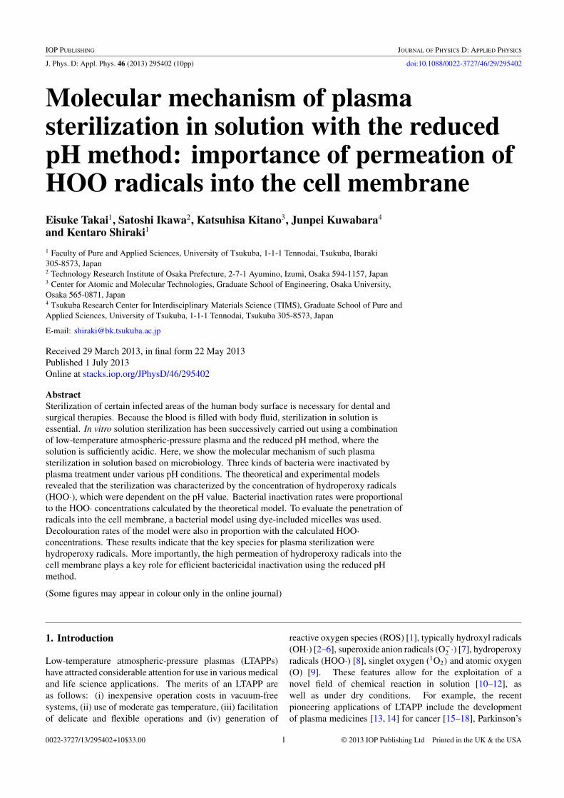
A low-frequency (LF) plasma jet was used in a manner similarto that described in a previous study [8]. The plasma shapeis elongated from the end of a quartz glass tube, in whichhelium gas flows, by the application of an alternating currenthigh voltage (ranging from −3.5 to +5.0 kV at a frequency of13.9 kHz) to a single-sided electrode. The electrode consists ofa small copper sheet wound around the glass tube (figure 2(a)).A helium plasma with a low gas temperature was generated inan elongated shape (figure 2(b)).
2.3. Bacterial inactivation assay
S. mutans was cultivated on an SCD agar plate for 48 h at 37 ◦C.C. rectus was cultivated on an SBA plate for 48 h at 37 ◦C underanaerobic conditions. Both these cultivated bacteria sampleswere harvested from the surfaces of the plates. E. coli wascultivated in LB broth for 18 h at 30 ◦C with 110 rpm reciprocalshaking and was subsequently harvested by centrifugation. Allharvested bacteria were suspended in an appropriate volumeof distilled water (DW). The bacterial suspension was diluted
2
J. Phys. D: Appl. Phys. 46 (2013) 295402 E Takai et al
He gas flow
LF/HVElectrode
Solution
Glass tube
(a) (b)
Figure 2. Plasma jet system. (a) Schematic representation of theplasma jet system with a sample solution. The plume-like structureof the plasma extends towards the surface of a solution containing abacterial suspension or surfactant micelles. (b) Photograph of theplasma jet. The plasma of helium gas flows through the glass tube.The discharge power of the plasma was 3 W.
with DW to OD600 = 0.1, which contained approximately7 × 107 cells ml−1 of bacteria.
Bacterial inactivation assays were performed in thefollowing manner using LF plasma jets. The prepared bacterialsuspension (OD600 = 0.1) was diluted 10-fold with a 2.0 mMsodium citrate buffer (pH range 6.5–3.7) and subsequently,500 µl of this diluted solution was distributed into 24-wellmicroplates. The plasma jet was applied to the surface ofthe bacteria suspension in each well for predetermined timeintervals in ambient air. The tip of the tube that producesthe plasma jets was located approximately 17 mm above thesolution surface. The helium gas flow rate was 2.0 slm.
The number of living bacteria after plasma application wasdetermined by a colony-forming unit (CFU) assay performedas follows. After plasma exposure, the suspensions wererecovered from the wells and DW was added to each recoveredsolution to replenish water lost by evaporation during theprocess. Subsequently, the solutions were serially dilutedfurther with DW, and 100 µl of each dilution was spread on theSCD agar, blood agar and LB agar plates used for S. mutans, C.rectus and E. coli, respectively. These plates were incubatedfor 48 h at 37 ◦C (only C. rectus was incubated under anaerobicconditions), thereby enabling the development and subsequentcounting of the bacterial colonies. The minimum detectionvalue of the CFU assay in this study was 10 CFU ml−1. Theoperations to plating for CFU assay from the suspension ofbacteria were terminated within 30 min.
2.4. Dye decolouration assay of surfactant micelles
Surfactant micelles that included a dyeing agent were preparedby agitating a solution containing 116µM Nile red and 20%v/v Tween 20 at 25 ◦C for 24 h. In the plasma exposureexperiments, a 300 µl micelle sample containing 5.8µM Nilered, 1% v/v Tween 20 and 100mM buffer was applied to thevessel as the target solution. The buffers of the target solutionswere potassium chloride at pH 1, glycine at pH 2, 3, 9 and 10,
citric acid at pH 4–6, phosphate at pH 7 and 8, and bicarbonateat pH 11.
The decolouration assay measurements were obtained asfollows. The plasma jet was generated within an airtightchamber (at its centre) using the plasma generation devicedescribed above. The ambient gas in the chamber wascontrolled via an oxygen (O2) gas supply port connected to theside of the plasma jet port to avoid the generation of nitrogenspecies that could affect the pH value. The flow rates of Heand O2 gas were 0.50 slm and 0.15 slm, respectively. To enablethe exchange of ambient gas, He and O2 gases were initiallymade to flow into the chamber for 5 min. The various ROSproduced from O2 gas by the helium plasma were supplied tothe solution in the chamber.
3. Results
3.1. Calculation of the concentration of radicals
Radical species are generally unstable with short lifetimes;hence, it is impossible to determine the concentration ofradicals in aqueous solution after the plasma treatment. Here,we introduce the theoretical calculation of the concentrationof O−
2 · and HOO·. Details were cited in [31].The concentration of HOO· and O−
2 · as a function ofpH was calculated considering not only acid dissociationequilibrium (equation 1)) but also dismutation of radicals(equations (2)–(4)). First, O−
2 · radicals related to HOO·, asdetermined by equation (1), contribute to bacterial inactivationin aqueous solution [40] because of a long lifetime of O−
2 · (5 sat 1.0×10⁻⁶M) [41]. O−
2 · and HOO· are consumed in solutionin the proportions indicated by equations (2)–(4) [32, 33].
O−2 ·+ HOO·+ H+ → H2O2+O2, k1 = 9.7×10⁷ M−1 s−1.
(2)HOO · + HOO· → H2O2 + O2, k2 = 8.3 × 10⁵ M−1 s−1.
(3)
O−2 · +O−
2 · +2H+ → H2O2 + O2, k3 = 0.3 M−1 s−1.
(4)
Here, k1, k2 and k3 denote the respective reaction rates. Thesupply of O−
2 · generated by the plasma in the gas phase to thesolution balances the consumption of O−
2 · and HOO· in thesolution. Consequently, using equations (1)–(4), the overallreaction can be written as below.
Vs = Vd = k1[O−2 ·][HOO·] + k2[HOO·]2 + k3[O−
2 ·]2
= k1 × 10(pH−4.8)[HOO·]2 + k2[HOO·]2 + k3
× 102(pH−4.8)[HOO·]2
= k1 × 10(1−4.8)([HOO·]pH=1)2 + k2([HOO·]pH=1)
2
+ k3 × 102(1−4.8)([HOO·]pH=1)2. (5)
Here, Vs and Vd denote the supply rate of O−2 · from the
gas phase to the solution by the plasma treatment and theconsumption rate of O−
2 · and HOO·, respectively. BecauseVs is constant at any pH, the fraction of HOO· concentration ateach pH value can be described using the following equation,derived from equation (5).
[HOO·]/[HOO·]pH=1 = (9.2 × 10²) × (k1 × 10(pH−4.8)
+ k2 + k3 × 102(pH−4.8))−1/2. (6)
3
J. Phys. D: Appl. Phys. 46 (2013) 295402 E Takai et al
100
80
60
40
20
0
[O2 -
] / [HO
O pH
=1
1197531
pH
1.0
0.8
0.6
0.4
0.2
0.0
[HO
O] /
[HO
O] p
H=1
0.6
0.4
0.2
0.06.25.75.24.74.23.7
0.6
0.4
0.2
0.0
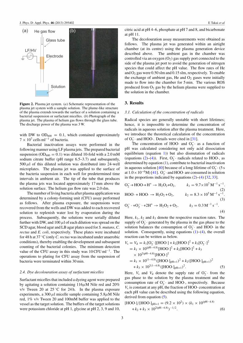
Figure 3. Relative concentration of HOO· and O−2 ·. The HOO·
concentration is described by equation (6) (with respect to the leftaxis, closed circle) and the O−
2 · concentration is described byequation (7) (with respect to the right axis, open square) for the pHrange 1.0–11.0. (inset) The relative concentration of HOO·, O−
2 · and(HOO · +O−
2 ·) at pH 3.7–6.5. The (HOO · +O−2 ·) concentration is
shown by asterisks. In the inset figure, the left and right axes showthe same range.
Consequently, using equations (1) and (6), the fraction of O−2 ·
concentration at each pH can be written as below.
[O−2 ·]/[HOO·]pH=1 = 9.2 × 102 × 10(pH−4.8)
×(k1 × 10(pH−4.8) + k2 + k3 × 102(pH−4.8))−1/2. (7)
The corresponding rate curve for equations (6) and (7) isplotted in figure 3; the obtained curve is in good agreementwith the results of the previous paper for spectrophotometricanalysis of spontaneous disproportionation of radicals [41].Therefore, the result indicates that the concentration of O−
2 · andHOO· can be arbitrarily controlled by the pH of the solution.
3.2. Bacterial inactivation
If figure 1 describes the mechanism of plasma sterilization,the bacterial inactivation rate should be proportional to theconcentration of HOO· in the solution. Here, we investigatedthe inactivation of three kinds of bacteria at various pH values.The efficiency of bacterial inactivation was evaluated using thedecimal reduction value (D value), which refers to the timerequired to inactivate 90% of bacteria in a given sample. Thereciprocal of the D value is also the death rate of the bacteria,which implies the sterilizing power.
The death rate of bacteria was examined at pH 3.7–6.5for typical types of bacteria using three different species:S. mutans, a Gram-positive acid-resistant bacterium causinghuman dental caries [42], C. rectus, a Gram-negative anaerobicbacterium causing periodontal diseases [43], and E. coli,a Gram-negative facultative anaerobic bacterium. Thesebacteria were not sterilized by the treatment in acid solutionfor several hours. Pseudomonas aeruginosa, which is anopportunistic human pathogen, was not used because of itshigh sensitivity to acidic solutions. Although S. mutans waspartially inactivated by the plasma treatment of a target solution
(a)
(b)
(c)
7
5
3
1log 1
0(C
FU/ m
)L
543210Plasma Exposure Time (min)
D (min)pH 6.5 17.9pH 5.0 0.86pH 4.7 0.50pH 4.5 0.39pH 3.7 0.15
7
5
3
1log 1
0(C
FU/m
)L
543210Plasma Exposure Time (min)
D (min)pH 6.5 2.11pH 5.8 2.42pH 5.5 1.12pH 5.2 0.94pH 5.0 0.76pH 4.7 0.41pH 3.7 0.16
7
5
3
1log 1
0(C
FU/m
)L
543210
Plasma Exposure Time (min)
D (min)pH 6.5 >20pH 5.2 1.92pH 4.7 0.96pH 4.2 0.59pH 3.7 0.21
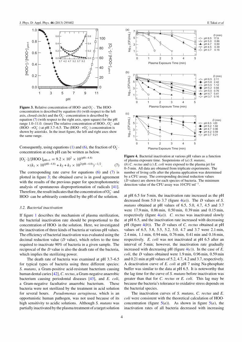
Figure 4. Bacterial inactivation at various pH values as a functionof plasma exposure time. Suspensions of (a) S. mutans,(b) C. rectus and (c) E. coli were exposed to the plasma jet for0–5 min. All data are obtained from triplicate experiments. Thenumber of living cells after the plasma application was determinedby a CFU assay. The corresponding decimal reduction values(D values) are shown for each species of bacteria. The minimumdetection value of the CFU assay was 10 CFU ml−1.
at pH 6.5 for 5 min, the inactivation rate increased as the pHdecreased from 5.0 to 3.7 (figure 4(a)). The D values of S.mutans obtained at pH values of 6.5, 5.0, 4.7, 4.5 and 3.7were 17.9 min, 0.86 min, 0.50 min, 0.39 min and 0.15 min,respectively (figure 4(a)). C. rectus was inactivated slowlyat pH 6.5, and the inactivation rate increased with decreasingpH (figure 4(b)). The D values of C. rectus obtained at pHvalues of 6.5, 5.8, 5.5, 5.2, 5.0, 4.7 and 3.7 were 2.1 min,2.4 min, 1.1 min, 0.94 min, 0.76 min, 0.41 min and 0.16 min,respectively. E. coli was not inactivated at pH 6.5 after aninterval of 5 min; however, the inactivation rate graduallyincreased with decreasing pH (figure 4(c)). In the case of E.coli, the D values obtained were 1.9 min, 0.96 min, 0.59 minand 0.21 min at pH values of 5.2, 4.7, 4.2 and 3.7, respectively.A deactivation curve of E. coli at pH 7 using Na-phosphatebuffer was similar to the data at pH 6.5. It is noteworthy thatthe lag time for the curve of S. mutans before inactivation wasgreater than that for C. rectus or E. coli. This lag may bebecause the bacteria’s tolerance to oxidative stress depends onthe bacterial species.
The inactivation curves of S. mutans, C. rectus and E.coli were consistent with the theoretical calculation of HOO·concentration (figure 5(a)). As shown in figure 5(a), theinactivation rates of all bacteria decreased with increasing
4
J. Phys. D: Appl. Phys. 46 (2013) 295402 E Takai et al
(a) (b)
6
5
4
3
2
1
0
Inac
tivat
i on
Rat
e ( m
in-1
)
6.56.05.55.04.54.0
pH
0.30
0.25
0.20
0.15
0.10
0.05
0.00
[HO
O]/[H
OO
] p H = 1
S. mutans C. rectus E. coli
6
5
4
3
2
1
0
Inac
tivat
ion
Rat
e ( m
in-1
)
0.300.200.100.00
[HOO ] / [HOO ] pH = 1
S. mutans R2 = 0.999 C. rectus R2 = 0.991 E. coli R2 = 0.950
Figure 5. Assessment of the bacterial inactivation. (a) Bacterial inactivation rate (reciprocal of the D value) for plasma treatment at variouspH values. The inactivation rates were calculated from each corresponding D value. The red line (with respect to the right axis) indicatesthe relative concentration of HOO· in the pH range 3.7–6.5. The HOO· concentration is expressed by equation (6). (b) Correlation betweenthe inactivation rate of S. mutans, C. rectus and E. coli and the HOO· concentration.
(a) (b) 0.20
0.15
0.10
0.05
0.00
Freq
uenc
y
12 4 6 8
102 4 6 8
1002 4
Diameter (nm)
N
O ON
CH2CH3
H3CH2C
OO
OOHO
OOH
OHO
O
W
X
YZw+x+y+z=20
: Nile Red
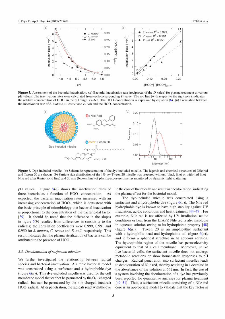
: Tween 20Dye-included micelle
Figure 6. Dye-included micelle. (a) Schematic representation of the dye-included micelle. The legends and chemical structures of Nile redand Tween 20 are shown. (b) Particle size distribution of the 1% v/v Tween 20 micelle was prepared without (black line) or with (red line)Nile red after 0 min (solid line) and 20 min (broken line) of plasma exposure time, as monitored by dynamic light scattering.
pH values. Figure 5(b) shows the inactivation rates ofthree bacteria as a function of HOO· concentration. Asexpected, the bacterial inactivation rates increased with anincreasing concentration of HOO·, which is consistent withthe basic principle of microbiology that bacterial inactivationis proportional to the concentration of the bactericidal factor[39]. It should be noted that the difference in the slopesin figure 5(b) resulted from differences in sensitivity to theradicals; the correlation coefficients were 0.999, 0.991 and0.950 for S. mutans, C. rectus and E. coli, respectively. Thisresult indicates that the plasma sterilization of bacteria can beattributed to the presence of HOO·.
3.3. Decolouration of surfactant micelles
We further investigated the relationship between radicalspecies and bacterial inactivation. A simple bacterial modelwas constructed using a surfactant and a hydrophobic dye(figure 6(a)). This dye-included micelle was used for the cellmembrane model that cannot be permeated by the O−
2 · chargedradical, but can be permeated by the non-charged (neutral)HOO· radical. After penetration, the radicals react with the dye
in the core of the micelle and result in decolouration, indicatingthe plasma effect for the bacterial model.
The dye-included micelle was constructed using asurfactant and a hydrophobic dye (figure 6(a)). The Nile redhydrophobic dye is known to have high stability against UVirradiation, acidic conditions and heat treatment [44–47]. Forexample, Nile red is not affected by UV irradiation, acidicconditions or heat from the LTAPP. Nile red is also insolublein aqueous solution owing to its hydrophobic property [48](figure 6(a)). Tween 20 is an amphipathic surfactantwith a hydrophilic head and hydrophobic tail (figure 6(a)),and it forms a spherical structure in an aqueous solution.The hydrophobic region of the micelle has permselectivityequivalent to that of a cell membrane. Moreover, unlikelive bacterial cells, the surfactant micelle does not undergometabolic reactions or show homeostatic responses to pHchanges. Radical penetration into surfactant micelles leadsto decolouration of Nile red, thereby resulting in a decrease inthe absorbance of the solution at 552 nm. In fact, the use ofa system involving the decolouration of a dye has previouslybeen reported for quantitative analyses for plasma treatment[49–51]. Thus, a surfactant micelle consisting of a Nile redcore is an appropriate model to validate that the key factor in
5
J. Phys. D: Appl. Phys. 46 (2013) 295402 E Takai et al
pH 2
pH 10
0 10 20 min(a) (b)
(c) (d)0.20
0.18
0.16
0.14
0.12
0.10
0.08
0.06
0.04Dec
olou
ratio
n R
ate
(µm
ol /
min
)
1197531
pH
1.0
0.8
0.6
0.4
0.2
0.0
[HO
O]/[H
OO
] p H= 1
0.18
0.16
0.14
0.12
0.10
0.08
0.06
0.04Dec
olou
rat io
n R
a te
(µm
ol /
mi n
)
1.00.80.60.40.20.0
[HOO ]/[HOO ] pH=1
R2 = 0.991
2
3
4
5
6789
1
Frac
tion
of N
il e R
ed (N
orm
aliz
ed)
20151050
Plasma exposure time (min)
pH 10.0 pH 7.0 pH 4.0 pH 2.0
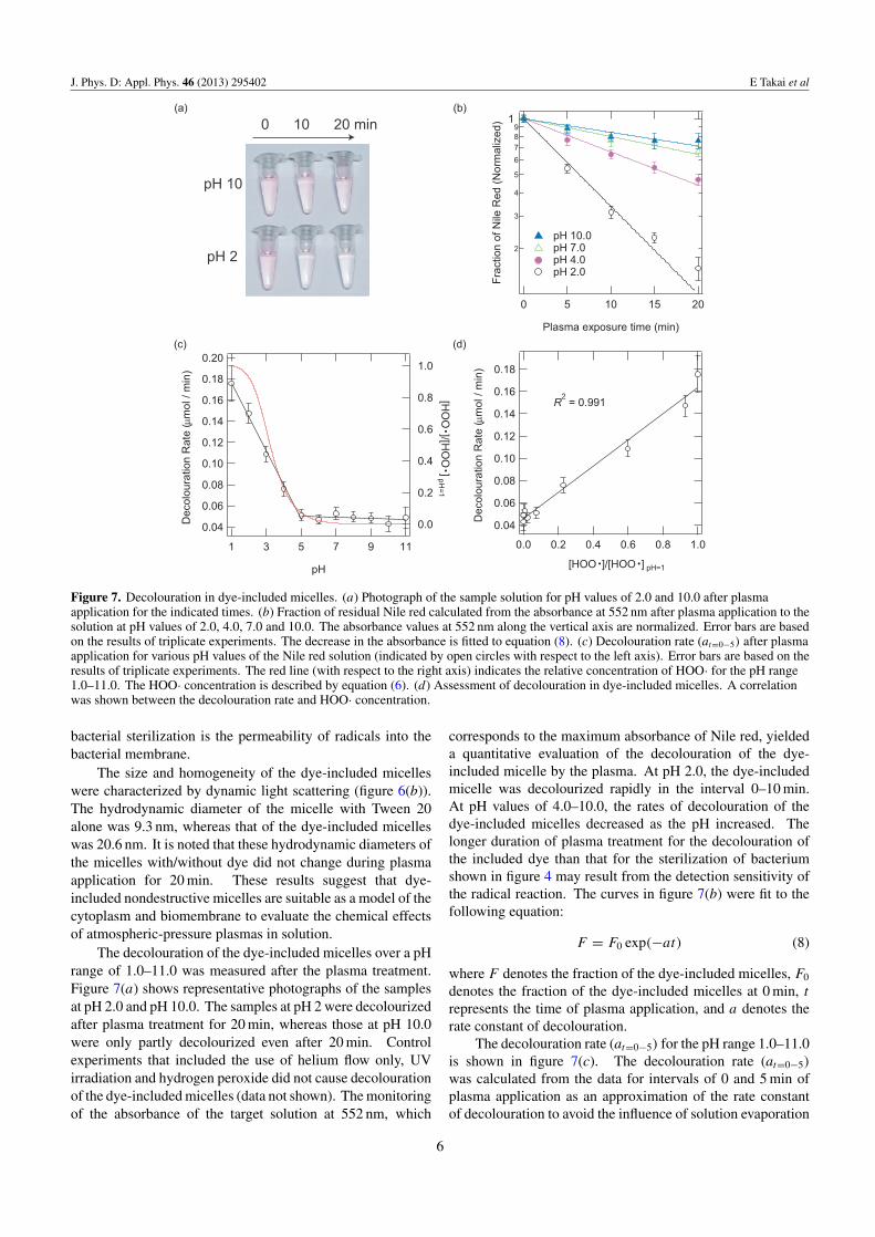
Figure 7. Decolouration in dye-included micelles. (a) Photograph of the sample solution for pH values of 2.0 and 10.0 after plasmaapplication for the indicated times. (b) Fraction of residual Nile red calculated from the absorbance at 552 nm after plasma application to thesolution at pH values of 2.0, 4.0, 7.0 and 10.0. The absorbance values at 552 nm along the vertical axis are normalized. Error bars are basedon the results of triplicate experiments. The decrease in the absorbance is fitted to equation (8). (c) Decolouration rate (at=0−5) after plasmaapplication for various pH values of the Nile red solution (indicated by open circles with respect to the left axis). Error bars are based on theresults of triplicate experiments. The red line (with respect to the right axis) indicates the relative concentration of HOO· for the pH range1.0–11.0. The HOO· concentration is described by equation (6). (d) Assessment of decolouration in dye-included micelles. A correlationwas shown between the decolouration rate and HOO· concentration.
bacterial sterilization is the permeability of radicals into thebacterial membrane.
The size and homogeneity of the dye-included micelleswere characterized by dynamic light scattering (figure 6(b)).The hydrodynamic diameter of the micelle with Tween 20alone was 9.3 nm, whereas that of the dye-included micelleswas 20.6 nm. It is noted that these hydrodynamic diameters ofthe micelles with/without dye did not change during plasmaapplication for 20 min. These results suggest that dye-included nondestructive micelles are suitable as a model of thecytoplasm and biomembrane to evaluate the chemical effectsof atmospheric-pressure plasmas in solution.
The decolouration of the dye-included micelles over a pHrange of 1.0–11.0 was measured after the plasma treatment.Figure 7(a) shows representative photographs of the samplesat pH 2.0 and pH 10.0. The samples at pH 2 were decolourizedafter plasma treatment for 20 min, whereas those at pH 10.0were only partly decolourized even after 20 min. Controlexperiments that included the use of helium flow only, UVirradiation and hydrogen peroxide did not cause decolourationof the dye-included micelles (data not shown). The monitoringof the absorbance of the target solution at 552 nm, which
corresponds to the maximum absorbance of Nile red, yieldeda quantitative evaluation of the decolouration of the dye-included micelle by the plasma. At pH 2.0, the dye-includedmicelle was decolourized rapidly in the interval 0–10 min.At pH values of 4.0–10.0, the rates of decolouration of thedye-included micelles decreased as the pH increased. Thelonger duration of plasma treatment for the decolouration ofthe included dye than that for the sterilization of bacteriumshown in figure 4 may result from the detection sensitivity ofthe radical reaction. The curves in figure 7(b) were fit to thefollowing equation:
F = F0 exp(−at) (8)
where F denotes the fraction of the dye-included micelles, F0
denotes the fraction of the dye-included micelles at 0 min, t
represents the time of plasma application, and a denotes therate constant of decolouration.
The decolouration rate (at=0−5) for the pH range 1.0–11.0is shown in figure 7(c). The decolouration rate (at=0−5)
was calculated from the data for intervals of 0 and 5 min ofplasma application as an approximation of the rate constantof decolouration to avoid the influence of solution evaporation
6
J. Phys. D: Appl. Phys. 46 (2013) 295402 E Takai et al
with long plasma treatment times. The decolouration ratesof the dye-included micelles at pH 1.0–4.0 increased withdecreasing pH, whereas they remained constant for the pHrange 5.0–11.0. The experimental results show that thedecolouration rate of dye-included micelles subject to plasmatreatment increases under acidic conditions, in a mannersimilar to the bacterial inactivation rates shown in figures 4and 5. The boundary region between pH values of 4.0and 5.0 showed good agreement with that described in aprevious study reporting that plasma application inactivatesbacteria effectively at pH levels below 4.8 [8]. Figure 7(c)shows the theoretical calculation curve of HOO· concentration(equation (6)). It is significant that the decolouration ratesstrongly correlate with the concentration of HOO· for the pHrange 1.0–11.0. Figure 7(d) shows the correlation between thedecolouration rate and the amount of HOO· for a correlationcoefficient of 0.991. This result indicates that decolourationin surfactant micelles can also be attributed to the presenceof HOO· ; the different permeability of radicals into the cellmembrane is a key factor in plasma sterilization in solution.
4. Discussion
4.1. Molecular mechanism of bacterial sterilization
In this study, we demonstrated the mechanism of bacterialsterilization using plasma-induced chemical processing withthe reduced pH method, as illustrated in figure 1. Molecularmechanisms of plasma sterilization achieved using the reducedpH method are discussed below. We can assume that bacterialsterilization results from O−
2 · and/or HOO· in the solution,which are generated by the LTAPP. As shown in figure 3, acomparison of pH-dependence on the concentration of O−
2 ·,HOO· and (HOO · +O−
2 ·) revealed that the profile of onlyHOO· was similar to that of the inactivation rates for the threebacteria tested (figure 5(a)). Furthermore, the concentrationof HOO· correlated well with all the inactivation rates ofall bacteria (figure 5(b)). These results indicate that HOO·predominantly contributes to the plasma sterilization achievedusing the reduced pH method.
These data raise the question as to why HOO· ratherthan O−
2 · plays a key role in plasma sterilization achievedusing the reduced pH method. It is known that chargedsubstances possess a low level of permeability across the cellmembrane. In fact, it has been reported that O−
2 · has an electriccharge of −1, which hinders its ability to permeate the cellmembrane [52]. On the other hand, HOO· is not charged,which allows it to easily cross the cell membrane. Therefore,bacterial sterilization by radicals may depend on differencesin permeability into the biomembrane.
To more clearly define the relationship betweenbacterial sterilization and permeability of radicals into thebiomembrane, we constructed the dye-included micellebacterial model (figure 6(a)). The decolouration system usingdye-included micelles allowed us to detect only the chemicalreactions of radicals in the micelle. Consequently, clear resultswere obtained; pH-dependence of the decolouration rate wassimilar to that of the concentration of HOO· (figure 7(c)),and the concentration of HOO· was well correlated with
the decolouration rates (figure 7(d)). Taken together, wesuccessfully demonstrated that plasma sterilization achievedusing the reduced pH method results only from HOO· due toits permeability into the biomembrane. We have reported thatplasma treatment induced inactivation of protein in solution,although the protein was dispersed in the solution, and it didnot remain in the micelle or cell membrane [38]. Furthermore,we have showed that the sterilization of bacteria by the plasmawas not related to the visible destruction of bacteria [8]. Thus,we concluded that the mechanism of sterilization using thereduced pH method is as follows: (step 1) penetration of HOO·into the cell (figure 1); (step 2) penetrated radicals react withand inactivate proteins in the cell; and (step 3) sterilizationis induced by inactivation of protein/s that are important forbacteria. We will try the challenge to control experiment usingthe radical species generated by other means but treatment ofacidic solution with the jet in future.
4.2. Assay of plasma sterilization
Several assays for plasma sterilization have been reported byother researchers, such as number of surviving CFUs plottedagainst dissipated power [27]. However, it is well known thatbacterial inactivation is proportional to the concentration of thebactericidal factor [39]. This study shows a new assessmentfor plasma sterilization (figures 5(b) and 7(d)). It is ofinterest that a different gradient was found for each bacterium,which results from the different sensitivity of the bacteriato the plasma treatment in solution. This result indicatesthat the gradient shows the sterilizing power of the plasmasystem for each bacterium. Consequently, the assay of plasmasterilization in solution is appropriate to plot the death rate ofbacterial inactivation with the concentration of key species. Inthe future, it is expected to elucidate the various situations ofsterilization under medical applications by determination andquantitative measurement of other possible radicals which maycause similar effects.
Various plasma systems have been reported forsterilization [23, 28] and medicines [13], although thesesystems have not assessed the relationship between plasmatreatment and radical behaviour. Among them, our dye-included micelle successfully evaluated bacterial inactivation,which has simple components of abundant detergent andNile red, which is known to have high stability against UVirradiation, acidic conditions and heat treatment. Thus, wepropose that the dye-included micelle system can be widelyused as an assay of plasma systems for plasma sterilization insolution.
4.3. Contribution to plasma medicines
We believe that our research contributes to plasma medicine.The inactivation rates of all bacteria tested increased witha decreasing pH (figure 5). Other researchers have alsoreported that plasma treatment effectively sterilizes bacteriaunder acidic conditions. For example, S. aureus waseffectively inactivated at pH 3.7–4.5 compared with pH 4.5–7.5[53], Bacillus subtilis spores were inactivated only in anacidic solution [54], and the eukaryotic microbe, Neurospora
7
J. Phys. D: Appl. Phys. 46 (2013) 295402 E Takai et al
crassa, showed a rapid decrease in spore germinationin acidic water [55]. Thus, we conclude that plasmatreatment generally effectively sterilizes bacteria under acidicconditions.
We assume that the sterilization in solution using plasma-induced chemical processing may be applicable to plasmamedicine. However, human lymph and blood are at neutral pHwith a buffering capacity. Therefore, the reduced pH method isnecessary for plasma sterilization. Actually, plasma treatmentfor 5 min had no effect at pH 6.5 but sterilized S. mutans,which is a model oral microorganism in dental plaques inbiofilms at pH 4.5 [30]. Thus, the reduced pH method maybe applicable to plasma medicine, such as for burn injury andbedsore.
There has been a growing interest in plasma medicine forits application in removing cancer cells [56]. For example,the permeability of radicals into the biomembrane can becontrolled by pore formation using Magainin 2 peptide [57],and the electroporation of cell membranes can be accomplishedusing electric pulses [58]. The combination of modifying cellmembranes and using atmospheric-pressure plasma [59, 60]can increase the efficiency of plasma therapy for canceroustumours by increasing the permeability of radicals into themembranes of cancer cells. Although it has been proposedthat plasma treatment is toxic because of the generation ofneutral reactive species, instead of ozone [61], the controversyis solved by our data. Our results can significantly contributetowards an increasingly efficient and safer application ofplasma sterilization for medicinal purposes. The effectivepermeation of radicals into the cell membrane implies thepossibility of using plasma therapy for the removal of cancercells.
5. Conclusion
In this study, we demonstrated the mechanism of efficientbactericidal inactivation with the reduced pH methodillustrated in figure 1, using three kinds of living bacteria anda simple bacterial model. Our data showed that the death rateof the bacteria tested was proportional to the concentration ofhydroperoxy radicals in the solution (figures 5(b) and 7(d)).These results indicate that the key species for the plasmasterilization with the reduced pH method is hydroperoxyradicals, and its key role is to bring about high permeationof radicals into the cell membrane in order to achieve efficientbactericidal activity of plasma treatment in solution. Our datacontribute to the development of not only plasma sterilizationbut also plasma medicine.
Acknowledgments
The authors thank Yoshida Dental Mfg Co., Ltd fortheir research collaboration and assistance in the bacterialinactivation experiments. The authors thank Professor YukioNagasaki for use of the light-scattering spectrometer. Thiswork was supported by the Japan Society for the Promotionof Science (JSPS) Fellows from the Ministry of Education,Culture, Sports, Science, and Technology (MEXT), Japan.
References
[1] Bruggeman P, Iza F, Lauwers D and Gonzalvo Y A 2010 Massspectrometry study of positive and negative ions in acapacitively coupled atmospheric pressure RF excited glowdischarge in He–water mixtures J. Phys. D: Appl. Phys.43 012003
[2] Locke B R, Sato M, Sunka P, Hoffmann M R and Chang J S2006 Electrohydraulic discharge and nonthermal plasma forwater treatment Indust. Eng. Chem. Res. 45 882–905
[3] Sahni M and Locke B R 2006 The effects of reactionconditions on liquid-phase hydroxyl radical production ingas–liquid pulsed-electrical-discharge reactors PlasmaProcess. Polym. 3 668–81
[4] Sahni M and Locke B R 2006 Quantification of hydroxylradicals produced in aqueous phase pulsed electricaldischarge reactors Indust. Eng. Chem. Res. 45 5819–25
[5] Bruggeman P and Leys C 2009 Non-thermal plasmas in and incontact with liquids J. Phys. D: Appl. Phys. 42 012003
[6] Burlica R, Shih K Y and Locke B R 2010 Formation of H2 andH2O2 in a water-spray gliding arc nonthermal plasmareactor Indust. Eng. Chem. Res. 49 6342–9
[7] Tani A, Ono Y, Fukui S, Ikawa S and Kitano K 2012 Freeradicals induced in aqueous solution by non-contactatmospheric-pressure cold plasma Appl. Phys. Lett.100 254103
[8] Ikawa S, Kitano K and Hamaguchi S 2010 Effects of pH onbacterial inactivation in aqueous solutions due tolow-temperature atmospheric pressure plasma applicationPlasma Process. Polym. 7 33–42
[9] Liu D X, Rong M Z, Wang X H, Iza F, Kong M G andBruggeman P 2010 Main species and physicochemicalprocesses in cold atmospheric-pressure He + O2 plasmasPlasma Process. Polym. 7 846–65
[10] Furusho H, Miyamoto D, Nagasaki Y, Kitano K andHamaguchi S 2007 Synthesis of uniformly dispersed metalnanoparticles with dispersion stability by nonequilibriumatmospheric plasma jets J. Photopolym. Sci. Technol.20 229–33
[11] Furusho H, Kitano K, Hamaguchi S and Nagasaki Y 2009Preparation of stable water-dispersible PEGylated goldnanoparticles assisted by nonequilibriumatmospheric-pressure plasma jets Chem. Mater. 21 3526–35
[12] Sumitani S, Murotani H, Oishi M, Kitano K, Hamaguchi S andNagasaki Y 2009 Nonequilibrium atmospheric plasma jetsassisted stabilization of drug delivery carriers: preparationand characterization of biodegradable polymericnano-micelles with enhanced stability J. Photopolym. Sci.Technol. 22 467–71
[13] Kong M G, Kroesen G, Morfill G, Nosenko T, Shimizu T,van Dijk J and Zimmermann J L 2009 Plasma medicine: anintroductory review New J. Phys. 11 115012
[14] Babaeva N Y and Kushner M J 2013 Reactive fluxes deliveredby dielectric barrier discharge filaments to slightly woundedskin J. Phys. D: Appl. Phys. 46 025401
[15] Sato T, Yokoyama M and Johkura K 2011 A key inactivationfactor of HeLa cell viability by a plasma flow J. Phys. D:Appl. Phys. 44 372001
[16] Kim K, Choi J D, Hong Y C, Kim G, Noh E J, Lee J-S andYang S S 2011 Atmospheric-pressure plasma-jet frommicronozzle array and its biological effects on living cellsfor cancer therapy Appl. Phys. Lett. 98 073701
[17] Kim S J, Chung T H, Bae S H and Leem S H 2010 Inductionof apoptosis in human breast cancer cells by a pulsedatmospheric pressure plasma jet Appl. Phys. Lett. 97 023702
[18] Kim C H, Kwon S, Bahn J H, Lee K, Jun S I, Rack P D andBaek S J 2010 Effects of atmospheric nonthermal plasmaon invasion of colorectal cancer cells Appl. Phys. Lett.96 243701
8
J. Phys. D: Appl. Phys. 46 (2013) 295402 E Takai et al
[19] Karakas E, Munyanyi A, Greene L and Laroussi M 2010Destruction of alpha-synuclein based amyloid fibrils by alow temperature plasma jet Appl. Phys. Lett.97 143702
[20] Fridman G, Peddinghaus M, Balasubramanian M, Ayan H,Fridman A, Gutsol A and Brooks A 2006 Blood coagulationand living tissue sterilization by floating-electrode dielectricbarrier discharge in air Plasma Chem. Plasma Process.26 425–42
[21] Gweon B, Kim D, Kim D B, Jung H, Choe W and Shin J H2010 Plasma effects on subcellular structures Appl. Phys.Lett. 96 101501
[22] Feng H Q, Wang R X, Sun P, Wu H Y, Liu Q, Fang J,Zhu W D, Li F T and Zhang J 2010 A study of eukaryoticresponse mechanisms to atmospheric pressure cold plasmaby using Saccharomyces cerevisiae single gene mutantsAppl. Phys. Lett. 97 131501
[23] Bussiahn R, Brandenburg R, Gerling T, Kindel E, Lange H,Lembke N, Weltmann K D, von Woedtke T and Kocher T2010 The hairline plasma: an intermittent negativedc-corona discharge at atmospheric pressure for plasmamedical applications Appl. Phys. Lett.96 143701
[24] Perni S, Shama G, Hobman J L, Lund P A, Kershaw C J,Hidalgo-Arroyo G A, Penn C W, Deng X T, Walsh J L andKong M G 2007 Probing bactericidal mechanisms inducedby cold atmospheric plasmas with Escherichia coli mutantsAppl. Phys. Lett. 90 073902
[25] Kolb J F, Mohamed A A H, Price R O, Swanson R J,Bowman A, Chiavarini R L, Stacey M and Schoenbach K H2008 Cold atmospheric pressure air plasma jet for medicalapplications Appl. Phys. Lett. 92 241501
[26] Zhang X H, Huang J, Liu X, Peng L, Guo L, Lv G, Chen W,Feng K and Yang S 2009 Treatment of Streptococcusmutans bacteria by a plasma needle J. Appl. Phys. 105063302
[27] Majumdar A, Singh R K, Palm G J and Hippler R 2009Dielectric barrier discharge plasma treatment on E. coli:Influence of CH4/N2, O2, N2/O2, N2 and Ar gases J. Appl.Phys. 106 084701
[28] Lu X, Ye T, Cao Y G, Sun Z Y, Xiong Q, Tang Z Y, Xiong Z L,Hu J, Jiang Z H and Pan Y 2008 The roles of the variousplasma agents in the inactivation of bacteria J. Appl. Phys.104 053309
[29] Burlica R, Grim R G, Shih K Y, Balkwill D and Locke B R2010 Bacteria inactivation using low power pulsed glidingarc discharges with water spray Plasma Process. Polym.7 640–9
[30] Yamazaki H, Ohshima T, Tsubota Y, Yamaguchi H,Jayawardena J A and Nishimura Y 2011 Microbicidalactivities of low frequency atmospheric pressure plasma jetson oral pathogens Dent. Mater. J. 30 384–91
[31] Kitano K, Ikawa S, Tani A, Ohshima T, Yamaguchi H,Yamazaki H, Arakawa R, Kitamura T and Ohnishi N 2011Innovative plasma disinfection of bacteria in water by thereduced pH method combined with free radicals suppliedby non-contact atmospheric plasma Proc. 20th Int. Symp. onPlasma Chemistry (ISPC-20) (Philadelphia, PA, 24–29 July2011) p 17
[32] Bielski B H J, Cabelli D E, Arudi R L and Ross A B 1985Reactivity of HO2/O2 radicals in aqueous-solution J. Phys.Chem. Ref. Data 14 1041–100
[33] Behar D, Czapski G, Rabani J, Dorfman L M and Schwarz H A1970 Acid dissociation constant and decay kinetics of theperhydroxyl radical J. Phys. Chem. 74 3209–13
[34] Li G, Li H P, Wang L Y, Wang S, Zhao H X, Sun W T,Xing X H and Bao C Y 2008 Genetic effects ofradio-frequency, atmospheric-pressure glow discharges withhelium Appl. Phys. Lett. 92 221504
[35] Yan X et al 2009 Effect of the atmospheric pressurenonequilibrium plasmas on the conformational changes ofplasmid DNA Appl. Phys. Lett. 95 083702
[36] Kurita H, Nakajima T, Yasuda H, Takashima K, Mizuno A,Wilson J I B and Cunningham S 2011 Single-moleculemeasurement of strand breaks on large DNA inducedby atmospheric pressure plasma jet Appl. Phys. Lett.99 191504
[37] Yasuda H, Miura T, Kurita H, Takashima K and Mizuno A2010 Biological evaluation of DNA damage inbacteriophages inactivated by atmospheric pressure coldplasma Plasma Process. Polym. 7 301–8
[38] Takai E, Kitano K, Kuwabara J and Shiraki K 2012 Proteininactivation by low-temperature atmospheric pressureplasma in aqueous solution Plasma Process. Polym.9 77–82
[39] Deindoerfer F H 1957 Calculation of heat sterilization timesfor fermentation media Appl. Microbiol. 5 221–8
[40] Kim A Y and Thayer D W 1995 Radiation-induced celllethality of Salmonella-TyphimuriumAtcc-14028—cooperative effect of hydroxyl radical andoxygen Radiat. Res. 144 36–42
[41] Marklund S 1976 Spectrophotometric study of spontaneousdisproportionation of superoxide anion radical and sensitivedirect assay for superoxide-dismutase J. Biol. Chem. 2517504–7
[42] Loesche W J 1986 Role of Streptococcus mutans in humandental decay Microbiol. Rev. 50 353–80
[43] Rams T E, Feik D and Slots J 1993 Campylobacter-Rectus inhuman periodontitis Oral Microbiol. Immunol. 8 230–5
[44] Mishra R, Sjolander D and Hammarstrom P 2011Spectroscopic characterization of diverse amyloid fibrils invitro by the fluorescent dye Nile red Mol. BioSyst.7 1232–40
[45] Greenspan P and Fowler S D 1985 Spectrofluorometric studiesof the lipid probe, Nile Red J. Lipid Res. 26 781–9
[46] Greenspan P, Mayer E P and Fowler S D 1985 Nile Red—aselective fluorescent stain for intracellular lipid dropletsJ. Cell Biol. 100 965–73
[47] Cser A, Nagy K and Biczok L 2002 Fluorescence lifetime ofNile Red as a probe for the hydrogen bonding strength withits microenvironment Chem. Phys. Lett. 360 473–8
[48] Krishna M M G 1999 Excited-state kinetics of thehydrophobic probe Nile red in membranes and micellesJ. Phys. Chem. A 103 3589–95
[49] Wang Z, Xu D, Chen Y, Hao C and Zhang X 2008 Plasmadecoloration of dye using dielectric barrier discharges withearthed spraying water electrodes J. Electrost. 66 476–81
[50] Chen G, Zhou M, Chen S and Chen W 2009 The differenteffects of oxygen and air DBD plasma byproducts on thedegradation of methyl violet 5BN J. Hazard. Mater.172 786–91
[51] Kuwahata H, Kimura K and Ohyama R-i 2010 Decolorizationof methylene blue aqueous solution byatmospheric-pressure plasma jet e-J. Surf. Sci. Nanotechnol.8 381–3
[52] Korshunov S S and Imlay J A 2002 A potential role forperiplasmic superoxide dismutase in blocking thepenetration of external superoxide into the cytosol ofGram-negative bacteria Mol. Microbiol. 43 95–106
[53] Liu F X et al 2010 Inactivation of bacteria in an aqueousenvironment by a direct-current, cold-atmospheric-pressureair plasma microjet Plasma Process. Polym. 7 231–6
[54] Sun P, Wu H, Bai N, Zhou H, Wang R, Feng H, Zhu W, Zhang Jand Fang J 2012 Inactivation of Bacillus subtilis spores inwater by a direct-current, cold atmospheric-pressure airplasma microjet Plasma Process. Polym. 9 157–64
[55] Park G, Ryu Y H, Hong Y J, Choi E H and Uhm H S 2012Cellular and molecular responses of Neurospora crassa to
9

J. Phys. D: Appl. Phys. 46 (2013) 295402 E Takai et al
non-thermal plasma at atmospheric pressure Appl. Phys.Lett. 100 063703
[56] Huang J, Chen W, Li H, Wang X-Q, Lv G-H, Khosa M L,Guo M, Feng K-C, Wang P-Y and Yang S-Z 2011Deactivation of A549 cancer cells in vitro by a dielectricbarrier discharge plasma needle J. Appl. Phys. 109 053305
[57] Tamba Y and Yamazaki M 2009 Magainin 2-induced poreformation in the lipid membranes depends on itsconcentration in the membrane interface J. Phys. Chem. B113 4846–52
[58] Tsong T Y 1991 Electroporation of cell-membranes Biophys.J. 60 297–306
[59] Srivastava N and Wang C 2011 Effects of water addition on OHradical generation and plasma properties in an atmosphericargon microwave plasma jet J. Appl. Phys. 110 053304
[60] Sousa J S, Niemi K, Cox L J, Algwari Q T, Gans T andO’Connell D 2011 Cold atmospheric pressure plasma jetsas sources of singlet delta oxygen for biomedicalapplications J. Appl. Phys. 109 123302
[61] Kalghatgi S, Fridman A, Azizkhan-Clifford J and Friedman G2012 DNA damage in mammalian cells by non-thermalatmospheric pressure microsecond pulsed dielectric barrierdischarge plasma is not mediated by ozone Plasma Process.Polym. 9 726–32
10