Embed Size (px)
Citation preview
ISO/DIS 20186-3:2018(E)
Fel! Hittar inte referenskälla. 1
ISO TC 212/N559
ISO/DIS 20186-3
ISO TC 212/WG 4
Secretariat: ANSI
Molecular in vitro diagnostic examinations — Specifications for pre-examination processes for venous whole blood — Part 3: Part 3 Isolated
circulating cell free DNA from plasma
DIS stage
Warning for WDs and CDs
This document is not an ISO International Standard. It is distributed for review and comment. It is subject to change without notice and may not be referred to as an
International Standard.
Recipients of this draft are invited to submit, with their comments, notification of any relevant patent rights of which they are aware and to provide supporting
documentation.
To help you, this guide on writing standards was produced by the ISO/TMB and is available at http://www.iso.org/iso/how-to-write-standards.pdf
ISO/DIS 20186-3:2018(E)
2 Fel! Hittar inte referenskälla.
© ISO 2014
All rights reserved. Unless otherwise specified, no part of this publication may be reproduced or utilized otherwise in any form or by any means, electronic or mechanical, including photocopying, or posting on the internet or an intranet, without prior written permission. Permission can be requested from either ISO at the address below or ISO's member body in the country of the requester.
ISO copyright office
Case postale 56 • CH-1211 Geneva 20
Tel. + 41 22 749 01 11
Fax + 41 22 749 09 47
E-mail [email protected]
Web www.iso.org
Published in Switzerland.
ISO/DIS 20186-3:2018(E)
Fel! Hittar inte referenskälla. 3
Contents
Foreword v
Introduction..................................................................................................................................................................... 7
1 Scope ............................................................................................................................................................................... 8
2 Normative references ............................................................................................................................................... 8
3 Terms and definitions .............................................................................................................................................. 8
4 General Consideration .......................................................................................................................................... 13
5 Outside the laboratory .......................................................................................................................................... 14
5.1 Specimen collection .......................................................................................................................................................... 14
5.1.1 Information about the specimen donor/patient ................................................................................. 14
5.1.2 Selection of the venous whole blood collection tube by the laboratory .................................... 14
5.1.3 Venous whole blood collection from the donor/patient and stabilization procedures ...... 15
5.1.4 Information about the specimen and storage requirements at the blood collection facility 15
5.2 Transport requirements .......................................................................................................................................... 16
6 Inside the laboratory ........................................................................................................................................ 17
6.1 Specimen reception ................................................................................................................................................... 17
6.2 Storage requirements for blood specimens .................................................................................................... 17
6.3 Plasma preparation ................................................................................................................................................... 17
6.4 Storage requirements for plasma samples ...................................................................................................... 18
6.5 Isolation of the ccfDNA ............................................................................................................................................ 19
6.5.1 General .................................................................................................................................................................. 19
6.5.2 Using blood collection tubes with stabilizers ....................................................................................... 20
6.5.3 Using blood collection tubes without stabilizers ................................................................................ 20
6.6 Quantity and quality assessment of isolated ccfDNA .................................................................................. 20
6.7 Storage of isolated ccfDNA ..................................................................................................................................... 21
6.7.1 General .................................................................................................................................................................. 21
6.7.2 ccfDNA isolated with commercially available kits .............................................................................. 21
ISO/DIS 20186-3:2018(E)
4 Fel! Hittar inte referenskälla.
6.7.3 ccfDNA isolated with the laboratory's own protocols ...................................................................... 21
Annex A (informative) Impact of pre-examination process steps on circulating cell free DNA profiles in venous whole blood plasma .............................................................................................................. 22
A1. Influence of venous whole blood storage duration on ccfDNA examination ................................. 22
A.1.1 Post collection changes of blood ccfDNA profiles ................................................................................ 22
A.1.2 Impact of ccfDNA profile changes on EGFR mutant detection in plasma..................................... 23
Bibliography ................................................................................................................................................................. 26
ISO/DIS 20186-3:2018(E)
Fel! Hittar inte referenskälla. 5
Foreword
ISO (the International Organization for Standardization) is a worldwide federation of national standards bodies (ISO member bodies). The work of preparing International Standards is normally carried out through ISO technical committees. Each member body interested in a subject for which a technical committee has been established has the right to be represented on that committee. International organizations, governmental and non-governmental, in liaison with ISO, also take part in the work. ISO collaborates closely with the International Electrotechnical Commission (IEC) on all matters of electrotechnical standardization.
The procedures used to develop this document and those intended for its further maintenance are described in the ISO/IEC Directives, Part 1. In particular the different approval criteria needed for the different types of ISO documents should be noted. This document was drafted in accordance with the editorial rules of the ISO/IEC Directives, Part 2 (see www.iso.org/directives).
Attention is drawn to the possibility that some of the elements of this document may be the subject of patent rights. ISO shall not be held responsible for identifying any or all such patent rights. Details of any patent rights identified during the development of the document will be in the Introduction and/or on the ISO list of patent declarations received (see www.iso.org/patents).
Any trade name used in this document is information given for the convenience of users and does not constitute an endorsement.
For an explanation on the meaning of ISO specific terms and expressions related to conformity assessment, as well as information about ISO's adherence to the World Trade Organization (WTO) principles in the Technical Barriers to Trade (TBT) see the following URL: www.iso.org/iso/foreword.html.
The committee responsible for this document is ISO/TC 212, Clinical laboratory testing and in vitro diagnostic test systems.
— A list of all parts in the ISO 20186- series can be found on the ISO website.
ISO/DIS 20186-3:2018(E)
6 Fel! Hittar inte referenskälla.
Introduction
Molecular in vitro diagnostics has enabled a significant progress in medicine. Further progress is expected by new technologies analyzing profiles of nucleic acids, proteins, and metabolites in human tissues and body fluids. However, the profiles of these molecules can change drastically during the pre-examination process, including the specimen collection, transport, storage and processing. Consequently, this makesthe outcome from diagnostics or research unreliable or even impossible because the subsequent examination might not determine real the situation in the patient but an artificial profile generated during the pre-examination processes.
CcfDNA profiles can change significantly after blood collection (e.g., release of genomic DNA from cells in blood, ccfDNA fragmentation and ccfDNA quantity change). Therefore, special measures have to be taken to secure good quality specimens for ccfDNA examination.
Standardization of the entire workflow from specimen collection to the circulating cell free DNA (ccfDNA) examination is needed due to release of DNA from cells in blood, thus changing the original native ccfDNA profile in the body, but also ccfDNA degradation and fragmentation after blood collection. Studies have been undertaken to determine the important influencing factors. This document draws upon such work to codify and standardize the steps for circulating cell free DNA examination from plasma prepared from human venous whole blood in what is referred to as the pre-examination phase.
In this document, the following verbal forms are used:
— "shall" indicates a requirement;
— "should" indicates a recommendation;
— "may" indicates a permission;
— "can" indicates a possibility or a capability.
Molecular in vitro diagnostic examinations — Specifications for pre-examination processes for venous whole blood — Part 3: Part 3 Isolated circulating cell free DNA from plasma
1 Scope
This document recommends the handling, storage, processing and documentation of venous whole blood specimens intended for circulating cell free DNA (ccfDNA) examination during the pre-examination phase before a molecular assay is performed. This document covers specimens collected in venous whole blood collection tubes.
This document is applicable to any molecular in vitro diagnostic examination performed by medical laboratories. It is also intended to be used by laboratory customers, in vitro diagnostics developers and manufacturers, biobanks, institutions and commercial organizations performing biomedical research, and regulatory authorities.
Different dedicated measures need to be taken for stabilizing blood genomic DNA, which are not described in this document. Blood genomic DNA is covered in ISO 20186-2, Molecular in vitro diagnostic examinations — specifications for pre-examination processes for venous whole blood — Part 2: Isolated genomic DNA.
Different dedicated measures need to be taken for preserving DNA in circulating exosomes, which are not described in this document.
NOTE 1 CcfDNA obtained from blood by the procedures suggested in this document can contain DNA present in exosomes[8][9].
ISO/DIS 20186-3:2018(E)
Fel! Hittar inte referenskälla. 7
DNA in pathogens present in blood is not covered by this document.
NOTE 2 International, national or regional regulations or requirements can also apply to specific topics covered in this document.
2 Normative references
The following documents are referred to in the text in such a way that some or all of their content constitutes requirements of this document. For dated references, only the edition cited applies. For undated references, the latest edition of the referenced document (including any amendments) applies.
ISO 15189:2012, Medical laboratories — Requirements for quality and competence (ISO 15189:2012, Corrected version 2014-08-15)
3 Terms and definitions
For the purposes of this document, the following terms and definitions apply.
ISO and IEC maintain terminological databases for use in standardization at the following addresses:
— ISO Online browsing platform: available at http://www.iso.org/obp
— IEC Electropedia: available at http://www.electropedia.org/
3.1 ambient temperature unregulated temperature of the surrounding air
3.2 analyte component represented in the name of a measurable quantity
[SOURCE: ISO 17511:2013, 3.2]
3.3 backflow flow of a liquid opposite to the usual or desired direction
3.4 blood collection set intravenous device specialized for venipuncture consisting of a stainless steel beveled needle and tube (tubing) with attached plastic wings and fitting connector
Note 1 to entry: The connector attaches to an additional blood collection device, e.g., a blood collection tube.
3.5 blood collection tube tube used for blood collection, usually in a vacuum which forces blood from the vein through the needle into the tube
3.6 ccfDNA circulating cell free DNA extracellular human DNA present in blood, serum and plasma
Note 1 to entry: ccfDNA can include DNA present in vesicles such as exosomes[8][9].
3.7 ccfDNA profile/s
ISO/DIS 20186-3:2018(E)
8 Fel! Hittar inte referenskälla.
circulating cell free DNA profile/s amounts of different ccfDNA molecules, that are present in blood and plasma that can be measured in the absence of any losses, inhibition and interference
3.8 ccfDNA proficiency testing program proficiency testing for ccfDNA based examinations
3.9 closed system non-modifiable system provided by the vendor including all necessary components for the examination (i.e., hardware, software, procedures and reagents)
3.10 cryo-precipitates for the purpose of this document an insoluble residue when frozen plasma is thawed
3.11 DNA deoxyribonucleic acid polymer of deoxyribonucleotides occurring in a double-stranded (dsDNA) or single-stranded (ssDNA) form
[SOURCE: ISO 22174:2005, 3.1.2]
3.12 DNase deoxyribonuclease enzyme that catalyzes the degradation of DNA into smaller components
3.13 examination analytical test set of operations having the object of determining the value or characteristics of a property
[SOURCE: ISO 15189:2012, 3.7, modified — The term and definition is used here without the original notes; an additional term was added.]
Note 1 to entry: Processes that start with the isolated analyte and include all kinds of parameter testing or chemical manipulation for quantitative or qualitative examination.
3.14 examination performance analytical test performance analytical performance ability of an examination procedure to measure or detect a particular analyte
Note 1 to entry: Analytical performance is determined from analytical performance studies used to assess the ability of an in vitro diagnostic examination procedure to measure or detect a particular analyte.
Note 2 to entry: Analytical performance includes such characteristics as analytical sensitivity, detection limit, analytical specificity (interference and cross-reactivity), trueness, precision and linearity.
[SOURCE: ISO/TS 17822-1:2014, 3.2, modified — Two terms were added.]
3.15 examination provider analytical test provider
ISO/DIS 20186-3:2018(E)
Fel! Hittar inte referenskälla. 9
entity that provides the specific analytical test
3.17 interfering substances endogenous (e.g., blood components, acidic polysaccharides) or exogenous (e.g., talc, anticoagulant) substances in clinical specimens that can alter an assay result
3.18 needle holder barrel used in routine venipuncture procedures to hold the blood collection tube in place and to protect the phlebotomist from direct contact with blood
3.19 pre-examination processes preanalytical phase preanalytical workflow processes that start, in chronological order, from the clinician's request and include the examination request, preparation and identification of the patient, collection of the primary sample(s), transportation to and within the medical laboratory, isolation of analytes, and end when the analytical examination begins
[SOURCE: ISO 15189:2012, 3.15, modified — An additional term was added and more detail was included.]
Note 1 to entry: The pre-examination phase includes preparative processes, e.g. ccfDNA isolation procedures, which influence the outcome of the intended examination.
3.20 primary sample specimen discrete portion of a body fluid, breath, hair or tissue taken for examination, study or analysis of one or more quantities or properties assumed to apply for the whole
[SOURCE: ISO 15189:2012, 3.16, modified — Term and definition are used here without the original notes.]
3.21 primary sample collection device apparatus specifically intended by an IVD manufacturer to obtain, contain and preserve a body fluid or tissue for in vitro diagnostic examination
Note 1 to entry: Includes devices intended to store a specimen prior to examination.
Note 2 to entry: Includes both vacuum and non-vacuum specimen collection devices.
[SOURCE: ISO 18113-1:2009, 3.55]
3.22 proficiency testing evaluation of participant performance against pre-established criteria by means of inter-laboratory comparisons
[SOURCE: ISO 17043:2010, 3.7, modified — Term and definition are used here without the original notes.]
3.23 RNA ribonucleic acid
ISO/DIS 20186-3:2018(E)
10 Fel! Hittar inte referenskälla.
polymer of ribonucleotides occurring in a double-stranded or single-stranded form
[SOURCE: ISO 22174:2005, 3.1.3]
3.24 RNase ribonuclease enzyme that catalyzes the degradation of RNA into smaller components
3.25 room temperature for the purposes of this document temperature in the range of 18 °C to 25 °C
Note 1 to entry: Local or national regulations can have different definitions.
3.26 sample one or more parts taken from a primary sample
[SOURCE: ISO 15189:2012, 3.24, modified — The examples were not taken over.]
3.27 stability ability of a sample material, when stored under specified conditions, to maintain a stated property value within specified limits for a specified period of time
[SOURCE: ISO Guide 30:2015, 2.1.15, modified — The words “reference material” were replaced by “sample material”.]
3.28 validation confirmation, through the provision of objective evidence, that the requirements for a specific intended use or application have been fulfilled
Note 1 to entry: The term "validated" is used to designate the corresponding status.
[SOURCE: ISO 9000:2015, 3.8.13, modified — Note 1 and 3 were not taken over.]
3.29 venous whole blood blood collected after directly puncturing a vein, usually with a needle and syringe, or other collection device
3.30 verification confirmation, through the provision of objective evidence, that specified requirements have been fulfilled
Note 1 to entry: The term "verified" is used to designate the corresponding status.
Note 2 to entry: Confirmation can comprise activities such as
— performing alternative calculations;
— comparing a new design specification with a similar proven design specification;
— undertaking tests and demonstrations;
ISO/DIS 20186-3:2018(E)
Fel! Hittar inte referenskälla. 11
— reviewing documents prior to issue.
[SOURCE: ISO 9000:2015, 3.8.12, modified — Note 1 and Note 2 were not taken over.]
3.31 workflow series of activities necessary to complete a task
4 General Consideration
For general statements on medical laboratory quality management systems and in particular on specimen collection, reception and handling (including avoidance of cross contaminations) see ISO 15189:2012, 4.2, 5.4.4, 5.4.6 or ISO/IEC 17020:2012, clause 8 and 7.2. The requirements on laboratory equipment, reagents, and consumables according to ISO 15189:2012, 5.3 shall be followed; ISO 15189:2012, 5.5.1.2 and 5.5.1.3 and ISO/IEC 17020, 6.2 can also apply.
All steps of a diagnostic workflow can influence the final examination result. Thus, the entire workflow, including specimen/sample storage and transport conditions, and its impact on the stability of biomolecules intended to be examined shall be verified and validated. Workflow steps which cannot always be controlled shall be documented and their impact on the examination performance shall be investigated and mitigation measures shall be established to enable the required examination performance. In these cases, risk assessment is recommended.
CcfDNA profiles can change significantly after blood collection. The post-collection release of genomic DNA from cells in blood can change the ccfDNA profile significantly. Additional post-collection effects can also occur, e.g., ccfDNA fragmentation.[10],[11],[12],[13] All these post-collection changes can vary individually in specimens from different donors or patients, and they can also depend on pathophysiological conditions.[10],[14],[15],[16] This can impact the validity and reliability of the examination results.
Before or during the design of an examination, it should therefore be investigated and assured that the ccfDNA profile/s intended to be analyzed is/are not compromized in a manner impacting the examination performance. This can be done, e.g., by applying the intended examination to specimens/samples which underwent time course studies reflecting the individual pre-examination process steps such as transport and storage.
Safety procedures for handling and transport shall be in place. Safety regulations on transport and handling shall be considered (see ISO 15189:2012, 5.2.3 and 5.4.5, and ISO 15190).
During the whole pre-examination process precautions shall be taken to avoid cross contamination between different samples/specimens, e.g., by using single-use material whenever feasible or appropriate cleaning procedures between processing of different specimens/samples.
If a commercial product is not used in accordance with the manufacturer's instructions, responsibility for its validation, verification, use and performance lies with the user.
5 Outside the laboratory
5.1 Specimen collection
5.1.1 Information about the specimen donor/patient
The documentation shall include the ID of the specimen donor/patient, which can be in the form of a code.
The documentation should include, but is not limited to:
a) the relevant health status of the specimen donor or patient (e.g., healthy, disease type, concomitant disease, demographics (e.g., age and gender));
ISO/DIS 20186-3:2018(E)
12 Fel! Hittar inte referenskälla.
NOTE In particular, e.g., cancer, inflammation, diabetes, hepatic disease, coronary disease, respiratory syndrome, trauma, after exhaustive exercise,[10] in elderly patients suffering from acute or chronic disease, first trimester of pregnancy, placental disorders as pre-term labour, pre-eclampsia and malimplantation have been reported to affect both ccfDNA quantity and fragmentation.[10],[14],[15],[16]
b) the information about medical treatment and special treatment prior to blood collection (e.g., anaesthetics, medications, fasting status);
c) the type and purpose of the proposed examination requested;
d) the appropriate consent from the specimen donor/patient.
See also ISO 15189:2012, 5.4.4.
5.1.2 Selection of the venous whole blood collection tube by the laboratory
The ccfDNA profile can be influenced by inadequate venous whole blood collection procedures and inappropriate storage/shipping conditions, plasma separation as well as by ccfDNA isolation procedures. Specifically, the post-collection release of genomic DNA from white blood cells can change the ccfDNA profile significantly. This can impact the validity of the examination results.
Venous whole blood should be collected in appropriate collection devices.
Blood should be collected in appropriate venous whole blood collection tubes containing ccfDNA profile stabilizers as post-collection release of genomic DNA from blood cells or other ccfDNA profile changes can cause impacts on the intended examination. The tubes' catalogue and lot number should be documented.
Blood collection tubes not containing any ccfDNA profile stabilizers should only be used, if allowed by the examination provider's instructions. EDTA blood collection tubes should be used in preference to other collection tubes.[12],[17] EDTA prevents clotting but not the release of DNA from blood cells.[17] Consult the specifications by the examination provider for details.
Induced clotting process in serum tubes can lead to a leucocytes lysis causing the release of DNA, thus changing the native ccfDNA profile. Therefore the use of serum tubes should be avoided.[18]
5.1.3 Venous whole blood collection from the donor/patient and stabilization procedures
e) The identity of the person collecting the specimen and the date and time of blood collection according to ISO 15189:2012, 5.4.4.3, f) shall be documented.
f) For the labelling (sample/specimen identification) of the blood collection tube a routine procedure (ISO 15189:2012, 5.4.4.3, e)) or a procedure with additional information (e.g., 2D-barcode) shall be used.
g) Standard venipuncture technique can be used. Steps for preventing possible backflow into the donor's/patient’s body can be required. The manufacturer's instructions for using the blood collection tubes shall be followed. A blood collection set and needle holder can be required when using ccfDNA profile stabilizer containing tubes. In this case, the instructions of the collection set and needle holder manufacturer shall be followed.
NOTE There is no known specific effect of venous whole blood draw procedure on the ccfDNA profile. Routine procedures can therefore be used.
h) Blood collection tubes shall be filled in accordance to the manufacturer's instructions and attention should be drawn to the correct positioning of the collection tube during the blood draw as well as the required blood volume.
i) Blood collection tube manufacturer’s instructions, for mixing or inverting the tube immediately after blood collection, shall be followed. Mixing or inverting the blood collection tube shall be done gently to avoid the destruction of cells in blood with subsequent release of DNA. If no information
ISO/DIS 20186-3:2018(E)
Fel! Hittar inte referenskälla. 13
about mixing or inverting is given by the manufacturer’s instructions, each tube should be inverted 8 times to 10 times.
NOTE Wrong and/or insufficient mixing can be one of the most important pre-examination variables. Unless additives in the blood collection tubes are homogenously mixed with the specimen, the ccfDNA profile and the ccfDNA quality can be compromized, which can impact the validity and reliability of the examination results.
j) Any tampering with and/or additions to the specimen shall be documented.
Until clean plasma is generated, special care has to be taken to avoid lysis of blood cells thus leading to the release of cellular DNA changing the original native ccfDNA profile of the donor/patient. Therefore, the specimen shall not be frozen or shaken vigorously.[11]
5.1.4 Information about the specimen and storage requirements at the blood collection facility
5.1.4.1 General
Ongoing blood cell lysis after blood collection contaminates the sample with cellular DNA [11] and can affect the validity and reliability of the examination result. The documentation regarding the specimen shall include the date and time of blood collection.[11],[18]
The required storage conditions shall be documented.
The temporary storage duration in the blood collection facility contributes to the total duration for storage.
5.1.4.2 Using blood collection tubes with stabilizers
Blood tubes with ccfDNA profile stabilizers should be used. For storing the specimens collected in blood tubes with ccfDNA profile stabilizers, the dedicated blood collection tube manufacturer’s instructions on storage conditions shall be followed (e.g., temperature and storage duration). Where the examination provider’s instructions are more stringent, these shall be followed. The storage conditions (temperature and duration etc.) shall be documented.
5.1.4.3 Using blood collection tubes without stabilizers
5.1.4.3.2 Blood collection tubes without ccfDNA profile stabilizers should only be used, if the ordered examination specifications allow the usage of such tubes. In these cases, the examination provider's instructions on storage conditions shall be followed. This can require documentation of storage conditions (temperature and duration etc.).
5.1.4.3.2 When using blood collection tubes without ccfDNA profile stabilizers and no requirements on the storage conditions are available, the specimen should be transferred immediately to 2 °C to 8 °C in order to minimize the release of DNA from cells into the blood. The storage duration allowed at 2 °C to 8 °C shall be validated by analyzing the potential impact on the examination. The storage conditions (temperature and duration etc.) shall be documented.
NOTE Some studies using blood collection tubes with EDTA have shown that storage at 2 °C to 8 °C for up to 6 h[11] had no negative impact on results obtained by the dedicated applied examination/s.[19],[20]
5.2 Transport requirements
The required transport conditions shall be documented including any deviations therefrom.
Temperature monitoring should be applied in a suitable manner.
When using blood collection tubes with ccfDNA profile stabilizers, the dedicated tube's manufacturer’s instructions on transport conditions shall be followed (transport and temperature etc.). Where the examination provider’s instructions are more stringent, these shall be followed. The transportation conditions (duration and temperature etc.) shall be documented.
ISO/DIS 20186-3:2018(E)
14 Fel! Hittar inte referenskälla.
When using blood collection tubes without ccfDNA profile stabilizers, the examination provider’s instructions on transport conditions shall be followed. This can require the documentation of transport conditions (duration and temperature etc.).
When using blood collection tubes without ccfDNA profile stabilizers and no from the examination provider’s instructions are available, the specimen should be transported at 2 °C to 8 °C in order to minimize the release of DNA from cells into the blood. The transport duration allowed at 2 °C to 8 °C shall be validated by analyzing the potential impact on the examination. The transportation conditions (duration and temperature etc.) shall be documented.
NOTE Some studies have shown that storage at 2 °C to 8 °C for up to 6 h [11] had no negative impact on results obtained by the applied examination/s. See also Table 1.
If the blood collection tube manufacturer or the examination provider requires a dedicated packaging, specimens shall be transported accordingly. If there are no such requirements, the specimens should be packed in tissues, airbags, paper or the like to protect it from shaking during transport, including accidental dropping of the package.
The specimen shall not be frozen or shaken strongly.[11]
See also ISO 15189:2012, 5.4.5.
The transport duration to the laboratory contributes to the total duration for storage.
6 Inside the laboratory
6.1 Specimen reception
The specimen reception date and time shall be documented as well as the name of the person receiving the specimen. Non-conformities of labeling, transport conditions and blood volume differences to specifications, leaking/broken tubes etc. shall be documented.
NOTE This includes for example a note, when specimens have been accidentally frozen or collected in another tube than the indicated one.
Where there are nonconformities in labelling, transport conditions, overall storage, and transport duration or blood volume that could affect the validity and reliability of the examination result, a new specimen should be obtained.
The correct identity of the specimen shall be checked. This should include the clinical information (see 5.1.1 and 5.1.3) of the specimen, hospital admission number and/or donor/patient ID, name of the patient, date of birth of the patient.
6.2 Storage requirements for blood specimens
The storage temperature and time interval between specimen receipt and sample processing for plasma generation shall be documented. Storage temperature and total storage duration shall not exceed specifications identified in 5.1.4, 5.2 and Table 1.
The specimen total storage duration shall include the duration for storage at the blood collection facility (5.1.4), for transportation to the laboratory (5.2) and for further storage at the laboratory or other institutions. Any specified maximum storage duration given by the blood collection tube manufacturer or the provider of the examination shall not be exceeded. If such specifications are not available, the maximum storage duration shall be validated and generally kept to a minimum.
6.3 Plasma preparation
When using blood collection tubes with ccfDNA profile stabilizers, the manufacturer's instructions to perform the plasma preparation shall be followed.
When using blood collection tubes without ccfDNA profile stabilizers, and if instructions for plasma preparation are available from the provider of the dedicated examination procedure, these shall be followed.
ISO/DIS 20186-3:2018(E)
Fel! Hittar inte referenskälla. 15
When using blood collection tubes without ccfDNA profile stabilizers, and no instructions are available from the provider of the dedicated examination procedure, EDTA blood specimen should be centrifuged at 1 600 g to 2 500 g at 2 °C to 8 °C for 10 min. Plasma shall be carefully transferred into a new tube without disturbing the plasma cellular interface layer in order to avoid contamination with genomic DNA and cellular RNA derived from leucocytes. A second centrifugation should be performed on the supernatant of the first centrifugation step at 14 000 g to 16 000 g at 2 °C to 8 °C for 10 min. If the second centrifugation step is performed, the supernatant shall be carefully transferred into a new tube without disturbing the pellet.[12],[21] If high g-force centrifugation is not possible, e.g., due to lack of appropriate centrifuges, the examination shall be validated when carrying out the second centrifugation at a lower g-force e.g., 3 000 g to 5 000 g for 20 min at 2 °C to 8 °C.
6.4 Storage requirements for plasma samples
The storage temperature and time interval between the plasma generation and the isolation of the ccfDNA shall be documented, including any deviations therefrom. Any specified maximum storage duration given by the blood collection tube manufacturer or the provider of the dedicated examination procedure shall not be exceeded. If such specification is not available, the maximum storage duration shall be validated and generally kept to a minimum.
The plasma samples should be processed for analytical down-stream tests immediately. Depending on the examination specifications, for short term storage, plasma may be stored at 2 °C to 8 °C for a maximum of 24 h. For long-term storage plasma should be stored frozen at ≤ –20 °C.[11],[21] The plasma storage conditions (i.e., duration and temperature) shall be documented.
Frozen plasma samples shall not be thawed more than once.[11],[21],[22] Therefore, the plasma samples should be aliquoted into cryo-vials or other suitable vials if further testing is needed.[10] See also Table 1.
Where dedicated examination provider's instructions on storage of plasma are available, these shall be followed and documented (see Table 1).
Table 1 — Summary of storage requirements for venous whole blood collection tubes with or without ccfDNA profile stabilizers
Blood collection tube
Blood collection, transport and storage Plasma Storage
Duration Temperature (°C) Duration Temperature (°C)
With ccfDNA profile stabilizer
According to blood collection tube manufacturer’s or examination provider's instructionsa, b, c
According to blood collection tube manufacturer’s or examination provider's instructionsa
EDTA blood collection tubes without ccfDNA profile stabilizer
Examination provider's instructionsc, d
Examination provider's instructionsc, d
2 °C to 8 °C c, e
Examination provider's instructionsf
≤ 24 h g
Examination provider's instructionsf
2 °C to 8 °C g
Long term storage Examination provider's instructionsf
≤ –20 °C g
a If more stringent than blood tube manufacturer’s instructions.
b Requirement according to 5.1.4.2.
ISO/DIS 20186-3:2018(E)
16 Fel! Hittar inte referenskälla.
c Requirement according to 5.2.
d Requirement according to 5.1.4.3.1.
e Requirement according to 5.1.4.3.2, if there are no examination provider's instructions.
f Requirement according to 6.4.
g Requirement according to 6.4, if there are no examination provider's instructions.
6.5 Isolation of the ccfDNA
6.5.1 General
To avoid a cross contamination with amplified material, the isolation of ccfDNA should not be performed in the same area as the amplification and post-amplification steps of the examination, unless a closed system is used, which is designed to avoid cross-contamination.
CcfDNA is usually of shorter length than genomic DNA, and contains ccfDNA lengths between a few bp (basepairs) up to several kbp. Therefore, specific dedicated ccfDNA isolation procedures should be used.
Different ccfDNA isolation procedures can show significantly different ccfDNA yields,[21],[23],[24] (Figure A.1), and size distribution patterns,[21],[23] (Figure A.1) from the same sample. Therefore, this aspect should be specifically considered during the validation process.
6.5.2 Using blood collection tubes with stabilizers
When processing blood from tubes containing blood ccfDNA profile stabilizers, kits specified by the manufacturer of the blood collection tube should be used for the isolation of ccfDNA. The blood collection tube and kit manufacturer's instructions for isolating the ccfDNA shall be followed.
If the specifications of the examination provider require the use of a dedicated commercially available kit, then this shall be used instead in accordance with the instructions of the examination provider.
Alternative isolation procedures can be used, if no examination provider's instructions are available and if they are verified for the same requirements and validated for the same intended use. In this case, the instructions for the validated alternative for isolating the ccfDNA shall be followed.
NOTE 1 When using alternative isolation procedures, dedicated measures and technologies can be needed in order to avoid carrying over ccfDNA stabilization molecules to the final ccfDNA eluate. Stabilization molecules carry over can lead to an inhibition of the examination reaction.
NOTE 2 Dedicated procedures can be included in the ccfDNA isolation kit manufacturer's instructions for processing frozen plasma samples.
6.5.3 Using blood collection tubes without stabilizers
6.5.3.1 When using blood collection tubes not containing any ccfDNA profile stabilizer, the examination provider’s instructions or validated alternatives for ccfDNA isolation shall be followed.
6.5.3.2 When using blood collection tubes not containing any ccfDNA profile stabilizers, and when there are no examination provider’s instructions available, the laboratory shall validate the entire ccfDNA isolation process. Specific ccfDNA isolation kits or alternatives validated to the same requirements shall be used for reliably isolating the different fragment lengths of the ccfDNA profile. The kit manufacturer's instructions or the instructions for the validated alternative for isolating the ccfDNA shall be followed.
The reagents and consumables coming in contact with the ccfDNA shall be DNase-free.
6.6 Quantity and quality assessment of isolated ccfDNA
The ccfDNA quantity and quality should be checked according to the examination provider’s instructions or according to validated procedures by generally accepted physical, chemical and biochemical procedures. These may include one or more of the following:
ISO/DIS 20186-3:2018(E)
Fel! Hittar inte referenskälla. 17
1) Quantity: CcfDNA is usually at very low concentration, which makes the use of UV absorbance reading such as spectrophotometers unreliable and therefore should be avoided. Often, ccfDNA isolation procedures use carrier nucleic acids (e.g., carrier RNA of a neutral sequence such as Poly(A) or Poly(C)), this carrier will additionally interfere with the UV absorbance reading. The currently most used method for ccfDNA quantification is therefore qPCR, targeting a known sequence of a single copy gene such as RNaseP,[25],[26] other target genes,[21] or a conserved non-coding sequence.[27] For the standard curve of the qPCR target gene assay Standard Reference Material (SRM) 2372 Human DNA Quantitation Standard [28] can be used.
2) Quality: Due to the low concentration and the heterogeneity of ccfDNA profiles, there is no generic method for quality assessment. Depending on the examination requirements dedicated quality assessment test may therefore be required to be performed, e.g., percentage of fetal DNA within the total ccfDNA.[29]
The ccfDNA isolation performance should be tested in a ccfDNA proficiency test program where available.[30]
6.7 Storage of isolated ccfDNA
6.7.1 General
For long-term storage, usually the isolated ccfDNA is frozen. However, for ccfDNA preservation other validated methods for archiving can also be used.
For long-term storage, aliquots of the isolated ccfDNA should be generated to avoid repeated freezing and thawing [11] or repeated recovery from other archiving systems.
For small ccfDNA amounts, storage vessels with reduced nucleic acid adsorption to the tube wall should be used.
Unintended freeze-drying of the isolated ccfDNA during long-term storage due to water evaporation should be avoided as the ccfDNA can degrade and the recovery from the tube can be difficult or even impossible. Therefore, appropriate storage vessels, such as screw-capped cryogenic vials, avoiding water evaporation during long-term storage, should be used and documented.
Traceability shall be ensured. For long-term storage, a validated system to organize and uniquely mark aliquots in the intended storage temperature making them easily retrievable and identifiable should be in place. Etiquettes suitable for storage temperature with readable 1D- or 2D-barcodes or pre-printed tubes with unique codes provided by manufacturers are recommended to avoid loss or confusion of sample identity.
6.7.2 ccfDNA isolated with commercially available kits
For storing the isolated ccfDNA before the examination, the ccfDNA isolation kit provider's specific instructions should be followed. Where the examination provider’s instructions are most stringent, these shall be followed.
6.7.3 ccfDNA isolated with the laboratory's own protocols
If the laboratory's own validated ccfDNA isolation procedures are used, the isolated ccfDNA should be assayed immediately. Where the isolated ccfDNA cannot be assayed immediately, the laboratory shall have verified procedures in place on how to store the isolated ccfDNA before the examination.
For long-term storage, isolated ccfDNA should be eluted in an appropriate buffer and stored at ≤ –20 °C.[11] Other validated methods for archiving can also be used.[31]
ISO/DIS 20186-3:2018(E)
18 Fel! Hittar inte referenskälla.
Annex A (informative)
Impact of pre-examination process steps on circulating cell free DNA
profiles in venous whole blood plasma
A.1 Influence of venous whole blood storage duration on ccfDNA examination
A.1.1 Post collection changes of blood ccfDNA profiles
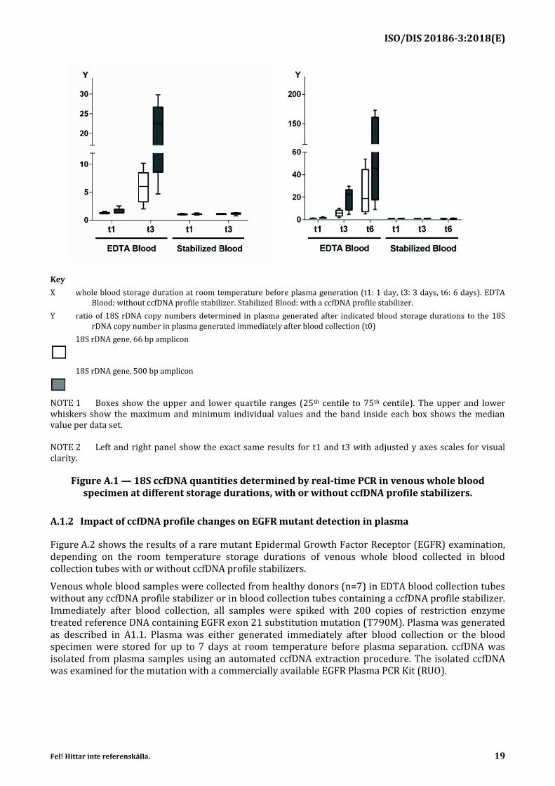
Upon storage and transport of venous whole blood collected in blood collection tubes without ccfDNA stabilizers, blood cells undergo apoptosis or mechanical lysis and release fragmented genomic DNA which thus becomes cell-free. These post collection changes artificially modify the native ccfDNA profile as it was in the donor’s body. This can lead to unreliable or wrong ccfDNA examination results. The artificial post collection release of ccfDNA can be measured by comparing ccfDNA quantities in plasma samples generated immediately after blood collection with ccfDNA quantities in plasma samples generated from stored blood.
Figure A.1 shows the post collection change of 18S ccfDNA quantities in venous whole blood specimen at different storage durations, with or without ccfDNA profile stabilizers.
Venous whole blood specimen were collected from healthy donors (n=6) in EDTA blood collection tubes without any ccfDNA profile stabilizer and either left untreated or were stabilized by a ccfDNA profile stabilizer reagent immediately after blood collection. Plasma was either generated immediately after blood collection/stabilization (t0) or after blood storage for 1, 3 or 6 days at room temperature (RT). From EDTA blood collection tubes, plasma was generated according to the examination provider’s instructions. A first centrifugation was performed at 1900 g for 15 min at room temperature. Plasma was transferred into a new tube. A second centrifugation of this plasma was performed at 1900 g for 10 min at room temperature. From stabilized blood, plasma was generated according to the manufacturer’s instruction which were identical to those for EDTA blood. ccfDNA was isolated from the generated plasma samples by using an automated ccfDNA extraction procedure. The 18S ribosomal DNA (rDNA) copy numbers in the isolated ccfDNA were determined by real-time PCR (66 bp/500 bp amplicon).
Blood collected and stored in EDTA blood collection tubes without any ccfDNA profile stabilizer showed a significant increase of 18S rDNA copies in the isolated ccfDNA in a range of 5-10 fold after 3 days and up to 20-150-fold after 6 days of RT storage. An increase was also observed after one day of storage. In contrast, blood preserved by a ccfDNA profile stabilizer did not show these artificial post collection changes.
The release of cellular DNA can be minimized by using blood collection tubes containing a ccfDNA profile stabilizing reagent, which prevents or minimizes the increase of ccfDNA quantities over time during blood storage and transport.
Blood collection tubes with ccfDNA profile stabilizers can be an important contributor to avoid impacts on examinations by artificial post collection changes in the native whole blood ccfDNA profile.
ISO/DIS 20186-3:2018(E)
Fel! Hittar inte referenskälla. 19
Key
X whole blood storage duration at room temperature before plasma generation (t1: 1 day, t3: 3 days, t6: 6 days). EDTA Blood: without ccfDNA profile stabilizer. Stabilized Blood: with a ccfDNA profile stabilizer.
Y ratio of 18S rDNA copy numbers determined in plasma generated after indicated blood storage durations to the 18S rDNA copy number in plasma generated immediately after blood collection (t0)
18S rDNA gene, 66 bp amplicon
18S rDNA gene, 500 bp amplicon
NOTE 1 Boxes show the upper and lower quartile ranges (25th centile to 75th centile). The upper and lower whiskers show the maximum and minimum individual values and the band inside each box shows the median value per data set.
NOTE 2 Left and right panel show the exact same results for t1 and t3 with adjusted y axes scales for visual clarity.
Figure A.1 — 18S ccfDNA quantities determined by real-time PCR in venous whole blood specimen at different storage durations, with or without ccfDNA profile stabilizers.
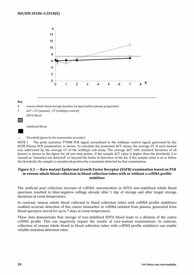
A.1.2 Impact of ccfDNA profile changes on EGFR mutant detection in plasma
Figure A.2 shows the results of a rare mutant Epidermal Growth Factor Receptor (EGFR) examination, depending on the room temperature storage durations of venous whole blood collected in blood collection tubes with or without ccfDNA profile stabilizers.
Venous whole blood samples were collected from healthy donors (n=7) in EDTA blood collection tubes without any ccfDNA profile stabilizer or in blood collection tubes containing a ccfDNA profile stabilizer. Immediately after blood collection, all samples were spiked with 200 copies of restriction enzyme treated reference DNA containing EGFR exon 21 substitution mutation (T790M). Plasma was generated as described in A1.1. Plasma was either generated immediately after blood collection or the blood specimen were stored for up to 7 days at room temperature before plasma separation. ccfDNA was isolated from plasma samples using an automated ccfDNA extraction procedure. The isolated ccfDNA was examined for the mutation with a commercially available EGFR Plasma PCR Kit (RUO).
ISO/DIS 20186-3:2018(E)
20 Fel! Hittar inte referenskälla.
Key
X venous whole blood storage duration (in days) before plasma preparation
Y ΔCT = CT (mutant) - CT (wildtype control)
EDTA Blood
Stabilized Blood
….. Threshold (given by the examination provider)
NOTE 1 The point mutation T790M PCR signal, normalized to the wildtype control signal, generated by the EGFR Plasma PCR examination, is shown. To calculate the presented ΔCT values, the average CT of each mutant was subtracted by the average CT of the wildtype sub-assay. The average ΔCT with standard deviation of all donors is shown in the figure for all test time points. If the sample ΔCT value is higher than the threshold, it is classed as “mutation not detected” or beyond the limits of detection of the kit. If the sample value is at or below the threshold, the sample is considered positive for a mutation detected by that examination.
Figure A.2 — Rare mutant Epidermal Growth Factor Receptor (EGFR) examination based on PCR in venous whole blood collection in blood collection tubes with or without a ccfDNA profile
stabilizer
The artificial post collection increase of ccfDNA concentration in EDTA non-stabilized whole blood specimen resulted in false-negative callings already after 1 day of storage and after longer storage durations at room temperature.
In contrast, venous whole blood collected in blood collection tubes with ccfDNA profile stabilizers enabled accurate detection of this cancer biomarker in ccfDNA isolated from plasma, generated from blood specimen stored for up to 7 days at room temperature.
These data demonstrate that storage of non-stabilized EDTA blood leads to a dilution of the native ccfDNA profile. This can negatively impact the results of rare-mutant examinations. In contrast, collection of venous whole blood in blood collection tubes with ccfDNA profile stabilizers can enable reliable mutation detection rates.
ISO/DIS 20186-3:2018(E)
Fel! Hittar inte referenskälla. 21
Bibliography
[1] ISO 17511:2013, In vitro diagnostic medical devices — Measurement of quantities in biological samples — Metrological traceability of values assigned to calibrators and control materials
[2] ISO 22174:2005, Microbiology of food and animal feeding stuffs — Polymerase chain reaction (PCR) for the detection of food-borne pathogens — General requirements and definitions
[3] ISO/TS 17822-1:2014, In vitro diagnostic test systems — Qualitative nucleic acid-based in vitro examination procedures for detection and identification of microbial pathogens — Part 1: General requirements, terms and definitions
[4] ISO 18113-1:2009, In vitro diagnostic medical devices — Information supplied by the manufacturer (labelling) — Part 1: Terms, definitions and general requirements
[5] ISO 17043:2010, Conformity assessment — General requirements for proficiency testing
[6] ISO Guide 30:2015, Reference materials — Selected terms and definitions
[7] ISO 9000:2015, Quality management systems — Fundamentals and vocabulary
[8] THAKUR B.K., ZHANG H., BECKER A. et al. Double- stranded DNA in exosomes: a novel biomarker in cancer detection. Cell Res. 2014, 24 pp. 766–769
[9] PETERS D.L., PRETORIUS P.J. Origin, translocation and destination of extracellular occurring DNA- A new paradigm in genetic behavior. Clin. Chim. Acta. 2011, 412 pp. 806–811
[10] JUNG K., FLEISHHACKER M., RABIEN A. Cell-free DNA in the blood as a solid tumor biomarker- A critical appraisal of the literature. Clin. Chim. Acta. 2010, 411 pp. 1611–1624
[11] EL MESSAOUDI S., ROLET F., MOULIERE F. et al. Circulating cell free DNA: preanalytical considerations. Clin. Chim. Acta. 2013, 424 pp. 222–230
[12] WONG D., MOTURI S., ANGKACHATCHAI V. et al. Optimizing blood collection, transport and storage conditions for cell free DNA increases access to prenatal testing. Clin. Biochem. 2013, 46 (12) pp. 1099–1104
[13] BARRETT A.N., ZIMMERMANN B.G., WANG D. et al. Implementing prenatal diagnosis cased on cell-free fetal DNA: Accurate identification of factors affecting fetal DNA yield. PLoS ONE. 2011, 6 (10) p. e25202
[14] SIFAKIS S., ZARAVINOS A., MAIZ N. et al. First-trimester maternal plasma cell-free fetal DNA and preeclampsia. Am. J. Obstet. Gynecol. 2009, 201 (5) pp. 472.e1–472.e7
[15] HAHN S., JACKSON L.G., KOLLA V. et al. Noninvasive prenatal diagnosis of fetal aneuploidies and Mendelian disorders: new innovative strategies. Expert Rev. Mol. Diagn. 2009, 9 (6) pp. 613–621
[16] SHARMA D., TRIVEDI S.S., BHATTACHARJEE J. Intergenotypic variation of endothelial dysfunction and inflammatory markers in eclampsia. Hypertens. Pregnancy. 2013, 32 (1) pp. 11–19
[17] NORTON S.E., LECHNER J.M., WILLIAMS T. et al. A stabilizing reagent prevents cell free DNA contamination by cellular DNA in plasma during blood storage and shipping as determined by digital PCR. Clin. Biochem. 2013, 46 pp. 1561–1565
ISO/DIS 20186-3:2018(E)
22 Fel! Hittar inte referenskälla.
[18] JUNG M., KLOTZEK S., LEWANDOWSKI M. et al. Changes in concentration of DNA in serum and plasma during storage of blood samples. Clin. Chem. 2003, 49 pp. 1028–1029
[19] LAM N.Y., RAINER T.H., CHIU R.W., LO Y.M. EDTA is a better anticoagulant than heparin or citrate for delayed blood processing for plasma DNA analysis. Clin. Chem. 2004, 50 pp. 256–257
[20] CHAN ALLEN K.C., YEUNG S.-W., LUI W.-B. et al. Effects of preanalytical factors on the molecular size of cell-free DNA in blood. Clinical Chemistry Journal. 2005, 51 (4) pp. 781–784
[21] PINZANI P., SALVIANTI F., PAZZAGLI M. et al. Circulating nucleic acids in cancer and pregnancy. Methods. 2010, 50 (4) pp. 302–307
[22] CHAN ALLEN K.C., YEUNG S.-W., LUI W.-B. et al. Effects of preanalytical factors on the molecular size of cell-free DNA in blood. Clin. Chem. 2005, 51 (4) pp. 781–784
[23] MALENTACCHI F., PIZZAMIGLIO S., VERDERIO P. et al. Influence of extraction methods on circulating cell free DNA (ccfDNA): the SPIDIA-DNAplas External Quality Assessment experience. Clin. Chem. Lab. Med. 2015, 53 (12) pp. 1935–1942
[24] HUNG E.C., CHIU R.W., LO Y.M. Detection of circulating fetal nucleic acids: a review of methods and applications. J. Clin. Pathol. 2009, 62 (4) pp. 308–313
[25] CASSINOTTI E., BONI L., SEGATO S. et al. Free circulating DNA as a biomarker of colorectal cancer. Int. J. Surg. 2013, ••• pp. 54–57
[26] TORACCHIO S., KOZINETZ C.A., KILLEN D.E. et al. Variable frequency f polyomavirus SV40 and herpesvirus EBV in lymphomas from two different urban population groups in Houston, TX. J. Clin. Virol. 2009, 46 (2) pp. 154–160
[27] Fleishhacker M., Schmidt B. Circulating nucleic acid (CNAS) and cancer- A survey. Biochimica et Biophysica Acta. 2007, 1775: pp. 181-232.
[28] SRM 2372 - Human DNA Quantitation Standard. National Institute of Standards and Technology.
[29] CHAN K.C., HUI A.B., WONG N. et al. Investigation of the genomic representation of plasma DNA in pregnant women by comparative genomic hybridization analysis: a feasibility study. Clin. Chem. 2005, 51 (12) pp. 2398–2401
[30] HOLDEN M.J., MADEJ R.M., MINOR P. et al. Molecular diagnostics: harmonization through reference materials, documentary standards and proficiency testing. Expert Rev. Mol. Diagn. 2011, 11 (7) pp. 741–755
[31] BONNET J., COLOTTE M., CLUODY D. et al. Chain and conformation stability of solid state DNA: implications form room temperature storage. Nucleic Acids Res. 2010, 28 pp. 1531–1546
[32] ISO 15190, Medical laboratories — Requirements for safety