Embed Size (px)
Citation preview
Molecular Genetics and Metabolism xxx (2010) xxx–xxx
ARTICLE IN PRESS
Contents lists available at ScienceDirect
Molecular Genetics and Metabolism
journal homepage: www.elsevier .com/locate /ymgme
Next generation sequencing in research and diagnostics of ocular birth defects
Gordana Raca a,b,*, Craig Jackson a, Berta Warman d, Tom Bair c, Lisa A. Schimmenti d
a UW Cytogenetic Services, Wisconsin State Laboratory of Hygiene, 465 Henry Mall, Madison WI 53706, USAb Department of Pathology and Laboratory Medicine, University of Wisconsin-Madison, 13000 University Avenue, Madison WI 53706, USAc The University of Iowa DNA Facility, John W. Eckstein Medical Research Building, 431 Newton Rd, Iowa City, IA, USAd Department of Pediatrics, Division of Genetics and Metabolism, University of Minnesota, 420 Delaware St. SE, Minneapolis, MN 55455, USA
a r t i c l e i n f o
Article history:Received 18 January 2010Received in revised form 8 March 2010Accepted 8 March 2010Available online xxxx
Keywords:Next generation sequencingSequence captureGS FLXAnophthalmiaMicrophthalmiaColoboma
1096-7192/$ - see front matter � 2010 Elsevier Inc. Adoi:10.1016/j.ymgme.2010.03.004
* Corresponding author at: UW Cytogenetics Servicof Hygiene, 465 Henry Mall, Madison, WI 53706, USA
E-mail address: [email protected] (G. Raca
Please cite this article in press as: G. Raca et al.,doi:10.1016/j.ymgme.2010.03.004
a b s t r a c t
Sequence capture enrichment (SCE) strategies and massively parallel next generation sequencing (NGS)are expected to increase the rate of gene discovery for genetically heterogeneous hereditary diseases, butat present, there are very few examples of successful application of these technologic advances in trans-lational research and clinical testing. Our study assessed whether array based target enrichment followedby re-sequencing on the Roche Genome Sequencer FLX (GS FLX) system could be used for novel mutationidentification in more than 1000 exons representing 100 candidate genes for ocular birth defects, and as acontrol, whether these methods could detect two known mutations in the PAX2 gene. We assayed twosamples with heterozygous sequence changes in PAX2 that were previously identified by conventionalSanger sequencing. These changes were a c.527G > C (S176T) substitution and a single basepair deletionc.77delG. The nucleotide substitution c.527G > C was easily identified by NGS. A deletion of one base in along polyG stretch (c.77delG) was not registered initially by the GS Reference Mapper, but was detectedin repeated analysis using two different software packages. Different approaches were evaluated for dis-tinguishing false positives (sequencing errors) and benign polymorphisms from potentially pathogenicsequence changes that require further follow-up. Although improvements will be necessary in accuracy,speed, ease of data analysis and cost, our study confirms that NGS can be used in research and diagnosticsettings to screen for mutations in hundreds of loci in genetically heterogeneous human diseases.
� 2010 Elsevier Inc. All rights reserved.
Introduction
The goal of our study was to evaluate the capacity of arraybased sequence capture target enrichment (SCE) and massivelyparallel, next generation sequencing (NGS) to successfully identifymutations in candidate genes for the developmental ocular birthdefects anophthalmia, microphthalmia and coloboma.
The advent of NGS technologies is expected to transform thepractice of medical genetics [1–3]. With the high throughput anddecreased sequencing costs achieved by NGS, it is no longer impos-sible to sequence hundreds or even thousands of exons and othergenomic sequences in an individual with a suspected genetic dis-ease. It is predicted that in the near future NGS might replace arraybased techniques and Sanger sequencing in their current clinicalapplications for the detection of mutations [1,3]. Additionally,NGS provides entirely new research and diagnostic capabilities,including whole genome screening for novel mutations andsequencing biological specimens for the genomic signature of no-vel infectious agents [4,5].
ll rights reserved.
e, Wisconsin State Laboratory. Fax: +1 608 265 7818.).
Next generation sequencing in
NGS could be particularly advantageous in research and testingfor genetically heterogeneous hereditary conditions. Common dis-orders evaluated by clinical geneticists are caused by heteroge-neous Mendelian loci and lend themselves to enrichmentstrategies followed by NGS. Examples include intellectual disabil-ity [6], deafness [7], familial cardiomyopathy [8] and retinitis pig-mentosa [9]. In these conditions there are often very subtlephenotypic differences between affected patients to guide molecu-lar diagnostics by indicating which gene is likely to be mutated in aparticular individual. Current diagnostic evaluation proceeds bysequencing a series of genes, individually or in small sets, basedon the relative frequency of the mutations and the sensitivity ofavailable assays. If there is no predominant mutation(s) causingthe disease, the pathogenic change often remains unknown evenafter very extensive and expensive molecular testing. With enrich-ment strategies followed by NGS, sequencing of all genes impli-cated in a particular genetic disorder could be performedsimultaneously, efficiently and at low cost.
While clearly superior to traditional Sanger sequencing, NGShas had little impact on clinical testing to date. There are veryfew examples of successful application of NGS in translational re-search and diagnostics. Clinical testing using NGS is currently of-fered for Hypertrophic Cardiomyopathy (HCM), Dilated
research and diagnostics of ocular birth defects, Mol. Genet. Metab. (2010),
2 G. Raca et al. / Molecular Genetics and Metabolism xxx (2010) xxx–xxx
ARTICLE IN PRESS
Cardiomyopathy (DCM) and Long QT syndrome (http://www.genedx.com/). NGS has also been explored as a method to performrapid human leukocyte antigen (HLA) typing for high resolutionallele identification [10,11], and to develop assays for Neurofibro-matosis Type 1 [12], autosomal recessive ataxia [13] and mito-chondrial disorders caused by mutations in the mitochondrialgenome and 362 nuclear genes controlling mitochondrial function[14]. Ng et al. applied targeted sequencing of all coding regions(‘‘exome”) to show the presence of causative mutations in fourunrelated individuals with a rare dominantly inherited disorder,Freeman–Sheldon syndrome (FSS) [15] and to discover the genefor a rare recessive disorder of previously unknown cause, Millerdisease [16]. Exciting applications have also been described in can-cer research, where NGS has been applied in discovering new can-didate genes for acute myeloid leukemia [17,18], glioblastomamultiforme [19] and other malignancies [20].
We describe the first study which showed the feasibility ofusing genomic enrichment by sequence capture followed by NGSto investigate genetic causes of the ocular birth defects anophthal-mia, microphthalmia and coloboma. These eye anomalies areamong the most prevalent causes of childhood blindness, affectingannually �2 per 10,000 newborns worldwide [21]. Although theycan be of different origins, the majority are caused by defects ingenes which regulate normal eye development [22–25]. There isincreased evidence that mutations in large numbers (possibly hun-dreds) of different genes can cause congenital eye malformations,but no single gene is responsible for a high percentage of cases[23–25]. Anophthalmia, microphthalmia and coloboma thereforerepresent disorders where simultaneous sequencing of large num-bers of candidate genes by NGS is an ideal approach to study genet-ic causes and demonstrate the feasibility of NGS for clinicaldiagnostics.
We showed that the combination of array based SCE with re-sequencing on the GS FLX instrument using Titanium chemistry al-lows concurrent sequencing of more than 100 candidate genes foranophthalmia, microphthalmia and coloboma. However, improve-ments will be necessary in several areas including accuracy, speed,ease of data analysis and cost to allow successful diagnostic imple-mentation of NGS for simultaneous mutation testing in hundredsof genes in genetically heterogeneous human diseases.
Materials and methods
We tested whether two known sequence changes in the renal-coloboma syndrome (a.k.a. Papillorenal syndrome, OMIM#120330) associated gene PAX2, which were previously character-ized by Sanger sequencing, can reliably be detected by NGS. Thefirst variant was a missense change in exon 5, c.527G > C, which re-sulted in serine to threonine amino-acid change S176T. This basesubstitution was identified in one of our previous studies in afather of a proband with ocular birth defects, but not in his affectedchild. Since detailed clinical information for the parent was notavailable, the change was described as a variant of unknown clin-ical significance.
Since short stretches of mono-, di-, and trinucleotide repeatsrepresent hotspots for disease causing frameshift mutations ingenomic DNA, we wanted to determine if this mutation type isdetectable by NGS. Therefore, the second sequence change selectedfor the study was a deletion of one base in a polyG stretch in exon 2of the PAX2 gene (c.77delG), which has previously been describedby Schimmenti et al. [26].
We designed a custom 385,000 probe SCE array with more than100 candidate genes for eye malformations. The list of selectedcandidate genes is provided in Table 1. The genes were chosenbased on reports of mutations identified in patients with
Please cite this article in press as: G. Raca et al., Next generation sequencing indoi:10.1016/j.ymgme.2010.03.004
coloboma, microphthalmia and anophthalmia [22,23,25]. Addi-tionally, a comprehensive literature search was performed for pub-lished mutations associated with ocular phenotypes in animalmodels [27,28]. Some genes were included based on their role insignaling and developmental pathways which are known as impor-tant for eye formation and function [24].
Appropriate SCE probes for the regions of interest were chosenin collaboration with Roche-NimbleGen design team (Roche-Nim-bleGen, Madison, WI). Only protein coding regions (coding exons)of the 112 candidate genes were targeted on the array. We selected385,000 long oligonucleotide probes (>60 bp) to tile the exons ofthe genes of interest with a very high density. All the probes hadthe uniqueness score of one (defined as having no match in thegenome other than itself longer than 38 bp, allowing up to fiveinsertions/deletions/mismatches in that match), to exclude repeti-tive regions from probe selection and avoid capturing pseudogenesequences [29]. Upon completion of the design and manufacturingof the array, the SCE on samples of genomic DNA from two patientswith known mutations in the PAX2 gene was performed at theNimbleGen service laboratory (Roche-NimbleGen, Madison, WI),following previously described protocols [30–32]. Briefly, �20 lgof each patient’s genomic DNA were randomly fragmented by neb-ulization to an average size of 500 bp. Linkers were ligated to theDNA fragments to provide a priming site for post-enrichmentamplification of the eluted fragment pool. The fragments weredenatured and hybridized to the custom SCE array. After a 72-hhybridization, unbound material was removed by stringent wash-ing. The arrays were transferred to the NimbleGen elution system,and the enriched fragment pool was eluted and recovered from thearray. The enriched fragments were amplified with 22mer linkersto generate enough DNA template for downstream applications.After amplification, the amount of captured DNA was measuredby spectrophotometry and the product was tested for enrichmentlevel by quantitative PCR with four proprietary QC control loci.These QC loci are conserved in both human and mouse genomesand have been empirically determined to accurately predictenrichment with several different array designs.
Sequencing of the two SCE prepared samples was performed onthe GS FLX instrument using Titanium chemistry, at the Universityof Iowa DNA Facility following standard protocols. Two samples,separated by gaskets, were sequenced independently on two re-gions of the picotiter-plate. Briefly, amplified fragments from SCEwere end-repaired and ligated to adapter oligonucleotides. The li-brary was diluted based on the results of a previously performedtitration, so that upon denaturation single DNA fragments hybrid-ized to individual beads containing sequences complementary toadapter oligonucleotides. The beads were compartmentalized intowater-in-oil microvesicles to allow clonal expansion of separateDNA molecules bound to the beads by emulsion PCR. After ampli-fication, the emulsion was disrupted, and the beads containingclonally amplified template DNA were enriched. The beads wereagain separated by limiting dilution, deposited into individualpicotiter-plate wells, and combined with sequencing enzymes.Iterative pyrosequencing was performed on the picotiter-plate bysuccessive flow addition of the four dNTPs. A nucleotide-incorpora-tion event in a well containing clonally amplified template pro-duced pyrophosphate release and picotiter-plate well-localizedluminescence, which recorded by a charge-coupled device (CCD)camera. With the flow of each dNTP reagent, wells were imaged,analyzed for their signal-to-noise ratio, filtered according to qual-ity criteria, and subsequently algorithmically translated into a lin-ear sequence output [33–35].
Data analysis was performed using the Roche proprietary soft-ware package for the GS FLX system. Image acquisition, imageprocessing and signal processing were performed during the run.Post run analysis was conducted using the GS Reference Mapper.
research and diagnostics of ocular birth defects, Mol. Genet. Metab. (2010),
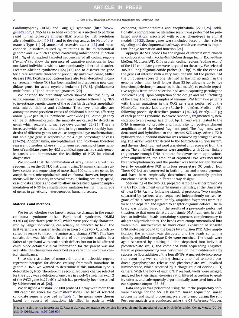
Table 1Candidate genes for anophthalmia, microphthalmia and coloboma represented on the SCE array.
Implicated in syndromic anophthalmia,microphthalmia and coloboma
Implicated innon-syndromicanophthalmia,microphthalmiaand coloboma
Code forproteins onthe SHHsignalingpathway
Code for proteins on theWNT signaling pathway
Other
DPYD, SIX3, RAB3GAP, ZFHX1B, ALG3, PITX2, SHH,PTCH, POMT1, KIAA1279, RBP4, PAX2, PAX6,PTPN11, CREBBP, SALL1 NDP, MKS1, SALL4,PQBP1, BCOR, PORCN, IGBP1, FLNA, CHD7,CC2D2A, HMX1, CLDN19, LRP2, TFAP2A, IGBP1,IKBKG, B3GALTL, GDF6, SOX2,OTX2, JAG1,BMP4, MITF, HESX1
CHX10, MAF,SIX6, RAX
GLI2, GLI3,SMO,VAX1,VAX2, SUFU,DRM, CER1
DVL1, WNT1, WNT16, WNT5A, WNT5B, WNT7B,WNT7A, WNT2, WNT8A, WNT10A, WNT6, WNT8B,WNT3, WNT3A, WNT9A, WNT9B, WNT11, WNT4,WNT2B, LRP5, LRP6, FZD6, FZD9, FZD2, FZD1, FZD7,FZD5, FZD10, AXIN2, GSK3A, APC, CTNNB1
CRYAA, CRYAB, CRYBA1,CRYBA2, CRYBA4, CRYBB1,CRYBB2, CRYBB3, CRYGA,CRYGB, CRYGC, CRYGD, CRYGS,CRYZSOX10, ZNF703, ZNF503CRX, FOXG1,BMP7, CHD2,DLX1, DLX2, TBX2,TBX5, FGF8,HES1, LHX1
40 loci 4 loci 8 loci 32 loci 28 loci
Table 2GS FLX sequencing summary.
Sample 1 Sample 2
Sequence yield 214 Mb 191 MbBases on targeta 76.1 Mb 89.6 MbAverage depthb 206 175.17Average coveragec 99.24% 99.28%% Covered P15� 97.35 97.35# Regions with <100% 15� coverage 54 54# Regions without coverage 3 3Total # of detected sequence changes 376 372# Changes in coding regions 110 114# Changes not corresponding to known SNPs 116 108
G. Raca et al. / Molecular Genetics and Metabolism xxx (2010) xxx–xxx 3
ARTICLE IN PRESS
Sequence runs were mapped against the human reference genome(hg18), using the default software settings (minimum base overlapof 40 bp and minimum overlap identity 90%). Sequence variationswere detected automatically during mapping, and were annotatedwith known gene (refSeq genes from http://genome.ucsc.edu/) andSNP information (dbSNP129 from http://genome.ucsc.edu/). Vari-ants were determined as high quality differences (HCDiffs) if thechange was present in at least three non duplicate reads which in-cluded at least one read from each direction (forward and reverse).Additional analysis was performed with the CLC Genomic Work-bench software (CLCbio, Aarhus, Denmark) and the NextGENe™software (SoftGenetics, State College, PA).
# Changes in coding regions not correspondingto known SNPs
26 26
# Changes likely to be real sequence alterationsd 5 3
a Bases on target were determined based on rigorous criteria, so that bases thatare a single-nucleotide outside of the captured region are not included in the count,even if they map to regions flanking the targets.
b Average depth is defined as average number of reads covering individual tar-geted nucleotides.
c Average coverage is the average proportion of bases within targeted exonswhich were covered by reads.
d These sequence variants met the following selection criteria: (1) they do notrepresent synonymous substitutions, (2) there is at least 30� coverage at the var-iant site and (3) the percentage of reads showing the variant allele exceeds 30%.
Results
We performed target enrichment by array based SCE andsequencing using GS FLX instrument on two DNA samples withknown mutations in the PAX2 gene. The sequencing was performedsimultaneously for 112 candidate genes for ocular birth defectswhich were selected based on extensive literature search. A cus-tom SCE array designed for target enrichment contained probesfor all coding regions (total of 1017 exons) of the selected 112genes. The size of the entire target region was 373,083 bp. Since385,000 probe SCE arrays are able to capture up to 5 Mb of targetsequence [32], our target region of �0.37 Mb only used a portion ofthe capacity of the 385 K array. This allowed the use of a largenumber of probes per each targeted region and helped in obtaininggood enrichment. DNA yield after elution from the SCE arrays andPCR amplification was approximately 10 lg for each sample; con-trol loci showed 721-fold enrichment for the sample with the mis-sense change (Sample 1), and 697-fold enrichment for the samplewith the frameshift mutation (Sample 2).
The run on the GS FLX instrument using Titanium chemistryyielded 214 Mb of sequence for the first sample and 191 Mb forthe second sample. Both met the Roche standard of at least150 Mb per sample. 76.1 Mb of sequence mapped to the targetedregions for Sample 1, and 89.6 Mb for Sample 2 (Table 2); this pro-portion of bases on the target is comparable to what has previouslybeen observed with array based SCE [12,15]. The average readlengths were 360 bp for Sample 1 and 318 bp for Sample 2. Thisis lower than the average read length for genomic samples but typ-ical for samples prepared by SCE (The Roche standard is at least300 bp). Details about the sequencing coverage obtained in the tar-get regions are shown in Table 2 and in Fig. 1A and B.
For both samples, 94.7% of the captured regions showed P15�coverage for all targeted bases (100%), with the average depth ofcoverage being �206 for Sample 1 and �175 for Sample 2. Onlythree regions showed complete lack of coverage. For 51 regions
Please cite this article in press as: G. Raca et al., Next generation sequencing indoi:10.1016/j.ymgme.2010.03.004
(out of a total of 1017) 15� coverage was not obtained for all thebases within a region. Most regions with low coverage (44/51)were identical between the two samples. The regions which com-pletely lacked coverage were in both cases the first coding exons ofthe genes WNT4, WNT9A and LRP5, which were noted to have avery high GC-content (83%, 81% and 83%, respectively). Addition-ally, low complexity GC rich repetitive elements were present inthe vicinity (100 bp upstream or downstream) of all the threeexons without coverage. Unusual sequence characteristics withhigh GC-content and a presence of repetitive elements are wellknown causes of poor enrichment by SCE and decreased sequenc-ing efficiency [12].
Studies have shown that the coverage in the 10–15-fold rangemay be sufficient for re-sequencing applications, but higher cover-age depths (50–60-fold) provide better alignment, assembly andaccuracy [36]. Therefore, most of our target exons have higher cov-erage than needed for a robust re-sequencing assay, allowing to ex-pand our target region by incorporating additional genes and tosequence our samples as a pool.
The missense change in Sample 1 (c.527G > C, S176T) was easilyidentified by NGS. A total of 151 reads covered the variant site,with 52% of the reads showing the wild-type base and 48% showingthe variant base, as would have been expected for a heterozygousallele. The c.527G > C change identified by Sanger sequencing is
research and diagnostics of ocular birth defects, Mol. Genet. Metab. (2010),
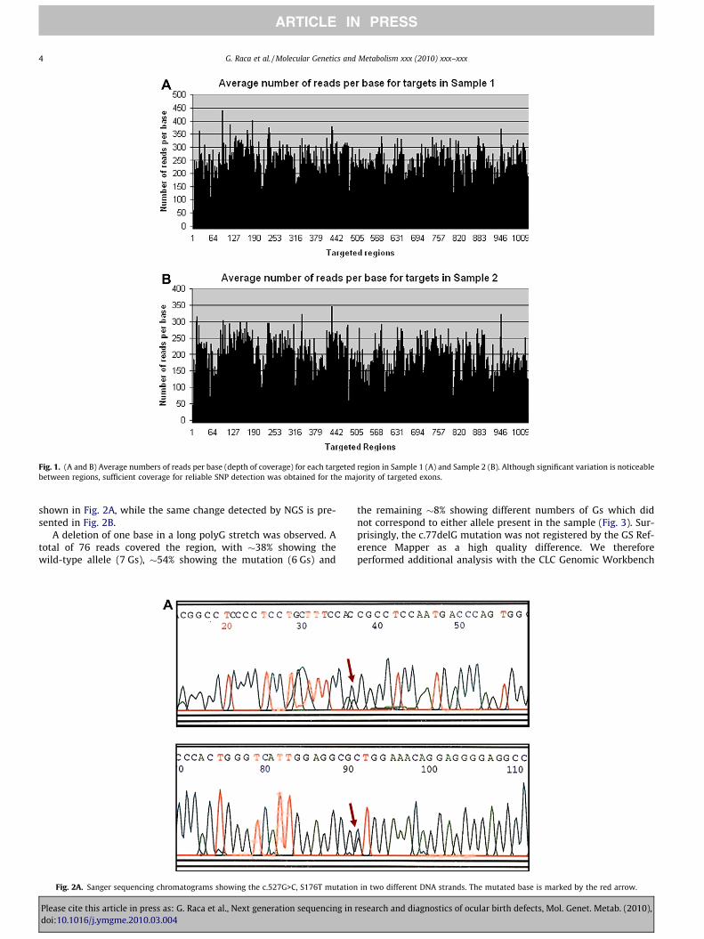
Fig. 1. (A and B) Average numbers of reads per base (depth of coverage) for each targeted region in Sample 1 (A) and Sample 2 (B). Although significant variation is noticeablebetween regions, sufficient coverage for reliable SNP detection was obtained for the majority of targeted exons.
4 G. Raca et al. / Molecular Genetics and Metabolism xxx (2010) xxx–xxx
ARTICLE IN PRESS
shown in Fig. 2A, while the same change detected by NGS is pre-sented in Fig. 2B.
A deletion of one base in a long polyG stretch was observed. Atotal of 76 reads covered the region, with �38% showing thewild-type allele (7 Gs), �54% showing the mutation (6 Gs) and
Fig. 2A. Sanger sequencing chromatograms showing the c.527G>C, S176T mutation
Please cite this article in press as: G. Raca et al., Next generation sequencing indoi:10.1016/j.ymgme.2010.03.004
the remaining �8% showing different numbers of Gs which didnot correspond to either allele present in the sample (Fig. 3). Sur-prisingly, the c.77delG mutation was not registered by the GS Ref-erence Mapper as a high quality difference. We thereforeperformed additional analysis with the CLC Genomic Workbench
in two different DNA strands. The mutated base is marked by the red arrow.
research and diagnostics of ocular birth defects, Mol. Genet. Metab. (2010),
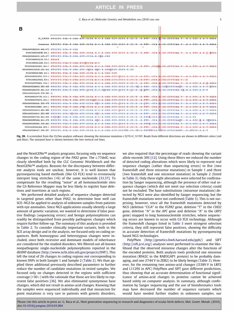
Fig. 2B. A screenshot from the CLCbio analysis software showing the missense mutation c.527G>C, S176T. Reads from different directions are shown in different colors (redand blue). The mutated base is shown between the two vertical red lines.
G. Raca et al. / Molecular Genetics and Metabolism xxx (2010) xxx–xxx 5
ARTICLE IN PRESS
and the NextGENe™ analysis programs, focusing only on sequencechanges in the coding region of the PAX2 gene. The c.77delG wasclearly identified both by the CLC Genomic Workbench and theNextGENe™ analysis. Reasons for the discrepancy between differ-ent analysis tools are unclear, however, it is well known thatpyrosequencing based methods (like GS FLX) tend to erroneouslyinterpret long stretches (>6) of the same nucleotide [33,37]. Tocompensate for the resulting ‘‘noise” at all homonucleotide sitesthe GS Reference Mapper may be less likely to register base dele-tions and insertions at such regions.
We performed detailed analysis of sequence changes detectedin targeted genes other than PAX2, to determine how well canSCE–NGS be applied to analysis of unknown samples from patientswith eye anomalies. Since NGS applications usually identify a largeamount of genetic variation, we wanted to determine if false posi-tive findings (sequencing errors) and benign polymorphisms canreadily be distinguished from possibly pathogenic changes whichrequire further follow-up. The summary of this analysis is providedin Table 2. To consider clinically important variants, both in theSCE array design and in the analysis, we focused only on coding se-quences. Both homozygous and heterozygous changes were in-cluded, since both recessive and dominant models of inheritanceare considered for the studied disorders. We filtered out all knownnonpathogenic single-nucleotide polymorphisms reported in thedbSNP database (http://www.ncbi.nlm.nih.gov/projects/SNP/). Thisleft the total of 26 changes in coding regions not corresponding toknown SNPs in both Sample 1 and Sample 2 (Table 2). We than ap-plied three additional previously described parameters to furtherreduce the number of candidate mutations in tested samples. Wefocused only on changes detected in the regions with sufficientcoverage (>30�) with the rationale that those are less likely to rep-resent false positives [36]. We also eliminated synonymous basechanges, which did not result in amino-acid changes. Knowing thatthe samples were sequenced individually and that mosaicism forpoint mutations is very rare in patients with genetic disorders,
Please cite this article in press as: G. Raca et al., Next generation sequencing indoi:10.1016/j.ymgme.2010.03.004
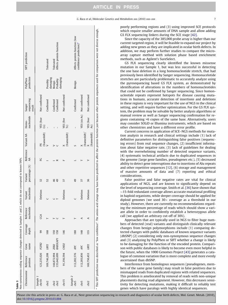
we also required that the percentage of reads showing the variantallele exceeds 30% [12]. Using these filters we reduced the numberof detected coding alterations which were likely to represent realsequence changes (rather than sequencing errors) to five (twoframeshift and three missense mutations) in Sample 1 and three(two frameshift and one missense mutation) in Sample 2 (listedin Table 3). Only these eight alterations were selected for confirma-tion by Sanger sequencing, although the presence of other real se-quence changes (which did not meet our selection criteria) couldnot be excluded. The base substitutions (missense mutations) de-tected by NGS were also identified by Sanger sequencing, but theframeshift mutations were not confirmed (Table 3). This is not sur-prising, however, since all the frameshift mutations detected byNGS (deletion ‘‘CGA” in the FGFR2 gene, deletion ‘‘T” in the CHD2gene, deletion ‘‘A” in the APC gene and deletion ‘‘A” in the LRP2gene) mapped to long homonucleotide stretches, where sequenc-ing errors are known to occur with GS FLX technology. Althoughthe frameshift changes listed in Table 3 meet stringent selectioncriteria, they still represent false positives, showing the difficultyin accurate detection of frameshift mutations by pyrosequencingbased NGS technologies.
PolyPhen (http://genetics.bwh.harvard.edu/pph/) and SIFT(http://sift.jcvi.org/) analyses were performed to examine the like-lihood that the observed missense changes alter the functions ofthe encoded proteins. Both analysis tools predicted one missensemutation (R942C in the RAB3GAP1 protein) to be probably dam-aging, and one (I744 V in ZEB2) to be likely benign (Table 3). How-ever, for the remaining two amino-acid changes (I3389 V in LRP2and L1129S in APC) PolyPhen and SIFT gave different predictions,thus showing that an accurate determination of functional signif-icance of amino-acid changes in proteins cannot be achievedbased solely on computer analysis. In summary, although confir-mation by Sanger sequencing and the use of bioinformatics toolsmay have decreased the number of sequence variants whichwould have needed further studies in unknown samples, our
research and diagnostics of ocular birth defects, Mol. Genet. Metab. (2010),
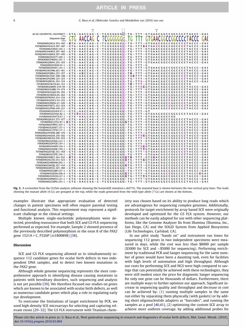
Fig. 3. A screenshot from the CLCbio analysis software showing the frameshift mutation c.del77G. The mutated base is shown between the two vertical grey lines. The readsshowing the mutant allele (6 Gs) are grouped at the top, while the reads generated from the wild-type allele (7 Gs) are shown at the bottom.
6 G. Raca et al. / Molecular Genetics and Metabolism xxx (2010) xxx–xxx
ARTICLE IN PRESS
examples illustrate that appropriate evaluation of detectedchanges in patient specimens will often require parental testingand functional analysis. This requirement may represent a signif-icant challenge in the clinical settings.
Multiple known single-nucleotide polymorphisms were de-tected, providing reassurance that both SCE and GS FLX sequencingperformed as expected. For example, Sample 2 showed presence ofthe previously described polymorphism in the exon 8 of the PAX2gene 1521A > C, P326P (rs1800898) [38].
Discussion
SCE and GS FLX sequencing allowed us to simultaneously se-quence 112 candidate genes for ocular birth defects in two inde-pendent DNA samples, and to detect two known mutations inthe PAX2 gene.
Although whole genome sequencing represents the most com-prehensive approach to identifying disease causing mutations inpatients with hereditary disorders, such sequencing and analysisis not yet possible [39]. We therefore focused our studies on geneswhich are known to be associated with ocular birth defects, as wellas numerous candidate genes which play a role in regulating earlyeye development.
To overcome the limitations of target enrichment by PCR, weused high-density SCE microarrays for selecting and capturing rel-evant exons [29–32]. The GS FLX instrument with Titanium chem-
Please cite this article in press as: G. Raca et al., Next generation sequencing indoi:10.1016/j.ymgme.2010.03.004
istry was chosen based on its ability to produce long reads whichare advantageous for sequencing complex genomes. Additionally,protocols for target enrichment by array based SCE were originallydeveloped and optimized for the GS FLX system. However, ourmethods can be easily adapted for use with other sequencing plat-forms, like the Genome Analyzer IIx from Illumina (Illumina, Inc.,San Diego, CA) and the SOLiD System from Applied Biosystems(Life Technologies, Carlsbad, CA).
In our pilot study ‘‘hands on” and instrument run times forsequencing 112 genes in two independent specimens were mea-sured in days, while the cost was less than $8000 per sample($3000 for SCE and �$5000 for sequencing). Performing enrich-ment by traditional PCR and Sanger sequencing for the same num-ber of genes would have been a daunting task, even for facilitieswith high levels of automation and high throughput. Althoughour costs for performing SCE and NGS were high compared to sav-ings that can potentially be achieved with these technologies, theywere still modest since the price for diagnostic Sanger sequencingfor only one gene can be thousands of dollars. Furthermore, thereare multiple ways to further optimize our approach. Significant in-crease in sequencing quality and throughput and decrease in costcan be achieved by: (1) running multiple samples in the samerun either by separating them physically (with gaskets) or by add-ing short oligonucleotide adapters as ‘‘barcodes”, and running thesamples as a pool [40,41], (2) optimizing the custom SCE array toachieve more uniform coverage, by adding additional probes for
research and diagnostics of ocular birth defects, Mol. Genet. Metab. (2010),
Tabl
e3
Non
-syn
onym
ous
codi
ngse
quen
cech
ange
sde
tect
edin
Sam
ples
1an
d2,
whi
chdo
not
corr
espo
ndto
know
nSN
Ps.
Ch
rom
osom
eR
efer
ence
posi
tion
Gen
en
ame
Cov
erag
ede
pth
Ref
eren
cen
ucl
eoti
deV
aria
nt
nu
cleo
tide
Var
ian
tpe
rcen
tage
(%)
Ref
eren
ceA
AV
aria
nt
AA
Hom
o-n
ucl
eoti
deSa
nge
rse
quen
cin
gre
sult
Poly
Phen
pred
icti
onSI
FTpr
edic
tion
Sam
ple
1ch
r10
123,
235,
026
FGFR
299
CG
AD
elet
ion
32FG
PX (fra
mes
hif
t)Y
ES(f
our
Cs)
Not
con
firm
ed
chr2
169,
746,
208
LRP2
168
TC
52I
V–
Con
firm
edPo
ssib
lyda
mag
ing
Tole
rate
d
chr2
144,
872,
994
ZEB2
150
TC
46I
V–
Con
firm
edB
enig
nTo
lera
ted
chr2
135,
642,
699
RA
B3G
AP1
158
CT
49R
C–
Con
firm
edPr
obab
lyda
mag
ing
Aff
ects
prot
ein
fun
ctio
n
chr1
591
,356
,594
CHD
214
6T
Del
etio
n37
FX (f
ram
esh
ift)
YES
(five
Ts)
Not
con
firm
ed
Sam
ple
2ch
r511
2,20
2,57
6A
PC18
8T
C49
LS
–C
onfi
rmed
Ben
ign
Aff
ects
prot
ein
fun
ctio
n(l
owco
nfi
den
cepr
edic
tion
)a
chr5
112,
203,
856
APC
148
AD
elet
ion
37T
X (fra
mes
hif
t)*
YES
(six
As)
Not
con
firm
ed
chr2
169,
750,
453
LRP2
155
AD
elet
ion
36Y
X (fra
mes
hif
t)Y
ES(f
our
As)
Not
con
firm
ed
aTh
isSI
FTpr
edic
tion
was
desc
ribe
das
‘‘low
con
fide
nce
”.Th
eam
ino-
acid
chan
gem
ayh
ave
been
pred
icte
dto
affe
ctth
epr
otei
nfu
nct
ion
just
beca
use
othe
rpr
otei
nse
quen
ces
use
dfo
rco
mpa
riso
nw
ere
not
dive
rse
enou
gh.
G. Raca et al. / Molecular Genetics and Metabolism xxx (2010) xxx–xxx 7
ARTICLE IN PRESS
Please cite this article in press as: G. Raca et al., Next generation sequencing in rdoi:10.1016/j.ymgme.2010.03.004
poorly performing regions and (3) using improved SCE protocolswhich require smaller amounts of DNA sample and allow addingGS FLX sequencing linkers during the SCE stage [42].
Since the capacity of the 385,000 probe array is higher than ourcurrent targeted region, it will be feasible to expand our project byadding new genes as they are implicated in ocular birth defects. Inaddition, we may perform further studies to compare the micro-array capture method with solution phase based enrichmentmethods, such as Agilent’s SureSelect.
GS FLX sequencing clearly identified the known missensemutation in our Sample 1, but was less successful in detectingthe one base deletion in a long homonucleotide stretch, that hadpreviously been identified by Sanger sequencing. Homonucelotidestretches are particularly problematic to accurately analyze usingthe pyrosequencing based GS FLX system, as demonstrated byidentification of alterations in the numbers of homonucleotidesthat could not be confirmed by Sanger sequencing. Since homon-ucleotide repeats represent hotspots for disease causing muta-tions in humans, accurate detection of insertions and deletionsin these regions is very important for the use of NGS in the clinicalsetting, and will require further optimization. For the GS FLX sys-tem, the problem may be solvable by better analysis algorithms ormanual review as well as Sanger sequencing confirmation for re-gions containing >6 copies of the same base. Alternatively, usersmay consider SOLiD or Illumina instruments, which are based onother chemistries and have a different error profile.
Current concerns in application of SCE–NGS methods for muta-tion analysis in research and clinical settings include (1) lack ofdefinitive parameters for distinguishing false positives (sequenc-ing errors) from real sequence changes, (2) insufficient informa-tion about false negative rate, (3) lack of guidelines for dealingwith the overwhelming number of detected sequence variants,(4) systematic technical artifacts due to duplicated sequences inthe genome (large gene families, pseudogenes etc.), (5) decreasedability to detect gene interruptions due to insertions of Alu repeatsand other repetitive sequences [12], (6) storage and managementof massive amounts of data and (7) reporting and ethicalconsiderations.
False positive and false negative rates are vital for clinicalapplications of NGS, and are known to significantly depend onthe level of sequencing coverage. Smith et al. [36] have shown that�15-fold redundant coverage allows accurate mutational profilingin haploid organisms, while deeper coverage should be applied fordiploid genomes (we used 30� coverage as a threshold in ourstudy). However, there are currently no recommendations regard-ing the minimum percentage of reads which should show a vari-ant allele in order to confidently establish a heterozygous allelecall (we applied an arbitrary cut-off of 30%).
Approaches that are typically used in NGS to filter huge num-bers of detected (real) variants and distinguish clinically relevantchanges from benign polymorphisms include (1) comparing de-tected changes with public databases of known sequence variants(dbSNP) (2) considering only non-synonymous sequence changesand (3) analyzing by PolyPhen or SIFT whether a change is likelyto be damaging for the function of the encoded protein. Compari-son with public databases is likely to become even more helpful inthe future, when the 1000 Genomes Project [43] generates a cata-logue of common variation that is more complete and more evenlyascertained than dbSNP.
Interference from homologous sequences (pseudogenes, mem-bers of the same gene family) may result in false positives due tomismapped reads from duplicated regions with related sequences.This problem is ameliorated by removal of reads with non-uniqueplacements during read alignment. However, this decreases sensi-tivity for detecting mutations, making it difficult to reliably testgenes which have paralogs with highly identical sequences.
esearch and diagnostics of ocular birth defects, Mol. Genet. Metab. (2010),
8 G. Raca et al. / Molecular Genetics and Metabolism xxx (2010) xxx–xxx
ARTICLE IN PRESS
Data storage and analysis are large challenges for individuallaboratories considering use of NGS. The issue of data storage isfurther complicated by the consideration of how much of the data-set to keep (image data, raw data or just fasta files). The bioinfor-matics challenges are alleviated by core facilities offering dataretention on their servers, access to analysis software and consul-tation with bioinformatics experts. Additionally, it has been pro-posed that cloud-computing solutions which provide internetaccess to large clusters of computers may allow investigators fromsmall laboratories to access data analysis tools and significanthardware power at a reasonable cost [44].
Large sample sets will need to be studied and specific, objective,evidence based guidelines and quality controls developed to ad-dress main technical issues and enable use of SCE and NGS technol-ogies to reliably detect pathogenic mutations in research anddiagnostics laboratories. Routine Sanger sequencing will likely berequired for a long time to confirm data generated by NGS. Evenwhen technical limitations get resolved, serious test reporting is-sues and ethical considerations are likely to remain. For example,each person is likely to carry multiple clinically relevant mutationswhich can be detected by genome-wide sequencing. It is unclearshould participants in NGS based studies receive all their sequenc-ing information (even when it is not possible to implement effec-tive medical treatments for clinically relevant variants) and howshould this information be delivered and interpreted to patients[45].
In conclusion, our study showed that even with existing limita-tions, individual researchers working with core sequencing facili-ties can use NGS as a research tool with confidence and ease. Inthe near future, thanks to continuing improvements in throughput,accuracy, cost and ease of data analysis, it will also become feasibleto apply NGS in the clinical setting.
Acknowledgments
We gratefully acknowledge Einat Snir and Jennifer Bair fromThe University of Iowa DNA Facility for technical assistance withGS FLX sequencing and manuscript review, and Eric Cabot fromthe University of Wisconsin-Madison Biotechnology Center forhelp with reviewing the sequencing data.
References
[1] J.R. ten Bosch, W.W. Grody, Keeping up with the next generation: massivelyparallel sequencing in clinical diagnostics, J. Mol. Diagn. 10 (2008) 484–492.
[2] K.V. Voelkerding, S.A. Dames, J.D. Durtschi, Next-generation sequencing: frombasic research to diagnostics, Clin. Chem. 55 (2009) 641–658.
[3] T. Tucker, M. Marra, J.M. Friedman, Massively parallel sequencing: the next bigthing in genetic medicine, Am. J. Hum. Genet. 85 (2009) 142–154.
[4] S.C. Schuster, Next-generation sequencing transforms today’s biology, Nat.Methods 5 (2008) 16–18.
[5] S. Marguerat, B.T. Wilhelm, J. Bahler, Next-generation sequencing: applicationsbeyond genomes, Biochem. Soc. Trans. 36 (2008) 1091–1096.
[6] H.H. Ropers, Genetics of intellectual disability, Curr. Opin. Genet. Dev. 18(2008) 241–250.
[7] W.E. Nance, The genetics of deafness, Ment. Retard. Dev. Disabil. Res. Rev. 9(2003) 109–119.
[8] A.J. Marian, Genetic determinants of cardiac hypertrophy, Curr. Opin. Cardiol.23 (2008) 199–205.
[9] P. Goodwin, Hereditary retinal disease, Curr. Opin. Ophthalmol. 19 (2008) 255–262.
[10] C. Gabriel, M. Danzer, C. Hackl, G. Kopal, P. Hufnagl, K. Hofer, H. Polin, S.Stabentheiner, J. Proll, Rapid high-throughput human leukocyte antigen typingby massively parallel pyrosequencing for high-resolution allele identification,Hum. Immunol. 70 (2009) 960–964.
[11] G. Bentley, R. Higuchi, B. Hoglund, D. Goodridge, D. Sayer, E.A. Trachtenberg,H.A. Erlich, High-resolution, high-throughput HLA genotyping by next-generation sequencing, Tissue Antigens 74 (2009) 393–403.
[12] L.S. Chou, C.S. Liu, B. Boese, X. Zhang, R. Mao, DNA sequence capture andenrichment by microarray followed by next-generation sequencing fortargeted resequencing: neurofibromatosis type 1 gene as a model, Clin.Chem. 56 (2010) 1–11.
Please cite this article in press as: G. Raca et al., Next generation sequencing indoi:10.1016/j.ymgme.2010.03.004
[13] A. Hoischen, C. Gilissen, P. Arts, N. Wieskamp, W. van der Vliet, S. Vermeer, M.Steehouwer, P. de Vries, R. Meijer, J. Seiqueros, N.V.A.M. Knoers, M.F. Buckley,H. Scheffer, J.A. Veltman, Massively parallel sequencing of ataxia genes afterarray-based enrichment, Hum. Mutat., doi:10.1002/humu.21221 (Publishedon-line 11 Feb 2010).
[14] V. Vasta, S.B. Ng, E.H. Turner, J. Shendure, S.H. Hahn, Next generation sequenceanalysis for mitochondrial disorders, Genome Med. 1 (2009) 100.
[15] S.B. Ng, E.H. Turner, P.D. Robertson, S.D. Flygare, A.W. Bigham, C. Lee, T. Shaffer,M. Wong, A. Bhattacharjee, E.E. Eichler, M. Bamshad, D.A. Nickerson, J.Shendure, Targeted capture and massively parallel sequencing of 12 humanexomes, Nature 461 (2009) 272–276.
[16] S.B. Ng, K.J. Buckingham, C. Lee, A.W. Bigham, H.K. Tabor, K.M. Dent, C.D. Huff,P.T. Shannon, E.W. Jabs, D.A. Nickerson, J. Shendure, M.J. Bamshad, Exomesequencing identifies the cause of a Mendelian disorder, Nat. Genet. 42 (2010)30–35.
[17] T.J. Ley, E.R. Mardis, L. Ding, B. Fulton, M.D. McLellan, K. Chen, D. Dooling, B.H.Dunford-Shore, S. McGrath, M. Hickenbotham, L. Cook, R. Abbott, D.E. Larson,D.C. Koboldt, C. Pohl, S. Smith, A. Hawkins, S. Abbott, D. Locke, L.W. Hillier, T.Miner, L. Fulton, V. Magrini, T. Wylie, J. Glasscock, J. Conyers, N. Sander, X. Shi,J.R. Osborne, P. Minx, D. Gordon, A. Chinwalla, Y. Zhao, R.E. Ries, J.E. Payton, P.Westervelt, M.H. Tomasson, M. Watson, J. Baty, J. Ivanovich, S. Heath, W.D.Shannon, R. Nagarajan, M.J. Walter, D.C. Link, T.A. Graubert, J.F. DiPersio, R.K.Wilson, DNA sequencing of a cytogenetically normal acute myeloid leukaemiagenome, Nature 456 (2008) 66–72.
[18] E.R. Mardis, L. Ding, D.J. Dooling, D.E. Larson, M.D. McLellan, K. Chen, D.C.Koboldt, R.S. Fulton, K.D. Delehaunty, S.D. McGrath, L.A. Fulton, D.P. Locke, V.J.Magrini, R.M. Abbott, T.L. Vickery, J.S. Reed, J.S. Robinson, T. Wylie, S.M. Smith,L. Carmichael, J.M. Eldred, C.C. Harris, J. Walker, J.B. Peck, F. Du, A.F. Dukes, G.E.Sanderson, A.M. Brummett, E. Clark, J.F. McMichael, R.J. Meyer, J.K. Schindler,C.S. Pohl, J.W. Wallis, X. Shi, L. Lin, H. Schmidt, Y. Tang, C. Haipek, M.E.Wiechert, J.V. Ivy, J. Kalicki, G. Elliott, R.E. Ries, J.E. Payton, P. Westervelt, M.H.Tomasson, M.A. Watson, J. Baty, S. Heath, W.D. Shannon, R. Nagarajan, D.C.Link, M.J. Walter, T.A. Graubert, J.F. DiPersio, R.K. Wilson, T.J. Ley, Recurringmutations found by sequencing an acute myeloid leukemia genome, N. Engl. J.Med. 361 (2009) 1058–1066.
[19] D.W. Parsons, S. Jones, X. Zhang, J.C. Lin, R.J. Leary, P. Angenendt, P. Mankoo, H.Carter, I.M. Siu, G.L. Gallia, A. Olivi, R. McLendon, B.A. Rasheed, S. Keir, T.Nikolskaya, Y. Nikolsky, D.A. Busam, H. Tekleab, L.A. Diaz Jr., J. Hartigan, D.R.Smith, R.L. Strausberg, S.K. Marie, S.M. Shinjo, H. Yan, G.J. Riggins, D.D. Bigner,R. Karchin, N. Papadopoulos, G. Parmigiani, B. Vogelstein, V.E. Velculescu, K.W.Kinzler, An integrated genomic analysis of human glioblastoma multiforme,Science 321 (2008) 1807–1812.
[20] E.R. Mardis, R.K. Wilson, Cancer genome sequencing: a review, Hum. Mol.Genet. 18 (2009) R163–R168.
[21] C. Stoll, Y. Alembik, B. Dott, M.P. Roth, Congenital eye malformations in212,479 consecutive births, Ann. Genet. 40 (1997) 122–128.
[22] J.R. Guercio, L.J. Martyn, Congenital malformations of the eye and orbit,Otolaryngol. Clin. North Am. 40 (2007) 113–140. vii.
[23] C.Y. Gregory-Evans, M.J. Williams, S. Halford, K. Gregory-Evans, Ocularcoloboma: a reassessment in the age of molecular neuroscience, J. Med.Genet. 41 (2004) 881–891.
[24] D.R. Fitzpatrick, V. van Heyningen, Developmental eye disorders, Curr. Opin.Genet. Dev. 15 (2005) 348–353.
[25] L. Chang, D. Blain, S. Bertuzzi, B.P. Brooks, Uveal coloboma: clinical and basicscience update, Curr. Opin. Ophthalmol. (2006) 447–470.
[26] L.A. Schimmenti, H.H. Shim, J.D. Wirtschafter, V.A. Panzarino, C.E. Kashtan, S.J.Kirkpatrick, D.S. Wargowski, T.D. France, E. Michel, W.B. Dobyns,Homonucleotide expansion and contraction mutations of PAX2 and inclusionof Chiari 1 malformation as part of renal-coloboma syndrome, Hum. Mutat. 14(1999) 369–376.
[27] J.M. Gross, B.D. Perkins, Zebrafish mutants as models for congenital oculardisorders in humans, Mol. Reprod. Dev. 75 (2008) 547–555.
[28] C. Alescaron, R.T. Ernst, Anterior eye development and ocular mesenchyme:new insights from mouse models and human diseases, Bioessays 26 (2004)374–386.
[29] Roche-NimbleGen Inc, Roche Nimble-Gen probe design fundamentals.Application Notes. Available from: <http://www.nimblegen.com/products/lit/probe_design_2008_06_04.pdf> (accessed December 2009).
[30] Roche-NimbleGen Inc, NimbleGen services user’s guides: sequence captureservice. Application Notes. Available from: <http://www.nimblegen.com/products/lit/SeqCap_UsersGuide_Service_v3p0.pdf> (accessed December2009).
[31] M. Olson, Enrichment of super-sized resequencing targets from the humangenome, Nat. Methods 4 (2007) 891–892.
[32] T.J. Albert, M.N. Molla, D.M. Muzny, L. Nazareth, D. Wheeler, X. Song, T.A.Richmond, C.M. Middle, M.J. Rodesch, C.J. Packard, G.M. Weinstock, R.A. Gibbs,Direct selection of human genomic loci by microarray hybridization, Nat.Methods 4 (2007) 903–905.
[33] M. Margulies, M. Egholm, W.E. Altman, S. Attiya, J.S. Bader, L.A. Bemben, J.Berka, M.S. Braverman, Y.J. Chen, Z. Chen, S.B. Dewell, L. Du, J.M. Fierro, X.V.Gomes, B.C. Godwin, W. He, S. Helgesen, C.H. Ho, G.P. Irzyk, S.C. Jando, M.L.Alenquer, T.P. Jarvie, K.B. Jirage, J.B. Kim, J.R. Knight, J.R. Lanza, J.H. Leamon,S.M. Lefkowitz, M. Lei, J. Li, K.L. Lohman, H. Lu, V.B. Makhijani, K.E. McDade,M.P. McKenna, E.W. Myers, E. Nickerson, J.R. Nobile, R. Plant, B.P. Puc, M.T.Ronan, G.T. Roth, G.J. Sarkis, J.F. Simons, J.W. Simpson, M. Srinivasan, K.R.Tartaro, A. Tomasz, K.A. Vogt, G.A. Volkmer, S.H. Wang, Y. Wang, M.P. Weiner,
research and diagnostics of ocular birth defects, Mol. Genet. Metab. (2010),

G. Raca et al. / Molecular Genetics and Metabolism xxx (2010) xxx–xxx 9
ARTICLE IN PRESS
P. Yu, R.F. Begley, J.M. Rothberg, Genome sequencing in microfabricated high-density picolitre reactors, Nature 437 (2005) 376–380.
[34] J.M. Rothberg, J.H. Leamon, The development and impact of 454 sequencing,Nat. Biotechnol. 26 (2008) 1117–1124.
[35] E.R. Mardis, Next-generation DNA sequencing methods, Annu. Rev. GenomicsHum. Genet. 9 (2008) 387–402.
[36] D.R. Smith, A.R. Quinlan, H.E. Peckham, K. Makowsky, W. Tao, B. Woolf, L. Shen,W.F. Donahue, N. Tusneem, M.P. Stromberg, D.A. Stewart, L. Zhang, S.S. Ranade,J.B. Warner, C.C. Lee, B.E. Coleman, Z. Zhang, S.F. McLaughlin, J.A. Malek, J.M.Sorenson, A.P. Blanchard, J. Chapman, D. Hillman, F. Chen, D.S. Rokhsar, K.J.McKernan, T.W. Jeffries, G.T. Marth, P.M. Richardson, Rapid whole-genomemutational profiling using next-generation sequencing technologies, GenomeRes. 18 (2008) 1638–1642.
[37] T. Wicker, E. Schlagenhauf, A. Graner, T.J. Close, B. Keller, N. Stein, 454sequencing put to the test using the complex genome of barley, BMC Genomics7 (2006) 275.
[38] H.H. Shim, B.N. Nakamura, R.M. Cantor, L.A. Schimmenti, Identification of twosingle nucleotide polymorphisms in exon 8 of PAX2, Mol. Genet. Metab. 68(1999) 507–510.
Please cite this article in press as: G. Raca et al., Next generation sequencing indoi:10.1016/j.ymgme.2010.03.004
[39] D. Summerer, Enabling technologies of genomic-scale sequence enrichmentfor targeted high-throughput sequencing, Genomics 94 (2009) 363–368.
[40] M. Meyer, U. Stenzel, S. Myles, K. Prufer, M. Hofreiter, Targeted high-throughput sequencing of tagged nucleic acid samples, Nucleic Acids Res. 35(2007) e97.
[41] M. Meyer, U. Stenzel, M. Hofreiter, Parallel tagged sequencing on the 454platform, Nat. Protoc. 3 (2008) 267–278.
[42] Roche-NimbleGen Inc., NimbleGen titanium optimized sequence capture 385 Karrays. Application Notes. Available from: <http://www.454.com/downloads/products-solutions/SeqCapTitan385K_Final.pdf> (accessed December 2009).
[43] N. Siva, 1000 genomes project, Nat. Biotechnol. 26 (2008) 256.[44] J.D. McPherson, Next-generation gap, Nat. Methods 6 (2009) S2–S5.[45] L.G. Biesecker, Exome sequencing makes medical genomics a reality, Nat.
Genet. 42 (2010) 13–14.
research and diagnostics of ocular birth defects, Mol. Genet. Metab. (2010),


![NEKI ASPEKTI EER - University of Belgradepoincare.matf.bg.ac.rs/~gordana/PRED3.pdf · Microsoft PowerPoint - Pred3-Neki-aspekti-EER [Compatibility Mode] Author: Gordana Created Date:](https://img.pdfslide.us/doc/110x75/60c06abd2cebee602c3b8a1d/neki-aspekti-eer-university-of-gordanapred3pdf-microsoft-powerpoint-pred3-neki-aspekti-eer.jpg)