Embed Size (px)
Citation preview

MOLECULAR GENETIC BACKGROUND OF
JUVENILE POLYPOSIS
Stina Roth
Department of Medical Genetics
Haartman Institute
University of Helsinki
Finland
Helsinki 2000

MOLECULAR GENETIC BACKGROUND OF
JUVENILE POLYPOSIS
Stina Roth
Department of Medical Genetics
Haartman Institute
University of Helsinki
Finland
Academic Dissertation
To be publicly discussed with the permission of the Medical Faculty
of the University of Helsinki, in the large lecture hall of the Haartman Institute,
Haartmaninkatu 3, Helsinki, on the 3rd of March at 12 o´clock.
Helsinki 2000

Supervised by
Lauri A. Aaltonen, MD, PhDDocentDepartment of Medical GeneticsHaartman InstituteUniversity of HelsinkiFinland
Albert de la Chapelle, MD, PhDProfessorDirector, Human Cancer Genetics ProgramComprehensive Cancer CenterThe Ohio State UniversityUnited States
Reviewed by
Anu Jalanko, PhDDocentDepartment of Human Molecular GeneticsNational Public Health InstituteFinland
Hannu Sariola, MD, PhDProfessorInstitute of BiotechnologyDevelopmental Biology Research ProgrammeUniversity of HelsinkiFinland
Official opponent:
Helena Kääriäinen, MD, PhDDocent, Chief PhysicianDepartment of Medical GeneticsThe Family Federation of FinlandFinland
ISBN 952-91-1792-2Cosmoprint OY, Helsinki 2000

Molecular genetic background of juvenile poyposis
4
TABLE OF CONTENTS
LIST OF ORIGINAL PUBLICATIONS 6
ABBREVIATIONS 7
INTRODUCTION 9
REVIEW OF THE LITERATURE 10
1. CLINICAL FEATURES AND CANCER RISK OF HEREDITARY 10
POLYPOSIS SYNDROMES
1.1 Adenomatous polyposes 10
1.1.1 Familial adenomatous polyposis, Gardner syndrome and
Turcot’s syndrome 10
1.2 Hamartomatous polyposes 12
1.2.1 Peutz-Jeghers syndrome 12
1.2.2 Cowden syndrome 13
1.2.3 Bannayan-Riley-Ruvalcaba syndrome 13
1.2.4 Juvenile polyposis 13
1.3 Hereditary mixed polyposis syndrome 15
2. MOLECULAR GENETIC BACKGROUND OF POLYPOSIS
SYNDROMES 15
2.1 Familial adenomatous polyposis, Gardner syndrome and 15
Turcot’s syndrome
2.2 Peutz-Jeghers syndrome 17
2.3 Cowden syndrome and Bannayan-Riley-Ruvalcaba syndrome 18
2.4 Juvenile polyposis 20
2.5 Hereditary mixed polyposis syndrome 21
AIMS OF THE STUDY 22
MATERIALS AND METHODS 23
1. Patients and tumor samples 23
1.1 JP families and patients 23
1.2 Hereditary non-polyposis colorectal cancer families 27
1.3 Tumor samples 27
2. DNA and RNA extraction 27
3. cDNA synthesis 28
4. Polymerase chain reaction (PCR) 28

Molecular genetic background of juvenile poyposis
5
4.1 PTEN gene 28
4.2 SMAD4 gene 28
4.3 SMAD2, SMAD3 and SMAD7 genes 28
4.4 ALK1 and endoglin genes 29
5. Denaturating gradient gel electrophoreis (DGGE) 29
6. Single strand conformation polymorphisms (SSCP) 29
7. Automated sequencing 29
8. Restriction enzyme analysis 29
9. Polyacrylamide gel electrophoresis (PAGE) analysis 30
10. Linkage analysis to 30
11. Methylation analysis 31
RESULTS 32
Linkage analysis to 10q22-24 32
PTEN mutation analysis 32
Mutation analysis of SMAD4 gene in JP 32
Linkage analysis to 18q21 34
Mutation analysis of SMAD2, SMAD3 and SMAD7 in JP patients 34
Mutation analysis of SMAD2, SMAD3 and SMAD4 in
HNPCC patients 34
SMAD4 promoter methylation 35
Mutation analysis of SMAD4 in sporadic colorectal carcinomas 35
Mutation analysis of SMAD4 promoter in JP patients 36
Mutation analysis of ALK1 and endoglin genes 36
DISCUSSION 37
Exclusion of PTEN gene as the gene predisposing to JP 37
SMAD4 is a gene predisposing to JP 38
Other candidate genes for JP 44
Mutation analysis of SMAD genes in HNPCC patients 46
SMAD4 is infrequently hypermethylated and mutated in sporadic 47
colorectal cancers
SUMMARY 51
CONCLUSIONS AND FUTURE PROSPECTS 53
ACKNOWLEDGEMENTS 54
REFERENCES 56

Molecular genetic background of juvenile poyposis
6
LIST OF ORIGINAL PUBLICATIONS
I Marsh, D.J.*, Roth, S.*, Lunetta, K.L., Hemminki, A., Dahia, P.L.M.,
Sistonen, P., Zheng, Z., Caron, S., van Orsouw, N.J., Bodmer, W.F., Cottrell,
S.E, Dunlop, M.G., Eccles, D., Hodgson, S.V., Järvinen, H., Kellokumpu, I.,
Markie, D., Neale, K., Phillips, R., Rozen, P., Syngal, S., Vijg, J., Tomlinson,
I.P.M., Aaltonen, L.A., Eng, C. Exclusion of PTEN and 10q22-24 as the
susceptibility locus for juvenile polyposis syndrome. Cancer Research, 57:
5017-5021, 1997.
II Howe, J.R., Roth, S., Ringold, J.C., Summers, R.W., Järvinen, H.J., Sistonen,
P., Tomlinson, I.P.M., Houlston, R.S., Bevan, S., Mitros, F.A., Stone, E.M.,
Aaltonen, L.A. Mutations in the SMAD4/DPC4 gene in juvenile polyposis.
Science, 280: 1086-1088, 1998.
III Roth, S., Sistonen, P., Salovaara, R., Hemminki, A., Loukola, A., Johansson,
A., Avizienyte, E., Cleary, K.A., Lynch, P., Amos, C.I., Kristo, P., Mecklin, J-
P., Kellokumpu, I., Järvinen, H., Aaltonen, L.A. SMAD genes in juvenile
polyposis. Genes, Chromosomes & Cancer, 26 (1): 54-61, 1999.
IV Roth, S., Johansson, M., Loukola, A., Peltomäki, P., Järvinen, H., Mecklin, J-
P., Aaltonen, L.A. Mutation analysis of SMAD2, SMAD3 and SMAD4 genes in
hereditary nonpolyposis colorectal cancer. Journal of Medical Genetics (in
press).
V Roth, S., Laiho, P., Salovaara, R., Launonen, V., Aaltonen, L. A. No SMAD4
hypermethylation in colorectal cancer. Submitted 1999.
* these authors contributed equally to the respective work

Molecular genetic background of juvenile poyposis
7
ABBREVIATIONS
AAPC attenuated adenomatous polyposis coliActR-IB activin A receptor, type I BALK1 activin receptor-like kinase 1 geneAPC adenomatous polyposis coli genebp base pairBRCA1 breast and ovarian cancer 1 geneBRCA2 breast and ovarian cancer 2 geneBMP bone morphogenetic proteinBRR Bannayan-Riley-Ruvalcaba syndromeBZS Bannayan-Zonana syndromeCS Cowden syndromeCDKN2 cyclin dependent kinase inhibitor gene 2cDNA complementary deoxyribonucleic acidCEPH Centre dÉtudes du Polymorphisme HumainCGH comparative genomic hybridizationCHRPE congenital hypertrophy of the retinal pigment epitheliumcM centiMorganc-MYC cellular proto-oncogene homologous to myelocytomatosis
virus oncogeneCRC colorectal carcinomadATP deoxyadenosine triphosphateDCC deleted in colorectal cancer genedCTP deoxycytosine triphosphateDGGE denaturate gradient gel electrophoresisdGTP deoxyguanosine triphosphateDNA deoxyribonucleic acidDPC4 deleted in pancreatic carcinoma 4 genedpp decapentaplegic gene (Drosophila BMP homolog)dTTP deoxythymidine triphosphateEDTA ethylene dinitrilotetra-acetic acidFAP familial adenomatous polyposisGI gastrointestinalGS Gardner syndromeHHT1 hereditary haemorrhagic telangiectasia type 1HHT2 hereditary haemorrhagic telangiectasia type 2HMPS hereditary mixed polyposis syndromeHNPCC hereditary non-polyposis colorectal cancerJP juvenile polyposisJP1 putative juvenile polyposis locus 1kb kilobaseKRAS2 Kirsten rat sarcoma 2 viral oncogene homologLKB1 serine/threonine protein kinase gene (also known as STK11)lod logarithm of oddsLOH loss of heterozygosityMad Drosophila mother against dpp geneMCC mutated in colorectal cancer geneMH1 Mad homology domain 1
Molecular genetic background of juvenile poyposis
8
MH2 Mad homology domain 2MLH1 mutator L homolog 1 geneMMAC1 mutated in multiple advanced cancers 1 genemRNA messenger ribonucleic acidM-MLV Moloney murine leukemia virusMSH2 mutator S homolog 2 geneMSH6 mutator S homolog 6 geneMSI microsatellite instabileMSS microsatellite stableN-CAM1 neural cell adhesion molecule 1p short arm of the chromosomep16 gene encoding protein 16 also know as CDKN2PAGE polyacrylamide gel electrophoresisPJS Peutz-Jeghers syndromePCR polymerase chain reactionPHTS PTEN-hamartoma-tumor syndromePI-3 phoshatidylinositol 3PKB protein kinase BPMS1 postmeiotic segregation 1 genePMS2 postmeiotic segregation 2 genePtd-Ins (3,4,5)P3 phosphotidylinositol-3,4,5-triphosphatePTEN phosphatase and tensin homolog deleted on chromosome ten
geneq long arm of the chromosomeRACE rapid amplification of cDNA endsRB1 retinoblastoma 1 geneRNA ribonucleic acidRT-PCR reverse transcription polymerase chain reactionSma C. elegans homolog of Drosophila Mad geneSMAD human homolog of Drosophila Mad geneSmad3 mouse homolog of SMAD3 geneSRP19 ribosomal signal recognition particle geneSSCP single strand conformation polymorphismsSTK11 serine-threonine kinase 11 geneTB2 gene locating in 5q21, also known as DP1 (deleted in
polyposis) geneTcf-4 T-cell factor-4TGFβ transforming growth factor betaTGFβRII transforming growth factor beta type II receptor geneTP53 tumor protein p53 geneUTR untranslated regionVHL von Hippel-Lindau syndrome gene

Molecular genetic background of juvenile poyposis
9
INTRODUCTION
Colorectal cancer is the second leading cause of cancer deaths in Finland. In 1995
there were 2022 new cases of colorectal cancers and 970 deaths from this disease
(Cancer Incidence in Finland 1995, Finnish Cancer Registry; Causes of Death 1996,
Statistics Finland). In general the incidence of colorectal cancer is high in Western
populations and low in Africa, Asia and South America.
Apart from its social and economical relevance, colorectal cancer represents an
ideal biological model for studying the molecular events responsible for tumor
initiation and progression. This feature has improved the identification of a number of
genetic and epigenetic changes such as hypo- and hypermethylation, activation of the
oncogenes and inactivation of the tumor suppressor genes, which are characteristic for
the different progression stages of tumorigenesis (Fearon and Vogelstein, 1990).
There are also a number of well-known inherited syndromes that are associated
with a high risk for the development of colorectal cancer. Among these are the
hereditary polyposis syndromes, which account for approximately 1% of all colorectal
cancer cases. Even rare, these syndromes represent an excellent model for studying
the tumorigenesis. First, they have premalignant lesion, the polyp, which may
progress through hamartoma/adenoma-carcinoma sequence. It is therefore possible to
study molecular genetic events at different stages of carcinogenesis. It is increasingly
recognized that the molecular genetic events responsible for these syndromes may
also lead to the better understanding of the mechanisms underlying the development
of sporadic colorectal cancer. Second, because these syndromes are inherited in an
autosomal dominant manner, linkage analysis can be efficiently used for the
localization of these genes.
From a more clinical point of view, the isolation of these genes allows
presymptomatic diagnosis of the disease in affected families, and the establishment of
genotype-phenotype correlation between the type and site of the mutation and its
corresponding clinical consequence.
The aim of this thesis is to clarify the molecular genetic background of juvenile
polyposis. First, the clinical features and genetic background of other known
hereditary polyposis syndromes will be reviewed.

Molecular genetic background of juvenile poyposis
10
REVIEW OF THE LITERATURE
1. CLINICAL FEATURES AND CANCER RISK OF HEREDITARY
POLYPOSIS SYNDROMES
The hereditary polyposis syndromes are a heterogeneous group of diseases
characterized by multiple intestinal polyps. Most of these syndromes predispose to
colorectal cancer, and they are divided into two groups on the basis of their pathology
and clinical features. The first group has a high risk of colorectal cancer and is
associated with the presence of adenomas of the bowel (adenomatous polyposis) and
the second group is associated with hamartomas, although adenomas may also occur
(hamartomatous polyposis).
1.1 Adenomatous polyposes
Hereditary adenomatous polyposes have been traditionally divided into three
syndromes; familial adenomatous polyposis (FAP), Gardner syndrome (GS) and
Turcot’s syndrome. Although FAP, GS and Turcot’s syndrome were originally
considered as three separate conditions, they are now generally thought to be different
manifestations of the same disorder, particularly as retinal lesions have been reported
in each of these syndromes (Stein and Brady, 1988; Berk et al., 1988; Munden et al.,
1991).
1.1.1 Familial adenomatous polyposis, Gardner syndrome and Turcot’s syndrome
FAP was first reported by Chargelaigue in 1859, when he described this condition
in two patients (Chargelaigue, 1859). FAP is characterized by an early onset of
multiple adenomatous polyps of the colon and rectum. The high risk of colorectal
cancer related to FAP was described as early as 1887 by Smith, and a few years later
Handford recognized that sporadic adenomas could give rise to cancers (Smith, 1887;
Handford, 1890). FAP patients develop colorectal cancer usually by the fourth decade
of life and they are also at increased risk for several extracolonic malignancies, like
cancers of the thyroid, small intestine, stomach, and brain (Jagelman et al., 1988;
Giardiello, 1995). Varieties of benign extracolonic features have also been reported,
the most common being polyps in the upper gastrointestinal tract (Ranzi et al., 1981;
Järvinen et al., 1983). Other important diagnostic features reported in FAP patients are

Molecular genetic background of juvenile poyposis
11
ocular, cutaneous, and skeletal manifestations (Bussey 1975; Krush et al. 1988).
Cutaneous lesions include epidermoid cysts, fibromas, lipomas and sebaceous cysts.
Desmoid tumors are fibromatous lesions occuring in the extremities, abdominal wall
and the mesentery of approximately 10% of FAP patients (Clark and Phillips, 1996;
Gardner and Richards, 1953). The most common skeletal manifestations are osteomas
which develop in the skull, long bones and characteristically in the mandible at the
angle of the jaw (Brett et al., 1994).
Although, approximately 1 in 10,000 individuals are affected with FAP, less than
one percent of all colon cancer cases occur in FAP patients, in part because of
prophylactic colectomies (Boland et al., 1995; Kinzler and Vogelstein, 1998).
The milder variant of FAP is the so-called attenuated adenomatous polyposis coli
(AAPC) which is characterized by a reduced number of intestinal polyps (less than
100 polyps) and delayed age of onset (approximately 10 to 15 years later than those
observed in patients with classical FAP), although the lifetime risk for colorectal
cancer remains unchanged (Leppert et al., 1990; Spirio et al., 1992).
The most common variant of FAP is the Gardner syndrome (GS). This
syndrome was first reported by Gardner and his colleagues when they described a
group of patients with multiple colonic adenomas and familial colon cancer like those
in FAP, but who also had osteomas of the skull and multiple epidermal cysts and
other skin lesions (Gardner and Richards, 1953; Gardner, 1962). Soft–tissue tumors,
dental abnormalities, and congenital hypertrophy of the retinal pigment epithelium
(CHRPE) were later added to the manifestations of the syndrome (Gardner and
Richards, 1953; Gardner, 1962; Lewis et al., 1984; Stein and Brady, 1988).
In 1959, Turcot and colleagues reported two siblings with adenomatous
polyposis coli, one of them also developed a medulloblastoma of the spinal cord and
the other developed glioblastoma of the frontal lobe (Turcot, 1959). Turcot’s
syndrome is characterized by adenomas of the colon, tumors of the central nervous
system and multiple skin lesions (Turcot et al., 1959; Everson and Fraumeni, 1976).
The majority of Turcot’s syndrome are thought to be variants of FAP, but in most
cases patients have less than 100 colonic adenomas which is not typical for FAP
(Hamilton et al., 1995).

Molecular genetic background of juvenile poyposis
12
1.2 Hamartomatous polyposes
Hamartomas are developmentally disorganized, benign tumors, which may occur
in many organ systems. Hamatomatous polypose syndromes are traditionally
classified into four subgroups; Peutz-Jeghers syndrome (PJS), juvenile polyposis (JP),
Cowden syndrome (CS) and Bannayan-Riley-Ruvalcaba syndrome (BRR), although
most recent studies have suggested that CS and BRR might be phenotypic variants of
the same disorder.
1.2.1 Peutz-Jeghers syndrome
In 1896 Hutchinson described twins with mucocutaneous pigmentation; one of
them died of an intussusception and the other developed breast cancer (Hutchinson,
1896). The disease was later more carefully described by Peutz and Jeghers (Peutz,
1921; Jeghers et al., 1949). The main feature of a Peutz-Jeghers syndrome (PJS) is the
hamartomatous polyps of the gastrointestinal tract. The polyps occur primaly in the
small intestine, but they can also be found in the colon and stomach. The
characteristic feature of Peutz-Jeghers polyp is a core of smooth muscle arising from
the muscular mucosa that extends into the polyp like the trunk and branches of a tree.
Another characteristic feature of PJS is mucocutaneous pigmentation. Usually these
melanin spots are flat and dark brown, and they are located on the lips, buccal mucosa
and hands (Spiegelman et al., 1995).
In the early literature cancer was not associated with PJS. Later, reports indicating
an increased risk of gastrointestinal cancer in PJS appeared (Utsunomiya et al., 1975;
Folley et al., 1988). It is now well documented that PJS patients are at an increased
risk for both gastrointestinal and extra-gastrointestinal cancer. It has been estimated
that PJS patients have an 18-fold increased risk of adenocarcinoma as compared with
the general population (Giardiello et al., 1987). The most common gastrointestinal
cancers in PJS are cancers of colon, stomach and small intestine, whereas cancers of
the pancreas, breast, ovary and testis are frequently diagnosed extraintestinal cancers
(Dozois et al., 1970; Wilson et al., 1986; Giardiello et al., 1987; Spigelman et al.,
1989, Hizawa et al., 1993; Boardman et al., 1998).

Molecular genetic background of juvenile poyposis
13
1.2.2 Cowden syndrome
Cowden syndrome (CS), also known as multiple hamartoma syndrome or multiple
hamartoma and neoplasia syndrome, was first described by Lloyd and Dennis (Lloyd
and Dennis 1963). CS is characterized by multiple hamartomatous lesions, especially
of the skin, mucous membranes, breast, thyroid and gastrointestinal tract, and by a
high incidence of malignant tumors of the breast and thyroid gland (Starink et al.,
1986; Eng, 1997). The life time risk of CS patients to develop breast cancer is
estimated to be 25-50% and the risk for thyroid cancer is 3-10% (Brownstein et al.,
1978; Starink et al., 1986; Hanssen and Fryns, 1995; Longy and Lacombe, 1996). CS
may also include central nervous system manifestations, like macrocephaly,
Lhermitte-Duclos disease or dysplastic gangliocytoma of the cerebellum, and
sometimes also mental retardation (Albrecht et al., 1992; Eng et al., 1994).
1.2.3 Bannayan-Riley-Ruvalcaba syndrome
This disorder was first described in a single patient by Bannayan and colleagues in
1971. Few years later Zonana and colleagues described the same disorder in a father
and 2 sons (Zonana et al., 1976). Bannayan-Riley-Ruvalcaba syndrome (BRR) which
is also known as Bannayan-Zonana syndrome (BZS), Ruvalcaba-Myhre-Smith
syndrome and Riley-Smith syndrome, is a congenital syndrome with characteristic
features of macrocephaly, cognitive and motor dysfunction, subcutaneous and visceral
lipomas and hemangiomas, pigmentary spotting of the penis, and juvenile type of
polyps in the colon (Bannayan, 1971; Zonana et al., 1976; Gorlin et al., 1992). Unlike
in CS, an increased risk of malignancy has not been documented of patients with BRR
(Marsh et al., 1998a).
Although CS and BRR have distinct phenotypic features, the presence of
macrocephaly, intestinal polyposis, and lipomas in both diseases suggests a partial
clinical overlap. Moreover, the presence of features common in CS and BRR within
the same family has been reported in a few kindred (Fargnoli et al., 1996).
1.2.4 Juvenile polyposis
Juvenile polyposis syndrome was first described by McColl and colleagues in
1964. Two years later Smilow and colleagues reported the first familial cases of this
syndrome (McColl et al., 1964; Smilow et al., 1966). Juvenile polyposis (JP) is a rare

Molecular genetic background of juvenile poyposis
14
condition characterized by the occurrence of multiple juvenile polyps in the
gastrointestinal tract. It is estimated that JP affects approximately 1 in 100,000 people
(Burt et al., 1993). The most widely accepted definition for JP requires any one of the
following: (a) more than five juvenile polyps of the colorectum; (b) juvenile polyps
throughout the gastrointestinal tract; (c) any number of juvenile polyps with a family
history of juvenile polyposis (Jass et al., 1988). The number of polyps in JP patients
can vary between few to few hundred, which is clearly less than in FAP (Veale et al.,
1966, Stemper et al., 1975; Järvinen et al., 1993). Macroscopically, the polyps are 5-
50 mm in size and have a spherical head and a narrow stalk. Microscopically, a polyp
contains dilated or cystic epithelial tubules and an excess of lamina propria. The
muscularis mucosa does not reach to the stalk, in contrast to Peutz-Jeghers polyps
(Järvinen, 1993; Desai et al., 1995).
Juvenile polyposis differs from PJS by the absence of consistent extraintestinal
manifestations, which could serve as diagnostic markers in symptom-free family
members. However, a variety of associated malformations have been detected in
many patients with juvenile polyposis. These include arteriovenous malformations,
porphyria, psoriasis, mental retardation, congenital heart disease, cleft lip/palate,
epilepsy, hereditary haemorrhagic telangiectasia (HHT), digital clubbing,
hypertrophic pulmonary osteoarthropathy and malrotation of the gut (Restrepo et al.,
1978; Cox at al., 1980; Järvinen et al., 1993; Desai et al., 1998, Inoue et al., 1999).
Juvenile polyps were originally defined as non-neoplastic hamartomatous epithelial
tumors with no potential for malignant or premalignant transformation, and this is still
largely true for solitary juvenile polyps (Rozen and Baratz, 1982; Järvinen and
Franssila, 1984; Jass et al., 1988; Nugent et al., 1993). The adenomatous and
carcinomatous changes in juvenile polyps were originally considered to be occasional
findings (Stemper et al., 1975; Liu et al., 1978; Billingham et al., 1980; Friedman et
al., 1982). The malignant potential of juvenile polyposis was first recognized in the
1970s and, at present, it is agreed that juvenile polyposis is a precancerous condition.
The risk of JP patients to develop gastrointestinal malignancy has been estimated to
be from 9 percent to as high as 50 percent (Järvinen and Franssila, 1984; Jass et al.,
1988; Howe et al., 1998a; Agnifili et al., 1999). Recently, 24 studies reporting
gastrointestinal cancers among JP patients were reviewed and it was concluded that
among 133 familial JP patients, there were 42 cases of colorectal cancer (31.5%), 15
cases of stomach cancer (11.3%), and one case each of pancreatic and duodenal

Molecular genetic background of juvenile poyposis
15
cancer (0.75%). Overall, 58 of these 133 patients (44.4%) developed gastrointestinal
cancer (Howe et al., 1998a).
1.3 Hereditary mixed polyposis sydrome
Hereditary mixed polyposis syndrome (HMPS) is a rare condition in which
patients develop characteristic polyps of the large bowel. These polyps closely
resemble juvenile polyps, but show significant histological differences. Also
adenomatous and hyperplastic polyps may occur in affected individuals. Typically
less than 15 polyps are found at colonoscopy and there is no extracolonic disease
associated with the development of the polyps (Whitelaw et al., 1997). HMPS is also
known to predispose to colorectal cancer. It is still unclear whether HMPS is a variant
of juvenile polyposis or a distinct disease (Murday and Slack, 1989).
2. MOLECULAR GENETIC BACKGROUND OF HEREDITARY POLYPOSIS
SYNDROMES
2.1 Familial adenomatous polyposis, Gardner syndrome and Turcot’s syndrome
The inherited nature of FAP was first noted by Bickersteth in 1890, when he
described a mother and son with the condition (Bickersteth, 1890). It took almost one
hundred years before Herrera and colleagues described a constitutional interstitial
deletion of chromosome arm 5q in a patient with Gardner syndrome (Herrera et al.,
1986). Like Herrera et al., also Kobayashi et al. (1991) described an interstitial
deletion of 5q in a boy with Gardner syndrome, mental retardation and multiple minor
anomalies. This deletion involved chromosome band 5q22.1-q31.1 (Kobayashi et al.,
1991). Similarly, Hockey and colleagues (1989) described an interstitial deletion of
5q15-q22 in two brothers with FAP (Hockey et al., 1989). These observations
suggested that a tumor suppressor gene on chromosome 5q could be responsible for
the condition of these patients. This proposal was later confirmed by linkage analysis,
which established the linkage of FAP to chromosome 5q21 markers in all the kindreds
analyzed (Leppert et al., 1987; Bodmer et al., 1987). At the same time, as the linkage
to chromosome 5q was found, Nakamura and colleagues (1988) made a linkage study
with three FAP and three Gardner syndrome families, and succeeded in refining the
genetic localization of the polyposis locus to a position at 5q21-q22 (Nakamura et al.,

Molecular genetic background of juvenile poyposis
16
1988). Four genes were later mapped to this region; MCC, TB2, SRP19 and APC
(Kinzler et al., 1991; Joslyn et al., 1991).
One of these genes, APC, was found to be mutated in the germline of FAP patients
(Nishisho et al., 1991; Groden et al., 1991) and later also in sporadic colorectal tumors
(Nishisho et al., 1991, Miyoshi et al., 1992; Powell et al., 1992). The APC gene has
been examined in over 500 FAP kindreds and coding region mutations are detected in
majority of them (Nagase and Nakamura, 1993; Powell et al., 1993). APC is a large
gene and the majority of mutations seen in FAP patients occur in the first half of the
last exon (the last exon contains a 6579 bp uninterrupted open reading frame)
(Miyoshi et al., 1992). The manifestations of FAP can vary considerably and in some
cases this is due to a specific mutation. CHRPE for example is associated with
truncating mutations between codons 463 and 1387 (Olschwang et al., 1993; Wallis et
al., 1994; Caspari et al., 1995). Another example is AAPC. Spirio and colleagues
(1993) have shown that terminating mutations located close to the 5’ end of APC gene
result in a milder phenotype of FAP (Spirio et al., 1993). However, patients with
identical mutations can develop dissimilar clinical features. For example some
patients with identical truncating mutations develop features of GS while others do
not (Giardiello et al., 1994).
Mutations in APC gene have also been found in patients with Turcot’s syndrome,
although the correlation between APC mutations and Turcot’s syndrome is not
straightforward (Van Meir, 1998). Hamilton and colleagues (1995) studied 14 Turcot
syndrome families and they detected germline APC mutation in ten of them. In
addition, germline mutations in the mismatch-repair gene MLH1 or PMS2 were found
in two families. Their findings indicate that Turcot’s syndrome can result from two
distinct types of germline defects: mutation of the APC gene or mutation of a
mismatch-repair gene (Hamilton et al., 1995).
The APC gene encodes a cytoplasmic protein that can bind to and promote the
degradation of β-catenin. β-catenin binds to members of the Tcf family of
transcription factors and activates gene transcription. Recently, the c-MYC oncogene
was identified as a target gene in this pathway (He et al., 1998). It was further
proposed that in normal colorectal epithelial cells, wild type APC prevents β-catenin
from forming a complex with Tcf-4 and activating c-MYC. In colorectal tumors with
APC mutations or activating β-catenin mutations an increased β-catenin/Tcf-4 activity

Molecular genetic background of juvenile poyposis
17
leads to overexpression of c-MYC, which then promotes neoplastic growth (He et al.,
1998).
2.2 Peutz-Jeghers syndrome
There has been few attempts to localize the gene for PJS. In 1996, Markie and
colleagues reported a pericentric inversion of chromosome 6 in a patient with PJS
(Markie et al., 1996), but a linkage was not detected in PJS families (Tomlinson et al.,
1996). Another locus was suggested on chromosome band 1p32-34, a result that was
obtained by the linkage analysis of two families. However, the linkage study with
extended pedigrees conflicted the previous results (Tomlinson and Houlston, 1997)
and the analysis of new set of families excluded linkage to 1p (Tomlinson et al.,
1996).
The PJS gene was finally localized in 1997 by Hemminki and colleagues. A novel
strategy was used for the localization. Multiple PJS polyps from a single PJS patient
were used in comparative genomic hybridization (CGH). It revealed a subtle loss at
19p in 6 of 16 polyps examined. LOH analysis using microsatellite markers from 19p
confirmed the CGH results. 12 PJS families of different ethnic origin were used for
linkage analysis and all families showed the linkage to 19p13.3. Other studies
confirmed 19p13.3 as the PJS predisposing locus (Amos et al., 1997; Mehenni et al.,
1997; Olschwang et al., 1998).
The gene predisposing to PJS was identified in 1998 (Hemminki et al., 1998).
First, the PJS region was narrowed down to 800 kilobases (kb) by meiotic
recombination mapping in PJS families (around markers D19S886 and D19S883).
Transcripts in this interval were identified by database searches and direct cDNA
selection. The 27 transcripts identified in this region were screened for mutations, and
truncating germline mutations were identified in LKB1(STK11) gene locating in
19p13.3. At the same time, Jenne and colleagues (1998), demonstrated mutations in
LKB1 (STK11) in five PJS patients (Jenne et al., 1998). These finding were confirmed
also by other groups. Resta and colleagues reported four germline mutations in nine
PJS cases and Wang and colleagues reported germline mutations in seven out of 12
PJS patients, respectively (Resta et al., 1998; Wang et al., 1998a). Gruber and
colleagues studied six families with PJS from the John’s Hopkins Polyposis Registry,
and they confirmed linkage to 19p13.3. They also identified germline mutations in
LKB1 in all six families studied (Gruber et al., 1998).

Molecular genetic background of juvenile poyposis
18
LKB1 was originally identified as a serine/threonine protein kinase expressed in
human fetal liver (Nezu, GenBank accession number U63333). LKB1 shows strong
homology with the cytoplasmic serine/threonine kinase XEEK1 of Xenopus laevis (Su
et al., 1996) and shows weaker similarity to many other protein kinases. Most of the
LKB1 mutations identified in PJS are truncating and are located in the kinase core
domain of the protein (Hemminki et al., 1998; Jenne et al., 1998; Mehenni et al.,
1998; Ylikorkala et al., 1999). It is likely that LKB1 is a tumor-suppressor gene, since
loss of the normal allele was observed in the polyps from a PJS patient with germline
mutation on the other allele (Hemminki et al., 1998). However, the frequency of
LKB1 somatic mutations in a wide range of sporadic tumors seem to be low
(Avizienyte et al., 1998; Avizienyte et al., 1999). Only a small number of somatic
changes have been reported in sporadic colorectal, breast and gastric cancers (Resta et
al., 1998; Bignell et al., 1998; Park et al., 1998). An exception was the study by Dong
and colleagues (1998), who reported frequent mutations in LKB1 in left-sided colon
cancers (Dong et al., 1998). LKB1 promoter hypermethylation has been demonstrated
in a few cases of colorectal and testicular tumors, but in general, it seems to be a rare
event in sporadic cancers (Esteller et al., 1999, submitted). LKB1 appears to be
restricted to the genetic pathway of tumorigenesis in gastrointestinal hamartomas and
adenocarcinomas of PJS patients and other tumor types developing in PJS patients.
2.3 Cowden syndrome and Bannayan-Riley-Ruvalcaba syndrome
CS and BRR are both autosomal dominantly inherited syndromes. The gene
responsible for CS was localized by a genome wide search using dinucleotide repeat
markers at 10-20 cM intervals. Nelen and colleagues (1996) examined 12 CS families
and a maximum lod score of 8.92 at theta = 0.02 was obtained with the marker
D10S573, locating on chromosome band 10q22-q23 (Nelen et al., 1996). It was
suggested that the susceptibility gene for CS is likely a tumor suppressor gene as
evidenced indirectly by loss of heterozygosity in the CS critical interval on 10q22-23
in various CS related tumors (Marsh et al., 1997; Marsh et al., 1998b).
At the time as the CS gene was localized to 10q22-23, a new putative tumor
suppressor gene was identified at this same region. PTEN/MMAC1 (phosphatase and
tensin homolog deleted on chromosome ten) was originally isolated from cancer cell
lines harboring homozygous deletions on the chromosome region 10q23. PTEN
encodes a dual-specificity protein phophatase, which shows homology to the focal

Molecular genetic background of juvenile poyposis
19
adhesion molecules tensin and auxillin (Li et al., 1997; Steck et al., 1997; Myers.,
1998). One of PTEN’s major endogenous substrates is phosphotidylinositol-3,4,5-
triphosphate [Ptd-Ins(3,4,5)P3], a phospholipid in the phoshatidylinositol 3-kinase (PI-
3 kinase) pathway, which previously has been shown to be important in cell growth
signaling (Stambolic et al., 1998; Myers et al., 1998; Dahia et al., 1999; Maehama and
Dixon, 1998). In this pathway, PTEN may act as a 3-phosphatase to dephosphorylate
Ptd-Ins(3,4,5)P3 to Ptd-Ins(3,4)P2. Mutant or decreased PTEN leads to the
accumulation of Ptd-Ins(3,4,5)P3, which is required for activation of protein kinase B
(PKB)/Akt, a known cell survival factor (Stambolic et al., 1998; Myers et al., 1998;
Dahia et al., 1999; Li et al., 1998).
Several PTEN mutations have been identified in sporadic tumors and cancer cell
lines from various tissues including brain, endometrium, prostate, breast, thyroid, and
melanoma (Risinger et al., 1997; Cairns et al., 1997; Rhei et al., 1997; Dahia et al.,
1997). Germline mutations of PTEN were first found to be associated with CS (Tsou
et al., 1997; Liaw et al., 1997; Nelen et al., 1997; Lynch et al., 1997) and later also
with BRR (Marsh et al., 1997). Up to now, germline mutations in the PTEN have
been found in 13-81% of CS patients (Tsou et al., 1997; Liaw et al., 1997; Nelen et
al., 1997; Lynch et al., 1997; Marsh et al., 1998a) and in 57-60% of BRR cases
(Marsh et al., 1998a; Longy et al., 1998). The PTEN mutation frequency in CS
patients varies widely in different studies. One possible explanation is that some
patients screened in these studies are not true CS patients. Marsh and colleagues
(1998c) analyzed mutations in the PTEN gene in 64 unrelated Cowden syndrome-like
families and identified only one mutation, in a male with follicular thyroid carcinoma.
It was concluded that germline PTEN mutations play a relatively minor role in
Cowden syndrome-like families (Marsh et al., 1998c).
Celebi and colleagues (1999) were the first to describe a family with two female
members phenotypically fulfilling the criteria for CS and two male members with the
phenotypic findings of BRR that all associated with a single germline mutation in the
PTEN coding sequence (Celebi et al., 1999). Marsh and colleagues (1999) analyzed
PTEN mutations among 43 unrelated BRR cases, including a subset of 11 families
with both BRR and CS, and they detected mutations in 26 of them (60%) (Marsh et
al., 1999). Interestingly, ten of 11 BRR/CS (91%) overlap families were shown to
have germline PTEN mutations. So, the overlap of a number of clinical features and
the sharing of identical PTEN mutations in CS and BRR patients suggests that CS and

Molecular genetic background of juvenile poyposis
20
BRR are different presentations of a single syndrome, which could be named “PTEN
hamartoma-tumor syndrome” (PHTS) (Marsh et al., 1999).
2.4 Juvenile polyposis
Linkage studies in JP families have been limited. There is one report excluding
APC and MCC as the genes for JP (Leggett et al., 1993). Other genetic studies,
originally stimulated by the finding of an interstitial deletion in the chromosomal
region 10q22-24 in an infant with multiple colonic juvenile polyps and several
congenital abnormalities, have focused on the region of the PTEN gene (Jacoby et al.,
1997a). Evaluation for LOH in 10q22 within juvenile polyps revealed somatic
deletions within the lamina propria in 83% of polyps derived from 16 unrelated
patients (13 familial and 3 patients with sporadic JP). Maps indicating the contiguous
extent of deletion for each individual polyp were constructed from LOH data,
showing that the shortest overlapping region was the 3-cM interval between
microsatellite markers D10S219 and D10S1696. This interval was considered as a
putative JP locus and was named JP1 (Jacoby et al., 1997b).
To further clarify the genetic background of JP, Howe and colleagues (1998b)
performed a focused genome screen to identify a gene locus predisposing to JP. The
genotyping was performed on 43 individuals, of whom 13 were affected (the Iowa
kindred). The linkage strategy involved the typing of markers at loci known to play an
important role in colorectal polyposis or cancer (including regions where MSH2,
MLH1, MCC, APC, HMPS, PTEN, KRAS2, TP53, DCC and LKB1 genes are located).
Linkage to JP was established with several markers from chromosome 18q21.1. The
maximum lod score was 5.00, with marker D18S1099. There was no evidence of
linkage to other markers. Analysis of critical recombinants placed the JP gene in an
11.9 cM interval between markers D18S1118 and D18S487 (Figure 1), a region that
also contains the tumor-suppressor genes DCC and SMAD4 (DPC4), both of these
being good candidates for the JP gene (Howe et al., 1998b).

Molecular genetic background of juvenile poyposis
21
Figure 1. Schematic representation of markers and genes in 18q21 (modified from
Howe et al., 1998b). JP gene is located between markers D18S1118 and D18S487.
This region includes two genes: DCC and SMAD4. SMAD2 is located outside of the
JP region.
2.5 Hereditary mixed polyposis syndrome
Like JP, hereditary mixed polyposis syndrome (HMPS) is inherited in an
autosomal dominant manner. It is still unclear whether HMPS is a variant of JP or a
distinct disease (Murday and Slack, 1989). To clarify the molecular genetic
background of HMPS, Thomas and colleagues (1996) genotyped one large HMPS
family. As a result, the linkage of HMPS to the APC, MSH2, TP53 and DCC loci
were excluded and evidence of linkage was found in chromosome 6q. Multipoint
linkage analysis gave a maximum lod score of 3.93 between markers D6S468 and
D6S283. (Thomas et al., 1996). Also Whitelaw and colleagues (1997) have reported
that HMPS is unlinked to candidate loci with importance in colorectal tumorigenesis,
such as APC, MSH2 and MLH1 (Whitelaw et al., 1997). The gene for HMPS has not
yet been identified and the genetic events behind HMPS are still unclear.

Molecular genetic background of juvenile poyposis
22
AIMS OF THE STUDY
1. To localize the gene/genes predisposing to JP
2. To identify the gene/genes predisposing to JP
3. To analyze the role of JP gene/genes in hereditary and sporadic colorectal
tumorigenesis
Molecular genetic background of juvenile poyposis
23
MATERIALS AND METHODS
1. Patients and tumor samples
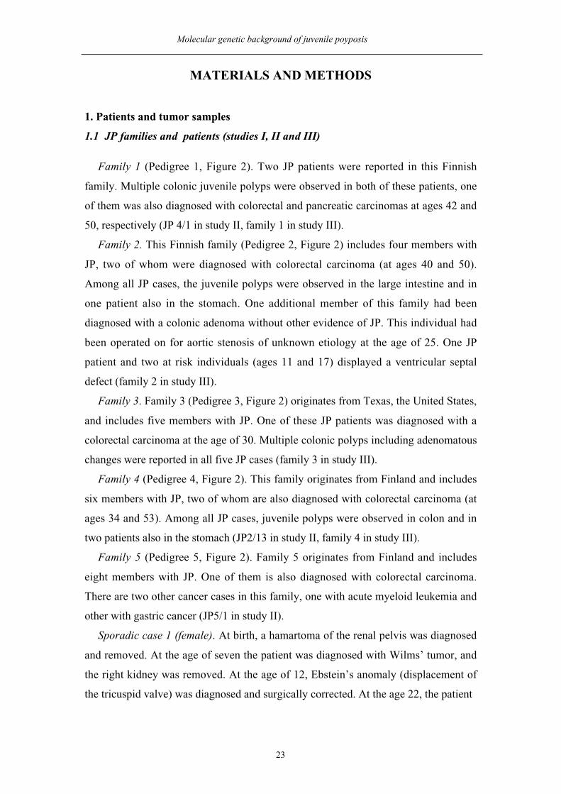
1.1 JP families and patients (studies I, II and III)
Family 1 (Pedigree 1, Figure 2). Two JP patients were reported in this Finnish
family. Multiple colonic juvenile polyps were observed in both of these patients, one
of them was also diagnosed with colorectal and pancreatic carcinomas at ages 42 and
50, respectively (JP 4/1 in study II, family 1 in study III).
Family 2. This Finnish family (Pedigree 2, Figure 2) includes four members with
JP, two of whom were diagnosed with colorectal carcinoma (at ages 40 and 50).
Among all JP cases, the juvenile polyps were observed in the large intestine and in
one patient also in the stomach. One additional member of this family had been
diagnosed with a colonic adenoma without other evidence of JP. This individual had
been operated on for aortic stenosis of unknown etiology at the age of 25. One JP
patient and two at risk individuals (ages 11 and 17) displayed a ventricular septal
defect (family 2 in study III).
Family 3. Family 3 (Pedigree 3, Figure 2) originates from Texas, the United States,
and includes five members with JP. One of these JP patients was diagnosed with a
colorectal carcinoma at the age of 30. Multiple colonic polyps including adenomatous
changes were reported in all five JP cases (family 3 in study III).
Family 4 (Pedigree 4, Figure 2). This family originates from Finland and includes
six members with JP, two of whom are also diagnosed with colorectal carcinoma (at
ages 34 and 53). Among all JP cases, juvenile polyps were observed in colon and in
two patients also in the stomach (JP2/13 in study II, family 4 in study III).
Family 5 (Pedigree 5, Figure 2). Family 5 originates from Finland and includes
eight members with JP. One of them is also diagnosed with colorectal carcinoma.
There are two other cancer cases in this family, one with acute myeloid leukemia and
other with gastric cancer (JP5/1 in study II).
Sporadic case 1 (female). At birth, a hamartoma of the renal pelvis was diagnosed
and removed. At the age of seven the patient was diagnosed with Wilms’ tumor, and
the right kidney was removed. At the age of 12, Ebstein’s anomaly (displacement of
the tricuspid valve) was diagnosed and surgically corrected. At the age 22, the patient

Molecular genetic background of juvenile poyposis
24
was operated on for bowel obstruction caused by a poorly differentiated
adenocarcinoma in the colon ascendens. In addition to the carcinoma, 20 to 30 polyps
were observed in the proximal colon up to hepatic flexure. Several polyps were
examined by an experienced pathologist, and all were hamartomatous polyps, which
could be designated as juvenile polyps. In one polyp, some features of Peutz-Jeghers
polyp were also seen. This patient died at the age of 25 (sporadic case 1 in study III).
Sporadic case 2 (female). This patient has congenital panhypopituitarism. JP was
diagnosed at the age of 38. Juvenile polyps were observed in the large and small
intestine and in stomach. Biological parents and relatives are unknown and the patient
has no children.
Sporadic case 3 (male). JP was diagnosed at the age of 13. Juvenile polyps were
observed in the large and small intestine. This patient has also been diagnosed with
empty cella-syndrome, Osler’s disease, and epilepsy. There is no family history of JP
(JP 1/1 in study II, sporadic case 3 in study III).
Sporadic case 4 (male). This patient was diagnosed with 30 to 40 colonic juvenile
polyps at age of 6, but there is no family history of JP (JP 10/1 in study II).
Sporadic case 5 (female). At the age of 18 ulcerative colitis (UC) was diagnosed,
later the diagnosis was confirmed as JP (age of 29). In addition to JP, this patient is
also mentally retardated. There is no clear evidence of family history of JP, however
the patient’s father has a history of gastrointestinal symptoms, but has not been
clinically evaluated.
In addition to patients listed above, studies I and II included JP families and
sporadic cases from United Kingdom (study I) and United States (studies I and II).
Clinical features and pedigree of the Iowa kindred (family I-13 in study II) have been
previously reported; first by Stemper and colleagues (1975) and later by Howe and
colleagues (1998a).
This study was approved by the Ethical Committee of the Department of Medical
Genetics, University of Helsinki.

Molecular genetic background of juvenile poyposis
25

Molecular genetic background of juvenile poyposis
26
Figure 2. Pedigrees of JP families. Those samples where DNA was available aremarked with an asterisk.

Molecular genetic background of juvenile poyposis
25
1.2 Hereditary non-polyposis colorectal cancer families (study IV)
Fourteen Finnish hereditary non-polyposis colorectal cancer (HNPCC) kindreds
from whom lymphoblastoid cell lines were available were selected for this study. One
affected individual per family was included in the study. Six kindreds fulfilled the
Amsterdam criteria for HNPCC (Vasen et al., 1991). Other patients represent familial
HNPCC-like colorectal cancer (CRC). The number of patients with CRC or
endometrial cancer ranged from 2 to 6 per family. All kindreds selected for this study
have been previously shown to be MLH1 and MSH2 mutation negative (Nyström-
Lahti et al., 1996; Holmberg et al., 1998). All except three kindreds displayed
microsatellite stable tumors (MSS). In these three kindreds DNA from tumor tissue
has not been available.
1.3 Tumor samples (study V)
Between May 1994 and June 1998 over one thousand fresh-frozen specimens of
colorectal adenocarcinoma have been collected at the Department of Medical
Genetics, Haartman Institute, University of Helsinki (Aaltonen et al., 1998; Salovaara
et al., in press). Among those, 26 microsatellite instable (MSI) and 16 MSS tumors
were selected for SMAD4 methylation analysis and 15 MSI and 7 MSS tumors for
SMAD4 mutation screening, respectively.
2. DNA and RNA extraction
In studies I and II, DNA was extracted from JP patient cell lines (cell pellets) or
blood samples with standard procedure. DNA extraction from paraffin-embedded
tumor or normal tissue was performed using the phenol and chloroform procedure
described in Kannio et al. (1996).
In studies III and IV, total cellular RNA was extracted from lymphoblasts by RNA
extraction kit (QIAGEN).
In study V, the tumor DNA was extracted from fresh frozen tumor specimens by
the standard procedure described by Lahiri and Nürnberger (1991). The corresponding
normal DNA was extracted from normal mucosa or blood.
Molecular genetic background of juvenile poyposis
25
3. cDNA synthesis (studies III and IV)
20 µl of cDNA was created from 0.8 µg of RNA using standard random priming
methods with 200 units of M-MLV reverse transcriptase (Promega), 1 × reaction
buffer (Promega), 10 µM random hexamer and 60 units of RNAse inhibitor
(Promega). The reaction was carried out at 42oC for 1 h and then 95oC for 10 min.
4. Polymerase chain reaction (PCR)
4.1 PTEN gene (study I)
Primers for genomic PTEN amplification have been previously described (Liaw et
al., 1997; Steck et al., 1997) except for primers for amplification and sequencing of
exons 2 and 4 which are shown in study I. The detailed PCR conditions are described
in study I.
4.2 SMAD4 gene (studies II, III, IV, V)
Primers used for genomic amplification of the SMAD4 gene have been previously
described (Moskaluk et al., 1997) except for new primers for exons 4, 7 and 8, which
are described in study III. Those primers were designed by using the Primer3
program. PCR conditions for genomic SMAD4 amplification are shown in study III.
The cDNA sequence for SMAD4 was derived from the GenBank (accession
number U44378). The gene was amplified in five fragments and PCR primers for
cDNA amplification were designed using the Primer3 server (http://www-
genome.wi.mit.edu/cgi-bin/primer/primer3.cgi) and they are listed in study IV. The
PCR reactions were carried out as described in studies II, III, IV and V.
4.3 SMAD2, SMAD3 and SMAD7 genes (studies III and IV)
The cDNA sequences for SMAD2 , SMAD3 and SMAD7 were derived from
GenBank database (accession numbers U65019, U76622 and AF010193,
respectively). The genes were amplified in five fragments and PCR primers for
cDNA amplification were designed using the Primer3 server. Primer sequences and
PCR conditions are shown in studies III and IV.

Molecular genetic background of juvenile poyposis
25
4.4 ALK1 and endoglin genes (unpublished data)
The genomic sequences for ALK1 and endoglin were derived from the GenBank
database (accession numbers U77707-U77713 for ALK1 and U37439-47, U17156-7,
and AF036969-71 for endoglin, respectively). The primers and PCR conditions for
amplification of all exons of ALK1 and endoglin genes have been previously
published. Those primers and conditions were also used here (Berg et al., 1997;
Gallione et al., 1998).
5. Denaturating gradient gel electrophoresis (DGGE) (study I)
DGGE was performed for all exons of PTEN, with the exception of exons 2 and 4,
in probands from 10 JPS families and 8 sporadic cases. The rest of the samples (4 JP
families and 3 sporadic cases) and exons 2 and 4 in all cases, were directly sequenced.
For DGGE conditions, see study I.
6. Single strand conformation polymorphism (SSCP) (study II)
In study II, the germline DCC and SMAD4 mutations were initially screened by
SSCP assay. The SSCP procedure is described in study II.
7. Automated sequencing (studies I, II, III, IV and V)
The PCR products were purified using the QIAquick PCR purification Kit
(QIAGEN). Direct sequencing of the purified PCR products was performed using the
ABI PRISM Dye Terminator or ABI PRISM dRhodamine cycle sequencing kits
(PE/ABI). Cycle sequencing products were electrophoresed on 6% Long Ranger gels
(FMC Bioproducts) and analyzed on an Applied Biosystems model 373A or 377
DNA sequencer (PE/ABI).
8. Restriction enzyme analysis (studies III, IV and V)
In study III, restriction enzyme digestion was used to screen for the presence of
two base substitutions in SMAD4 in control individuals. EcoRI (New England
BioLabs) digestion was used to detect a C to G change at codon 177 (exon 4). For the
analysis of an A to C change at codon 353 AlwI (New England BioLabs) digestion
was performed. The detailed digestion procedure is described in study III.

Molecular genetic background of juvenile poyposis
25
In study IV, the presence of an A to G change at codon 170 (SMAD3 exon 3) in
control individuals was analyzed with HgaI (New England BioLabs) digestion. For
the detailed procedure, see study IV.
In study V, NsiI (New England BioLabs) digestion was used to detect a G to A
change at codon 118 (SMAD4 exon 2). The PCR was performed as described is study
V. The detailed digestion procedure is described in study V.
9. Polyacrylamide gel electrophoresis (PAGE) (study III)
In study III, PAGE analysis was performed to analyze the presence of 4-base-pair
deletion in control individuals. A new set of primers was designed for amplification of
SMAD4 exon 9. Primers were forward: GGTTGCACATAGGCAAAGGT and
reverse: TTGGGTAGATCTTATGAACAGCA (5’ to 3’). With these primers, a 156-
bp fragment, containing the site of the 4-base-pair deletion, was amplified from exon
9. For PCR conditions, see study III. 10 µl of denaturing loading buffer (95%
formamide, 20 mM EDTA, 0.05 % bromphenol blue, 0.05% xylene cyanole FF) was
added to 10 µl of PCR sample, and the sample was denaturated for 5 min at 80oC. A 5
µl aliquot of the mixture was loaded in 6% polyacrylamide gels containing 8.3 M urea
and run at 2.5 kV for 50 min. Finally, the gels were dried and autoradiographed.
10. Linkage analysis (studies I and III)
Eight JP families originating from the United States and the United Kingdom were
included in the linkage analysis of study I. The microsatellite markers, PCR
conditions and statistical methods used for this analysis are described in detail in the
original study (study I).
In study III, those two JP families, in which the germline mutations of SMAD4
gene were not detected, were tested for linkage to 18q21. Microsatellite markers
D18S970, D18S474, D18S1099, D18S851, D18S484, D18S858 and D18S977
(SMAD4 is located close to markers D18S474 and D18S1099) were used for the
analysis. PCR reactions were carried out in a volume of 10 µl containing 50 ng DNA,
1 × PCR buffer, 1.5 mM MgCl2, 200 µM each of dATP, dGTP, dTTP and 0.7 µl of
[α-32P] dCTP (3000Ci/mmol, Amersham), 0.5 µM of each primer and 0.25 units of
AmpliTaqGOLD polymerase (PE/ABI). After PCR amplification in standard
conditions, PAGE analysis was performed (see above).

Molecular genetic background of juvenile poyposis
25
Multipoint linkage analyzes were performed using the GENEHUNTER program
(Kruglyak et al. 1996). The JP locus was defined as an affection status locus with
dominant inheritance. Three liability classes and age dependent penetrances were set
as follows: liability class 1 assigned to age <21 years, with heterozygote penetrance of
50%; liability class 2, age 21-50 years, heterozygote penetrance 70%; liability class 3,
>50 years heterozygote penetrance 80%. The JP locus frequency was assumed to be
1/50,000 and the marker loci frequencies and their genetic distances were obtained
from the CEPH database V8.1 (h t tp : / /www.cephb. f r and h t t p : / /www-
genome.wi.mit.edu/cgi-bin/contig/phys_map).
11. Methylation analysis (study V)
In study V, the methylation status of the SMAD4 promoter was studied in tumors
from patients with CRC. The fragment selected for this analysis was CG-rich region,
including the non-coding exon 1. To determine whether this particular promoter
region was hypermethylated, a PCR-based HpaII and MspI restriction enzyme assay
was used. Both tumor and normal DNA were digested and the reactions contained
either no enzyme, 25 units of HpaII, or 20 units of MspI. Samples were incubated for
16 h at 37οC. To analyze cleavage of the SMAD4 promoter region, 12.5 ng of DNA
from each digest was analyzed by PCR in 25 µl reaction volume. Primers were
designed (Primer3) to amplify a 408 bp fragment of SMAD4 promoter containing six
HpaII/MspI restriction sites and the primer sequences were: forward: 5’-
CAAGTTGGCAGCAACAACAC; and reverse: 5’- ACATGGCGCGGTTACCT.

Molecular genetic background of juvenile poyposis
32
RESULTS
Linkage analysis to 10q22-24 (study I)
Four microsatellite markers flanking the PTEN locus (markers D10S219, D10S551,
D10S579, and D10S541) were used to generate haplotypes for 47 individuals of eight
informative JP families originating from the United States and the United Kingdom.
For the microsatellite markers D10S219 and D10S541, the maximum two-point lod
score was 0 at a recombination fraction of θ = 0.5 for both models (model 1: allele
frequency 0.002 and penetrance 0.5; model 2: allele frequency 0.0002 and penetrance
0.85). For markers D10S551 and D10S579, the maximum two-point lod scores were
0.50 and 0.72, respectively, at θ = 0 for the first model and 0.63 and 0.20 at θ = 0 and
θ = 0.4, respectively for the second model. Multipoint analysis revealed lod scores
< -2.0 over the entire region, so the linkage of JP to PTEN locus was excluded in the
eight families studied.
PTEN mutation analysis (study I)
PTEN mutations were analyzed among probands from 14 JP families and 11
sporadic cases by either direct sequencing or DGGE analysis and mutations were not
detected. A frequent polymorphism in intron 8 was observed in 36 % (4 of 11) of
sporadic JPs cases. The presence of this sequence variant in its heterozygous state in
four cases excludes whole gene deletion as a cause of JP in these patients.
Mutation analysis of SMAD4 gene in JP (studies II, III)
To localize the gene predisposing to JP, a whole genome wide search was done in
three Finnish JP families (families 2, 4 and 5, data not shown). Simultaneously and
independently, Howe and colleagues (1998b) succeeded in localizing the gene
predisposing to JP in chromosome band 18q21.1 (Howe et al., 1998b). This
chromosomal region contains two putative tumor suppressor genes: DCC and
SMAD4. Since both of these genes were good candidates for JP, the mutation
screening was performed for both of them. After sequencing 14 DCC exons and 11
SMAD4 exons, a 4 base pair (bp) deletion was detected in SMAD4 exon 9 (between
nucleotides 1372 and 1375, GenBank accession number U44378). This mutation was
Molecular genetic background of juvenile poyposis
33
first detected in one affected individual from the Iowa kindred. Next, the SMAD4
exon 9 was screened from all 46 members of the Iowa JP kindred, the same 4 bp
deletion being present in all 13 affected and 4 of 26 at risk individuals, but not in any
of 7 spouses (study II).
Then, eight additional unrelated JP patients were analyzed for mutations of all
exons of SMAD4 by SSCP or genomic sequencing, and the mutation was found in
four of them. Two JP kindreds (originating from United States and Finland) were
segregating the same 4 bp deletion in exon 9 that was detected in the Iowa kindred.
This deletion causes a frameshift that creates a new stop codon at codon 434. In total,
242 controls were analyzed for the presence of this alteration and the altered allele
was not observed in any of them (study II). Mutations were also detected in two
sporadic JP cases. The first one was a 2 bp deletion in exon 8 at nucleotides 1170 to
1171 (codon 348). This deletion causes a frameshift that creates a stop codon at codon
350. The change was not detected in any of 101 controls analyzed. Another patient
was found to have a 1 bp insertion between nucleotides 815 and 820 of exon 5, this
change added a guanine to a stretch of six sequential guanines in the wild-type
sequence and created a frameshift and a new stop codon at codon 235. For this
change, 107 controls were analyzed and again, this change was not detected in any of
them (study II).
In study III, seven unrelated JP families or sporadic patients were analyzed for
mutations of all exons of SMAD4 by genomic sequencing. Four out of these seven
cases were previously analyzed for SMAD4 mutations in study II (by SSCP), and were
then reported mutation negative (JP1/1, JP2/13, JP4/1, and JP6/1, study II). In this
study, three different germline defects were detected. In family 3, we detected the
same 4-base-pair deletion in exon 9, which has been previously described in three JP
kindreds. In the sporadic case number 2 (JP6/1 in study II) a C to a G transversion at
nucleotide 661 was detected. This mutation changes serine to a stop codon at codon
177. Forty-nine controls were analyzed (AlwI digestion) and the change was not
detected in any of them. The change detected in JP family 1 (JP 4/1 in study II) was
an A to C transition at nucleotide 1186, which is predicted to convert tyrosine to
serine at amino acid 353. This variant was present in both cases with JP (see Material
and Methods, Patients) but also in one unaffected at risk individual. For this variant,
55 Finnish controls were analyzed by restriction enzyme digestion (EcoRI), and none
of them displayed the change. These two base pair changes were missed in study II,

Molecular genetic background of juvenile poyposis
34
because they did not show in SSCP analysis. No SMAD4 mutations were detected in
families 2 and 4, or in sporadic cases number 1 and 3.
In total, a set of 12 independent JP cases were analyzed for SMAD4 mutations
(studies II and III). Among these, mutations were detected in 5 families and 3
sporadic cases.
Linkage analysis to 18q21 (study III)
Linkage analysis using markers D18S970 , D18S474, D18S1099, D18S851,
D18S484, D18S858 and D18S977 (SMAD4 is located close to markers D18S474 and
D18S1099) resulted in a clear exclusion (Z ≤ -2) of the SMAD4 region in family 2
(see Materials and Methods, Patients). However, formal exclusion could be obtained
only at, and in the near vicinity of, marker D18S858 in family 4 while the rest of the
marker map produced inconclusive lod scores.
Mutation analysis of SMAD2, SMAD3 and SMAD7 in JP patients (study III)
SMAD2, SMAD3 and SMAD7 mutations were analyzed among those JP families or
sporadic cases where SMAD4 or PTEN mutations had not been detected (families 2
and 4 and patients 1 and 3). Mutation analysis was performed by automated
sequencing covering the translated region of these genes. No SMAD2, SMAD3 or
SMAD7 mutations were detected in any of these patients. The only variant identified
was an A to G change at the third position of codon 103 in the SMAD3 gene. The
change was homozygous in all of our four samples. This polymorphism has been
reported earlier and the variant does not cause any amino acid change (Arai et al.,
1998).
Mutation analysis of SMAD2, SMAD3 and SMAD4 in HNPCC patients
(study IV)
SMAD2, SMAD3 and SMAD4 mutations were analyzed among 14 familial colon
cancer kindreds, eleven of these displaying at least one MSS tumor. Previous studies
had evaluated MLH1 and MSH2 mutations in these families, all of them being
mutation negative (Nyström-Lahti et al., 1996; Holmberg et al., 1997). SMAD gene
mutation analysis was performed by automated sequencing covering the translated
region of the genes. Genetic alterations were not detected in SMAD2 or SMAD4 genes
in any of these patients.

Molecular genetic background of juvenile poyposis
35
In the SMAD3 gene, three discrepancies were detected between GenBank sequence
(U76622) and sequences from our patients. The first was an A to G change at the third
position of codon 103 (exon 2), this silent change has been reported earlier (study II).
A second, silent, change detected was C to T transition at nucleotide 907 (exon 6).
The frequency of these two variants in the normal population was not analyzed, as the
changes were silent.
The third change was an adenine to guanine transition at nucleotide 545, which is
predicted to convert isoleucine to valine at amino acid 170. This change was detected
in two patients. For this variant, 110 Finnish controls were analyzed by restriction
enzyme digestion (HgaI). Seven out of 110 control individuals displayed the change
(6.4 %). To further compare the frequency of this polymorphism in colon cancer
patients versus control individuals, 132 patients were included in analysis. Taken
together the 14 HNPCC patients and 132 colon cancer patients the frequency of this
polymorphism was 8.9% (13/146). From those 13 cancer patients who had valine
instead of isoleucine at codon 170, four turned out to be familial. Segregation of the
polymorphism was analyzed in two of these families where DNA from multiple
family members was available, and the polymorphism was not segregating with
cancer in these families.
SMAD4 promoter methylation (study V)
In this study, the possible hypermethylation of the SMAD4 promoter was analyzed
by using a HpaII and MspI digestion. Using this assay, we examined the methylation
status for SMAD4 promoter region in a group of 26 MSI and 16 MSS colorectal
tumors. The fragment selected for this analysis was a CG-rich region published by
Hagiwara and colleagues (submitted), and the amplified sequence contained
altogether 55 CpG dinucleotides. It was possible to determine the methylation status
for six CCGG sites by restriction. No PCR product was detected from any of HpaII
digested DNA, suggesting that the SMAD4 promoter is unmethylated in all the cases
studied.
Mutation analysis of SMAD4 in sporadic colorectal carcinomas (study V)
Twenty-two primary colon cancers were analyzed for mutations of all exons of
SMAD4 by genomic sequencing. The only change detected was a G to A transition at

Molecular genetic background of juvenile poyposis
36
the third position of codon 118 (exon 2). This silent change was present in one tumor
sample and also in corresponding normal DNA.
Mutation analysis of SMAD4 promoter in JP patients (unpublished data)
Mutations in SMAD4 5’-untranslated region (331 bp fragment downstream from
the transcription start site) were analyzed among those JP families/cases where
SMAD4 or PTEN mutations had not been detected (families 2 and 4 and sporadic
cases 1 and 3). Mutation analysis was performed by automated sequencing, and no
mutations were detected in any of patients analysed.
Mutation analysis of ALK1 and endoglin genes (unpublished data)
Genomic ALK1 and endoglin mutation screening was performed among those four
JP patient, where SMAD2, SMAD3, SMAD4, SMAD7 or PTEN mutations were not
detected. Mutations were not detected in either one of these genes in any of those four
patients.

Molecular genetic background of juvenile poyposis
37
DISCUSSION
Exclusion of PTEN gene as the gene predisposing to JP (study 1)
Previous studies have suggested that the gene predisposing to JP is located in
chromosomal band 10q22-24, in the region where the PTEN, gene associated with CS
and BRR is located. Jacoby and colleagues (1997) first reported a de novo interstitial
deletion of 10q22.3-24.1 in a single JP patient with multiple congenital abnormalities
(Jacoby et al., 1997a). To further analyze the relevance of this finding, they studied
allelic loss at 10q22 in 47 juvenile polyps from 16 unrelated JP patients. As a result,
83% of these polyps were found to have somatic deletion at 10q22 (Jacoby et al.,
1997b). These findings were interpreted as evidence for a tumor suppressor gene on
10q.
In our study, PTEN mutations were analyzed among probands from 14 JP families
and 11 sporadic cases and no mutations were detected. The lack of germline PTEN
mutations in a total of 25 unrelated JP patients argues that PTEN is not the JP
susceptibility gene. Also the possibility of whole gene deletion was excluded among
those four cases in which heterozygous sequence variant in intron 8 of PTEN was
present.
The possible linkage of JP to chromosomal region 10q22-24, which includes PTEN
gene and the putative JP locus (JP1, Jacoby et al., 1997b) was also analyzed in this
study. Four microsatellite markers covering the PTEN locus and flanking 20 cM were
used to generate haplotypes for members of eight JP families. The linkage analysis
excluded linkage to PTEN locus and, hence excluded the possibility of both a PTEN
promoter and intron mutations in at least these eight families. Thus, our data
suggested that PTEN is not the gene responsible of JP.
Later, another negative study was published by Riggins and colleagues (1997). In
their study, 11 JP cases were analyzed for PTEN mutations and no mutations were
found (Riggins et al., 1997).
There are however two reports where PTEN mutations have been described in JP
patients. Lynch and colleagues (1997) reported one family, which was thought to have
both JP and CS, as having nonsense mutation in PTEN (Lynch, et al., 1997).
In the study by Olschwang and colleagues (1998) 14 JP patients were screened for
PTEN mutations and three variants were observed in three JP patients. The first

Molecular genetic background of juvenile poyposis
38
variant was a 1 bp deletion at codon 232. This frameshift mutation leads to a stop
codon at position 255. The patient having this variant was a 74 year old man
diagnosed with anemia, hypoalbuminaemia, polyps in the GI tract and laryngeal
cancer. The second variation was a T to G transversion at codon 35, predicted to
substitute arginine for methionine. This patient was ten years old and had a history of
rectal bleeding. Juvenile polyps were found throughout the GI tract. The third variant
was a T to G transversion at the second position of the consensus splicing sequence of
the donor site of exon 6 (intronic variant). This change was predicted to lead to the
skipping of at least exon 6 in the mRNA, resulting in a shift of the translational
reading frame. The patient with this variant was 14 years old when he underwent
colonoscopy that revealed juvenile polyps (Olschwang et al., 1998).
These reports showing that four individuals with juvenile polyposis had PTEN
germline mutations would appear to confirm that PTEN is the predisposing gene on
10q in some families with JP (Lynch et al., 1997; Olschwang et al., 1998). However,
one of these patients was described as having both CS and JP (Lynch et al., 1997),
whereas the other three had no family history of JP (Olschwang et al., 1998). This
raises the question whether these patients were truly affected with JP or CS (Eng and
Ji, 1998).
SMAD4 is a gene predisposing to JP (studies II and III)
To localize the gene predisposing to JP, a whole genome wide search was
performed in three Finnish JP families (families 2, 4 and 5, data not shown).
Simultaneously and independently, Howe and colleagues succeeded in localizing the
gene predisposing to JP on chromosome band 18q21.1, between markers D18S1118
and D18S487 (Howe et al., 1998b). This interval contains two putative tumor
suppressor genes, DCC and SMAD4 (Figure 1).
The DCC gene was cloned in 1990, and it encodes a cell-surface protein containing
homology with N-CAM (Fearon et al. 1990). There have been many reports on the
loss of heterozygosity at the DCC gene locus in human colon cancers (Kikuchi-
Yanoshita et al., 1992; Thiagalingam et al., 1996), suggesting that DCC might be a
tumor suppressor gene. However, mutations in the coding region of DCC seem to be
rare (Cho et al. 1994) and the position of DCC as a candidate tumor suppressor is not
clear.
Molecular genetic background of juvenile poyposis
39
SMAD4 (DPC4) was first identified through deletion mapping in sporadic
pancreatic cancers (Hahn et al., 1996). It is inactivated in nearly half of sporadic
pancreatic carcinomas and in a subset of breast, ovarian, and colorectal tumors
(Schutte et al., 1996; Thiagalingam et al., 1996). The SMAD4 gene was first called
DPC4 (deleted in pancreatic carcinoma, locus 4), since it was the fourth marker to be
investigated for homozygous deletions in pancreatic cancer. Later the name SMAD4
has been used in addition to DPC4, as this gene is a member of the SMAD family of
genes related to the Drosophila Mad and C. elecans Sma genes (Sekelsky et al., 1995;
Hahn et al., 1996; Savage et al., 1996).
The mutation screening was performed for both of these genes simultaneously.
After sequencing 14 DCC exons and all 11 SMAD4 exons, a 4 base pair deletion in
exon 9 of SMAD4 was detected. After this finding the mutation screening was limited
only to the SMAD4 gene. In total, a set of 12 independent JP cases were analyzed for
SMAD4 mutations (studies II and III). Among these, mutations were detected in 5
families and 3 sporadic cases (62%).
Interestingly, the mutation was the same in four out of five familial cases (4 bp
deletion in exon 9). This deletion causes a frameshift that creates a new stop codon at
codon 434. In the recent study SMAD4 mutations were analyzed from 11 unrelated JP
patients and mutations were found in three of them (Friedl et al., 1999). In two of
these patients the mutation was the same 4 bp deletion in exon 9, described earlier in
four unrelated patients (studies II and III). According to the haplotype analysis, these
two patients did not share common alleles for the markers D18S363 and D18S1110
(markers located close to SMAD4) (Friedl et al., 1999). Since this 4 bp deletion has
been detected in one kindred originating from Finland, in three kindreds originating
from the United States, and in two kindreds originating from Germany, the defect
seems to represent a mutational hotspot rather than an ancestral founder mutation.
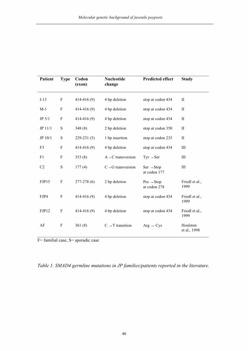
Other germline SMAD4 mutations detected in JP are a 1 bp insertion in exon 5
(study I), a 2 bp deletion in exon 8 (study I), an A to C transition in exon 8 (study III),
a C to G transversion in exon 4 (study III), a 2 bp deletion in exon 6 (Friedl et al.,
1999) and a C to T transition in exon 8 (Houlston et al., 1998). Mutations detected in
studies II and III, or reported in the literature are summarized in Table 1.
Molecular genetic background of juvenile poyposis
40
Patient Type Codon(exon)
Nucleotidechange
Predicted effect Study
I-13 F 414-416 (9) 4 bp deletion stop at codon 434 II
M-1 F 414-416 (9) 4 bp deletion stop at codon 434 II
JP 5/1 F 414-416 (9) 4 bp deletion stop at codon 434 II
JP 11/1 S 348 (8) 2 bp deletion stop at codon 350 II
JP 10/1 S 229-231 (5) 1 bp insertion stop at codon 235 II
F3 F 414-416 (9) 4 bp deletion stop at codon 434 III
F1 F 353 (8) A→C transversion Tyr →Ser III
C2 S 177 (4) C→G transversion Ser →Stopat codon 177
III
FJP15 F 277-278 (6) 2 bp deletion Pro →Stopat codon 278
Friedl et al.,1999
FJP4 F 414-416 (9) 4 bp deletion stop at codon 434 Friedl et al.,1999
FJP12 F 414-416 (9) 4 bp deletion stop at codon 434 Friedl et al.,1999
AF F 361 (8) C →T transition Arg → Cys Houlstonet al., 1998
F= familial case, S= sporadic case
Table 1. SMAD4 germline mutations in JP families/patients reported in the literature.

Molecular genetic background of juvenile poyposis
41
On the basis of structural and functional criteria, the SMAD family can be divided
into three subgroups. The first group includes so called receptor regulated SMADs
that act as direct substrates for receptors (in human those are SMADs 1, 2, 3, 5 and 8)
(Liu et al., 1996; Eppert et al., 1996; Chen et al., 1997; Nishimura et al., 1998). The
second group includes SMADs that are not direct receptor substrates, but whose
function is essential for signalling by the receptor regulated SMADs. SMAD4 belongs
to the second group. The third group includes the inhibitory SMADs (SMADs 6 and
7) (Hata et al., 1998; Hayashi et al., 1997; Nakao et al., 1997). All these SMAD
proteins have a domain structure composed of highly conserved N-terminal and C-
terminal domains known as MH1 and MH2 (Mad homology domains 1 and 2) which
are joined by a linker region (Figure 3). The receptor regulated SMADs are directly
phosphorylated at their carboxyl terminus by type I TGFβ superfamily receptors.
SMAD4 lacks this carboxy-terminal phosphorylation sequence and thus can not
behave as direct substrate for the type I receptors (Macias-Silva et al., 1996; Zhang et
al., 1996; Krezschmar et al., 1997). In the inactive state, SMAD4 is located in the
cytoplasm as a homo-oligomer and remains in an inactive conformation through an
interaction between the MH1 and MH2 domains. After ligand stimulation and
phosphorylation of the receptor regulated SMADs, SMAD4 forms hetero-oligomers
with receptor regulated SMADs and translocates into the nucleus (Lagna et al., 1996;
Liu et al., 1997; Whitman, 1998). In the nucleus this SMAD hetero-oligomer complex
may specifically associate with a nuclear transcription factor, which then targets the
SMAD transcriptional activation function to a specific promoter (Liu et al., 1997). In
addition to targeting promoters via trancription factors, SMADs have a site-specific
DNA-binding activity. It has been demonstrated that the MH1 domain of SMAD
protein may directly bind to the promoter or enhancer region of the genes, thus
regulating the transcription (Whitman, 1998). The third mechanism for SMAD protein
signalling in cells is transcriptional activation through MH2 domain (Liu et al., 1997)
A summary of the domain structure of SMADs and their interaction is presented in
Figure 3.

Molecular genetic background of juvenile poyposis
42
Figure 3. The domain structure of SMADs and their interactions (modified from
Hata et al., 1998). In basal state, SMADs form homo-oligomers and remain in an
inactive conformation through an interaction between the MH1 and MH2 domains.

Molecular genetic background of juvenile poyposis
43
The majority of somatic, as well as germline, mutations described in SMAD4 map
to the carboxyl-terminus between codons 330-526 (MH2 domain), and half of
mutations are located in exons 8 and 9 (Hahn et al., 1996, Hahn et al., 1998; Kim et
al., 1996; Schutte et al., 1996; Takagi et al., 1996) (Figure 3). The tumorigenic
mutations in SMADs fall in three major categories according to their effect on protein
function. The missense mutations in the MH1 domain interrupt DNA binding
function, truncating mutations of the MH2 domain remove its ability to activate gene
expression, and the missense mutations of the MH2 domain appear to interfere with
its nuclear localization (Dai et al., 1998). The location of SMAD4 germline mutations
and the hotspots of sporadic SMAD4 mutations are shown in Figure 4.
Figure 4. Summary of the locations of germline SMAD4 mutations in JP patients. The
hotspots of somatic mutations in sporadic cancer are also shown.

Molecular genetic background of juvenile poyposis
44
Other candidate genes for JP (study III and unpublished data)
So far, 12 SMAD4 mutations have been identified in 46 familial and sporadic JP
cases (26%) (studies II and III; Friedl et al., 1999; Houlston et al., 1998). In our
studies (studies II and III) SMAD4 mutations were detected in eight of 12 JP patients
analysed (66%). However, in two recent studies a considerably lower proportion of
SMAD4 involvement in JP were reported. Friedl and colleagues analysed 11 unrelated
patients with JP and found SMAD4 mutation in three of them (27%). Similarly,
Houlston and colleagues analysed 21 JP patients and found a mutation only in one of
them (5%). Taken together these results suggest that mutations in SMAD4 predispose
to JP, but account only for approximately 25% of cases. Further studies will hopefully
clarify the true frequency, though issues such as diagnosis of JP and different
mutation detection method utilized (genomic sequencing in studies II, III, and in the
study by Friedl and colleagues and conformation-specific gel elctrophoresis in the
study by Houlston) may contribute to the discrepancy.
Other explanation for the relatively low SMAD4 mutation frequency among JP
patients may be the existence of other gene/genes predisposing to JP. Houlston and
colleagues (1998) analyzed eight JP families for linkage to SMAD4 and found no
evidence for linkage. It could be excluded in two of the eight kindreds and was
unlikely in two others. Of those four families which provided good evidence against
linkage to 18q21.1, two were also unlinked to the PTEN locus (Houlston et al., 1998),
one was compatible with PTEN linkage but no PTEN mutation was detected (study I)
and one has not been tested for PTEN mutations. None of these families had notable
features of CS or BRR.
In our studies (studies II and III) SMAD4 mutations were not detected in two
families (families 2 and 4; family 2 also excluding linkage to 18q21) and two sporadic
cases (sporadic cases 1 and 3), these being negative also in the previous PTEN
mutation analysis (study I). Two of these (one family and one sporadic case) display a
JP phenotype with heart anomalies; the sporadic case also has a history of Wilms’
tumor. The other sporadic case was diagnosed with hereditary hemorrhagic
telangiectesia (HHT) and epilepsy.
There is good evidence for the importance of the TGF-β signaling pathway in
colorectal carcinogenesis. A large proportion of sporadic colon cancers with MSI
have TGF-β type II receptor mutations (Myeroff et al., 1995) and germline variants in
the TGF-β type II receptor may predispose to colon cancer (Lu et al., 1998). Also the

Molecular genetic background of juvenile poyposis
45
downstream targets of TGF-β have been implicated in colorectal carcinogenesis.
Somatic mutations of SMAD2 and SMAD4 have been found in colorectal cancer in
humans and germline Smad3 mutations cause colon cancer in mice (Zhu et al., 1998).
It has been hypothesized that the germline mutations in genes (other than SMAD4)
encoding different components of the TGF-β signaling pathway may be present in JP.
To study this hypothesis further, we focused on SMAD genes, which are involved
in TGF-β signaling via TGF-β type II and I receptors (TGFβR-II and TGFβR-I).
SMAD4 is known to be the co-SMAD and its function is essential for signaling by the
receptor-regulated SMADs. This group includes SMADs 2 and 3, which are substrates
of the TGF-β and activin type I receptors (TGFβR-I and ActR-IB) and mediators of
TGF-β and activin signals (Whitman, 1998; Hata et al., 1998). The same pathway also
includes SMAD7, which is an inhibitory SMAD. Expression of SMAD7 inhibits the
receptor-regulated phosphorylation of SMAD2 or SMAD3 (Hayashi et al., 1997;
Nakao et al., 1997). In this study a cDNA based mutation screening was performed
for SMAD2, SMAD3 and SMAD7 genes and no mutations were detected in these
genes in any of our patients (study III). Recently, similar results were reported by
Bevan and colleagues (1999). In their study, 30 unrelated JP patients, who were
negative in previous SMAD4 mutation analyses (Houlston et al., 1998) were screened
for germline mutations at SMAD1, SMAD2, SMAD3 and SMAD5. No mutations were
found in any of the patients screened (Bevan et al., 1999).
Previous results together with the occurrence of other genetic syndromes with
juvenile polyposis, suggest genetic heterogeneity in the etiology of JP. BRR, Gorlin
syndrome and HHT are genetic syndromes, which commonly are associated with JP.
The genes responsible for these conditions may also be considered as candidate genes
for JP. The gene predisposing to BRR (and CS) is PTEN, and as discussed in previous
chapters it is unlikely that it would also be the gene predisposing to JP. Gorlin
syndrome is characterized by multiple basal cell carcinomas of the skin. The gene for
Gorlin syndrome has been localized to chromosome 9 and shown to be the human
homolog of the Drosophila developmental gene patched (Johnson et al., 1996). The
third syndrome, which is occasionally described among JP patients, is HHT (also
known as Osler’s disease or Osler-Rendu-Weber disease). HHT is an autosomal
dominant disorder characterized by multisystemic vascular dysplasia and recurrent

Molecular genetic background of juvenile poyposis
46
haemorrhage (Guttmacher et al., 1995). Two predisposing genes have been identified
for HHT. Endoglin, which maps to chromosome 9q33-34 has been identified as the
gene for HHT1 (HHT type 1), and ALK1 (activin receptor–like kinase 1 gene), which
maps to chromosomal locus 12q11-14, is the gene for HHT2 (HHT type 2)
(McDonald et al., 1994; Shovlin et al., 1994; Johnson et al., 1995; Vincent et al.,
1995).
One of our JP patients was diagnosed with HHT (study III, sporadic case 3).
Similar cases have previously been reported in some other studies. In 1980 two
patients (mother and daughter) with HHT and polyposis were reported (Cox et al.,
1980). Two years later an autosomal dominant syndrome with juvenile
gastrointestinal polyposis, cutaneous telangiectesia, and pulmonary arteriovenous
malformations in a father and two children were described. The father died from
colon cancer at the age of 36 (Conte et al., 1982). Recently, Desai and colleagues
(1998) described one JP patient with HHT and two patients with some features of
HHT (Desai et al., 1998).
The association of HHT with juvenile polyposis in a subset of patients raises the
possibility that JP could occur as part of a 9q or 12q microdeletion syndrome in some
cases, or JP with HHT might be a unique syndrome due to mutation in a single gene.
To further study this hypothesis, endoglin and ALK1 mutation screening was
performed among those JP patient, where SMAD2, SMAD3, SMAD4, SMAD7 or
PTEN mutations were not detected. Both endoglin and ALK1 are members of the
TGF-β receptor superfamily and are therefore cosidered as good candidates for JP
gene. No mutations were detected in either of these genes (unpublished data). Our
data suggest that there are still unidentified genes predisposing to JP, perhaps
associated with developmental defects such as cardiovascular anomalies.
Mutation analysis of SMAD genes in HNPCC patients (study IV)
HNPCC is an autosomal dominant cancer susceptibility syndrome. The genes
responsible for the disease are MSH2, MLH1, PMS1, PMS2 and MSH6 (Leach et al.,
1993; Bronner et al., 1994; Papadopoulos et al., 1994; Nicolaides et al., 1994, Miyaki
et al., 1997). Inactivation of both alleles of one of these genes results in microsatellite
instability (MSI) that is a hallmark of HNPCC tumors. The genes responsible for
Molecular genetic background of juvenile poyposis
47
microsatellite stable (MSS) HNPCC are still largely unknown (Peltomäki et al., 1997;
Aaltonen et al., 1993, Nyström-Lahti et al., 1996).
Loss of growth inhibition by transforming growth factor β(TGF-β) is an important
step in colorectal tumorigenesis. In HNPCC tumors with MSI, this is mainly due to
frameshift mutations within a polyadenine sequence repeat in the TGFβRII gene. It
has been proposed that germline mutations in TGFβRII underlie a subset of MSS
HNPCC cases (Lu et al., 1998). Other genes involved in the TGF-β pathway could
also be candidates for MSS HNPCC (Grady et al., 1999).
SMAD proteins play a key role in intracellular TGF-β signaling and mutations in
some of the SMAD genes disrupt this pathway. To investigate whether germline
mutations in SMAD2, SMAD3 or SMAD4 could predispose to MSS HNPCC, we
analyzed mutations in these genes in 14 HNPCC kindreds in which germline
mutations in MSH2 and MLH1 had been ruled out (Nyström-Lahti et al., 1996;
Holmberg et al., 1997). The only changes detected were three polymorphisms in
SMAD3. One of these was an A to G transition at nucleotide 545, which changes
isoleucine to valine at amino acid 170, in the so called linker-region of SMAD3
protein. This variant has not been reported earlier. We analyzed 110 Finnish controls
and 132 additional colon cancer patients by restriction enzyme digestion to clarify the
frequency of this polymorphism. In total, seven control individuals (6.4%) and 13
colon cancer patients (8.9 %, including two HNPCC cases) displayed the variant,
suggesting that this polymorphism is rather neutral than related to colorectal cancer.
Even SMAD2 and SMAD4 genes are mutated in a subset of human colorectal tumors,
our results suggest that these genes are not involved in the MSS HNPCC. To date,
SMAD3 mutations have not been reported in human cancers. In a recent study SMAD3
mutations were analysed among 35 sporadic colorectal and 15 HNPCC cancers and
no mutations were found (Arai et al., 1998). Further work is necessary to unravel the
molecular background of MSS HNPCC.
SMAD4 is infrequently hypermethylated and mutated in sporadic colorectal
cancers (study V)
SMAD4 seems to be mutated in only less than 10% of cancers analysed. The
only exception is pancreatic cancer, where the mutation frequency is approximately
Molecular genetic background of juvenile poyposis
48
20% (Takagi et al., 1996; Thiagalingham et al., 1996; Schutte et al., 1996; Hahn et al.,
1996; Nagatake et al., 1996; Rozenblum et al., 1997).
Knudson’s hypothesis that two hits are required for the full inactivation of a tumor-
suppressor gene has been shown to be correct in most human cancers. Traditionally, it
has been thought that tumor suppressor genes are inactivated by intragenic mutations
and loss of chromosomal material. However, the fact that methylation of CpG islands
located in the promoters of genes can cause transcriptional silencing, has led to the
suggestion that hypermethylation of tumor-suppressor gene promoters may be one of
the mechanisms leading to malignant transformation (Jones and Laird, 1999). Several
tumor suppressor genes contain CpG islands in their promoters and some of them, like
RB1 and VHL also show evidence of methylation silencing (Herman et al. 1994; Sakai
et al., 1991). On the other hand, promoters of some tumor suppressor genes are very
rarely hypermethylated. For example, TP53 is mostly inactivated by point mutations,
whereas p16 is generally inactivated by homozygous deletion (Hussain and Harris,
1998; Kamb et al. 1994).
There has been two studies on the possible SMAD4 promoter region. The first
candidate for the SMAD4 promoter was reported by Minami and colleagues (1998),
when they cloned a 1.4 kb fragment of the SMAD4 5’-flanking region from a phage
library by using the first coding exon’s sequence as a primer. This SMAD4 promoter
lacks typical TATA boxes and CpG islands, but contains some TATA-like structures
(TAAAAT) as well as some binding sites for transcription factors (Minami et al.,
1998). Later, another candidate for the SMAD4 promoter was published by Hagiwara
and colleagues (submitted). First, they identified a new non-coding exon (exon 1) by
using the 5’-rapid amplification of the cDNA ends (5’-RACE) (Hagiwara et al.,
submitted). Then SMAD4 promoter was cloned using the exon 1 sequence as a probe.
The SMAD4 promoter published by Hagiwara and colleagues is a typical CpG island
having a high G+C content (Hagiwara et al., submitted).
In study V, we examined whether hypermethylation of the promoter could be an
alternative mechanism for SMAD4 inactivation. The CpG island near non-coding exon
1 was selected for this analysis, since it is well documented that methylation has
important regulatory effects especially when involving these CpG rich areas (Bird,
1986). In total, 26 MSI and 16 MSS tumors and corresponding normal DNAs were
selected for the analysis and no evidence of hypermethylation was found.

Molecular genetic background of juvenile poyposis
49
In the recent study SMAD4 mutations were analysed from 176 colorectal tumors
and SMAD4 mutation frequencies were found to be 0% in adenoma, 10% in
intramucosal carcinoma, 7% in primary invasive carcinoma without metastasis, 35%
in primary invasive carcinoma with metastasis and 31% in distant metastasis. So, the
frequency of SMAD4 mutations was strongly correlating with the stage of colorectal
cancer (Miyaki et al., 1999). This correlation was further confirmed by Koyma and
colleagues (1999). They analysed SMAD4 mutations among 64 advanced colorectal
cancers and found them in seven tumors. They also demonstrated that in all these
seven tumors carrying intragenic mutations in one allele, LOH analysis showed the
loss of the other allele. Their results suggested that inactivation of both alleles of the
SMAD4 gene occurs in a substantial portion of advanced colorectal cancers (Koyama
et al., 1999).
In our study (study V), 22 primary CRCs were taken into SMAD4 mutation
screening and the only change detected was a polymorphism in exon 2. The tumors
included in our mutation screening study were mainly classified as Dukes’ stages A
and B (15/22,) and one explanation for the lack of SMAD4 mutations could be that the
tumors were mainly limited to mucosa and submucosa and were not metastasizing.
There are many studies reporting SMAD4 mutation analyses for different tumors.
In most of them, the mutation screening is limited to the coding exons and small
fragments of the introns around these exons. In one recent study, Zhou and colleagues
(1999) analysed six endometrial tumors that do not express normal SMAD4, and
found mutation in the 5’-untranslated region in two of them (Zhou et al., 1999). The
region included in mutation screening was a 331 bp long fragment, which spanned
nucleotides –262 to +69 from the transcription start site of SMAD4. This fragment
contains most of the important transcription factor binding sites (Minami et al., 1998).
To further study whether this 5’-untranslated region is also mutated in colorectal
carcinomas, we sequenced the same fragment from 15 MSS and 7 MSI tumor
samples. We however found no changes.
It has generally been considered that the genes involved in hereditary cancer
syndromes are also mutated in the same type of sporadic tumors (Fearon, 1998). For
example APC, the gene responsible for FAP, is involved in up to 80% of all sporadic
colon cancers (Beroud and Soussi, 1996). There is good evidence that the hamartomas
in PJS, JP and CS can progress to colorectal carcinoma. However, mutations in PTEN,
LKB1 or SMAD4 genes in the sporadic colon cancers occur at very low frequencies

Molecular genetic background of juvenile poyposis
50
(Kong et al., 1997; Avizienyte, et al., 1998; Wang et al., 1998b; MacGrogan et al.,
1997; Takagi et al., 1996).
There are also examples of cancer syndromes, where a gene, which is mutated in
germline is not frequently mutated in the corresponding somatic cancers. The familial
breast/ovarian cancer genes, BRCA1 and BRCA2, cause cancer when mutated in the
germline, but are only seldom mutated in sporadic cancers (Futreal et al., 1994; Miki
et al., 1996; Teng et al., 1996). There is, however, evidence that BRCA1 is inactivated
by promoter methylation in some sporadic breast cancers (Dobrovic and
Simpfendorfer, 1997). TP53 gene is an another example of discrepancy in somatic
and germline mutations. The TP53 mutations were first described in sporadic
colorectal carcinomas (Baker et al., 1989), in which they are frequent, however
families with germline TP53 mutations (Li-Fraumeni syndrome) are not predisposed
to colorectal carcinomas (Knudson, 1993).
Our results together with previously published data strongly suggests that the
SMAD4 is not frequently mutated in primary colorectal carcinoma and that the
hypermethylation of SMAD4 promoter region is not a crucial mechanism in colorectal
tumorigenesis.
Molecular genetic background of juvenile poyposis
51
SUMMARY
The aims of this study were to localize and identify the gene predisposing to JP,
and to evaluate its role in hereditary and sporadic colorectal tumorigenesis.
In study I, we analysed whether PTEN, gene frequently mutated in CS and BRR,
could also be the gene predisposing to JP. In total, 25 unrelated JP patients were
screened for the presence of PTEN mutations and no mutations were found.
Furthermore, linkage analysis in eight informative families excluded linkage to PTEN,
and hence, excluded the possibility of PTEN promoter or 3’-UTR mutations and
whole gene deletions at least these eight JP families. Thus, although germline
mutation has been detected both in CS and BRR (also characterized by
hamartomatous gastrointestinal polyps), our results suggest that JP is not allelic to
these syndromes.
To localize the gene predisposing to JP, a whole genome wide search was done in
three Finnish JP families (families 2, 4 and 5, data not shown). However,
simultaneously and independently, Howe and colleagues (1998b) succeeded in
localizing the gene predisposing to JP in chromosome band 18q21.1 (Howe et al.,
1998b). This chromosomal region contains two putative tumor suppressor genes:
DCC and SMAD4, and in collaboration with Howe and colleagues (study II) we
managed to identify germline mutations in SMAD4 gene in JP patients. Conclusively,
SMAD4 mutations were found in 8 of 12 JP cases studied (5 familial and 3 sporadic).
SMAD4 mutations were not detected in two families and in two sporadic cases, that
were negative also in the previous PTEN mutation analysis. JP is obviously a
genetically heterogeneous condition, as evidenced by the fact that not all families are
linked to 18q markers and not all patients studied had germline SMAD4 mutations. It
has been hypothesized that the germline mutations in genes (other than SMAD4)
encoding different components of the TGF-β signalling pathway may be present in JP.
To study this hypothesis further, we focused our screening on SMAD genes, which are
involved in TGF-β signaling via TGF-β type II and I receptors. In study III cDNA
based mutation screening was performed for SMAD2, SMAD3 or SMAD7 genes and
no mutations were detected in these genes in any of our patients.

Molecular genetic background of juvenile poyposis
52
One syndrome, which is occasionally detected among JP patients, is HHT. Two
genes have been identified for HHT. Endoglin, which maps to chromosome 9q3 is
identified as the gene for HHT1 and ALK1, which maps to chromosomal locus 12q11-
14 is the gene for HHT2, respectively. Both endoglin and ALK1 are members of TGF-
β receptor superfamily and are therefore considered as good candidates for JP gene.
We screened endoglin and ALK1 mutations among those JP patients, where SMAD2,
SMAD3, SMAD4, SMAD7 or PTEN mutations have not been detected. No mutations
were detected in either one of these genes in any of patients analyzed (unpublished
data).
Loss of growth inhibition by transforming growth factor β (TGF-β) is an important
step in colorectal tumorigenesis. As SMAD proteins play a key role in intracellular
TGF-β signaling, mutations in some of the SMAD genes may disrupt this pathway. To
investigate whether germline mutations in SMAD2, SMAD3 or SMAD4 could
predispose to HNPCC, we analyzed mutations in these genes from 14 HNPCC
kindreds in which germline mutations of MSH2 and MLH1 had been ruled out. SMAD
gene mutations were not found in any of patient analyzed, suggesting that these genes
do not have a major role in HNPCC (study IV).
It is generally considered that the genes involved in hereditary cancer syndromes
are also mutated in the same type of sporadic tumors. However, SMAD4 mutations
seem to be relatively rare in the sporadic colon cancers. In study V, we examined
whether hypermethylation of the promoter could be an alternative mechanism for
SMAD4 inactivation in colorectal cancers. In total, 26 MSI and 16 MSS tumors and
corresponding normal DNAs were analyzed and no evidence of hypermethylation was
found. SMAD4 mutation screening was also performed among 22 primary CRCs and
the only change detected was a polymorphism in exon 2. Our results together with
previously published data suggests that SMAD4 is not frequently mutated in primary
colorectal carcinoma and that the hypermethylation of SMAD4 promoter region is not
a crucial mechanism in colorectal tumorigenesis.

Molecular genetic background of juvenile poyposis
53
CONCLUSIONS AND FUTURE PROSPECTS
Although it has been reported that some patients with JP have germline PTEN
mutations, this connection remains controversial and until now the only proven cause
of JP, is germline mutation in the SMAD4 gene. However, SMAD4 mutations seem to
account for only less than half of the JP cases suggesting that juvenile polyposis is a
genetically heterogeneous disease. The identification of further JP loci by linkage
analysis might be problematic, since most of the existing JP families are relatively
small, and it is possible that there is still more than one gene to be identified. There
have been a few attempts to identifying other JP genes by “candidate gene”- strategy,
but so far these efforts have failed.
Identification of further gene/genes predisposing to JP will, however, be important,
since it may help in the diagnosis, and especially asymptomatic at-risk individuals
may benefit from gene testing. In particular, when a mutation in a predisposing gene
(like SMAD4) is known to be segregating in the family, at-risk persons who are
negative for the mutation, may be relieved of years of colonoscopic surveillance.
Similarly, persons who are test positive for the mutation, may approach the screening
regimen with a greater willingness to participate.
Further work related to the function of SMAD4 gene in juvenile polyposis is also
needed. Especially the role of the 4 bp deletion detected in six JP families is highly
interesting, since it seems to represent a mutational hotspot rather than an ancestral
founder mutation. On the other hand, the fact that a single mutation in SMAD4 is
responsible for 50% of the clinically classified JP cases might simplify the genetic
testing.
The role of the SMAD4 gene in colorectal tumorigenesis is still largely unknown.
There is some evidence suggesting that the SMAD4 gene is mutated in advanced
stages of human colorectal cancers, but otherwise the SMAD4 mutations seem to be
rare in these cancers. Our results also suggest that the hypermethylation of the
SMAD4 promoter is not a crucial mechanism in colorectal carcinogenesis. However,
final conclusions about the significance of SMAD4 in colorectal tumorigenesis can not
be drawn before the expression of SMAD4 has been carefully studied in these tumors.

Molecular genetic background of juvenile poyposis
54
ACKNOWLEDGEMENTS
This work was carried out at the Department of Medical Genetics, Haartman Institute,
University of Helsinki during 1997-2000. I wish to express my warmest gratitude to all those
who made it possible:
Lauri Aaltonen, my supervisor, for introducing me to the field of medical genetics. I am very
grateful that I had the opportunity to join his research group and learn so much during this
period. Besides the scientific discussions, I will always remember those intense debates
concerning ice-hockey.
Albert de la Chapelle, my second supervisor, for his enthusiasm for the research, his guidance
and encouraging attitude towards this work.
Juha Kere, Leena Peltonen and Pertti Aula, the former and present heads of the Department of
Medical Genetics for providing me with excellent working facilities.
Anu Jalanko and Hannu Sariola, the official pre-examiners of this study for their valuable
comments.
Anu Loukola and Virpi Launonen for their encouragement and friendship, and for helping me
through all those days, when smad went mad and everything was unlinked. We really had
many (extremely!) enjoyable moments at work as well as outside our working hours!
Egle Avizienyte, Akseli Hemminki and Reijo Salovaara for their friendship during these
years. I am especially grateful to them for their warm welcome when I first came to the
“Peutz-Jeghers team”.
Heikki Järvinen, Jukka-Pekka Mecklin, Pertti Sistonen, Ilmo Kellokumpu, Päivi Peltomäki,
and Paula Kristo, whose help and experience were of great value during this study.
Marie Johansson, Päivi Laiho, Kirsi Laukkanen and Annika Lahti for their skillful technical
assistance in the lab as well as many cheerful lunch breaks in the UniCafe.
Sinikka Lindh and Siv Lindroos for their sincere friendship, and for helping me patiently no
matter how busy they were.

Molecular genetic background of juvenile poyposis
55
Elina Kirjavainen and Minna Maunula for their help in many practical matters. Elvi Karila,
Anita Hottinen and Susanna Anjala for sequencing service. Kaija Collin, Liisa Suksi and
Taija Poikonen for cell culturing and DNA extraction. Tuula Lehtinen for helping me with the
JP patient data.
Charis Eng, James Howe, Christopher I. Amos, Ian Tomlinson and Debbie Marsh for
excellent collaboration.
The co-authors in different studies: Steven Bevan, Walter F. Bodmer, Stacey Caron, Karen A.
Cleary, Sally E. Cottrell, Patricia L.M. Dahia, Malcom G. Dunlop, Diana Eccles, Shirley V.
Hodgson, Richard S. Houlston, Kathryn L. Lunetta, Patrick Lynch, David Markie, Frank A.
Mitros, Kay Neale, Robin Phillips, John C. Ringold, Paul Rozen, Edwin M. Stone, Robert W.
Summers, Sapna Syngal, Nathalie J. van Orsouw, Jan Vijg, and Zimu Zheng.
My previous colleagues and my present friends; Minna Veini, Sari Rantanen and Terhi
Kiviranta for sharing many hobbies and unforgettable moments with me.
All my colleagues at the Department of Medical Genetics for creating such a nice atmosphere.
Donald Smart for the language revision of this thesis.
My parents Aila-Riitta and Rolf for their love and support; and my brother Kristian and my
sister Emma for their friendly criticism and many enjoyable moments together.
My godfather Arvo for his encouragement and great interest in my career.
Mikko Tammiruusu for his love, support, and understanding during these years. I really didn’t
make it easy...
Aki Ylä-Soininmäki for his care, optimism and generous help during the last few months.
This work was financially supported by the Finnish Cancer Society, the Ida Montin
Foundation, the Emil Aaltonen Foundation, the Finnish Cultural Foundation, the Academy of
Finland and the Research and Science Foundation of Farmos.

Molecular genetic background of juvenile poyposis
56
REFERENCES
Aaltonen, L.A., Peltomäki, P., Leach, F.S., Sistonen, P., Pylkkänen, L., Mecklin, J.-P.,Järvinen, H., Powell, S.M., Jen, J., Hamilton, S.R., Petersen, G.M., Kinzler, K.W.,Vogelstein, B., and de la Chapelle, A. Clues to the pathogenesis of familial colorectal cancer.Science, 260: 812-816, 1993.
Aaltonen, L.A., Salovaara, R., Kristo, P., Canzian, F., Hemminki, A., Peltomäki, P.,Chadwick, R., Kääriäinen, H., Eskelinen, M., Järvinen, H., Mecklin, J-P., and de la Chapelle,A. Incidence og hereditary nonpolyposis colorectal cancer and the feasibility of molecularscreening for the disease. The New England Journal of Medicine, 338: 1481-1487.
Agnifili, A., Verzaro, R., Gola, P., Marino, M., Mancini, E., Garducci, G., Ibi, I., and Valenti,M. Juvenile polyposis: case report and assessmant of the neoplastic risk in 271 patientsreported in the literature. Digestive Surgery, 16: 161-166, 1999.
Albrecht, S., Haber, R.M., Goodman, J.C., and Duvic, M. Cowden syndrome and Lhermitte-Duclos disease. Cancer, 70: 869-876, 1992.
Amos, C.I., Bali, D., Thiel, T.J., Anderson, J.P., Gourley, I., Frazier, M.L., Lynch, P.M.,Luchtefeld, M.A., Young, A., McGarrity, T.J., and Seldin, M.F. Fine mapping of a geneticlocus for Peutz-Jeghers syndrome on chromosome 19p. Cancer Research, 57: 3653-3656,1997.
Arai, T., Akiyama, Y., Okabe, S., Ando, M., Endo, M., and Yuasa, Y. Genomic structure ofthe human Smad3 gene and its infrequent alterations in colorectal cancers. CancerLetters,122: 157-163, 1998.
Avizienyte, E., Roth, S., Loukola, A., Hemminki, A., Lothe, R.A., Stenwig, A.E., Fosså, S.D.,Salovaara, R., and Aaltonen L.A. Somatic mutations in LKB1 are rare in sporadic colorectaland testicular tumors. Cancer Research, 58: 2087-2090, 1998.
Avizienyte, E., Loukola, A., Roth, S., Hemminki, A., Tarkkanen, M., Salovaara, R., Arola, J.,Butzow, R., Husgafvel-Pursiainen, K., Kokkola, A., Järvinen, H., and Aaltonen L.A. LKB1somatic mutations in sporadic tumors. American Journal of Pathology, 154: 677-681, 1999.
Bannayan, G.A. Lipomatosis, angiomatosis, and macrencephalia: a previously undescribedcongenital syndrome. Archieves of Pathology, 92: 1-5, 1971.
Baker, S.J., Fearon, E.R., Nigro, J.M., Hamilton, S.R., Preisinger, A.C., Jessup, J.M.,vanTuinen, P., Ledbetter, D.H., Barker, D.F., Nakamura, Y. Chromosome 17 deletions andp53 gene mutations in colorectal carcinomas. Science, 244: 217-221, 1989.
Berg, J.N., Gallione, C.J., Stenzel, T.T., Johnson, D.W., Allen, W.P., Schwartz, C.E.,Jackson, C.E., Porteous, M.E.M., and Marchuk, D.A. The activin receptor-like kinase 1 gene:Genomic Structure and Mutations in hereditary hemorrhagic telangiectasia type 2. AmericanJournal of Human Genetics, 61: 60-67, 1997.
Berk, T., Cohen, Z., McLeod, R.S., and Parker, J.A. Congenital hypertrophy of the retinalpigment epithelium as a marker for familial adenomatous polyposis. Diseases of the Colon &Rectum, 31: 253-257, 1988.
Beroud C., and Soussi, T. APC gene: database of germline and somatic mutations in humantumors and cell lines. Nucleic Acids Research, 24: 121-124, 1996.
Molecular genetic background of juvenile poyposis
57
Bevan, S., Woodford-Richens, K., Rozen, P., Eng, C., Young, J., Dunlop, M., Neale, K.,Phillips R., Markie, D., Rodrigues-Bigas, M., Legget, B., Sharidan, E., Hodgson, S., Iwama,T., Eccles, D., Bodmer, W., Houlston, R., and Tomlinson, I. Screening SMAD1, SMAD2,SMAD3, and SMAD5 for germline mutations in juvenile polyposis syndrome. Gut, 45: 406-408, 1999.
Bignell, G.R., Barfoot, R., Seal, S., Collins, N., Warren, W., and Stratton, M. Low frequencyof somatic mutations in the LKB1/Peutz-Jeghers syndrome gene on sporadic breast cancer.Cancer Research, 58: 1384-1387, 1998.
Billingham, R.P., Bowman, H.E., and MacKeigan, J.M., Solitary adenomas in juvenilepatients. Diseases of the Colon & Rectum, 23: 26-30, 1980.
Bird, A.P. CpG-rich islands and the function of DNA methylation. Nature 321: 209-213,1986.
Birkersteth, R.A. Multiple polypi of the rectum occurring in a mother and child. S tBartholomew’s Hospital Reports, 26: 299-301, 1890.
Boardman, L.A., Thibodeau, S.N., Schaid, D.J., Lindor, N.M., McDonnell, S.K., Bugart, L.J.,Ahlquist, D.A., Podratz, K.C., Pittelkow, M., and Hartmann, L.C. Increased risk for cancer inpatients with the Peutz-jeghers syndrome. Annals of Internal Medicine, 128: 896-899, 1998.
Bodmer, W.F., Bailey, C.J., Bodmer, J., Bussey, H.J.R., Ellis, A., Gorman, P., Lucibello, F.C., Murday, V. A., Rider, S.H., Scambler, P., Sheer, D., Solomon, E., and Spurr, N.K.Localization of the gene for familial adenomatous polyposis on chromosome 5. Nature, 328:614-616, 1987.
Boland C.R. Neoplasia og the Gastrointestinal Tract, in Yamada T (ed): Textbook ofGastroenterology, 2nd ed. Philadelphia, Lippincott, pp. 578-595, 1995.
Brett, M.C.A., Hershman, M.J., and Glazer, G. Other manifestations of familial adenomatouspolyposis. In: Phillips, R.K.S., Spiegelman, A.D., Thomson, J.P.S. eds. Familial adenomatouspolyposis and other polyposis syndromes. London: Edward Arnold. 1994: 143-160.
Bronner, C.E., Baker, S.M., Morrison, P.T., Warren, G., Smith, L.G., Lescoe, M.K., Kane,M., Earebino, C., Lipford, J., Lindblom, A., Tannergård, P., Bollag, R.J., Godwin, A.R.,Ward, D.C., Nordenskjöld, M., Fishel, R., Kolodner, R., and Liskay, M. Mutation in the DNAmismatch repair gene homologue hMLH1 is associated with hereditary non-polyposis coloncancer. Nature, 368: 258-261, 1994.
Brownstein, M.H., Wolf, M., and Bilowski, J. B. Cowden’s disease. Cancer, 41: 2393-2398,1978.
Burt, R.W., Bishop, D.T., Lynch, H.T., Rozen, P., and Winawer, S.J. Risk and surveillance ofindividuals with heritable factors for colorectal cancer. WHO Bulletin, 68: 655-664, 1993.
Bussey, H.J.R. Familial Polyposis Coli. Baltimore: The Johns Hopkins University Press,1975.
Cairns, P., Okami, K., Halachmi, S., Halachmi, N., Esteller, M., Herman, J.G., Jen, J., Isaacs,W.B., Bova, G.S., and Sidransky, D. Frequent inactivation of PTEN/MMAC1 in primaryprostate cancer. Cancer Research, 57: 4997-5000, 1997.

Molecular genetic background of juvenile poyposis
58
Cancer Incidence in Finland 1995. Finnish Cancer Registry- Institute for Statistical andEpidemiological Cancer Research. Cancer Society of Finland. Publication No. 58. VammalanKirjapaino Oy, Helsinki 1997.
Caspari, R., Olschwang, S., Friedl, W., Mandl, M., Boisson, C., Boker, T., Augustin, A.,Kadmon, M., Moslein, G., Thomas, G., and Propping, P. Familial adenomatous polyposis:desmoid tumours and lack of ophthalmic lesions (CHRPE) associated with APC mutationsbeyond codon 1444. Human Molecular Genetics, 4: 337-340, 1995.
Causes of Death 1996. Statistics Finland. Tummavuoren kirjapaino Oy, Vantaa 1999.
Celebi, J.T., Tsou, H.C., Chen, F.F., Zhang, H., Ping, X.L., Lebwohl, M.G., Kezis, J., andPeacocke, M. Phenotypic findings of Cowden syndrome and Bannayan-Zonana syndrome in afamily associated with a single germline muatation in PTEN. Journal og Medical Genetics,36: 360-364, 1999.
Chargelaigue, A. Des polyposes du rectum. Thesis, Paris, 1859.
Chen, Y., Bhushan, A., and Vale, W. Smad8 mediates the signaling of the receptor serinakinase. Proceedings of National Academy of Science, USA, 94: 12938-12943, 1997.
Cho, K.R., Oliner, J.D., Simons, J.W., Hedrick, L., Fearon, E.R., Preisinger, A.C., Hedge, P.,Silverman, G.A., and Vogelstein B. The DCC gene: structural analysis and mutations incolorectal carcinomas. Genomics, 19: 525-531, 1994.
Clark, S.K., and Phillips, R.K. Desmoids in familial adenomatous polyposis. The BritishJournal of Surgery, 83: 1494-1504, 1996.
Conte, W.J., Rotter, J.I., Schwartz, A.G., and Congleton, J.E. Hereditary generalized juvenilepolyposis, arteriovenous malformations and colonic carcinoma. Abstract in Clinical Research,30: 93A, 1982.
Cox, K.L., Frates, R.C. Jr, Wong, A., and Gandhi, G. Hereditary generalized juvenilepolyposis associated with pulmonary arteriovenous malformation. Gastroenterology, 78:1566-1570, 1980.
Dahia, P.L., Marsh, D.J., Zheng, Z., Zedenius, J., Komminoth, P., Frisk, T., Wallin, G.,Parsons, R., Longy, M., Larsson, C., and Eng, C. Somatic deletions and mutations in theCowden disease gene, PTEN, in sporadic thyroid tumors. Cancer Research, 57: 4710-4713,1997.
Dahia, P.L.M., Aguiar, R.C.T., Alberta, J., Kum, J.B., Caron, S., Sill, H., Marsh, D.J., Ritz, J.,Freedman, A., Stiles, C., and Eng, C. PTEN is inversely correlated with the cellsurvival factorAkt/PKB and is inactivated via multiple mechanisms in hematological malignancies. HumanMolecular Genetics, 6: 185-193, 1999.
Dai, J.L., Turnacioglu, K.K., Schutte, M., Sugar, A.Y., and Kern, S.E. Dpc4 transcriptionalactivation and dysfunction in cancer cells. Cancer Research, 58: 4592-4597, 1998.
Desai, D.C., Neale, K.F., Talbot, I.C., Hodgson, S.V., and Phillips, K.S. Juvenile polyposis.British Journal of Surgery; 82: 14-17, 1995.
Desai, D.C., Murday, V., Phillips, R.K.S., Neale, K.F., Milla, P., and Hodgson, S.V. A surveyof phenotypic features in juvenile polyposis. Journal of Medical Genetics, 35: 476-481, 1998.

Molecular genetic background of juvenile poyposis
59
Dobrovic, A., and Simpfendorfer, D. Methylation of the BRCA1 gene in sporadic breastcancer. Cancer Research, 57: 3347-3350, 1997.
Dong, S.M., Kim, K.M., Kim, S.Y., Shin, M.S., Na, E.Y., Lee, S.H., Park, W.S., Yoo, N.J.,Jang, J.J., Yoon, C.Y., Kim, J.W., Kim, S.Y., Yang, Y.M., Kim, S.H., Kim, C.S., and Lee,J.Y. Frequent somatic mutations in serine/threonine kinase 11/Peutz-Jeghers syndrome genein left-sided colon cancer. Cancer Research, 58: 3787-3790, 1998.
Dozois, R.R., Kempers, R.D., Dahlin, D.C., and Bartholomew, L.G. Ovarian tumorsassociated with the Peutz-Jeghers syndrome. Annals of Surgery, 172: 233-238, 1970.
Eng, C., Murday, V., Seal, S., Mohammed, S., Hodgson, S.V., Chaudary, M.A., Fentiman,I.S., Ponder, B.A.J., and Eeles, R.A. Cowden syndrome and Lhermitte-Duclos disease in afamily: a single genetic syndrome with pleiotropy? Journal of Medical Genetics, 31: 458-461,1994.
Eng, C. Cowden syndrome. Journal of Genetic Counseling, 6: 181-191, 1997.
Eng, C., and Ji, H. Molecular classification of the inherited hamartoma polyposis syndromes:clearing the muddied waters. American Journal of Human Genetics, 62:1020-1022, 1998.
Eppert, K., Scherer, S.W., Ozcelik, H., Pirone, R., Hoodless, P., Kim, H., Tsui, L.-C., Bapat,B., Gallinger, S., Andrulis, I.L., Thomsen, G.H., Wrana, J.L., and Attisano, L. MADR2 mapsto 18q21 and encodes a TGFβ-regulated MAD-related protein that is functionally mutated incolorectal carcinoma. Cell, 86: 543-552, 1996.
Esteller, M., Avizienyte, E., Corn, P.G., Lothe, R.A., Baylin, S.B., Aaltonen L.A. and HermanJ.G. Epigenetic inactivation of LKB1 in primary tumors associated with the Peutz-Jegherssyndrome. Submitted. 1999.
Everson, R. B., and Fraumeni, J. F., Jr. Familial glioblastoma with hepatic focal nodularhyperplasia. Cancer 38: 310-313, 1976.
Fargnoli, M.C., Orlow, S.J., Semel-Concepcion, J., and Bolognia, J.L. Clinicopathologicfinding in the Bannayan-Riley-Ruvalcaba syndrome. Archieves of Dermatology, 132: 1214-1218, 1996.
Fearon, E.R. Tumor suppressor genes in Vogestein, B. and Kinzler, K. W. (ed) The GeneticBasis of Human Cancer, McGraw-Hill, pp- 565-587, 1998.
Fearon, E.R., Cho, K.R., Nigro, J.M., Kern, S.E., Simons, J.W., Ruppert, J.M., Hamilton,S.R., Preisinger, A.C., Thomas, G., Kinzler, K.W., and Vogelstein, B. Identification of achromosome 18q gene that is altered in colorectal cancers. Science, 247: 49-56, 1990.
Fearon E.R., and Vogelstein, B. A genetic model for colorectal tumorigenesis. Cell, 61: 759-767, 1990.
Folley, T.R., McGarrity, T.J., and Abt, A.B. Peutz-Jeghers syndrome: a clinicopathologicsurvey of the “Harrisburg family” with a 49-year follow-up. Gastroenterology, 95: 1535-1540, 1988.
Friedl, W., Kruse, R., Uhlhaas, S., Stolte, M., Schartmann, B., Keller, K.M., Jungck, M.,Stern, M., Loff, S., Back, W., Propping, P., and Jenne, D.E. Frequent 4-bp deletion in exon 9of the SMAD4/MADH4 gene in familial juvenile polyposis patients. Genes,Chromosomes &Cancer, 25: 403-406, 1999.

Molecular genetic background of juvenile poyposis
60
Friedman, C.J., and Fechner, R.E. A solitary juvenile polyp with hyperplastic andadenomatous glands. Digestive Diseases and Sciences, 27: 946-948, 1982.
Futreal, P.A., Liu, Q., Shattuck-Eidens, D., Cochran, C., Harshman, K., Tavtigian, S.,Bennett, L.M., Haugen-Strano, A., Swensen, J., Miki, Y., Eddington, K., McClure, M., Frye,C., Weaver-Fedhaus, J., Ding, W., Gholami, Z., Söderkvist, P., Terry, L., Jhanwar, S.,Berchuck, A., Iglehart, J.D., Marks, J., Ballinger, D.G., Barret, J.C., Skolnick, M.H., Kamb,A., and Wiseman, R. BRCA1 mutations in primary breast and ovarian carcinomas. Science,266: 120-122, 1994.
Gallione, C.J., Klaus, D.J., Yeh, E.Y., Stenzel, T.T., Xue, Y., Anthony, K.B., McAllister,K.A., Baldwin, M. A., Berg, J.N., Lux, A., Smith, J.D., Vary, C.P.H., Craigen, W.J.,Westermann, C.J.J., Warner, M.L., Miller, Y.E., Jackson, C.E., Guttmacher, A.E., andMarchuk, D.A. Mutation and expression analysis of the endoglin gene in hereditaryhemorrhagic telangiectasia reveals null alleles. Human Mutation, 11: 286-294, 1998.
Gardner, E. J. Follow-up study of a family group exhibiting dominant inheritance for asyndrome including intestinal polyps, osteomas, fibromas and epidermal cysts. AmericanJournal of Human Genetics, 14: 376-390, 1962.
Gardner, E., and Richards, R. Multiple cutaneous and subcutaneous lesions occurringsimultaneously with hereditary polyposis and osteomatosis. American Journal of HumanGenetics, 5: 139-143, 1953.
Giardiello, F.M., Welsh, S.B., Hamilton, S.R., Offerhaus, G.J.A., Gittelsohn, A.M., Booker,S. V., Krush, A. J., Yardley, J. H., and Luk, G.D. Increased risk of cancer in the Peutz-Jegherssyndrome. The New England Journal of Medicine, 316: 1511-1514, 1987.
Giardiello, F.M., Krush, A.J., Petersen, G.M., Booker, S.V., Kerr, M., Tong, L.L., andHamilton, S.R. Phenotypic variability of familial adenomatous polyposis in 11 unrelatedfamilies with identical APC gene mutation. Gastroenterology, 106: 1542-1547, 1994.
Giardiello, F.M. Gastrointestinal polyposis syndromes and hereditary nonpolyposis colorectalcancer, in Rustigi A.K. (eds): Gastrointestinal Cancers: Biology, Diagnosis, and Therapy.Philadelphia, Lippincott-Raven, pp. 367-377, 1995.
Gorlin, R.J., Cohen, M.M., Condon, L.M., and Burke, B.A. Bannayan-Riley-Ruvalcabasyndrome. American Journal of Medical Genetics, 44: 307-314, 1992.
Grady, W.M., Myeroff, L.L., Swinler, S.F., Rajput, A., Thiagalingam, S., Lutterhaugh, J.D.,Neumann, A., Brattain, M.G., Chang, J., Kim, S.-J., Kinzler, K.W., Vogelstein, B., Willson,J.K.V., and Markowitz, S. Mutational inactivation of transforming growth factor βreceptortype II in microsatellite stable colon cancers. Cancer Research, 59: 320-324, 1999.
Groden, J., Thliveris, A., Samowitz, W., Carlson, M., Gelbert, L., Albertsen, H., Joslyn, G.,Stevens, J., Spirio, L., Robertson, M., Sargeant, L., Krapcho, K., Wolff, E., Burt, R., Hughes,J. P., Warrington, J., McPherson, J., Wasmuth, J., Le Paslier, D., Abderrahim, H., Cohen, D.,Leppert, M., and White, R. Identification and characterization of the familial adenomatouspolyposis coli gene. Cell, 66: 589-600, 1991.
Gruber, S.B., Entius, M.M., Petersen, G.M., Laken, S.J., Longo, P.A., Boyer, R., Levin, A.M.,Mujumdar, U.J., Trent, J.M., Kinzler, K.W., Vogelstein, B., Hamilton, S.R., Polymeropoulos,M.H., Offerhaus, G.J., and Giardiello, F.M. Pathogenesis of adenocarcinoma in Peutz-Jegherssyndrome. Cancer Research, 58: 5267-5270, 1998.

Molecular genetic background of juvenile poyposis
61
Gumbiner, B.M. Signal transduction by β-catenin. Current Opinion on Cell Biology, 7: 634-640, 1995.
Guttmacher, A.E., Marchuk, D.A., and White, R.I. Current concepts: hereditary hemorrhagictelangiectasia. New England Journal of Medicine, 333: 918-924, 1995.
Hahn, S.A., Schutte, M., Hoque, A.T.M.S., Moskaluk, C.A., da Costa, L.T., Rozenblum, E.,Weinstein, C.L., Fischer, A., Yeo, C.J., Hruban, R.H., and Kern, S.E. DPC4, a candidatetumor suppressor gene at human chromosome 18q21.1. Science, 271: 350-353, 1996.
Hahn, S.A., Bartsch, D., Schroers, A., Galehdari, H., Becker, M., Ramaswamy, A. Schwarte-Waldhoff, I., Maschek, H., and Schmiegel, W. Mutations of the DPC4/Smad4 gene in biliarytract carcinoma. Cancer Research, 58: 1124-1126, 1998.
Hamilton, S.R., Liu, B., Parsons, R.E., Papadopoulos, N., Jen, J., Powell, S.M., Krush, A. J.,Berk, T., Cohen, Z., Tetu, B., Burger, P.C., Wood, P.A., Taqi, F., Booker, S.V., Petersen,G.M., Offerhaus, G.J.A., Tersmette, A.C., Giardiello, F.M., Vogelstein, B., and Kinzler, K.W.The molecular basis of Turcot's syndrome. The New England Journal of Medicine, 332: 839-847, 1995.
Handford, H. Disseminated polypi of the lower bowel occurring in one family. S tBartholomew’s Hospital Report: 23: 225-229, 1890.
Hanssen, A.M.N., and Fryns, J.P. Cowden syndrome. Journal of Medical Genetics, 32: 117-119, 1995.
Hata, A., Shi, Y., and Massague, J. TGF-β signalling and cancer: structural and functionalconsequences of mutations in Smads. Molecular Medicine Today, 4: 257-262, 1998.
Hayashi, H., Abdollah, S., Qui, Y., Cai, J., Xu, Y., Grinnel, B., Richardson, M., Topper, J.,Gimbrone, M., Wrana, J., and Falb, D. The Mad related protein Smad7 associates with theTGFβ receptor and functions as an antagonist of TGFβ signalling. Cell, 89: 1165-1173, 1997.
He, T.-C., Sparks, A.B., Rago, C., Hermeking, H., Zawel, L., da Costa, L.T., Morin, P.J.,Vogelstein, B. and Kinzler, K. W. Identification of c-MYC as a target of the APC pathway.Science, 281: 1509-1512, 1998.
Hemminki, A., Tomlinson, I., Markie, D., Järvinen, H., Sistonen, P., Björkqvist, A.-M.,Knuutila, S., Salovaara, R., Bodmer, W., Shibata, D., de la Chapelle, A., and Aaltonen, L.A.Localization of a susceptibility locus for Peutz-Jeghers syndrome to 19p using comparativegenomic hybridization and targeted linkage analysis Nature Genetics, 15: 87-90, 1997.
Hemminki, A., Markie, D., Tomlinson, I., Avizienyte, E., Roth, S., Loukola, A., Bignell, G.,Warren, W., Aminoff, M., Höglund, P., Järvinen, H., Kristo, P., Pelin, K., Ridanpää, M.,Salovaara, R., Toro, T., Bodmer, W., Olschwang, S., Olsen, A.S., Stratton, M.R., de laChapelle, A., and Aaltonen, L.A. A serine/threonine kinase gene defective in Peutz-Jegherssyndrome. Nature, 391: 184-187, 1998.
Herman, J.G., Latif, F., Weng, Y., Lerman, M.I., Zbar, B., Liu, S., Samid, D., Duan, D.S.,Gnarra, J.R., Linehan, W.M. Silencing of the VHL tumor suppressor gene by DNAmethylation in renal carcinoma. Proceedings of the National Acacdemy of Science, USA, 91:9700-9704, 1994.

Molecular genetic background of juvenile poyposis
62
Herrera, L., Kakati, S., Gibas, L., Pietrzak, E., and Sandberg, A.A. Gardner syndrome in aman with an interstitial deletion of 5q. American Journal of Medical Genetics, 25: 473-476,1986.
Hizawa, K., Iida, M., Matsumoto, T., Kohrogi, N., Yao, T., and Fujishima, M. Neoplastictransformation arising in Peutz-Jeghers polyposis. Diseases of the Colon & Rectum, 36: 953-957, 1993.
Hockey, K. A., Mulcahy, M.T., Montgomery, P., and Levitt, S. Deletion of chromosome 5qand familial adenomatous polyposis. Journal of Medical Genetics, 26: 61-62, 1989.
Holmberg, M., Kristo, P., Chadwicks, R.B., Mecklin, J.-P., Järvinen, H., de la Chapelle, A.,Nyström-Lahti, M., and Peltomäki, P. Mutation sharing, predominant involvement of theMLH1 gene and description of four novel mutations in hereditary nonpolyposis colorectalcancer. Human Mutation, (Mutations in brief no. 144. Online): 11: 482, 1998.
Houlston, R., Bevan, S., Williams, A., Young, J., Dunlop, M., Rozen, P., Eng, C., Markie, D.,Woodford-Richens, K., Rodriguez-Bigas, M.A., Leggett, B., Neale, K., Phillips, R., Sheridan,E., Hodgson, S., Iwama, T., Eccles, D., Bodmer, W., and Tomlinson, I. Mutations in DPC4(SMAD4) cause juvenile polyposis syndrome, but only account for a minority of cases.Human Molecular Genetics, 7: 1907-1912, 1998.
Howe, J.R., Mitros, F.A., Summers, and R.W. The risk of gastrointestinal carcinoma infamilial juvenile polyposis. Annals of Surgical Oncology, 5: 751-756, 1998a.
Howe, J.R., Ringold, J.C., Summers, R.W., Mitros, F.A., Nishimura, D.Y., and Stone, E.M. Agene for familial juvenile polyposis maps to chromosome 18q21.1. American Journal ofHuman Genetics, 62: 1129-1136, 1998b.
Hussain, S.P., and Harris, C.C. Molecular epidemiology of human cancer: contribution ofmutation spectra studies of tumor suppressor genes. Cancer Research, 58: 4023-4037, 1998.
Hutchinson, J. Notes of demonstration at the clinical museum. Pigmentation of lips andmouth. Archives of Surgery (London), 7: 290-294, 1896.
Inoue, S., Matsumoto, T., Iida, M., Hoshika, K., Shimizu, M., Hisamoto, N., and Kihara, T.Juvenile polyposis occurring in hereditary hemorrhagic telangiectasia. American Journal o fMedical Science, 317: 59-62, 1999.
Jacoby, R., Schlack, S., Sekhon, G., and Laxova, R. Del (10q22.3-24.1) associated withjuvenile polyposis. American Journal of Medical Genetics, 70: 361-364, 1997a.
Jacoby, R.F., Schlack, S., Cole, C.E., Skarbek, M., Harris, C., and Meisner, L.F. A juvenilepolyposis tumor suppressor locus at 10q22 is deleted from nonepithelial cells in the laminapropria. Gastroenterology, 112: 1398-1403, 1997b.
Jagelman, D.G., DeCosse, J.J., and Bussey, H.J.R., the Leeds Castle Polyposis Group. Uppergastrointestinal cancer in familial adenomatous polyposis. Lancet, 1: 1149-1151, 1988.
Jass, J.R., Williams, C.B., Bussey, H.J.R., and Morson, B.C. Juvenile polyposis – aprecancerous condition. Histopathology, 13: 619-630, 1988.
Jeghers, H. McCusick, V.A., and Katz, K.H. Generalized intestinal polyposis and melaninspots of the oral mucosa, lips and digits: a syndrome of diagnostic significance. New EnglandJournal of Medicine, 241: 1031-1036, 1949.

Molecular genetic background of juvenile poyposis
63
Jenne, D.E., Reimann, H., Nezu, J., Friedel, W., Loff, S., Jeschke, R., Muller, O., Back, W.,and Zimmer, M. Peutz-Jeghers syndrome is caused by mutations in a novel serine threoninekinase. Nature Genetics, 18: 38-43, 1998.
Johnson, D.W., Berg, J.N., Gallione, C.J., McAllister, K.A., Warner, J.P., Helmbold, E.A.,Markel, D.S., Jackson, C.E., Porteous, M.E., and Marchuk, D.A. A second locus forhederitary hemorrhagic telangiectaisa maps to chromosome 12. Genome Research, 1: 21-28,1995.
Johnson, R.L., Rothman, A.L., Xie, J. Goodrich, L.V., Bare, J.W., Bonifas, J.M., Quinn,A.G., Myers, R.M., Cox, D.R., Espstein, E.H. Jr, and Scott, M.P. Human homolog of patched,a candidate gene for basal cell nevus syndrome. Science, 272: 1668-1771, 1996.
Jones, P.A. and Laird, P.W. Cancer epigenetics comes of age. Nature Genetics, 21: 163-167,1999.
Joslyn, G., Carlson, M., Thliveris, A., Albertsen, H., Gelbert, L., Samowitz, W., Groden, J.,Stevens, J., Spirio, L., Robertson, M., Sargeant, L., Krapcho, K., Wolff, E., Burt, R., Hughes,J. P., Warrington, J., McPherson, J., Wasmuth, J., Le Paslier, D., Abderrahim, H., Cohen, D.,Leppert, M., and White, R. Identification of deletion mutations and three new genes at thefamilial polyposis locus. Cell, 66: 601-613, 1991.
Järvinen, H., Nyberg, M., and Peltokallio, P. Upper gastrointestinal tract polyps in familialadenomatosis coli. Gut, 24: 333-339, 1983.
Järvinen, H., and Franssila, K.O. Familial juvenile polyposis coli: Increased risk of colorectalcancer. Gut, 25: 792-800, 1984.
Järvinen, H. Juvenile gastrointestinal polyposis. Problems in General Surgery, 10: 749-757,1993.
Kamb, A., Gruis, N.A., Weaver-Feldhaus, J., Liu, Q., Harshman, K., Tavtigian, S.V.,Stockert, E., Day, R.S 3rd, Johnson B.E., and Skolnick, M.H. A cell cycle regulatorpotentially involved in genesis of many tumor types. Science, 264: 436-440, 1994.
Kannio, A., Ridanpää, M., Koskinen, H., Partanen, T., Anttila, S., Collan, Y., Hietanen, E.,Vainio, H., and Husgafvel-Pursiainen, K. A molecular and epidemiological study on bladdesrcancer: p53 mutations , tobacco somoking, and occupational exposure to asbestos. CancerEpidemiology, Biomarkers and Prevention, 5: 33-39, 1996.
Kikuchi-Yanoshita R., Konishi, M., Fukunari, H., Tanaka, K., and Miyaki, M. Loss ofexpression of the DCC gene during progression of colorectal carcinomas in familialadenomatous polyposis and non-familial adenomatous polyposis patients. Cancer Research,52: 3801-3803, 1992.
Kim, S.K., Fan, Y., Papadimitrakopoulou, V., Clayman, G., Hittelman, W.N., Hong, W.K.,Lotan, R., and Mao, L. DPC4, a candidate tumor suppressor gene, is altered infraquently inhead and neck squamous cell carcinoma. Cancer Research, 56: 2519-2521, 1996.
Kinzler, K.W., Nilbert, M.C., Su, L.-K., Vogelstein, B., Bryan, T.M., Levy, D.B., Smith, K.J., Preisinger, A. C., Hedge, P., McKechnie, D., Finniear, R., Markham, A., Groffen, J.,Boguski, M. S., Altschul, S. F., Horii, A., Ando, H., Miyoshi, Y., Miki, Y., Nishisho, I., andNakamura, Y. Identification of FAP locus genes from chromosome 5q21. Science, 253: 661-665, 1991.
Molecular genetic background of juvenile poyposis
64
Kinzler, K.W., and Vogelstein. Colorectal tumors in Vogestein, B. and Kinzler, K. W. (ed)The Genetic Basis of Human Cancer, McGraw-Hill, pp- 565-587, 1998.
Knudson, A.G. Antioncogenes and human cancer. Proceedings of the National Academy ofSciences, USA, 90: 10914-10921, 1993.
Kobayashi, T., Narahara, K., Yokoyama, Y., Ueyama, S., Mohri, O., Fujii, T., Fujimoto, M.,Ohtsuki, S., Tsuji, K., and Seino, Y. Gardner syndrome in a boy with interstitial deletion ofthe long arm of chromosome 5. American Journal of Medical Genetics, 41: 460-463, 1991.
Kong, D., Suzuki, A., Zou, T.-T., Sakurada, A., Kemp, L.W., Wakatsuki, S., Yokoyama, T.,Yamakawa, H., Furukawa, T., Sato, M., Ohuchi, N., Sato, S., Yin, J., Wang, S., Wang, S.,Abraham, J.M., Souza, R.F., Smolinski, K.N., Meltzer, S.J., and Horii, A. PTEN1 isfrequently mutated in primary endometrial carcinomas. Nature Genetics, 17: 143-144.
Koyama, M., Ito M., Nagai, H., Emi, M. and Moriyama, Y. Inacativation of both alleles ofthe DPC4/SMAD4 gene in advanced colorectal cancers: identification of seven novel somaticmutations in tumors from Japanese patients. Mutation Research, 406: 71-77, 1999.
Kretzschmar, M., Liu, F., Hata, J., Doody, J., and Masssague, J. The TGFβ family mediatorSmad1 is phosphorylated directly and activated functionally by the BMP receptor kinase.Genes & Development, 11: 984-995, 1997.
Krush, A. J., Traboulsi, E. I., Offerhaus, G.J.A., Maumenee, I.H., Yardley, J.H., and Levin,L.S. Hepatoblastoma, pigmented ocular fundus lesions and jaw lesions in Gardner syndrome.American Journal of Medical Genetics, 29: 323-332, 1988.
Kruglyak, L., Daly, M.J., Reeve-Daly, M.P., and Lander, E.S.. Parametric and nonparametriclinkage analysis: A Unified Multipoint Approach. American Journal of Human Genetics,58:1347-1363, 1996.
Lahiri, D.K. and Nürnberger J.I. Jr. A rapid non-enzymatic method for the preparation ofHMW DNA from blood for RFLP studies. Nucleic Acids Research, 19: 5444, 1991.
Lagna, G., Hata, A., Hemmati-Brivanlou, and Massaque, J. Partnership between DPC4 andSMAD proteins in TGF-β signalling pathways, Nature, 383: 832-836, 1996.
Leach, F.S., Nicolaides, N.C., Papadopoulos, N., Liu, B., Jen, J., Parsons, R., Peltomäki, P.,Sistonen, P., Aaltonen, L.A., Nyström-Lahti, M., Guan, X.-Y., Fournier, R.E.K., Todd, S.,Lewis, T., Leach, R.J., Naylor, S.L., Weissenbach, J., Mecklin, J.-P., Järvinen, H., Petersen,G.M., Hamilton, S.R., Green, J., Jass, J., Watson, P., Lynch, H.T., Trent, J.M., de la Chapelle,A., Kinzler, K.W., and Vogelstein, B. Mutations of mutS homolog in hereditary nonpolyposiscolorecral cancer. Cell, 75: 1215-1225, 1993.
Leggett, B.A., Thomas, L.R., Knight, N., Healey, S., Chenevix-Trench, G., and Searle, J.Exclusion of APC and MCC as the gene defect in one family with familial juvenile polyposis.Gastroenterology, 105: 1313-1316, 1993.
Leppert, M., Dobbs, M., Scambler, P., O'Connell, P., Nakamura, Y., Stauffer, D., Woodward,S., Burt, R., Hughes, J., Gardner, E., Lathrop, M., Wasmuth, J., Lalouel, J.-M., and White, R.The gene for familial polyposis coli maps to the long arm of chromosome 5. Science, 238:1411-1413, 1987.

Molecular genetic background of juvenile poyposis
65
Leppert, M., Burt, R., Hughes, J. P., Samowitz, W., Nakamura, Y., Woodward, S., Gardner,E., Lalouel, J.-M., and White, R. Genetic analysis of an inherited predisposition to coloncancer in a family with a variable number of adenomatous polyps. The New England Journalof Medicin, 322: 904-908, 1990.
Lewis, R.A., Crowder, W.E., Eierman, L.A., Nussbaum, R.L., and Ferrell, R.E. The Gardnersyndrome. Significance of ocular features. Ophthalmology, 91: 916-925, 1984.
Li, J., Simpson, L., Takashi, M., Miliaresis, C., Myers, M.P., Tonks, N.K., and Parsons, R.The PTEN/MMAC1 tumor suppressor induces cell death that is rescued by the AKT/proteinkinase B oncogene. Cancer Research, 58: 5667-5672, 1998.
Liaw, D., Marsh, D.J., Li, J., Dahia, P.L.M., Wang, S.I., Zheng, Z., Bose, S., Call,K.M., Tsou, H.C., Peacocke, M., Eng, C., and Parsons, R. Germline mutations of the PTENgene in Cowden disease, an inherited breast and thyroid cancer syndrome. Nature Genetics,16: 64-67, 1997.
Liu, T., Chen, M., and Tseng, H. Malignant change of juvenile polyp of colon. ChineseMedical Journal, 4: 434-439, 1978.
Liu, F., Hata, A., Baker, J.C., Doody, J., Carcamo, R.M., Harland, R.M. and Massaque, J. Ahuman Mad protein acting as a BMP regulated trancriptional activator. Nature 381: 620-623,1996.
Liu, F., Pouponnot , and Massaque, J. Dual role of the Smad4/DPC4 tumor suppressor inTGFβ inducible transcriptional complexes. Genes & Development, 11: 3157-3167, 1997.
Lloyd, K. M., and Dennis, M. Cowden's disease: a possible new symptom complex withmultiple system involvement. Annals of International Medicine, 58: 136-142, 1963.
Longy, M., and Lacombe, D. Cowden disease. Report of a family and review. Annals ofGenetics, 39: 35-42, 1996.
Longy, M., Coulon, V., Duboue, B., David, A., Larregue, M., Eng, C., Amati, P., Kraimps, J.-L., Bottani, A., Lacombe, D., and Bonneau, D. Mutations of PTEN in patients withBannayan-Riley-Ruvalcaba phenotype. Journal of Medical Genetics, 35: 886-889, 1998.
Lu, S.-L., Kawabata, M., Imamura, T., Akiyama, Y., Nomizu, T., Miyazono, K., and Yuasa,Y. HNPCC associated with germline mutation in the TGF-β type II receptor gene. NatureGenetics, 19: 17-18, 1998.
Lynch, E.D., Ostermeyer, E.A., Lee, M.K., Arena, F., Ji, HL., Dann, J., Swisshelm, K.,Suchard, D., MacLeod, P.M., Kvinnsland, S., Gjertsen, B.T., Heimdal, K., Lubs, H., Möller,P., and King M.-C. Inherited mutations in PTEN that are associated with breast cancer,cowden disease, and juvenile polyposis. American Journal of Human Genetics, 61: 1254-1260, 1997.
MacGrogan, D., Pegram, M., Slamon, D., and Bookstein, R. Comparative mutational analysisof DPC4 (SMAD4) in prostatic and colorectal carcinomas. Ongogene, 15: 1111-1114, 1997.
Macias-Silva, M., Abdollah,S., Hoodless, P. A., Pirone, R., Attisano, L., and Wrana, J.L.Madr2 is a substrate of the Tgfβ receptor and its phosphorylation is required for nuclearaccumulation and signalling. Cell, 87: 1215-1224, 1996.
Molecular genetic background of juvenile poyposis
66
Maehama, T., and Dixon, J. E. The tumor suppressor, PTEN/MMAC1, dephosphorylates thelipid second messenger, phosphatidylinositol 4,4,5-triphosphate. Journal of BiologicalChemistry, 273: 13375-13378, 1998.
Markie, D., Huson, S., Maher, E., Davies, A., Tomlinson, I., and Bodmer, W.F. A pericentricinversion of chromosome six an a patient with Peutz-Jeghers’ syndrome and the use of FISHto localice the breakpoints on a genetic map. Human Genetics, 98: 125-128, 1996.
Marsh, D.J., Dahia, P.L.M., Zheng, Z., Parsons, R., Gorlin, R.J., and Eng, C. Germlinemutations in the Cowden disease gene, PTEN, are present in Bannayan-Zonana syndrome.Nature Genetics, 16: 333-334, 1997.
Marsh, D.J., Coulon V., Lunetta, K.L., Rocca-Serra, P., Dahia, P.L.M., Zheng, Z., Liaw, D.,Caron, S., Duboue, B., Lin, A.Y., Richardson, A.L., Bonnetblanc, J.M., Bressieux, J.M.,Moreau, A.C., Chompret, A., Demange, L., Eeeles, R.A., Yohanda, A.M., Fearon, E.R.,Fricker, J.P., Gorlin, R.J., Hodgson, S.V., Huson, S., Lacombe, D., LePrat, F., Odent, S.,Toulouse, C., Olopade, O.I., Sobol, H., Tishler, S., Woods, C.G., Robinson, B.G., Weber, C.,Parsons, R., Peacocke, M., Longy, M., and Eng, C. Mutation spectrum and genotype-phenotype analyses in Cowden disease and Bannayan-Zonana syndrome, two hamartomasyndromes with germline PTEN mutation. Human Molecular Genetics, 7: 507-515, 1998a.
Marsh, D.J., Dahia, P.L., Coulon, V., Zheng, Z., Dorion-Bonnet, F., Call, K.M., Little, R.,Lin, A.Y., Eeles, R.A., Goldstein, A.M., Hodgson, S.V., Richardson, A.L., Robinson, B.G.,Weber, H.C., Longy, M., and Eng, C. Allelic imbalance, including deletion ofPTEN/MMACI, at the Cowden disease locus on 10q22-23, in hamartomas from patients withCowden syndrome and germline PTEN mutation. Genes Chromosomes Cancer, 21: 61-69,1998b.
Marsh, D.J., Dahia, P.L.M., Caron, S., Kum, J.B., Frayling, I.M., Tomlinson, I.P.M., Hughes,K.S., Eeles, R.A., Hodgson, S.V., Murday, V.A., Houlston, R., and Eng, C. (1998c) GermlinePTEN mutations in Cowden syndrome-like families. Journal of Medical Genetics, 35: 881-885, 1998c.
Marsh, D. J., Kum, JB., Lunetta, K.L., Bennett, M.J., Gorlin, R.J., Ahmed, S.F., Bodurtha, J.,Crowe, C., Curtis, M.A., Dasouki, M., Dunn, T., Feit, H., Geraghty, M.T., Graham Jr., J.M.,Hodgson, S.V., Hunter, A., Korf, B.R., Manchester, D., Miesfeldt, S., Murday, V.,Nathanson, K.L., Parisi, M., Pober, B., Romano, C., Tolmie, J.L., Trembath, R., Winter,R.M., Zackai, E.H., Zori, R.T., Weng, L-P., Dahia, P.L., and Eng, C. PTEN mutationspectrum and genotype-phenotype correlations in Bannayan-Riley-Ruvalcaba syndromesuggest a single entity with Cowden syndrome. Human Molecular Genetics, 8: 1461-1472,1999.
McColl, I., Bussey, H.J.R., Veale, A.M.C., and Morson, B.C. Juvenile polyposis coli.Proceedings of the Royal Society of Medicine, 57: 896-897, 1964.
McDonald, M.T., Papenberg, K.A., Ghosh, S., Glatfelter, A.A., Biesecker B.B., Helmbold,E.A., Markel, D.S., Zolotor, A., McKinnon, W.C., Vanderstoep, J.L, Jackson, C.E., Iannuzzi,M., Colins, F.S., Boehnke, M., Porteous, M.E., Guttmacher, A.E., and Marchuk, D.A. Adisease locus for hereditary haemorrhagic telangiectasia maps to chromosome 9q33-34.Nature Genetics, 6: 197-204, 1994.
Mehenni, H., Blouin, J.L., Radhakrishna, U., Bhardwaj, S.S., Dixit, V.B., Richards, K.F.,Bermejo-Fenoll, A., Leal, A.S., Raval, R.C., and Antonorakis, S.E. Peutz-Jeghers syndrome:Confirmation of linkage to chromosome 19p13.3 and identification of a potential secondlocus, on 19q13.4. American Journal of Human Genetics, 61: 1327-1334, 1997.

Molecular genetic background of juvenile poyposis
67
Mehenni, H., Gehrig, C., Nezu, J., Oku, A., Shimane, M., Rossier, C., Guex, N., Blouin, J.-L.,Scott, H.S., and Antonorakis, S.E. Loss of LKB1 kinase activity in Peutz-Jeghers syndrome,and evidence for allelic and locus heterogeneity, American Journal of Human Genetics, 63:1641-1650, 1998.
Miki, Y., Katagiri, T., Kasumi, F., Yoshimoto, T., and Nakamura, Y. Mutation analysis in theBRCA2 gene in primary breast cancers. Nature Genetics, 13: 245-247, 1996.
Minami, R., Kitazawa, R., Maeda, S., and Kitazawa, S. Analysis of 5’-flanking region ofhuman Smad4 (DPC4) gene. Biochimica et Biophysica Acta, 1443: 182-185, 1998.
Miyaki, M., Konishi, M., Tanaka, K., Kikuchi-Yanoshita, R., Muraoke, M., Yasuno, M.,Igari, T., Koike, M., Chiba, M., and Mori, T. Germline mutation of MSH6 as the cause ofhereditary nonpolyposis colorectal cancer. Nature Genetics, 17: 271-272, 1997.
Miyaki, M., Iijima, T., Konishi, M., Sakai, K., Ishii, A., Yasuno, M., Hishima, T., Koike, M.,Shitara, N., Iwama, T., Utsunomiya, J., Kuroki, T., and Mori, T. Higher frequency of Smad4gene mutation in human colorectal cancer with distant metastasis. Oncogene, 20: 3098-3103,1999.
Miyoshi, Y., Nagase, H., Ando, H., Horii, A., Ichii, S., Nakatsuru, S., Aoki, T., Miki, Y.,Mori, T., and Nakamura, Y. Somatic mutations of the APC gene in colorectal tumors:mutation cluster region in the APC gene. Human Molecular Genetics, 1: 229-233, 1992.
Moskaluk, C.A., Hruban, R.H., Schutte, M., Lietman, A.S., Smyrk, T., Fusaro, L., Fusaro, R.,Lynch, J., Yeo, C.J., Jackson, C.E., Lynch, H.T., and Kern, S.E. Genomic sequencing ofDPC4 in the analysis of familial pancreatic carcinoma. Diagnostic in Molecular Pathololy, 6:85-90, 1997.
Munden, P.M., Sobol, W.M. and Weingeist, T.A. Ocular findings in Turcot syndrome(glioma-polyposis). Ophthalmology, 98: 111-114, 1991.
Murday, V., and Slack, J. Inherited disorders associated with colorectal cancer. CancerSurvey, 8: 139-157, 1989.
Myeroff, L.L., Parsons, R., Kim, S.J., Hedrick, L., Cho, K.R., Orth, K., Mathis, M., Kinzler,K.W., Lutterbaugh, J., and Park, K. A transforming growth factor beta receptor type II genemutation common in colon and gastric but rare in endometrial cancers with microsatelliteinstability. Cancer Research, 55: 5545-5547, 1995.
Myers, M.P., Pass, I., Batty, I.H., Van der Kaay, J., Stolarov, J.P., Hemmings, B.A., Wigler,M.H., Doenes, C.P. and Tonks, N.K. The lipid phospholipid phosphatase activity of PTEN iscritical for its tumor suppressor function. Proceedings of the National Academy of Sciences,USA, 95: 13513-13518, 1998.
Nagase, H., and Nakamura, Y. Mutations of the APC (adenomatous polyposis coli) gene.Human Mutation, 2: 425-434, 1993.
Nagatake, M., Takagi, Y., Osada, H., Uchida, K., Mitsudomi T., Saji, S., Shimokata, K., andTakahashi, T. Somatic in vivo alterations of the DPC4 gene at 18q21 in human lung cancers.Cancer Research, 56: 2718-2720, 1996.
Nakamura, Y., Lathrop, M., Leppert, M., Dobbs, M., Wasmuth, J., Wolff, E., Carlson, M.,Fujimoto, E., Krapcho, K., Sears, T., Woodward, S., Hughes, J., Burt, R., Gardner, E.,

Molecular genetic background of juvenile poyposis
68
Lalouel, J.-M., and White, R. Localization of the genetic defect in familial adenomatouspolyposis within a small region of chromosome 5. American Journal of Human Genetics, 43:638-644, 1988.
Nakao, A., Afrekhte, M., Moren, A., Nakayama, T., Christian, J., Heuchel, R., Itoh, S.,Kawabata, M., Helsin, N., Helsin, C. and ten Dijke, P. Identification of Smad7, a TGFβinducible antagonist of tgfβ signalling. Nature: 389: 631-635, 1997.
Nelen, M.R., Padberg, G.W., Peeters, E.A.J., Lin, A.Y., van den Helm, B., Frants, R. R.,Coulon, V., Goldstein, A.M., van Reen, M.M.M., Easton, D.F., Eeles, R.A., Hodgson, S.,Mulvihill, J.J., Murday, V.A., Tucker, M.A., Mariman, E.C.M., Starink, T.M., Ponder, B.A.J.,Ropers, H.H., Kremer, H., Longy, M., and Eng, C. Localization of the gene for Cowdendisease to chromosome 10q22-23. Nature Genetics, 13: 114-116, 1996.
Nelen, M.R., van Staveren, W.C.G., Peeters, E.A.J., Hassel, M.B., Gorlin, R.J., Hamm, H.,Lindboe, C.F., Fryns, J.-P., Sijmons, R.H., Woods, D.G., Mariman, E.C.M., Padberg, G.W.,and Kremer, H. Germline mutations of the PTEN/MMAC1 gene in patients with Cowdendisease, Human Molecular Genetics, 6: 1383-1387, 1997.
Nezu, J. Unpublished submission to GenBank, 1996, http://www.ncbi.nlm.nih.gov; accessionnumber U63333.
Nicolaides NC, Papadopoulos N, Liu B, Wei YF, Carter KC, Ruben SM, Rosen CA,Haseltine WA, Fleischmann RD, Fraser CM, Adams MD, Venter JC, Dunlop MG, HamiltonSR, Petersen GM, de la Chapelle A, Vogelstein B, and Kinzler KW. Mutations of two PMShomologues in hereditary nonpolyposis colon cancer. Nature, 371: 75-80, 1994.
Nishimura, R., Kato, Y., Chen, D., Harris, S.E., Mundy, G.R. and Yoneda, T. Smad5 andDPC4 are key molecules in mediating BMP-2-induced osteoblastic differentiation of thepluripotent mesenchymal precursor cell line C2C12. Journal of Biological Chemistry, 273:1872-1879, 1998.
Nishisho, I., Nakamura, Y., Miyoshi, Y., Miki, Y., Ando, H., Horii, A., Koyama, K.,Utsunomiya, J., Baba, S., and Hedge, P. Mutations of chromosome 5q21 genes in FAP andcolorectal cancer patients. Science, 9: 665-669, 1991.
Nugent, K.P., Talbot, I.C., Hodgson, S.V., XX Solitary juvenile polyps: Not a marker forsubsequent malignancy. Gastroenterology, 105: 698-700, 1993.
Nyström-Lahti, M., Wu, Y., Moisio, A-L., Hofstra, R.MW., Osinga, J., Mecklin J-P.,Järvinen, H.J., Leisti, J., Buys, C.H.C.M., de la Chapelle, A., and Peltomäki, P. DNAmismatch repair gene mutations in 55 kindreds with verified or putative hereditary non-polyposis colorectal cancer. Human Molecular Genetics, 5: 763-769, 1996.
Olschwang, S., Tiret, A., Laurent-Puig, P., Muleris, M., Parc, R., and Thomas, G. Restrictionof ocular fundus lesions to a specific subgroup of APC mutations in adenomatous polyposiscoli patients. Cell, 75: 959-968, 1993.
Olschwang ,S., Markie, D,. Seal, S., Neale, K., Phillips, R., Cottrell, S., Ellis, I., Hodgson, S.,Zauber, P., Spigelman, A., Iwama, T., Loff, S., McKeown, C., Marchese, C., Sampson, J.,Davies, S., Talbot, I., Wyke, J., Thomas, G., Bodmer, W., Hemminki, A., Avizienyte, E., dela Chapelle, A., Aaltonen, L., and Tomlinson, I. Peutz-Jeghers disease: most, but not all,families are compatible with linkage to 19p13.3. Journal of Medical Genetics, 35: 42-44,1998.

Molecular genetic background of juvenile poyposis
69
Papadopoulos, N., Nicolaides, N.C., Wei, Y.F., Ruben, S.M., Carter, K.C., Rosen, C.A.,Haseltine, W.A., Fleischmann, R.D., Fraser, C.M., Adams, M.A., Venter, J.C., Hamilton,S.R., Petersen, G.M., Watson, P., Lynch, H.T., Peltomäki, P., Mecklin, J.-P., de la Chapelle,A., Kinzler, K.W., and Vogelstein B. Mutation of mutL homolog in hereditary colon cancer.Science, 263: 1625-1629, 1994.
Park, W.-S., Moon, Y.-W., Yang, Y.-M., Kim, Y.-S., Kim, Y.D., Fuller, B.G., Vortmeyer,A.O., Fogt, F., Lubensky, L.A., and Zhuang, Z. Mutations in the STK11 in sporadic gastriccarcinoma. International Journal of Oncology, 13: 601-604, 1998.
Peltomäki and Vasen HF. Mutations predisposing to hereditary nonpolyposis colorectalcancer: database and results of a collaborative study. The International Collaborative Groupon Hereditary Nonpolyposis Colorectal Cancer. Gastroenterology,113: 1146-1158, 1997.
Peutz, J.L.A. On a very remarkable case of familial polyposis of the mucous membrane of theintestinal tract and nasopharynx accompanied by peculiar pigmentation of the skin andmucous mebrane. Nederlandsch Tijdschrift voor Geneeskunde Jaar, 10: 136-146, 1921.
Powell, S.M., Zilz, N., Beazer-Barclay, Y., Bryan, T.M., Hamilton, S.R., Thibodeau, S.N.,Vogelstein, B., and Kinzler, K.W. APC mutations occur early during colorectaltumorigenesis. Nature, 359: 235-237, 1992.
Powell, S.M., Petersen, G.M., Krush, A.J., Booker, S., Jen, J., Giardiello, F.M., Hamilton,S.R., Vogelstein, B., and Kinzler, K.W. Molecular diagnosis of familial adenomatouspolyposis. New England Journal Medicine, 30; 329: 1982-1987, 1993.
Ranzi, T., Castagnone, D., Velio, P., Bianchi, P., and Polli, E.E. Gastric and duodenal polypsin familial polyposis coli. Gut, 22: 363-367, 1981.
Resta, N., Simone, C., Mareni, C., Montera, M., Gentile, M., Susca, F., Gristine, R., Pozzi, S.,Bertario, L., Bufo, P., Carlomago, N., Ingrosso, M., Rossini, F.P., Tenconi, R., and Guanti, G.STK11 mutations in Peutz-jeghers syndrome and sporadic colon cancer, Cancer Research,58: 4799-4801, 1998.
Restrepo, C., Moreno, J., Duque, E., Cuello, C., Amsel, J., and Correa, P. Juvenile colonicpolyposis in Colombia, Diseases of the Colon & Rectum, 11-12: 600-608, 1978
Rhei, E., Kang, L., Bogomolniy, F., Federici, M.G., Borgen, P.I., and Boyd, J. Mutationanalysis of the putative tumor suppressor gene PTEN/MMAC1 in primary breast carcinomas.Cancer Research, 57: 3657-3659, 1997.
Risinger, J.I., Hayes, A.K., Berchuck, A., and Barret, J.C. PTEN/MMAC1 mutations inendometrial cancers. Cancer Research, 57: 4736-4738, 1997.
Riggins, G.J., Hamilton, S.R., Kinzler, K.W., and Vogelstein, B. Normal PTEN gene injuvenile polyposis. Journal of Negative Observations in Genetic Oncology (online) 1:1.Available at http://pathology.jhu.edu/nogo/, 1997.
Rozen, P., and Baratz M. Familial juvenile colonic polyposis with associated colon cancer.Cancer, 49: 1500-1503, 1982.
Rozenblum E., Schutte, M., Goggins, M., Hahn, S.A., Panzer, S., Zahurak, M., Goodman,S.N., Sohn, T.A., Hruban, R.H., Yeo, C.J., and Kern, S.E. Tumor-suppressive pathways inpancreatic carcinoma. Cancer Research, 57: 1731-1734, 1997.

Molecular genetic background of juvenile poyposis
70
Sakai, T., Toguchida, J., Ohtani, N., Yandell, D.W., Rapaport, J.M., and Dryja, T.P. Allele-specific hypermethylation of the retinoblastoma tumorsuppressor gene. American Journal ofHuman Genetics, 48: 880-888, 1991.
Salovaara R, Loukola A, Kristo P, Kääriäinen H, Ahtola H, Eskelinen M, Härkönen N,Julkunen R, Kangas E, Ojala S, Tulikoura J, Valkamo E, Järvinen H, Mecklin J-P, AaltonenLA, de la Chapelle A. Population-wide molecular detection of hereditary nonpolyposiscolorectal cancer. Journal of Clinical Oncology (in press).
Savage, C., Das, P., Finelli, A.L., Townsend, S.R., Sun, C.Y., Baird, S.E., and Padgett, R.W.caenorhabditis elegans genes Sma2, Sma3, and Sma4 define a conserved family oftransforming growth factor β pathway components. Proceedings of the National Academy ofSciences, 93: 790-794, 1996.
Sekelsky, J.J., Newfeld, S.J.,Raftery, L.A., Chartoff, E.H., and Gelbart, W. M. Geneticcharacterization and cloning of Mother against dpp, a gene required for decapantapleigfunction in Drosophila melanogaster, Genetics, 139: 1347-1358, 1995.
Schutte, M., Hruban, R.H., Hedrick, L., Cho, K.R., Nadasdy, G.M., Weinstein, C.L., Bova,G.S., Isaacs, W.B., Cairns, P., Nawroz, H., Sidransky, D. Jr, Casero, R.A., Meltzer, P.S.,Hahn, S.A., and Kern SE. DPC4 gene in various tumor types. Cancer Research, 56: 2527-2530, 1996.
Shovlin, C., Huges, J.M.B., Tuddenham, E.G.D., Temperley, I., Perembelon, Y.F.N., Scott, J.,Seidman, C.E., and Seidman, J.G. A gene for hederitary haemorrhagic telangiectasia maps tochromosome 9q3. Nature Genetics, 6: 205-209, 1994.
Smilow, P.C., Pryor, C.A., Jr., and Swinton, N.W. Juvenile polyposis coli: a report of threepatients in three generations of one family. Diseases of the Colon & Rectum 9: 248-254, 1966.
Smith, T. Three cases of multiple polypi of the lower bowel occuring in one family. StBartholomew’s Hospital Report, 23: 225-229, 1887.
Spigelman, A.D., Murday, V., and Phillips, R.K.S. Cancer and the Peutz-Jegherssyndrome.Gut, 30: 1588-1590, 1989.
Spigelman, A.D., Arese, P., and Phillips, R.K.S. Polyposis: the Peutz-Jeghers syndrome.British Journal of Surgery, 82: 1311-1314, 1995.
Spirio, L.B., Otterud, D., Stauffer, H., Lynch, P., and Watson, P. Linkage of a variant orattenuated form of adenomatous polyposis coli to the adenomatous polyposis coli (APC)locus. American Journal of Human Genetics, 51: 92-100, 1992.
Spirio, L., Olschwang, S., Groden, J., Robertson, M., Samowitz, W., Joslyn, G., Gelbert, L.,Thliveris, A., Carlson, M., Otterud, B., Lynch, H., Watson, P., Lynch, P., Laurent-Puig, P.,Burt, R., Hughes, J.P., Thomas, G., Leppert, M., and White, R. Alleles of the APC gene: anattenuated form of familial polyposis. Cell, 75: 951-957, 1993.
Stambolic, V., Suzuki, A., de la Pompa, J.L., Brothers, G.M., Mirtsos, C., Sasaki, T., Ruland,J., Penninger, J.M., Siderovski, D.P., and Mak, T.W. Negative regulation of PKB/Akt-dependent cell survival by the tumor suppressor PTEN. Cell, 95: 29-39, 1998.
Starink, T. M., van der Veen, J.P.W., Arwert, F., de Waal, L.P., de Lange, G.G., Gille, J.J.P.,and Eriksson, A.W. The Cowden syndrome: a clinical and genetic study in 21 patients.Clinical Genetics, 29: 222-233, 1986.

Molecular genetic background of juvenile poyposis
71
Steck, P.A., Pershouse, M.A., Jasser, S.A., Slfred Yung, W.K., Lin, H., Ligon, A.H.,Langford, L.A., Baumgard, M.L., Hattier, T., Davis, T., Frye, C., Hu, R., Swedlund, B., Teng,D. H. F., and Tavtigian, S. V. Identification of a candidate tumour suppressor gene MMAC1,at chromosome 10q23.3 that is mutated in multiple advanced cancers. Nature Genetics, 15:356-362, 1997.
Stein E.A., and Brady K.D. (1988) Ophthalmologic and electro-oculographic findings inGardners syndrome. American Journal of Ophthalmology, 106: 326-331, 1988.
Stemper, T.J., Kent, T.H., and Summers, R.W. Juvenile polyposis and gastrointestinalcarcinoma. Annals of Internal Medicine, 83: 639-646, 1975.
Su, J-Y., Erikson, E., and Maller, J.L. Cloning and characterization of a novelserina/threonine protein kinase expressed in early Xenopus embryos. Journal of BiologicalChemistry, 271: 14430-14437, 1996.
Takagi, Y., Kohmura, H., Futamura, M., Kida, H., Tanemura, H., Shimokawa, K., and Saji, S.Somatic alterations of the DPC4 gene in human colorectal cancers in vivo. Gastroenterology,111: 1369-1372, 1996.
Teng, D.H., Bodgen, R., Mitchell, J., Baumgard, M., Bell, R., Berry, S., Davis, T., Ha, P.C.,Kehrer, R., Jammulapati, S., Chen, Q., Offit, K., Scolnick, M.H., Tavtigian, S.V., Jhanwar, S.,Swedlund, B., Wong, A.K.C., and Kamb, A. A low insidence of BRCA2 mutations in breastcarcinoma and other cancers. Nature Genetics, 13: 241-244, 1996.
Thiagalingam, S., Lengauer, C., Leach, F.S., Schutte, M., Hahn, S.A., Overhauser, J.,Willson, J.K., Markowitz, S., Hamilton, S.R., Kern, S.E., Kinzler, K.W., and Vogelstein, B.Evaluation of candidate tumour suppressor genes on chromosome 18 in colorectal cancers.Nature Genetics, 13: 343-346, 1996.
Thomas, H.J.W., Whitelaw, S.C., Cottrell, S.E., Murday, V.A., Tomlinson, I.P.M., Markie,D., Jones, T., Bishop, D.T., Hodgson, S.V., Sheer, D., Northover, J.M.A., Talbot, I.C.,Solomon, E., and Bodmer, W.F. Genetic mapping of the hereditary mixed polyposissyndrome to chromosome 6q. American Journal of Human Genetics, 58: 770-776, 1996.
Tomlinson, I.P., Olschwang, S., Abelovich, D., Nakamura, Y., Bodmer, W.F., Thomas, G.,and Markie, D. Testing candidate loci on chromosomes 1 anf 6 for genetic linkage to Peutz-Jeghers disease. Annals of Human Genetics, 60: 377-384, 1996.
Tomlinson I.., and Houlston, R.S. Peutz-Jeghers syndrome. Journal of Medical Genetics, 34:1007-1011, 1997.
Tsou, H.C., Teng, D., Ping, X.I., Broncolini, V., Davis, T., Hu, R., Xie, X.-X., Gruener, A.C.,Schrager, C.A., Christiano, A.M., Eng, C., Steck, P.A., Tavitigian, S.V., and Peacocke, M.Mutations in early onset breast cancer associated with Cowden syndrome and absence ofmutations in other individuals with early onset breast cancer. American Journal of HumanGenetics, 61: 1036-1043, 1997.
Turcot, J., Despres, J. P., and St. Pierre, F. Malignant tumors of the central nervous systemassociated with familial polyposis of the colon: report of two cases. Diseases of Colon &Rectum 2: 465-468, 1959.

Molecular genetic background of juvenile poyposis
72
Utsunomiya, J., Gocho, H., Miyanaga, T., Hamaguchi, E., and Kashimure, A. Peutz-Jegherssyndrome: its natural course amd management. Johns Hopkins Medical Journal, 136: 71-82,1975.
Van Meir, E. G. 'Turcot's syndrome': phenotype of brain tumors, survival and mode ofinheritance. (Letter) International Journal of Cancer, 75: 162-164, 1998.
Vasen, H.F., Mecklin, J-P., Khan, P.M., and Lynch HT. The International CollaborativeGroup on Hereditary Non-Polyposis Colorectal Cancer (ICG-HNPCC). Diseases of Colon &Rectum, 34: 424-425, 1991.
Veale, A.M., McColl, I., Bussey, H.J.R., and Morson, B.C. Juvenile polyposis coli. Journalof Medical Genetics. 3: 5-16, 1966.
Vincent, P. Plauchu, H., Hazan, J., Faure, S., Weissenbach, J., and Godet, J. A third locus forhereditary heamorrhagic telangiectasia maps to chromosome 12q. Human MolecularGenetics, 4: 945-949, 1995.
Wallis, Y.L., Macdonald, F., Hulten, M., Morton, J.E., McKeown, C.M., Neoptolemos, J.P.,Keighley, M., and Morton, D.G. Genotype-phenotype correlation between position ofconstitutional APC gene mutation and CHRPE expression in familial adenomatous polyposis.Human Genetics, 94: 543-548, 1994.
Wang, Z.J., Churchman, M., Avizienyte., McKeown, C., Davies, S., Evans, D.G:, Ferguson,A., Ellis, I., Xu, W.-H., Yan, Z.-Y., Aaltonen, L.A., and Tomlinson, I.P.M. Germlinemutations of the LKB1(STK11) gene in Peutz-Jeghers patients. Journal of Medical Genetics,153: 363-366, 1998a.
Wang, Z.J., Taylor, F., Churchman, M., Norbury, G., and Tomlinson, I. Genetic pathways ofcolorectal carcinogenesis rarely involve the PTEN and LKB1 genes outside the inheritedhamartoma syndromes. The American Journal of Pathology, 153: 363-366, 1998b.
Whitelaw, S.C., Murday, V.A., Tomlinson, I.P.M., Thomas, H.J.W., Cottrell, S., Ginsberg,A., Bukofzer, S., Hodgson, S.V., Skudowitz, R.B., Jass, J.R., Talbot, I.C., Northover, J.M.A.,Bodmer, W.F. and Solomon, E. Clinical and molecular features of the hereditary mixedpolyposis syndrome. Gastroenterology, 112: 327-334, 1997.
Whitman, M. Smads and early developmental signaling by the TGFβ superfamily.Genes & Development, 12: 2445-2462, 1998.
Wilson, D.M., Pitts, WC., Hintz, RL., and Rosenfeld, RG. Testicular tumors with Peutz-Jeghers syndrome. Cancer, 57: 2238-2240, 1986.
Ylikorkala, A., Avizienyte, E., Tomlinson, I.P.M., Tiainen, M., Roth, S., Loukola, A.,Hemminki, A., Johansson, M., Sistonen, P., Markie, D., Neale, K., Phillips, R., Zauber, P.,Twama, T., Sampson, J., Järvinen, H., Mäkelä, T.P., and Aaltonen, L.A. Mutations andimpaired funcrion of LKB1 in familial and non-familial Peutz-Jeghers syndrome and asporadic testicular cancer. Human Molecular Genetics, 8: 45-51, 1999.
Zhang, Y., Feng, X.H., Wu, R.Y., and Derynck, R. Rcerptor associated Mad homologuessynergize as effectors of the Tgfβ response. Nature, 383:168-172, 1996
Zhou, Y., Kato, H., Shan, D., Minami, R., Kitazawa, S., Matsuda, T., Arima, T., Barrett, J.C.,and Wake, N. Involvement of mutations in the DPC4 promoter in endometrial carcinomadevelopment. Molecular Carcinogenesis, 25: 64-72, 1999.

Molecular genetic background of juvenile poyposis
73
Zhu, Y., Richardson, J.A., Parda, L.F., and Graff, J.M. SMAD3 mutant mice developmetastatic colorectal cancer. Cell, 94: 703-714, 1998.
Zonana, J., Rimoin, D.L., and Davis, D.C. Macrocephaly with multiple lipomas andhemangiomas. Journal of Pediatrics, 89: 600-603, 1976.