Embed Size (px)
Citation preview
2017
UNIVERSIDADE DE LISBOA
FACULDADE DE CIÊNCIAS
DEPARTAMENTO DE BIOLOGIA VEGETAL
Molecular epidemiology and antibiotic resistance of Clostridium
difficile: focus on toxin A-negative/toxin B-positive strains
isolated in Portugal
Juliana Cruz de Oliveira de Menezes
Mestrado em Microbiologia Aplicada
Dissertação
Dissertação orientada por:
Doutora Mónica Oleastro
Prof. Doutora Ana Tenreiro


Focus on toxin A-negative/toxin B-positive strains isolated in Portugal
I
Acknowledgements
Conducting a master's thesis implies not only a lot of us but a lot of the others around, therefore,
there are contributions of a diverse nature that must be highlighted. First, I would like to thank Dr.
Mónica Oleastro, my supervisor at INSARJ. Thank you to receive me at your laboratory, for the
scientific competence and follow-up throughout the work, demanding high quality of work and patiently
encouraging me to improve my skills. As well as for the relevant criticisms, corrections and suggestions
made during the orientation.
I would like to express my gratitude to all those who took part in this journey, to the Masters
Andrea Santos and Joana Isidro for all the support, whenever necessary, transmitted knowledge,
encouragement, help and friendship, it would not have been possible to carry out this thesis without you
two.
I am grateful to Dr. Vítor Borges for all the support with bioinformatic issues, having always
been available for any clarification and willing to help.
To the Master Leonor Silveira and Dr. Ângela Pista, thank you for the tireless moral support,
for the friendship, strength and affection that you always demonstrated to me.
To the Microbial Development Lab from Instituto de Tecnologia Química e Biológica - António
Xavier, Universidade Nova de Lisboa that promptly contributed to this work with the transformation of
strains, thanks to all the people involved.
I thank all my family for their unconditional support, without you everything would certainly
be more complicated. Especially to my mother that was always there for me reminding that I am capable
and that has taught me the meaning of hard-work and personal responsibility.
Last but not least, I thank to all my friends who contribute directly to my happiness, I thank you
for the support and motivation.
Molecular epidemiology and antibiotic resistance of Clostridium difficile:
II
Molecular epidemiology and antibiotic resistance of Clostridium
difficile: focus on toxin A-negative/toxin B-positive strains
isolated in Portugal
Juliana Cruz de Oliveira de Menezes
2017
This thesis was fully performed at the National Reference Laboratory for Gastrointestinal
Infections, Department of Infectious Diseases, of the National Health Institute Doutor Ricardo
Jorge, Lisbon, Portugal under the direct supervision of Dr. Mónica Oleastro in the scope of the
Master in Applied Microbiology of the Faculty of Sciences of the University of Lisbon.
Focus on toxin A-negative/toxin B-positive strains isolated in Portugal
III
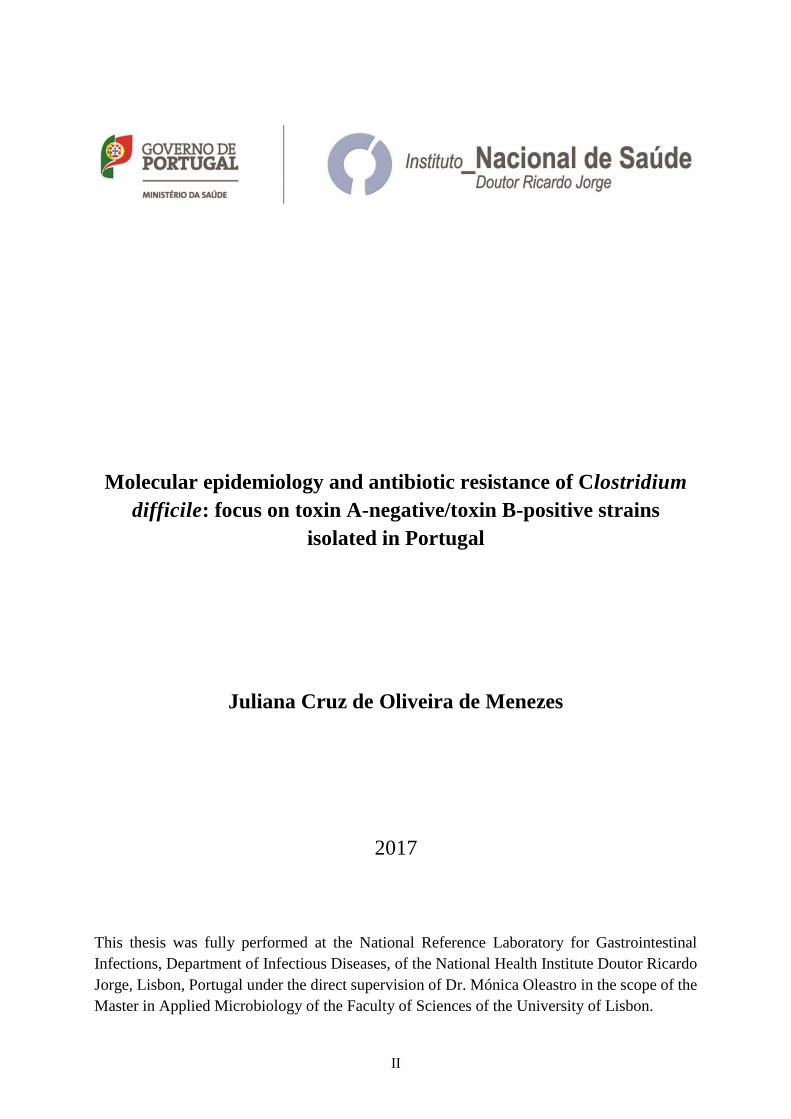
Abstract
Clostridium difficile affects patients in hospitals and communities worldwide, is responsible for
significant annual mortalities and represents a considerable economic burden on healthcare systems.
This bacterium might also be carried asymptomatically in the gut, potentially leading to ‘silent’ onward
transmission. Treatment has always been difficult, because the disease is both caused and resolved by
antibiotic intake. The two main C. difficile virulence factors are toxins A and B, both of which are
pro-inflammatory and enterotoxic in human intestine. Clinically relevant toxin A-negative/toxin
B-positive strains that cause diarrhoea and colitis in humans have been isolated with increasing
frequency worldwide, namely the multidrug resistant PCR-ribotype (RT) 017. Previous studies
documented changes in C. difficile infection (CDI) epidemiology associated with the rapid emergence
of antibiotic-resistant strains, highlighting the importance of antimicrobial susceptibility surveillance.
The present work describes epidemiological and antimicrobial susceptibility data of C. difficile
strains isolated in Portugal. A total of 378 C. difficile strains from 11 Portuguese hospital centres were
characterized regarding toxin profile and RT, and part of these strains were also evaluated for its
susceptibility to moxifloxacin, vancomycin, metronidazole, rifampicin and imipenem and determinants
of antimicrobial resistance. Multilocus variable tandem repeat analysis (MLVA) and whole genome
sequencing (WGS) analysis was also performed in a subgroup of epidemic, multidrug isolates.
Most of the isolates were toxigenic (91.3%), of which 94.2% had both toxins A and B and 25.8%
of them also had a binary toxin. Seventy-five different RTs were identified. RT027, RT014, RT106 and
RT017 were the most frequently isolated. There was no evidence of resistance to vancomycin among
the 183 tested strains, and reduced susceptibility to metronidazole was rare (2.2%). Resistance to
moxifloxacin was evident in multiple RTs, and were mainly from RTs positive for the three toxins,
RT027 (18/18), RT126 (8/9) and RT078 (6/12), except the RT017 (19/19), which is toxin
A-negative/toxin B-positive. All moxifloxacin-resistant strains exhibited a known mutation in GyrA
(Thr82Ile). Rifampicin resistance was found in 11.5% of the 183 strains tested, most from RT017
(19/19) but also in one strain from RT241 and other from RT043. Most rifampicin-resistant strains
harbour the previously described mutations in RpoB (His502Asn and Arg505Lys), although one
mutation, Ser507Leu, found alone in a resistant strain was not previously described. Of the 181 strains
belonging to 57 RTs tested for imipenem susceptibility, only strains from RT017 showing high level of
resistance to this antibiotic (MIC > 32 mg/L). The resistance determinants, ermB and tetM genes were
present in 34 (10.1%) and in 63 (20.12%) strains, respectively, being that 22 (6.5%) contained both
genes.
Twenty strains were toxin A-negative/toxin B-positive, 19 of them belonging to the well-known
emerging RT017, 11 from hospital A isolated in a short period of time suggesting that an outbreak have
occurred, the remaining eight from hospital B, isolated between 2016 and 2017, where this RT seems to
be endemic. Overall, these strains were multiresistant, presenting resistance to six of the 10 antibiotics
tested: moxifloxacin, rifampicin, imipenem, tetracycline, clindamycin and erythromycin (these two
belonging to the MLSB group), with high level of resistance. PCR screening of the resistance
determinants showed that all strains harboured the tetM gene, but only the eight strains from hospital B
were positive for ermB. Analysis by WGS revealed the presence of the ermG gene in the
ermB-negative/MLSB-resistant strains. This gene was found to be in a putative mobile element of 63 kb
exclusive of the hospital A clonal cluster. Transformation of a susceptible strain (C. difficile 630Δerm),
with a plasmid containing the ermG gene, proved that the presence of this gene provides high resistance
to clindamycin and erythromycin in C. difficile. Mutations in penicillin-binding proteins were also
observed in all imipenem resistant strains. Phylogenetic analysis of single nucleotide polymorphisms
Molecular epidemiology and antibiotic resistance of Clostridium difficile:
IV
(SNPs) of RT017 isolates collected from 2012 to 2017 revealed three clusters, each from a single hospi-
tal. Subtyping by MLVA was also applied to detect the clonal spread of C. difficile belonging to toxin
A-negative/toxin B-positive, and the results were overall similar with the WGS analysis. In addition,
74 SNPs variations were found among RT017 strains, namely in proteins involved in antimicrobial
resistance and in hypothetical proteins.
The current work gives a contribution to the knowledge of the molecular epidemiology and
resistance patterns of C. difficile in Portugal. The results presented herein alert to the presence of
multidrug resistant strains of RT017 in Portuguese hospitals in endemic and outbreak situations, and
indicates the need for adequate use of antimicrobial agents, especially carbapenems, whose resistance
was only observed among the strains of this emerging RT. The lineage of strains from RT017 appears
to be constantly evolving, acquiring new resistance determinants, which highlights the need for
continued epidemiological and antimicrobial surveillance.
The wide variety of RTs found suggests that there are other routes of transmission beyond
nosocomial transmission, raising concern about the epidemiological change in this pathogen. As such,
other potential sources, particularly in animals, which may also act as a reservoir for C. difficile and
antimicrobial resistance determinants, should be investigated in the future.
Finally, this study provides the basis for investigating important factors for the spread and
persistence of toxin A-negative/toxin B-positive strains, such as studies on the importance of proteins
that distinguish these strains, namely the hypothetical proteins.
Keywords: Clostridium difficile infection, molecular epidemiology, antimicrobial susceptibility,
resistance determinants, ribotype 017.
Focus on toxin A-negative/toxin B-positive strains isolated in Portugal
V
Resumo
Clostridium difficile é uma bactéria Gram-positiva, anaeróbia estrita e formadora de esporos,
que coloniza o cólon. A infeção por C. difficile (ICD) encontra-se principalmente associada ao meio
hospitalar e consumo recente de antibiótico, representando um fardo económico considerável para os
sistemas de saúde. No entanto, este quadro tem estado a alterar-se, verificando-se um acréscimo na
incidência de infeção em populações anteriormente pensadas em baixo risco e sem contacto prévio com
o ambiente hospitalar. Esta bactéria foi descoberta em 1935 como parte da microdiota intestinal normal
de recém-nascidos, no entanto, a sua importância em doenças em humanos só foi identificada mais tarde,
na sequência de múltiplos trabalhos conduzidos na década de 1970, quando esta patologia se tornou
mais frequente devido ao aumento no consumo de antibióticos.
O espectro da doença clínica varia de diarreia leve a megacólon tóxico, perfuração do colón e
morte. No entanto, esta bactéria também pode ser transportada de forma assintomática no intestino,
potencialmente levando a transmissão silenciosa. O tratamento da ICD sempre foi difícil, porque a
doença é causada e resolvida pela toma de antibióticos. Até a recente introdução da fidaxomicina, o
tratamento estava limitado a toma de metronidazol e vancomicina.
Nos últimos anos, a ICD surgiu como uma doença proeminente devido a um aumento súbito na
ocorrência de surtos, acompanhada por um aumento da gravidade da doença e mortalidade. Esta
alteração foi principalmente associada à disseminação de uma estirpe epidémica denominada ribotipo
(RT) 027, principalmente caracterizada por uma elevada resistência às fluoroquinolonas, cujo pico de
consumo coincidiu com o início da sua propagação epidémica. Ao mesmo tempo, porém, outras estirpes
também começaram a emergir com maior virulência.
Os dois principais fatores de virulência, e responsáveis pelo desenvolvimento da doença, são as
toxinas A e B, ambas pró-inflamatórias e enterotóxicas no intestino humano, algumas estirpes são
também caracterizadas pela produção de uma toxina binária. No entanto estirpes de C. difficile
clinicamente relevantes com fenótipo toxina A-negativa/toxina B-positiva que causam diarreia e colite,
têm sido isoladas com maior frequência em todo o mundo, nomeadamente pertencentes ao RT017,
resistentes a múltiplos antibióticos.
Embora os agentes antimicrobianos sejam fatores de grande relevância para o desenvolvimento
da ICD, a resistência da bactéria não é um pré-requisito para tal, podem sim antecipar uma rápida
disseminação dessas estirpes resistentes em ambiente hospitalar, uma vez que um fenótipo de resistência
pode conferir uma vantagem seletiva significativa dentro do ecossistema intestinal, facilitando o on-set
da ICD logo no início da toma de antibióticos. Deste modo, em termos de prevenção da ICD, é
imperativo limitar a propagação de estirpes resistentes de C. difficile, para tal, uma vigilância de
fenótipos e genótipos de resistência é essencial.
O presente trabalho tem como objetivo fornecer informação atualizada relativamente à
epidemiologia molecular e suscetibilidade antimicrobiana de estirpes de C. difficile isoladas de hospitais
Portugueses. Para esse fim, um total de 378 amostras de fezes (correspondentes a 374 pacientes
diagnosticados com ICD) provenientes de 11 centros hospitalares portugueses foram sujeitas a cultura
anaeróbica. As estirpes de C. difficile isoladas destas amostras foram caracterizadas em relação ao perfil
de toxinas e RTs; um subgrupo dessas estirpes foi também avaliado quanto à suscetibilidade à
moxifloxacina, vancomicina, metronidazol, rifampicina e imipenemo. Alguns determinantes de
resistência foram também estudados.
A maioria dos isolados eram toxigénicos (91,3%), dos quais 94,2% apresentavam as toxinas A
e B sendo que 25,8% desses também possuíam a toxina binária; no total 33 estirpes eram
Molecular epidemiology and antibiotic resistance of Clostridium difficile:
VI
não-toxigénicas. Foram identificados 75 RTs diferentes, sendo os RTs mais frequentes o RT027
(13,8%), RT014 (8,2%), RT106 (6,1%) e RT017 (5,1%). Não houve evidência de resistência à
vancomicina entre 183 estirpes toxigénicas testadas, a menor suscetibilidade ao metronidazol também
foi rara verificando-se em apenas 4 estirpes (2,2%). A resistência à moxifloxacina foi evidente em
múltiplos RTs, 55 de 183 (30,1%) estirpes testadas apresentaram resistência, pertencentes
principalmente a RTs positivos para três toxinas, RT027 (18/18), RT126 (8/9) e RT078 (6/12), exceto
o RT017 (19/19), toxina A-negativa/toxina B-positiva. Todas as estirpes resistentes à moxifloxacina
continham uma mutação já descrita na GyrA (Thr82Ile). A resistência à rifampicina foi encontrada em
11,5% das 183 estirpes testadas, na sua maioria do RT017 (19/19), mas também numa estirpe do RT241
e uma do RT043. A maioria das estirpes resistentes à rifampicina continham mutações na RpoB já
descritas (His502Asn e Arg505Lys); a mutação, Ser507Leu, descrita neste trabalho pela primeira vez,
foi a única encontrada numa estirpe resistente do RT043. De 181 estirpes, pertencentes a 57 RTs
diferentes, testadas quanto à suscetibilidade ao imipenemo, apenas as estirpes do RT017 (19/19)
apresentaram resistência a este antibiótico com alto nível de resistência (concentração mínima inibitória
≥ 32 mg/L). Os genes ermB e tetM, determinantes da resistência aos MLSB e à tetraciclina,
respetivamente, estavam presentes em 34 (10,1%) e 68 (20,12%) das estirpes testadas, respetivamente,
sendo que 22 estirpes (6,5%) continham ambos os genes; a maioria das estirpes (258/338) não continham
nenhum destes genes nomeadamente, as estirpes do RT027 (40/41). O gene catD, que confere resistência
ao cloranfenicol, não foi encontrado em nenhuma estirpe.
Foram identificadas 20 estirpes toxina A-negativa/toxina B-positiva, 19 delas pertencentes ao
RT emergente RT017, 11 do hospital A isoladas num curto período de tempo sugerindo que ocorreu um
surto, as oito restantes do hospital B, isoladas entre 2016 e 2017 onde esta estirpe parece ser endémica.
No geral, essas estirpes revelaram-se multi-resistentes, apresentando resistência a seis dos 10
antibióticos testados: moxifloxacina, rifampicina, imipenemo, tetraciclina, clindamicina e eritromicina
(os dois últimos pertencentes ao grupo MLSB). A triagem por PCR dos determinantes de resistência
mostrou que todas possuíam o gene tetM, mas apenas os oito isolados do hospital B eram positivos para
o ermB. A análise dos dados gerados por sequenciação total do genoma (WGS) das estirpes
ermB-negativas/MLSB-resistentes revelou a presença do gene ermG, pela primeira vez descrito em
estirpes clínicas toxigénicas de C. difficile. Este gene foi encontrado num putativo elemento móvel de
63 kb e somente nas estirpes associadas ao surto no hospital A. Foi possível verificar que este gene está
associado a um alto nível de resistência à clindamicina e eritromicina em C. difficile através da inserção
de um plasmídeo contendo o ermG em uma estirpe sensível (C. difficile 630Δerm). Outros genes
associados a resistências foram também encontrados neste elemento móvel, como os genes mefA e msrD
que conferem resistência aos macrólidos, e um gene que codifica para uma estreptogramina A
acetiltransferase que confere resistência à estreptogramina A em outras bactérias, no entanto este grupo
de antibióticos não foi aqui testado. Também foram observadas mutações em genes que codificam para
as proteínas de ligação à penicilina (PBPs) nas estirpes resistentes ao imipenemo; estas mutações
localizam-se próximo dos motivos conservados da PBP1 (Ala555Thr) e da PBP3 (Tyr721Ser).
A análise filogenética de polimorfismos de nucleotídeos únicos (SNPs) de isolados do RT017
obtidos de 2012 a 2017 revelou a existência de três grupos clonais, cada um com isolados de um único
hospital. A análise de multilocus variable number tandem repeat (MLVA) também foi aplicada para
detetar a disseminação clonal de C. difficile toxina A-negativa/toxina B-positiva, e os resultados foram
concordantes com a análise de WGS. Além disso, 74 variações de SNPs foram encontradas entre as
estirpes do RT017, compreendendo mutações sinónimas e não-sinónimas, nomeadamente em proteínas
envolvidas em resistência a antimicrobianos e em proteínas hipotéticas.
Em conclusão, embora grande parte do foco de C. difficile tenha sido o RT027, novas estirpes
virulentas continuam a emergir, o que requer consideração na vigilância futura. Os resultados aqui
Focus on toxin A-negative/toxin B-positive strains isolated in Portugal
VII
apresentados alertam para a presença de estirpes multirresistentes do RT017 nos hospitais portugueses,
sugerindo um potencial epidémico para as mesmas. A presença destas estirpes multirresistentes indica
a necessidade de uma utilização adequada dos agentes antimicrobianos, nomeadamente dos
carbapenemos, cuja resistência só foi observada entre as estirpes deste RT. A linhagem das estirpes do
RT017 parece estar em constante evolução, adquirindo novos determinantes de resistência, o que realça
a necessidade de uma vigilância epidemiológica e antimicrobiana contínua, bem como a imposição de
medidas de prevenção da transmissão de C. difficile no contexto hospitalar, nomeadamente através do
diagnóstico e tratamento de todos os pacientes com ICD e restrição de alguns antibióticos.
Contudo, a grande variedade de RTs encontrada no geral, sugere que existem outras vias de
transmissão para além da transmissão nosocomial, suscitando preocupação com a mudança na
epidemiologia deste agente patogénico. Como tal, outras potenciais fontes de infeção, nomeadamente
em animais, que também podem atuar como um reservatório de C. difficile e de determinantes da
resistência antimicrobiana, devem ser investigadas futuramente.
Por fim, este estudo proporciona a base para investigar fatores importantes para a disseminação
e persistência de estirpes toxina A-negativa/toxina B-positiva, como por exemplo estudos futuros sobre
a importância das proteínas que distinguem estas estirpes, nomeadamente as proteínas hipotéticas.
Palavras-chave: Infeção por Clostridium difficile, epidemiologia molecular, suscetibilidade aos
antimicrobianos, determinantes de resistência, ribotipo 017.
Molecular epidemiology and antibiotic resistance of Clostridium difficile:
VIII
Publications in the scope of this work
Menezes, J., Santos, A., Isidro, J., Paixão, P., Toscano, C., Oleastro, M. (2017, June). Study of
Clostridium difficile multiresistant strain from ribotype 017 in a hospital centre in Portugal. Poster
session presented at 2nd International Caparica Conference in Antibiotic Resistance, Costa da Caparica,
Portugal.
Menezes, J., Isidro, J., Serrano, M., Santos, A., Paixão, P., Toscano, C., Oleastro, M. (2017).
Outbreak of a Clostridium difficile multidrug resistant strain from ribotype 017 in a hospital centre in
Portugal. In preparation.
Focus on toxin A-negative/toxin B-positive strains isolated in Portugal
IX
Table of Contents
Acknowledgements ................................................................................................................................. I
Abstract ................................................................................................................................................ III
Resumo .................................................................................................................................................. V
Publications in the scope of this work ............................................................................................ VIII
List of Tables ......................................................................................................................................... X
List of Figures ....................................................................................................................................... X
List of Symbols and Abbreviations .................................................................................................... XI
1. Introduction ................................................................................................................................... 1
1.1. Clostridium difficile, the organisms and infection cycle ......................................................... 1
1.2. Virulence factors ..................................................................................................................... 2
1.3. Epidemiology changing and risk factors ................................................................................. 3
1.4. Toxin A-negative/toxin B-positive ribotypes .......................................................................... 4
1.5. Diagnosis and treatment .......................................................................................................... 4
1.6. Antimicrobial susceptibility and mechanisms of resistance .................................................... 5
2. Aims and objectives ....................................................................................................................... 8
3. Methods .......................................................................................................................................... 9
3.1. Samples and data collection .................................................................................................... 9
3.2. Clostridium difficile culture ..................................................................................................... 9
3.3. Antimicrobial susceptibility test .............................................................................................. 9
3.4. Molecular characterization of strains .................................................................................... 10
3.4.1. Identification, toxin profiling and ribotyping ................................................................ 10
3.4.2. Detection of genes and point mutations associated with antibiotic resistance .............. 10
3.4.3. Multilocus variable number tandem repeat analysis ..................................................... 12
3.5. Whole Genome Sequencing .................................................................................................. 13
3.6. Cloning and expression of ermG gene .................................................................................. 13
4. Results .......................................................................................................................................... 15
4.1. Toxin profile and ribotyping of Clostridium difficile clinical strains .................................... 15
4.2. Patients characteristics and clinical data ............................................................................... 16
4.3. Antimicrobial susceptibility .................................................................................................. 16
4.4. Determinants of antimicrobial resistance .............................................................................. 18
4.5. Study of toxin A-negative/toxin B-positive strains (RT017 and RT265) ............................. 19
4.5.1. Phylogenetic analysis by multilocus variable number tandem repeat analysis ............. 19
4.5.1. Whole genome sequencing analysis .............................................................................. 21
5. Discussion ..................................................................................................................................... 27
6. References .................................................................................................................................... 34
7. Supplementary information ....................................................................................................... 44
Molecular epidemiology and antibiotic resistance of Clostridium difficile:
X
List of Tables
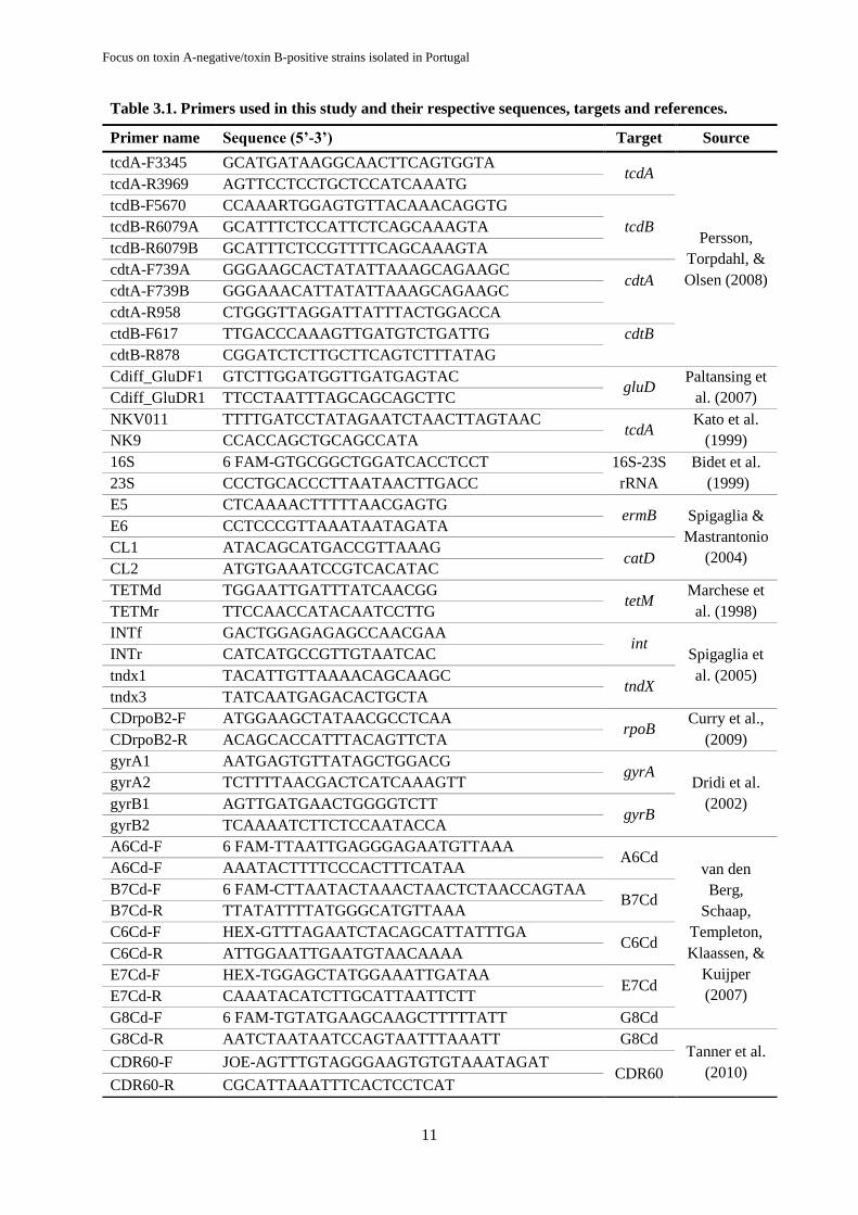
Table 3.1. Primers used in this study and their respective sequences, targets and references. ............. 11
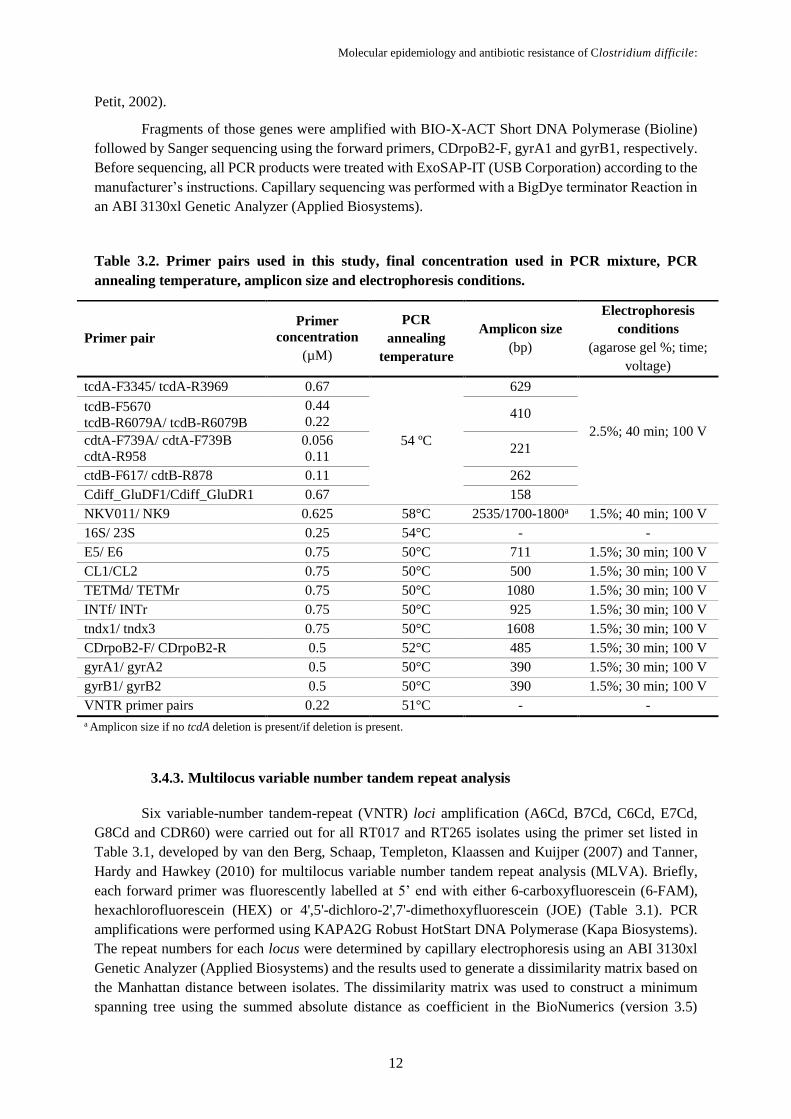
Table 3.2. Primer pairs used in this study, final concentration used in PCR mixture, PCR annealing
temperature, amplicon size and electrophoresis conditions. ................................................................. 12
Table 4.1. Toxin gene contents of 345 toxigenic Clostridium difficile clinical isolates. ...................... 15
Table 4.2. Demographic and clinical data of patients with positive culture for Clostridium difficile. . 16
Table 4.3. Antimicrobial susceptibility of Clostridium difficile clinical strains, determined by disk
diffusion. ............................................................................................................................................... 17
Table 4.4. Antimicrobial susceptibility of 19 Clostridium difficile RT017 strains determined by
E-test®. ................................................................................................................................................... 17
Table 4.5. Distribution of antibiotic resistance determinants according to PCR-ribotype. .................. 18
Table 4.6. Distribution of predicted amino acid substitutions in RpoB, GyrA and GyrB associated with
antibiotic resistance according to PCR-ribotypes. ................................................................................. 19
Table 4.7. Unique single nucleotide polymorphisms only found at RT017 strains from this work from
RT017 strains previously isolated. ........................................................................................................ 23
Table 4.8. Open reading frames and putative proteins that compose the mobile element carrying the
ermG gene. ............................................................................................................................................ 25
Supplementary table 1. Multilocus variable number tandem repeat analysis profiles for Clostridium
difficile tcdA-/tcdB+ isolates……………………………………………………………….…………..44
List of Figures
Figure 4.1. Distribution of PCR-ribotypes among 376 Clostridium difficile clinical strains isolated
from eleven Portuguese hospital centres during 2015-2017 ................................................................. 15
Figure 4.2 Minimum spanning tree of multilocus variable number tandem repeat analysis data for one
Clostridium difficile RT265 and 49 RT017 isolates .............................................................................. 20
Figure 4.3. Phylogeny of Clostridium difficile RT017 clinical isolates and genetic determinants of
antibiotic resistance based on WGS sequences ..................................................................................... 22
Figure 4.4. Genetic organization of the putative transposon in Clostridium difficile ........................... 25
Focus on toxin A-negative/toxin B-positive strains isolated in Portugal
XI
List of Symbols and Abbreviations
Δ deletion CO-HCFA Community onset health-care
facility-associated
% percentage DNA Deoxyribonucleic acid
°C Celsius degree EIA Enzyme immunoassay
bp base pair EUCAST
European Committee on
Antimicrobial Susceptibility
Testing
CFU colony-forming unit GDH Glutamate dehydrogenase
h hour HO-HCFA Healthcare facility–onset,
health-care facility-associated
kb kilobase HP Hypothetical protein
L liter HMW High-molecular-weight
min minute INSARJ Portuguese National Institute of
Health Dr. Ricardo Jorge
mg milligram MIC Minimum inhibitory
concentration
mL milliliter MLSB Macrolide, Lincosamide and
Streptogramin B
mm millimeter MLST Multilocus sequence typing
ng nanogram MLVA
Multilocus variable number
tandem repeat analysis
no. number NAAT Nucleic acid amplification tests
V volt ORF Open reading frame
μg microgram PBP Penicillin-binding protein
μL microliter PCR Polymerase chain reaction
μM micromolar QRDR Quinolone resistance-
-determining region
AMR Antibiotic resistance RNA Ribonucleic acid
BHI Brain Heart Infusion Agar RT PCR-ribotype
CA Community-associated SNP Single nucleotide polymorphism
CCNA Cell cytotoxicity neutralization
assay STRD
Summed tandem-repeat
difference
CDI Clostridium difficile infection TC Toxigenic culture
CDT Clostridium difficile transferase VNTR Variable-number tandem-repeat
BA Brucella Agar WGS Whole-genome sequencing

Focus on toxin A-negative/toxin B-positive strains isolated in Portugal
1
1. Introduction
1.1. Clostridium difficile, the organisms and infection cycle
Clostridium difficile is a Gram-positive bacterium, strict anaerobe, frequently implicated in
cases of nosocomial diarrhoea. The infection is caused by toxigenic strains, although there are naturally
non-toxin producing strains capable of colonizing their hosts (Martin, Monaghan, & Wilcox, 2016).
C. difficile was recently renamed as Clostridioides difficile (Lawson, Citron, Tyrrell, &
Finegold, 2016), however the still more familiar designation of C. difficile will be used throughout the
present document.
This bacterium was initially identified in 1935 as part of the normal gut microbiota of neonates
(Hall & O’Toole, 1935). The spectrum of C. difficile-associated disease varies from mild diarrhoea to
severe colitis, and it may lead to toxic megacolon, perforation of the colon, sepsis and death. Its
association with disease was not described until the 1970s following several studies. In those studies, a
causal effect of antibiotic exposure and gut diseases was also demonstrated as patients receiving
antibiotic treatment developed pseudomembranous colitis (Bartlett, Chang, Gurwith, Gorbach, &
Onderdonk, 1978; Tedesco, Barton, & Alpers, 1974).
C. difficile is a spore-forming bacterium, which, in turn, is able to tolerate extreme conditions
that the vegetative form cannot, assuming an essential role in the transmission of the pathogen, as well
as in environmental persistence. In this way, the spores are considered as the infectious form, since they
allow the survival of the microorganism in the acidic environment of the stomach of the host. Upon
reaching the duodenum, the spores get into contact with bile acids which along with glycine and some
other cofactors initiate germination (Howerton, Ramirez, & Abel-Santos, 2011; Janoir et al., 2013).
The organism is transmitted from person to person by the fecal-oral route. The cycle of C.
difficile infection (CDI) begins with an uncolonized individual who is exposed to bacterial spores. Then,
upon a disturbance of the normal gut microbiota, occurs the colonization and proliferation of C. difficile.
The infected individual will also be spore excretor, restarting the infectious cycle. The resulting
vegetative cells then penetrate the mucus layer and adhere to intestinal epithelial cells (Martin et al.,
2016).
Several factors can lead to modification of the intestinal microbiota, such as surgical incisions,
nasogastric tube feeding and medication intake. Antibiotic therapy is the major risk factor for CDI
because it disrupts the microbial community in the gut that forms a protective barrier, thus weakening
resistance to colonization (Dharmarajan, Sipalay, Shyamsundar, Norkus, & Pitchumoni, 2000; Manges
et al., 2010).
Pultz and Donskey (2005) have demonstrated that antibiotics can influence the growth of C.
difficile in different ways depending on its activity against the pathogen and the extent to which the
agent disrupts the gut microbiota; CDI can be virtually triggered by any antibiotic, as it promotes
damaged in the protective microbiota. However, if the strain of C. difficile is resistant to the antibiotic
taken it has an additional selective advantage. This is because, even in susceptible strains, the formation
of spores allows the bacteria to persist in the gut, germination only occurs when the drug is no longer
present at levels sufficient to inhibit growth and multiplication of C. difficile. But resistant strains can
germinate while the therapy is in progress, so they have an advantage in their spread due to the use of
antibiotics (Coia, 2009). In fact, mounting evidence suggests that antibiotic resistance is a key player in
the epidemiology of CDI (Gerding, 2004). Fluoroquinolone prescribing correlated highly with incidence
of CDI cause by fluoroquinolone-resistant strain, having been observed decline in incidence of CDI
Molecular epidemiology and antibiotic resistance of Clostridium difficile:
2
after restriction of this antibiotic (Dingle et al., 2017). Therefore, it is essential to monitor the resistance
profile of this microorganism.
1.2. Virulence factors
The main virulence factors associated with C. difficile infection are two large enteric toxins, A
and B toxins, whose action on the colonic intestinal epithelium are responsible for an intense
inflammatory response causing acute inflammation of the large intestine. Those toxins are encoded by
two genes, tcdA and tcdB situated on the C. difficile chromosome in a 19.6 kilobase (kb) pathogenicity
locus (PaLoc), that also contains three additional regulatory genes, tcdR, tcdC and tcdE (Kuehne et al.,
2010). TcdR is a sigma factor that facilitates the binding of RNA polymerase to the promoters of the
tcdA and tcdB genes, therefore promoting toxin production (Mani & Dupuy, 2001); TcdC is an
TcdR-specific anti-sigma factor that appears to negatively regulate toxin A and B expression through
disruption of TcdR interaction with RNA polymerase (Matamouros, England, & Dupuy, 2007); finally,
tcdE encodes a protein that has similarity to phage holin involved in toxin secretion (Govind & Dupuy,
2012).
Both toxins A and B catalyse the glucosylation and, hence, inactivation of Rho protein family
of GTPases in intestinal epithelial cells, mediating disorganization of the cell cytoskeleton and cell
death. Structurally these toxins have three other domains: i) the cysteine protease domain, involved in
the autocatalytic processing of the toxin protein that releases the enzymatic domain in the cytosol after
receptor-mediated endocytosis; ii) the translocation domain that mediates entry of the toxin into the
target cell cytoplasm; iii) the receptor-binding domain, responsible for binding to and the uptake into
the target cell (Just & Gerhard, 2004; Reineke et al., 2007).
Several studies tried to figure out the relative contributions of both toxins in the pathogenesis;
toxin A alone was first believed to induce most of the pathology observed after infection, but Lyras et
al. (2009) using isogenic tcdA and tcdB mutants in a hamster model showed that, in fact, toxin B is
essential for virulence while toxin A is dispensable. However the studies conduct by Kuehne et al. (2010)
using a similar method re-establish the importance of both toxins A and B in CDI, since they reveal to
be important mediators of in vitro cytotoxicity and in vivo virulence. Many factors differentiate both
studies which could lead to different interpretations. Although both groups used derived strains of C.
difficile 630, these strains had been separately passaged numerous times over a long period of time (>15
years) and there may be genetic differences between them, in fact, the strain used by Kuehne et al. (2010)
produces significantly more toxin A than the equivalent strain used in the earlier study (Carter, Awad,
Kelly, Rood, & Lyras, 2011; Lyras et al., 2009). The end points used to determine the in vivo
pathogenicity of the strains were also different in both studies. Death was an endpoint for Lyras et al.
(2009) study, by contrast a clinical scoring system comprising weight loss, behavioural changes, and
wet tail, followed by sacrifice of moribund and sick animals, was used by Kuehne et al. (2010). In any
case, both toxins are essential for disease because non-toxigenic strains are avirulent (Kuehne et al.,
2010).
Some strains of C. difficile only produce a functional toxin B, due to deletion in the repetitive
region of tcdA gene (tcdA-/tcdB+). Studies showed that, toxin B from those strains is able to modify
more substrates than wild-type toxin B from toxin A-positive/toxin B-positive (tcdA+/tcdB+) strains due
to polymorphisms in its tcdB gene, giving rise to altered glucosylation of Rho proteins and inducing a
cytopathic effect on cells (Chaves-Olarte et al., 1999; Rupnik, Kato, Grabnar, & Kato, 2003).
In addition to these two toxins, some strains produce a binary toxin known as C. difficile
transferase (CDT), encoded by two genes, cdtA and cdtB, and located in a different locus, the Binary
Focus on toxin A-negative/toxin B-positive strains isolated in Portugal
3
Toxin Encoding Locus. CDT induces redistribution of microtubules and formation of long
microtubule-based protrusions at the surface of intestinal epithelial cells that increase bacterial
adherence; this toxin also causes death of beneficial eosinophils that would play an important role in
promoting a healthy immune response during infection (Cowardin et al., 2016; Schwan et al., 2009).
Although the toxins are regarded as the primary virulence factors, other factors intervene in the
pathogenic process, notably to allow the establishment of the bacterium in its colonic niche, such as the
sporulation. Since C. difficile is a strict anaerobe, its virulence is linked to the ability to form spores,
being these able to survive in aerobic environments and resist to cleaning measures and disinfection
agents (Dawson, Valiente, Donahue, Birchenough, & Wren, 2011; Janoir et al., 2013).
1.3. Epidemiology changing and risk factors
CDI is primarily regarded as a healthcare-related disease that is most prominent among the
elderly population, representing a huge clinical and economic burden. This is most likely due to a
reduced immune status, concomitant comorbidities and several hospitalizations as these increases the
risk of exposure to the microorganism and clinical outcome (Dharmarajan et al., 2000). Also, this
population is more prompt to take antibiotics, which are the main risk for CDI. Use of drugs that reduce
gastric acid (such as proton pump inhibitors) also increase the likelihood of infection (Janarthanan,
Ditah, Adler, & Ehrinpreis, 2012). However, in the past years, the proportion of CDI occurring in
patients outside the hospital setting has increased, targeting also healthy young people and peripartum
women, two groups previously thought to be at low risk (Centers for Disease Control and Prevention,
2005; van Dorp et al., 2017). C. difficile is very widespread in the environment, and can be found in
diverse reservoirs such as soils, river waters, lakes, and also in various animal, such as animals in the
food chain (cattle, pigs) and also in pets (Álvarez-Pérez, Blanco, Harmanus, Kuijper, & García, 2017;
Thitaram et al., 2016); a genomic similarity has been found between strains isolated from humans and
from animals, mostly pigs (Janezic, Ocepek, Zidaric, & Rupnik, 2012; Knight, Squire, Collins, & Riley,
2017).
Changing C. difficile epidemiology is noted worldwide and is variable across countries,
particularly in relation to prevalent strains and its antimicrobial resistance pattern. Historically low rates
of severe disease and death (3% or less) may have led to an under estimation of the importance of C.
difficile-associated disease as a healthcare-associated infection (McDonald et al., 2005); nowadays, on
average, seven CDI cases occur for every 10,000-overnight patient stays in European hospitals, making
it the most commonly reported pathogen associated with hospital-associated gastrointestinal disease in
Europe (Davies et al., 2014). In the United States a population-based study by Lessa et al. (2015)
estimated almost a half million cases in 2011, with 83,000 recurrences and 29,300 deaths, while the
National and State Healthcare Associated Infections progress report, based on 2014 data, that CDI
incidence is higher than methicillin resistant Staphylococcus aureus (MRSA) incidence in U.S., and
although an 8% decrease in CDI has been observed between 2011 and 2014, U.S. hospitals reported a
significant increase in CDI between 2013 and 2014 (Centers for Disease Control and Prevention, 2016).
This shift in epidemiology has been noted from 2,000 onwards, when there was a further increase on
number and severity of CDI, with higher transmission rates and greater mortality, mainly due to the
spread of PCR-ribotype (RT) 027. This epidemic strain is characterized by high resistance to
fluoroquinolones, whose peak of consumption in North America coincides with the beginning of its
epidemic spread (McDonald et al., 2005; Muto et al., 2005; Pépin, Valiquette, & Cossette, 2005).
In Portugal the first CDI outbreak was reported in 2014 with a mortality rate of 11.3%, been
attributed to the RT027 that was also reported as the most frequent RT in the country, mainly due to
Molecular epidemiology and antibiotic resistance of Clostridium difficile:
4
healthcare facility-associated cases (Oleastro et al., 2014; Santos et al., 2016).
Although the increased rates of CDI have been primarily attributed to RT027, CDI cases
attributed to other emerging RTs, such as RT017 have been reported worldwide (Dobreva et al., 2013;
Drudy, Harnedy, Fanning, Hannan, & Kyne, 2007; Lee, Lee, Lee, Riley, & Kim, 2014).
1.4. Toxin A-negative/toxin B-positive ribotypes
A number of tcdA-/tcdB+ RTs have been identified to date (Rupnik et al., 2003), but the most
clinically significant is RT017 (also known as toxinotype VIII) that is been isolated with increasing
frequency worldwide.
The first RT017 outbreak reported occurred in a Canadian hospital in 1998 over a three month
period (Alfa et al., 2000). Since that time, cases associated with this RT have been described by other
groups. In Poland between 2004 and 2006 and in Bulgaria between 2008 and 2012, RT017 was identified
to be the most prevalent, infecting 44 and 28% of the patients, respectively (Dobreva et al., 2013; Pituch
et al., 2011).
This trend is also observed in Asia, where RT017 is one of the most prevalent RTs. A Korean
study, where sampling was performed over a 10-year period, showed that RT017 was the most dominant
strain type among hospitalized patients during 2004–2008. These results coincided with the initial use
of imipenem and moxifloxacin at the hospitals, making it feasible that the introduction of these drugs
would be related to the emergence of this RT, once the resistance levels among their RT017 isolates
were 12 and 85% for these drugs, respectively. Additionally, they were also resistant to clindamycin,
erythromycin and ciprofloxacin (Lee et al., 2014).
In other studies, the increased prevalence of RT017 strains has also been associated with
increased antibiotic resistance. Among the RT017 Polish isolates, 86.8% were resistant to imipenem
and 91% were erythromycin-resistant whereas only 7% of the other RTs with the most common toxin
profile (tcdA+/tcdB+) exhibited resistance to this antibiotic (Pituch et al., 2011). Isolates from an
outbreak in Ireland were all resistant to multiple antibiotics including macrolides, lincosamide and
newer classes of fluoroquinolones (Drudy et al., 2007). In Portugal RT017 is one of the most common
RTs, having been isolated endemic multidrug resistant strains in one hospital (Isidro et al., 2017; Santos
et al., 2016).
Although RT017 strains are negative for the toxin A encoding gene tcdA, they are capable of
causing the full spectrum of clinical illness usually associated with tcdA+/tcdB+ strains (ranging from
asymptomatic colonization through to severe disease), and cases of fulminant colitis have been
documented (Alfa et al., 2000; Arvand, Hauri, Zaiss, Witte, & Bettge-Weller, 2009; Drudy et al., 2007).
The C. difficile strain M68 is the representative strain of the RT017, and has the full genome
sequenced. The tcdA gene of this type strain contains a 1.8 kb deletion at the 3′ end and a nonsense
mutation at the amino acid 47 (He et al., 2010; Rupnik et al., 2003).
1.5. Diagnosis and treatment
There are many differences in CDI surveillance systems, infection detection and laboratory
diagnosis between and within countries (Davies et al., 2014). Two reference tests are available, the cell
cytotoxicity neutralization assay (CCNA) and toxigenic culture (TC). TC demonstrates the presence of
C. difficile isolates with the ability to produce toxin when cultured, while CCNA detects the presence of
toxin in stool samples. Both these techniques are however time consuming,
requiring specialized staff and equipment. Therefore, in hospital laboratories other standards tests are
Focus on toxin A-negative/toxin B-positive strains isolated in Portugal
5
often used for patients with diarrhoea, such as the toxin A/B commercial enzyme immunoassay (EIA)
that directly detects C. difficile toxins in stool samples or EIAs that detect the glutamate dehydrogenase
(GDH), an enzyme that is produced by both toxigenic and non-toxigenic strains. Nucleic acid
amplification tests (NAAT), such as PCR assay and loop-mediated isothermal amplification of DNA,
targeting toxin genes, are also used. However, all these tests have poor diagnostic accuracy as single
tests and are no longer recommended as a stand-alone test to diagnose CDI (Crobach et al., 2016;
Planche et al., 2013).
One strategy to overcome this problem is to use two- or three-step screening algorithms. In this
approach, an initial test is performed, which can be GDH EIA or nucleic acid amplification tests
(NAAT), and if negative, specimens are reported as negative with no further testing done. Positive
specimens must undergo additional testing for C. difficile by toxin A/B EIA and eventually for TC or
NAAT (in case first test was a GDH EIA test) when toxin A/B EIA result is negative (Crobach et al.,
2016).
Because many cases of CDI are hospital acquired, once CDI is diagnosed in a patient immediate
implementation of appropriate infection control measures such as appropriate handwashing, gloving and
improved environmental decontamination is mandatory to prevent further spread within the hospital
(Vonberg et al., 2008).
The mainstay of treatment is discontinuation of the offending antibiotic and administration of
metronidazole or vancomycin; oral metronidazole is indicated in cases of mild to moderate disease, oral
vancomycin for serious CDI, and combination therapy with enteral (oral/intracolonic) vancomycin and
intravenous metronidazole in cases of ileus or toxic megacolon. When oral treatment is not possible,
parenteral metronidazole is recommended, preferably combined with intracolonic or nasogastric
administration of vancomycin. For severe, complicated and recurrent CDI cases, treatment with
fidaxomicin is recommended, and has been found to be at least as effective as vancomycin and may be
more effective for achieving symptomatic cure, since also targets the spores of C. difficile. Faecal
transplantation following antibiotic treatment with an oral glycopeptide is reported to be highly effective
in treating multiple recurrent CDI. For relapsing CDI, rifaximin use is also proposed as a chaser therapy
(Debast et al., 2014; Nelson, Suda, & Evans, 2017).
1.6. Antimicrobial susceptibility and mechanisms of resistance
Apart from the classic virulence determinants, other factors, such as antibiotic resistance,
promotes increased pathogenicity and spread of C. difficile strains. This pathogen presents an enormous
capacity of adaptation to the environment, being able to become resistant to multiple drugs through
metabolic and genetic alterations, which includes mutations in specific genes and gene acquisition. In
fact, in a study conducted in 14 European countries, 47% of the strains analysed were resistant to at least
one antibiotic and multidrug resistant was observed among the predominantly RTs as result of
antibiotic-selective pressure (Spigaglia et al., 2011).
Antibiotic resistance may be generated by different mechanisms and the surveillance of those
different mechanisms and respective susceptibility patterns, for drugs frequently used in hospital
environment and in the community, are needed.
The resistance mechanism for vancomycin, the first-line of treatment for severe CDI, is still
unclear, with only a few strains reported with reduced susceptibility to this drug (Freeman et al., 2015).
In in vitro studies, C. difficile mutants exhibiting decreased susceptibility to vancomycin harboured
mutations leading to a P108L substitution in murG gene, responsible for the conversion of lipid I to lipid
II during the stage of peptidoglycan biosynthesis where the antibiotic acts, which could lead to antibiotic
Molecular epidemiology and antibiotic resistance of Clostridium difficile:
6
resistance. Nonsense mutation in an RNA/single-stranded DNA exonuclease (homologous locus tag
CD630_3659 in C. difficile 630), missense mutation causing a Asp244Tyr substitution in the β-subunit
of RNA polymerase (encoded by rpoC) and a single amino acid deletion in L-serine dehydrogenase
(encoded by sdaB) were also observed in those mutants (Leeds, Sachdeva, Mullin, Whitney Barnes, &
Ruzin, 2014).
C. difficile strains showing resistance to metronidazole are rare and when observed, the
phenotype has been transient and lost after exposure of the bacteria to freeze. Therefore, the
investigation of resistance mechanisms has proved to be difficult. Studies suggest that different C.
difficile strains resistant to metronidazole can show peculiar alterations in their enzymes or metabolic
pathways, such as decrease in the concentration of aminoacyl-tRNAs proteins, which likely result in
post translational variations of proteins relevant for metronidazole activation, increased expression of
proteins involved in DNA repair and variations in the electron transport, that could lead to alterations in
both the energy production and intracellular redox potential, which influences the efficiency of
metronidazole entry in the bacterial cell (Lynch et al., 2013; Moura et al., 2014).
Genetic mutations are involved in resistance to both fluoroquinolones and rifamycin. Resistance
to fluoroquinolones in C. difficile is due to point mutations in the quinolone-resistance determinant
region (QRDR) of GyrA or GyrB, the DNA gyrase subunits target of these agents. Several amino acid
substitutions have been identified in both GyrA and/or GyrB among moxifloxacin-resistant strains
(Spigaglia et al., 2008). Rifamycin’s are a class of antibiotics including rifampicin that inhibit the RNA
synthesis. Resistance to this group of antibiotics are associated with point mutations in rpoB, the gene
encoding for the β-subunit of RNA polymerase, thus changing the antibiotic target and preventing its
action (Curry et al., 2009). Resistance rates varied by geographic locations (Freeman et al., 2015),
however rifamycin agents have been proposed as therapy for treatment of relapsing CDI (Debast et al.,
2014).
Recently, mutations in two high-molecular-weight (HMW) penicillin-binding proteins (PBPs)
were suggest as being involved in imipenem resistance in C. difficile strains (Isidro et al., 2017). This
carbapenem antibiotic binds to PBPs inhibiting peptidoglycan synthesis, so these mutations would affect
the antibiotic binding to its targets. Reports of imipenem resistance have been attributed to RT017 strains
which have an additional PBP that could also play an important role in carbapenem resistance (Isidro et
al., 2017; Lee et al., 2014).
Resistance to macrolide-lincosamide-streptogramin B (MLSB) group of antibiotics is a common
phenotype in certain RTs, most notable among RT017, RT001, RT012, RT046, RT126 , RT053, and
RT078 (Freeman et al., 2015; Tenover, Tickler, & Persing, 2012). In C. difficile, MLSB is highly related
to the presence of the ermB gene carried by mobile elements (Spigaglia & Mastrantonio, 2004). The
ermB gene encodes for an rRNA methyltransferase that catalyse the methylation of specific adenine
residue in 23S rRNA, altering the antibiotic binding site (Leclercq, 2002). However, ermB was not
identified in all C. difficile clinical strains expressing high-level resistance to either erythromycin or
clindamycin, two antibiotics belonging to MLSB group (Nyc et al., 2016; Spigaglia et al., 2011).
Recently, a cfr-like gene named cfrC was described in C. difficile strains, and appears to be
associated with a transposon similar to Tn6218, a novel Tn916-like transposon (Candela, Marvaud,
Nguyen, & Lambert, 2017; Marín et al., 2015). This gene encodes for a 23S rRNA methyltransferase
which causes C-8 modification in A2503 located in the peptidyl transferase region of bacterial ribosome,
which modifies the antibiotic target. This mechanism confers resistance to several antimicrobial classes
comprising phenicol’s (as chloramphenicol), oxazolidones and pleuromutilins, lincosamides and
streptogramin A drugs (Candela et al., 2017).
Focus on toxin A-negative/toxin B-positive strains isolated in Portugal
7
Besides the cfr-like gene, chloramphenicol resistance is usually due to the presence of the catD
gene, which encodes for a chloramphenicol acetyltransferase (CAT) inactivating the antibiotic, and is
usually located in the Tn4451/Tn4453 transposons family (Spigaglia et al., 2011). This phenotype is not
very common in C. difficile clinical strains, varying between countries (Freeman et al., 2015).
Resistance to tetracycline in C. difficile is also related to a transposon-associated resistance
determinant, which is, in most of the cases, the tetM gene, encoding for a ribosomal protection protein
that binds to the ribosome and thereby preventing the drug from attaching to its binding site (Dönhöfer
et al., 2012; Spigaglia, Carucci, Barbanti, & Mastrantonio, 2005). In C. difficile, tetM is usually found
on conjugative Tn916-like elements and Tn5397 (Spigaglia et al., 2005). These two transposons
functionally differ from each other by its integration/excision module; Tn5397 harbours the tndX gene
that appears to encode a member of the large resolvase family of site-specific recombinases. The Tn916
harbours the the int and xis genes that are required for its excision and integration (Spigaglia et al., 2005;
H. Wang et al., 2000). The widespread use of tetracyclines during the past 60 years has led to an increase
in acquired tetracycline resistance determinants among clinically important pathogenic bacteria, making
important to study the susceptibility pattern of this antibiotic (Dönhöfer et al., 2012). In C. difficile,
resistance to tetracyclines varies widely between countries and with RT. In a European study published
in 2007, tetracycline resistance was observed in C. difficile in isolates from the UK and the Netherlands,
14.3% resistance in isolates from Poland, 21.4% resistance in isolates from Hungary, and 38.9%
resistance in isolates from Greece; compared to 9.2% across the entire study (Barbut et al., 2007).
Molecular epidemiology and antibiotic resistance of Clostridium difficile:
8
2. Aims and objectives
C. difficile is well recognized as the leading cause of antibiotic-associated diarrhoea, having a
significant impact in both healthcare and community settings. In view of this situation, knowledge about
the circulation dynamics of different strains in Portugal is of great importance. Modern technologies,
such as whole-genome sequencing and multilocus variable number tandem repeat analysis, are helping
to track C. difficile transmission.
In order to contribute to the knowledge about the current paradigm of CDI in the country, it was
intended with this work to accomplish the following objectives:
(i) Identify and characterize strains isolated from Portuguese hospitals, through phenotypic and
molecular techniques, namely the identification of predominant RTs and detection of main
virulence factors;
(ii) Evaluate antimicrobial susceptibility patterns, identify mechanisms of resistance and investigate
possible associations of specific genotypes with antimicrobial resistance phenotypes;
(iii) Evaluate the frequency of toxin A-negative/toxin B-positive strains and their epidemiological
characteristics;
(iv) Perform a molecular study in order to unravel relevant features of this group of strains as well
evaluate clonal relationships;
(v) Correlate genotypic and phenotypic data regarding antimicrobial resistance through a whole
genome approach.
Focus on toxin A-negative/toxin B-positive strains isolated in Portugal
9
3. Methods
3.1. Samples and data collection
Between October 2015 and June 2017, the National Reference Laboratory for Gastrointestinal
infections (NRL_GI) from the Portuguese National Institute of Health Dr. Ricardo Jorge (INSARJ)
received, under the scope of CDI surveillance, stool samples from patients with suspected CDI from 11
Portuguese hospital centres. The samples had positive enzyme immunoassay (EIA) test for the presence
of toxin A/toxin B and/or positive EIA for GDH. Each sample was accompanied by a short questionnaire
on the patient’s clinical and epidemiological data, including age, sex, place (hospital or community) and
time of symptoms onset. CDI cases were classified as healthcare facility–onset healthcare
facility-associated (HO-HCFA) when the symptoms onset occurred > 48 h after admission to a
health-care facility, community-associated (CA) when the onset of symptoms occurred outside a
health-care facility or less than 48 h before admission, and unknown when no data was available (Cohen
et al., 2010). A total of 455 samples were studied.
3.2. Clostridium difficile culture
Faecal samples were subject to alcohol shock treatment: 1 g of stool was mixed with 1 mL of
ethanol 100%; for liquid stools, 1 mL was mixed with 1 mL of ethanol, and left to rest for 60 min at
room temperature. Then, 1-2 drops of the faeces/ethanol suspension were inoculated on a selective
chromogenic medium chromID™ C. difficile agar (CDIF) (bioMérieux) for the growth of C. difficile.
The CDIF plates were examined after incubation at 37 °C for 48 h, under anaerobic atmosphere
generated by AnoxomatTM (MART Microbiology BV) with a catalyst.
Suspected C. difficile colonies were selected based on colour and morphology (5-7 mm in
diameter, flat with a filamentous edge); for each isolate one colony was cultured onto a Brain Heart
Infusion (BHI) agar plate and its identity confirmed by PCR targeting the glutamate dehydrogenase gene
(gluD gene).
3.3. Antimicrobial susceptibility test
Antimicrobial susceptibility testing was performed for the antibiotics moxifloxacin (5 g),
vancomycin (5 g), metronidazole (5 g) and rifampicin (5 g) by disk diffusion (Oxoid). For a
subgroup of strains, the minimum inhibitory concentration (MIC) of imipenem, chloramphenicol,
tetracycline, tigecycline, clindamycin and erythromycin were determined by diffusion gradient, using
Etest® strips (bioMérieux) according to the manufacturer’s instructions. Briefly, one colony from each
cultured sample was selected and subcultured on BHI plates and incubated anaerobically at 37 °C for
24 h. Overnight cultures of C. difficile strains were suspended in Schaedler Broth with Vitamin K1 (BD,
BBL™) to a density equivalent to 1.0 McFarland (~3x10⁸ CFU/mL), and then a sterile cotton swab was
used to spread the inoculum evenly onto the Brucella Agar (BA) supplemented with 5% Sheep Blood,
Hemin and Vitamin K1 (BD, BBL™) plates. After the surface was completely dry, antibiotic disks or
Etest® strips were applied to each plate. The plates were incubated anaerobically at 37 °C for 24 h for
the ones containing the disks and the imipenem Etest® strips, and for 48 h for the remaining plates
containing Etest® strips. For antibiotic disks, the following zone diameter ecological cut-off proposed
by Erikstrup et al. (2012) was used: ≥ 20 mm, ≥ 23 mm and ≥ 19 mm, to moxifloxacin, vancomycin,
metronidazole respectively. For rifampicin, and in order to make the transition to disk diffusion, a
comparative test was performed using both antibiotic disks (5 g) and Etest® strips, and strains were
categorized as susceptible if inhibition zone diameter was ≥ 30 mm, corresponding to MIC < 0.004
mg/L, according to the European Committee on Antimicrobial Susceptibility Testing (EUCAST)
Molecular epidemiology and antibiotic resistance of Clostridium difficile:
10
guidelines (2017, http://www.eucast.org/clinical_breakpoints/); resistant strains all had inhibition
zone diameter ≤ 6 mm, corresponding to MIC > 32 mg/L.
MICs breakpoints were defined according to Freeman et al. (2015) for: imipenem (≥ 16 mg/L),
chloramphenicol (≥ 32 mg/L), tigecycline (> 0.25 mg/L) and for clindamycin (≥ 8 mg/L), and according
to Clinical and Laboratory Standards Institute (CLSI) breakpoint guideline (M11-A7, 2012) for
tetracycline (≥ 16 mg/L) and erythromycin (≥ 8 mg/L). To optimize growth of C. difficile, all necessary
medium were reduced for 18–24 h in an anaerobic atmosphere before use (CLSI, 2012). The preparation
of inoculum, inoculation and incubation was accomplished within 30 min in order to avoid prolonged
exposure to aerobic atmosphere.
3.4. Molecular characterization of strains
DNA was extracted from a 24 h culture using the NucliSENS® easyMAG® (bioMérieux)
automated system (bioMérieux) according to the manufacturer’s instructions. In brief, samples were
placed in the sample vessel and were followed by lysis incubation. Magnetic silica was added to the
samples followed by automatic extraction with the Generic 2.0.1 protocol.
The primers used in this study, their respective sequences, targets and references are presented
in Table 3.1. The primers’ final concentrations used in the PCR mixtures, the expected PCR product
length, annealing temperature and gel electrophoresis conditions are all described in Table 3.2.
3.4.1. Identification, toxin profiling and ribotyping
The extracted DNA was used for amplification of tcdA, tcdB, cdtA, cdtB, toxin-encoding genes
as well as of for the gluD gene of C. difficile in a single multiplex PCR by using the respective primers
listed in Table 3.1. In order to investigate deletions in tcdA gene repeating regions, an additional PCR
was performed, using the primer pair NKV011/NK9, resulting in a 2535 bp amplicon when no deletion
is present, and 1700-1800 bp amplicon when deletions are present.
Multiplex PCR was carried out using HotStarTaq Master Mix (Qiagen) while PCR targeting
only the tcdA gene was carried out using RANGER DNA Polymerase (Bioline).
PCR ribotyping of isolates was determined using the primer set described by Bidet, Frédéric,
Valérie and Petit (1999) and a capillary gel electrophoresis-based approach as described by Indra et al.
(2008). The reaction was performed using KAPA2G Robust HotStart DNA Polymerase (Kapa
Biosystems) and employed a 16S rRNA primer labelled at the 5’ end with 6-carboxyfluorescein
(6-FAM). The PCR RTs were determined by uploading the data to the online database Webribo
(https://webribo.ages.at/).
3.4.2. Detection of genes and point mutations associated with antibiotic resistance
The primer pairs E5-E6, CL1-CL2 and TETMd/TETMr were used to identify the presence of
ermB, catD and tetM gene, respectively. For identification of transposons carrying the tetM, the int and
the tndX genes were used as markers of Tn916 and Tn5397 elements, respectively, according to
Spigaglia et al. (2005).
BioTaq DNA polymerase (Bioline) was used for screening of these genes and transposons
carrying the tetM.
Point mutations associated with resistance to rifampicin and moxifloxacin were screened on
rpoB and on gyrA/gyrB genes, respectively (Curry et al., 2009; Dridi, Tankovic, Burghoffer, Barbut, &
Focus on toxin A-negative/toxin B-positive strains isolated in Portugal
11
Table 3.1. Primers used in this study and their respective sequences, targets and references.
Primer name Sequence (5’-3’) Target Source
tcdA-F3345 GCATGATAAGGCAACTTCAGTGGTA tcdA
Persson,
Torpdahl, &
Olsen (2008)
tcdA-R3969 AGTTCCTCCTGCTCCATCAAATG
tcdB-F5670 CCAAARTGGAGTGTTACAAACAGGTG
tcdB tcdB-R6079A GCATTTCTCCATTCTCAGCAAAGTA
tcdB-R6079B GCATTTCTCCGTTTTCAGCAAAGTA
cdtA-F739A GGGAAGCACTATATTAAAGCAGAAGC cdtA
cdtA-F739B GGGAAACATTATATTAAAGCAGAAGC
cdtA-R958 CTGGGTTAGGATTATTTACTGGACCA
ctdB-F617 TTGACCCAAAGTTGATGTCTGATTG cdtB
cdtB-R878 CGGATCTCTTGCTTCAGTCTTTATAG
Cdiff_GluDF1 GTCTTGGATGGTTGATGAGTAC gluD
Paltansing et
al. (2007) Cdiff_GluDR1 TTCCTAATTTAGCAGCAGCTTC
NKV011 TTTTGATCCTATAGAATCTAACTTAGTAAC tcdA
Kato et al.
(1999) NK9 CCACCAGCTGCAGCCATA
16S 6 FAM-GTGCGGCTGGATCACCTCCT 16S-23S
rRNA
Bidet et al.
(1999) 23S CCCTGCACCCTTAATAACTTGACC
E5 CTCAAAACTTTTTAACGAGTG ermB Spigaglia &
Mastrantonio
(2004)
E6 CCTCCCGTTAAATAATAGATA
CL1 ATACAGCATGACCGTTAAAG catD
CL2 ATGTGAAATCCGTCACATAC
TETMd TGGAATTGATTTATCAACGG tetM
Marchese et
al. (1998) TETMr TTCCAACCATACAATCCTTG
INTf GACTGGAGAGAGCCAACGAA int
Spigaglia et
al. (2005)
INTr CATCATGCCGTTGTAATCAC
tndx1 TACATTGTTAAAACAGCAAGC tndX
tndx3 TATCAATGAGACACTGCTA
CDrpoB2-F ATGGAAGCTATAACGCCTCAA rpoB
Curry et al.,
(2009) CDrpoB2-R ACAGCACCATTTACAGTTCTA
gyrA1 AATGAGTGTTATAGCTGGACG gyrA
Dridi et al.
(2002)
gyrA2 TCTTTTAACGACTCATCAAAGTT
gyrB1 AGTTGATGAACTGGGGTCTT gyrB
gyrB2 TCAAAATCTTCTCCAATACCA
A6Cd-F 6 FAM-TTAATTGAGGGAGAATGTTAAA A6Cd
van den
Berg,
Schaap,
Templeton,
Klaassen, &
Kuijper
(2007)
A6Cd-F AAATACTTTTCCCACTTTCATAA
B7Cd-F 6 FAM-CTTAATACTAAACTAACTCTAACCAGTAA B7Cd
B7Cd-R TTATATTTTATGGGCATGTTAAA
C6Cd-F HEX-GTTTAGAATCTACAGCATTATTTGA C6Cd
C6Cd-R ATTGGAATTGAATGTAACAAAA
E7Cd-F HEX-TGGAGCTATGGAAATTGATAA E7Cd
E7Cd-R CAAATACATCTTGCATTAATTCTT
G8Cd-F 6 FAM-TGTATGAAGCAAGCTTTTTATT G8Cd
G8Cd-R AATCTAATAATCCAGTAATTTAAATT G8Cd Tanner et al.
(2010) CDR60-F JOE-AGTTTGTAGGGAAGTGTGTAAATAGAT
CDR60 CDR60-R CGCATTAAATTTCACTCCTCAT
Molecular epidemiology and antibiotic resistance of Clostridium difficile:
12
Petit, 2002).
Fragments of those genes were amplified with BIO-X-ACT Short DNA Polymerase (Bioline)
followed by Sanger sequencing using the forward primers, CDrpoB2-F, gyrA1 and gyrB1, respectively.
Before sequencing, all PCR products were treated with ExoSAP-IT (USB Corporation) according to the
manufacturer’s instructions. Capillary sequencing was performed with a BigDye terminator Reaction in
an ABI 3130xl Genetic Analyzer (Applied Biosystems).
Table 3.2. Primer pairs used in this study, final concentration used in PCR mixture, PCR
annealing temperature, amplicon size and electrophoresis conditions.
3.4.3. Multilocus variable number tandem repeat analysis
Six variable-number tandem-repeat (VNTR) loci amplification (A6Cd, B7Cd, C6Cd, E7Cd,
G8Cd and CDR60) were carried out for all RT017 and RT265 isolates using the primer set listed in
Table 3.1, developed by van den Berg, Schaap, Templeton, Klaassen and Kuijper (2007) and Tanner,
Hardy and Hawkey (2010) for multilocus variable number tandem repeat analysis (MLVA). Briefly,
each forward primer was fluorescently labelled at 5’ end with either 6-carboxyfluorescein (6-FAM),
hexachlorofluorescein (HEX) or 4',5'-dichloro-2',7'-dimethoxyfluorescein (JOE) (Table 3.1). PCR
amplifications were performed using KAPA2G Robust HotStart DNA Polymerase (Kapa Biosystems).
The repeat numbers for each locus were determined by capillary electrophoresis using an ABI 3130xl
Genetic Analyzer (Applied Biosystems) and the results used to generate a dissimilarity matrix based on
the Manhattan distance between isolates. The dissimilarity matrix was used to construct a minimum
spanning tree using the summed absolute distance as coefficient in the BioNumerics (version 3.5)
Primer pair
Primer
concentration
(µM)
PCR
annealing
temperature
Amplicon size
(bp)
Electrophoresis
conditions
(agarose gel %; time;
voltage)
tcdA-F3345/ tcdA-R3969 0.67
54 ºC
629
2.5%; 40 min; 100 V
tcdB-F5670
tcdB-R6079A/ tcdB-R6079B
0.44
0.22 410
cdtA-F739A/ cdtA-F739B
cdtA-R958
0.056
0.11 221
ctdB-F617/ cdtB-R878 0.11 262
Cdiff_GluDF1/Cdiff_GluDR1 0.67 158
NKV011/ NK9 0.625 58°C 2535/1700-1800a 1.5%; 40 min; 100 V
16S/ 23S 0.25 54°C - -
E5/ E6 0.75 50°C 711 1.5%; 30 min; 100 V
CL1/CL2 0.75 50°C 500 1.5%; 30 min; 100 V
TETMd/ TETMr 0.75 50°C 1080 1.5%; 30 min; 100 V
INTf/ INTr 0.75 50°C 925 1.5%; 30 min; 100 V
tndx1/ tndx3 0.75 50°C 1608 1.5%; 30 min; 100 V
CDrpoB2-F/ CDrpoB2-R 0.5 52°C 485 1.5%; 30 min; 100 V
gyrA1/ gyrA2 0.5 50°C 390 1.5%; 30 min; 100 V
gyrB1/ gyrB2 0.5 50°C 390 1.5%; 30 min; 100 V
VNTR primer pairs 0.22 51°C - -
a Amplicon size if no tcdA deletion is present/if deletion is present.
Focus on toxin A-negative/toxin B-positive strains isolated in Portugal
13
software (Applied Maths, Sint-Martens-Latem, Belgium). The minimum spanning tree was built with
VNTR information of RT017 strains from INSARJ database.
For each locus, the summed tandem-repeat difference (STRD) were also calculated and MLVA
types associated based on the smallest STRDs: isolates with STRD ≤ 10 were considered genetically
related regardless the number of different loci; the clonal complexes were defined by a STRD ≤ 2
between two isolates that were either single or double locus variants of each other.
3.5. Whole Genome Sequencing
Whole genome sequencing (WGS) was performed for eight RT017 strains and one RT265 strain
for genomic comparison between the two groups.
For each strain, DNA sample was extracted using the Isolate II Genomic DNA kit (Bioline, UK)
according to the manufacturer’s protocol, and further quantified using a fluorometric method (Qubit
dsDNA HS Assay Kit). For each isolate, paired-end (2 x 250 bp) WGS was carried out in a MiSeq
equipment (Illumina Inc., CA, USA) available at INSARJ. FastQC version 0.11.5 was subsequently
applied to check the reads’ quality before and after quality improvement measures carried out using
Trimmomatic version 0.36. Then, high-quality processed reads were subjected to de novo genome
assembly using SPAdes (version 3.10.1). Draft genome sequences were analysed in order to: i) perform
genome annotation to identify functional properties and biological roles of genes using RAST server
version 2.0 (http://rast.nmpdr.org/); ii) perform in silico Multilocus Sequence Typing (MLST) and allele
determination of well-known virulence-associated genes, using the online platform available at
PUBMLST (http://pubmlst.org/); iii) search for the presence of putative antimicrobial resistance (AMR)
genes using both CARD (https://card.mcmaster.ca/) and ResFinder version 2.1
(http://www.genomicepidemiology.org/) database; iv) identify potential dissimilarities enrolling AMR
genes; and v) verify the genomic context of potential horizontally-transferable AMR genes. Open
reading frames (ORFs) visualisation was conducted using SnapGene Viewer (version 4.0.5) software.
Sequence reads (after quality improvement using Trimmomatic) of all C. difficile RT017 clinical
isolates were mapped to the published genome of the RT017 reference strain, C. difficile M68 (GenBank
accession number: NC_017175) using Snippy v3.1 tool (https://github.com/tseemann/snippy); base-pair
calls at each position in the genome were used to identify single nucleotide polymorphisms (SNP)
between the clinical isolates and the reference genome. MEGA5 software
(http://www.megasoftware.net) was applied to calculate matrices of nucleotide distances. RAxML (NG
v0.4, beta) was applied to perform maximum likelihood phylogenetic reconstructions over the obtained
core-genome SNP alignment (enrolling 74 variant sites) by using the general time-reversible model
(GTR+G) with bootstrapping (1000 replicates). Microreact platform (https://microreact.org/) was used
to visualize the phylogenetic three linked to antimicrobial resistance data.
3.6. Cloning and expression of ermG gene
In order to verify if the presence of ermG gene by itself leads to MLSB resistance, a multicopy
plasmid carrying the gene was inserted in C. difficile 630∆erm, and antimicrobial susceptibility of the
transformed strains were evaluated.
To express the ermG gene under the control of a Ptet inducible promoter, the ermG gene with
its ribosome-binding site (positions -12 to +793 from the translational start codon) was amplified using
primers ermG850D (GGATTCGGAGAGGTTATAATGAACAAAG) and ermG1660R
(ATAGTTTAGCGGCCGCATTTTAACTTATGCTACCCTACC). The resulting PCR product (810
bp) was digested with EcoRI and NotI and cloned into pAM25, a derivative of pRPF185 lacking the
Molecular epidemiology and antibiotic resistance of Clostridium difficile:
14
gusA gene (Fagan & Fairweather, 2011). Using the Escherichia coli HB101 (RP4) strain containing
either pRPF185 or pMS534 (pAM25-ermG), these plasmids were transferred by conjugation into C.
difficile 630∆erm.
Transformable strains were incubated at 37°C for 24 h in BHI plates with 15 µg/mL of
thiamphenicol. Overnight cultures were suspended in Schaedler Broth with Vitamin K1 (BD, BBL™),
to a density of 1.0 McFarland and mixture with 250 ng/mL of anhydrotetracycline and 15 µg/mL of
thiamphenicol; 500 µL of the mixture were scattered onto BA plates, and the excess suspension was
aspirated with a sterile disposable Pasteur pipette. MIC determination of erythromycin and clindamycin
was performed using Etest® strips (bioMérieux). A C. difficile 630∆erm strain carrying the multicopy
plasmid, but without the ermG insertion was used as control.
The Sanger sequencing, capillary electrophoresis and WGS were performed at the Unidade de
Tecnologia e Inovação, of the Departamento de Genética Humana.
The construction of ermG inducible strains was carried at the Microbial Development Lab,
Instituto de Tecnologia Química e Biológica - António Xavier, Universidade Nova de Lisboa, Portugal.
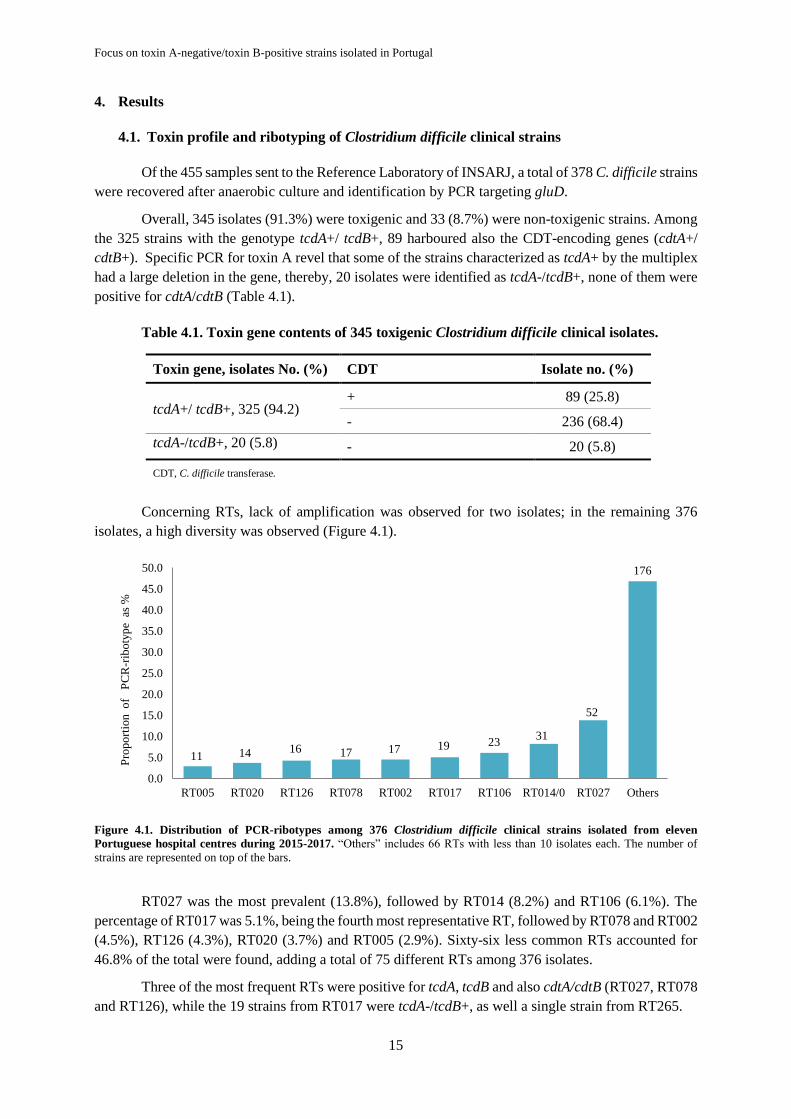
Focus on toxin A-negative/toxin B-positive strains isolated in Portugal
15
4. Results
4.1. Toxin profile and ribotyping of Clostridium difficile clinical strains
Of the 455 samples sent to the Reference Laboratory of INSARJ, a total of 378 C. difficile strains
were recovered after anaerobic culture and identification by PCR targeting gluD.
Overall, 345 isolates (91.3%) were toxigenic and 33 (8.7%) were non-toxigenic strains. Among
the 325 strains with the genotype tcdA+/ tcdB+, 89 harboured also the CDT-encoding genes (cdtA+/
cdtB+). Specific PCR for toxin A revel that some of the strains characterized as tcdA+ by the multiplex
had a large deletion in the gene, thereby, 20 isolates were identified as tcdA-/tcdB+, none of them were
positive for cdtA/cdtB (Table 4.1).
Table 4.1. Toxin gene contents of 345 toxigenic Clostridium difficile clinical isolates.
Concerning RTs, lack of amplification was observed for two isolates; in the remaining 376
isolates, a high diversity was observed (Figure 4.1).
Figure 4.1. Distribution of PCR-ribotypes among 376 Clostridium difficile clinical strains isolated from eleven
Portuguese hospital centres during 2015-2017. “Others” includes 66 RTs with less than 10 isolates each. The number of
strains are represented on top of the bars.
RT027 was the most prevalent (13.8%), followed by RT014 (8.2%) and RT106 (6.1%). The
percentage of RT017 was 5.1%, being the fourth most representative RT, followed by RT078 and RT002
(4.5%), RT126 (4.3%), RT020 (3.7%) and RT005 (2.9%). Sixty-six less common RTs accounted for
46.8% of the total were found, adding a total of 75 different RTs among 376 isolates.
Three of the most frequent RTs were positive for tcdA, tcdB and also cdtA/cdtB (RT027, RT078
and RT126), while the 19 strains from RT017 were tcdA-/tcdB+, as well a single strain from RT265.
Toxin gene, isolates No. (%) CDT Isolate no. (%)
tcdA+/ tcdB+, 325 (94.2) + 89 (25.8)
- 236 (68.4)
tcdA-/tcdB+, 20 (5.8) - 20 (5.8)
CDT, C. difficile transferase.
1911
52
17
3123
16 17
176
14
0.0
5.0
10.0
15.0
20.0
25.0
30.0
35.0
40.0
45.0
50.0
RT005 RT020 RT126 RT078 RT002 RT017 RT106 RT014/0 RT027 Others
Pro
po
rtio
n o
f
PC
R-r
ibo
typ
e a
s %
Molecular epidemiology and antibiotic resistance of Clostridium difficile:
16
The distribution of the RTs varied widely between and within hospitals. However, 11 of the 19
isolates from RT017 were detected in 11 patients hospitalized in the same ward of the same hospital
(herein designated as Hospital A), with the following temporal distribution: 1 patient in January, 6
patients in February, 1 in March, 1 in April, 1 in June and 1 in July, all in 2016, suggesting the occurrence
of nosocomial transmission.
4.2. Patients characteristics and clinical data
The 378 C. difficile strains were isolated from 374 different patients, as four patients were
colonized by two different strains (two different RTs). Table 4.2 summarize the relevant demographic
and clinical data of the patients.
The ratio female:male was 1.3 and the majority of CDI cases (70.1%) was from older patients
(age ≥ 65 years).
Most cases corresponded to hospital-acquired CDI (63.1%) but there were 90 cases associated
with the community setting (24.1%) and 48 cases (12.8%) classified as unknown due to lack of
information.
Table 4.2. Demographic and clinical data of patients with positive culture for Clostridium
difficile.
Patients characteristics no. (%)
Gender (n = 371) Males 158 (42.6)
Age (n = 264)
Mean age in years 69
< 18 15 (5.7)
18 - 65 years 64 (24.2)
≥ 65 years 185 (70.1)
CDI classification (n = 374)
HO-HCFA 236 (63.1)
CA 90 (24.1)
Unknown 48 (12.8)
Antibiotics exposure within three months
before CDI diagnosis (n = 374)
Yes 111 (29.7)
No 30 (8)
Unknown 233 (66.3)
HO-HCFA, healthcare facility–onset health-care facility-associated;
CA, community associated.
Regarding previous antibiotherapy, at least 29.7% of the patients received antibiotics within
three months before CDI diagnosis, being that 8% had not been exposed to this risk factor. However
most of the patients (66.3%) had no data available.
4.3. Antimicrobial susceptibility
The antimicrobial susceptibility for moxifloxacin, vancomycin, metronidazole and rifampicin
was investigated for 183 toxigenic strains (Table 4.3).
Overall, 30.1% (55/183) were moxifloxacin-resistant. All the strains from the frequently
detected RT027 and RT017 were resistant to moxifloxacin, while for others an important frequency was
observed, such as 89% (8/9) for RT126, 50% (6/12) for RT078.
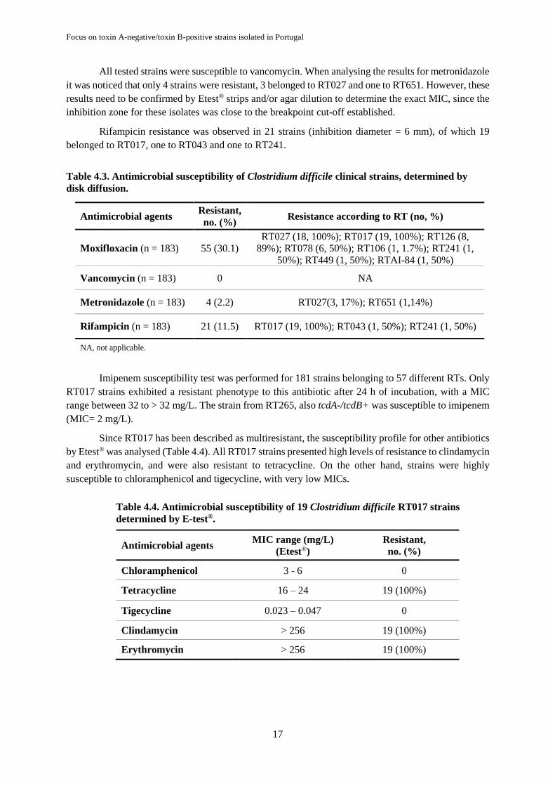
Focus on toxin A-negative/toxin B-positive strains isolated in Portugal
17
All tested strains were susceptible to vancomycin. When analysing the results for metronidazole
it was noticed that only 4 strains were resistant, 3 belonged to RT027 and one to RT651. However, these
results need to be confirmed by Etest® strips and/or agar dilution to determine the exact MIC, since the
inhibition zone for these isolates was close to the breakpoint cut-off established.
Rifampicin resistance was observed in 21 strains (inhibition diameter = 6 mm), of which 19
belonged to RT017, one to RT043 and one to RT241.
Table 4.3. Antimicrobial susceptibility of Clostridium difficile clinical strains, determined by
disk diffusion.
Imipenem susceptibility test was performed for 181 strains belonging to 57 different RTs. Only
RT017 strains exhibited a resistant phenotype to this antibiotic after 24 h of incubation, with a MIC
range between 32 to > 32 mg/L. The strain from RT265, also tcdA-/tcdB+ was susceptible to imipenem
(MIC= 2 mg/L).
Since RT017 has been described as multiresistant, the susceptibility profile for other antibiotics
by Etest® was analysed (Table 4.4). All RT017 strains presented high levels of resistance to clindamycin
and erythromycin, and were also resistant to tetracycline. On the other hand, strains were highly
susceptible to chloramphenicol and tigecycline, with very low MICs.
Table 4.4. Antimicrobial susceptibility of 19 Clostridium difficile RT017 strains
determined by E-test®.
Antimicrobial agents Resistant,
no. (%) Resistance according to RT (no, %)
Moxifloxacin (n = 183) 55 (30.1)
RT027 (18, 100%); RT017 (19, 100%); RT126 (8,
89%); RT078 (6, 50%); RT106 (1, 1.7%); RT241 (1,
50%); RT449 (1, 50%); RTAI-84 (1, 50%)
Vancomycin (n = 183) 0 NA
Metronidazole (n = 183) 4 (2.2) RT027(3, 17%); RT651 (1,14%)
Rifampicin (n = 183) 21 (11.5) RT017 (19, 100%); RT043 (1, 50%); RT241 (1, 50%)
NA, not applicable.
Antimicrobial agents MIC range (mg/L)
(Etest®)
Resistant,
no. (%)
Chloramphenicol 3 - 6 0
Tetracycline 16 – 24 19 (100%)
Tigecycline 0.023 – 0.047 0
Clindamycin > 256 19 (100%)
Erythromycin > 256 19 (100%)
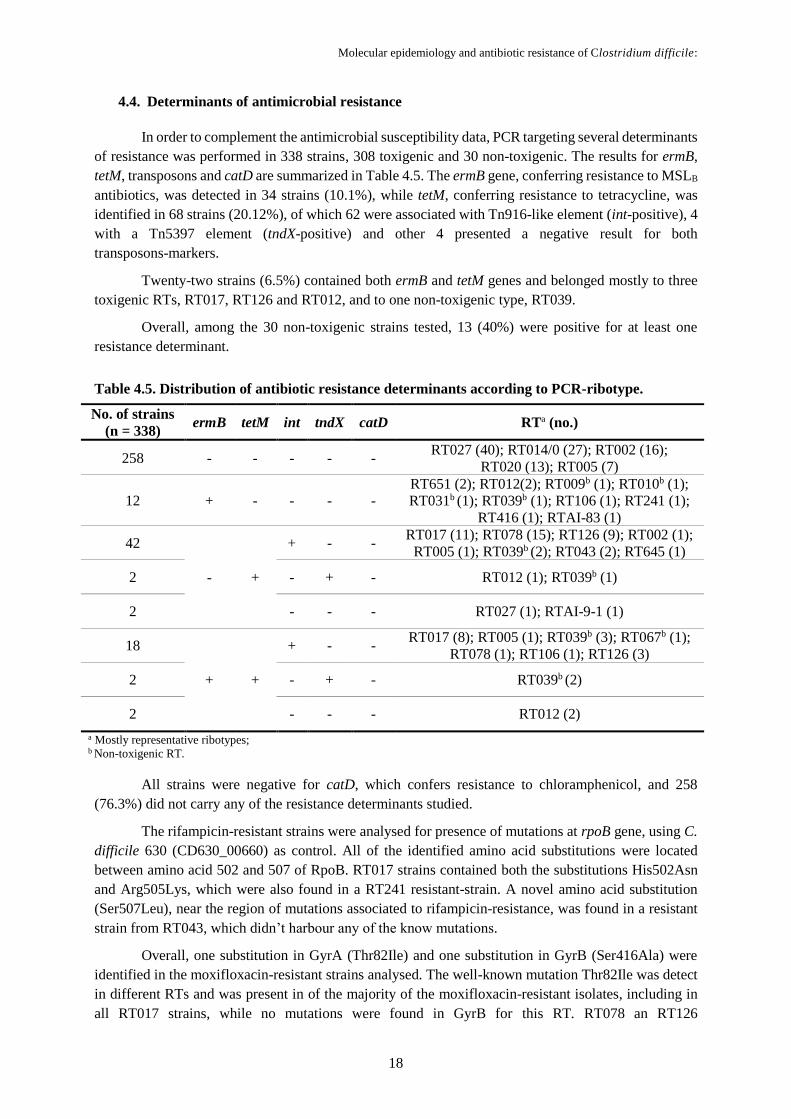
Molecular epidemiology and antibiotic resistance of Clostridium difficile:
18
4.4. Determinants of antimicrobial resistance
In order to complement the antimicrobial susceptibility data, PCR targeting several determinants
of resistance was performed in 338 strains, 308 toxigenic and 30 non-toxigenic. The results for ermB,
tetM, transposons and catD are summarized in Table 4.5. The ermB gene, conferring resistance to MSLB
antibiotics, was detected in 34 strains (10.1%), while tetM, conferring resistance to tetracycline, was
identified in 68 strains (20.12%), of which 62 were associated with Tn916-like element (int-positive), 4
with a Tn5397 element (tndX-positive) and other 4 presented a negative result for both
transposons-markers.
Twenty-two strains (6.5%) contained both ermB and tetM genes and belonged mostly to three
toxigenic RTs, RT017, RT126 and RT012, and to one non-toxigenic type, RT039.
Overall, among the 30 non-toxigenic strains tested, 13 (40%) were positive for at least one
resistance determinant.
Table 4.5. Distribution of antibiotic resistance determinants according to PCR-ribotype.
All strains were negative for catD, which confers resistance to chloramphenicol, and 258
(76.3%) did not carry any of the resistance determinants studied.
The rifampicin-resistant strains were analysed for presence of mutations at rpoB gene, using C.
difficile 630 (CD630_00660) as control. All of the identified amino acid substitutions were located
between amino acid 502 and 507 of RpoB. RT017 strains contained both the substitutions His502Asn
and Arg505Lys, which were also found in a RT241 resistant-strain. A novel amino acid substitution
(Ser507Leu), near the region of mutations associated to rifampicin-resistance, was found in a resistant
strain from RT043, which didn’t harbour any of the know mutations.
Overall, one substitution in GyrA (Thr82Ile) and one substitution in GyrB (Ser416Ala) were
identified in the moxifloxacin-resistant strains analysed. The well-known mutation Thr82Ile was detect
in different RTs and was present in of the majority of the moxifloxacin-resistant isolates, including in
all RT017 strains, while no mutations were found in GyrB for this RT. RT078 an RT126
No. of strains
(n = 338) ermB tetM int tndX catD RTa (no.)
258 - - - - - RT027 (40); RT014/0 (27); RT002 (16);
RT020 (13); RT005 (7)
12 + - - - -
RT651 (2); RT012(2); RT009b (1); RT010b (1);
RT031b (1); RT039b (1); RT106 (1); RT241 (1);
RT416 (1); RTAI-83 (1)
42
- +
+ - - RT017 (11); RT078 (15); RT126 (9); RT002 (1);
RT005 (1); RT039b (2); RT043 (2); RT645 (1)
2 - + - RT012 (1); RT039b (1)
2 - - - RT027 (1); RTAI-9-1 (1)
18
+ +
+ - - RT017 (8); RT005 (1); RT039b (3); RT067b (1);
RT078 (1); RT106 (1); RT126 (3)
2 - + - RT039b (2)
2 - - - RT012 (2)
a Mostly representative ribotypes; b Non-toxigenic RT.
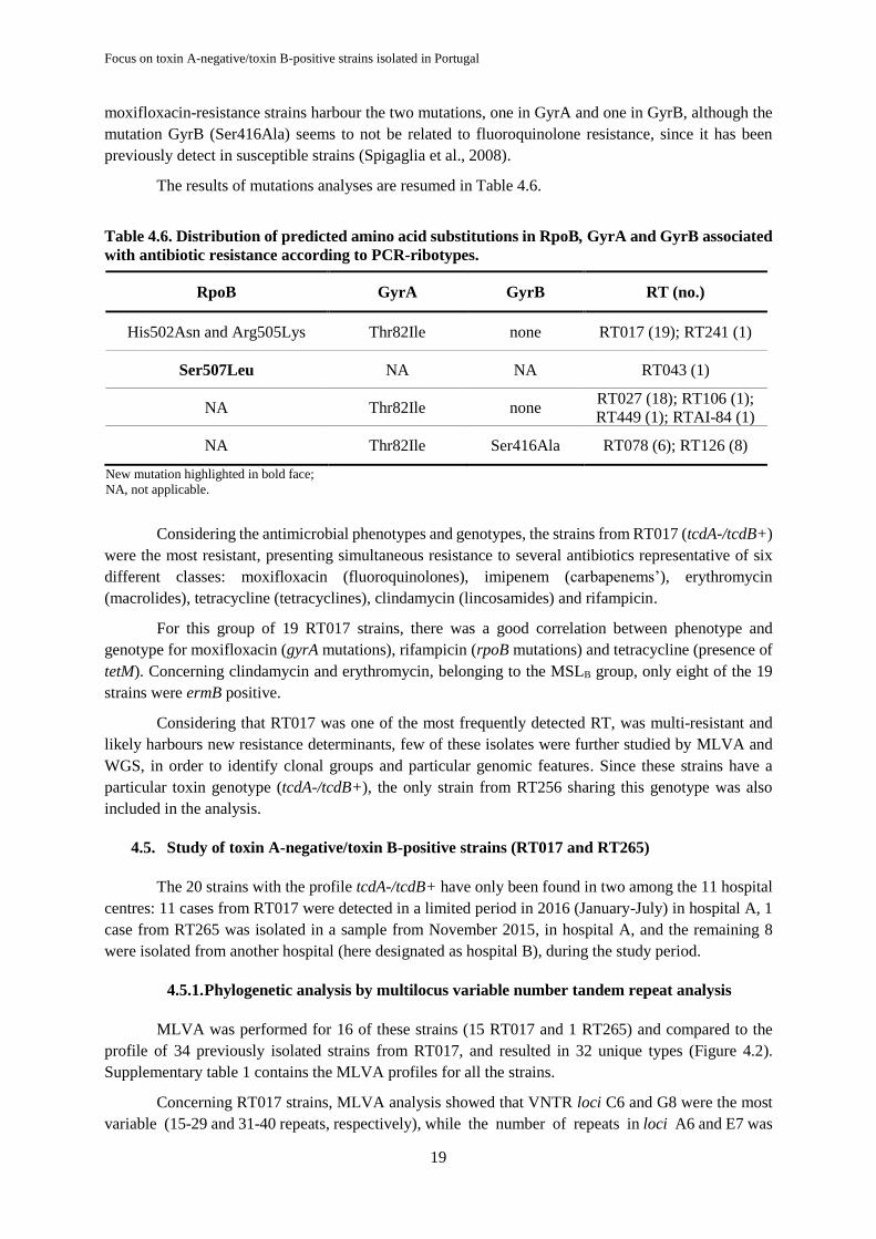
Focus on toxin A-negative/toxin B-positive strains isolated in Portugal
19
moxifloxacin-resistance strains harbour the two mutations, one in GyrA and one in GyrB, although the
mutation GyrB (Ser416Ala) seems to not be related to fluoroquinolone resistance, since it has been
previously detect in susceptible strains (Spigaglia et al., 2008).
The results of mutations analyses are resumed in Table 4.6.
Table 4.6. Distribution of predicted amino acid substitutions in RpoB, GyrA and GyrB associated
with antibiotic resistance according to PCR-ribotypes.
Considering the antimicrobial phenotypes and genotypes, the strains from RT017 (tcdA-/tcdB+)
were the most resistant, presenting simultaneous resistance to several antibiotics representative of six
different classes: moxifloxacin (fluoroquinolones), imipenem (carbapenems’), erythromycin
(macrolides), tetracycline (tetracyclines), clindamycin (lincosamides) and rifampicin.
For this group of 19 RT017 strains, there was a good correlation between phenotype and
genotype for moxifloxacin (gyrA mutations), rifampicin (rpoB mutations) and tetracycline (presence of
tetM). Concerning clindamycin and erythromycin, belonging to the MSLB group, only eight of the 19
strains were ermB positive.
Considering that RT017 was one of the most frequently detected RT, was multi-resistant and
likely harbours new resistance determinants, few of these isolates were further studied by MLVA and
WGS, in order to identify clonal groups and particular genomic features. Since these strains have a
particular toxin genotype (tcdA-/tcdB+), the only strain from RT256 sharing this genotype was also
included in the analysis.
4.5. Study of toxin A-negative/toxin B-positive strains (RT017 and RT265)
The 20 strains with the profile tcdA-/tcdB+ have only been found in two among the 11 hospital
centres: 11 cases from RT017 were detected in a limited period in 2016 (January-July) in hospital A, 1
case from RT265 was isolated in a sample from November 2015, in hospital A, and the remaining 8
were isolated from another hospital (here designated as hospital B), during the study period.
4.5.1. Phylogenetic analysis by multilocus variable number tandem repeat analysis
MLVA was performed for 16 of these strains (15 RT017 and 1 RT265) and compared to the
profile of 34 previously isolated strains from RT017, and resulted in 32 unique types (Figure 4.2).
Supplementary table 1 contains the MLVA profiles for all the strains.
Concerning RT017 strains, MLVA analysis showed that VNTR loci C6 and G8 were the most
variable (15-29 and 31-40 repeats, respectively), while the number of repeats in loci A6 and E7 was
RpoB GyrA GyrB RT (no.)
His502Asn and Arg505Lys Thr82Ile none RT017 (19); RT241 (1)
Ser507Leu NA NA RT043 (1)
NA Thr82Ile none RT027 (18); RT106 (1);
RT449 (1); RTAI-84 (1)
NA Thr82Ile Ser416Ala RT078 (6); RT126 (8)
New mutation highlighted in bold face;
NA, not applicable.
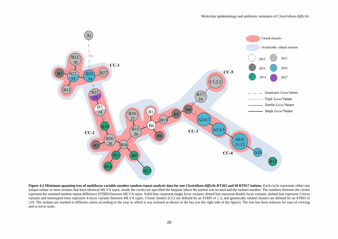
Molecular epidemiology and antibiotic resistance of Clostridium difficile:
20
Figure 4.2 Minimum spanning tree of multilocus variable number tandem repeat analysis data for one Clostridium difficile RT265 and 49 RT017 isolates. Each circle represents either one
unique isolate or more isolates that have identical MLVA types, inside the circles are specified the hospital where the patient was located and the isolates number. The numbers between the circles
represent the summed tandem-repeat difference (STRD) between MLVA types. Solid lines represent single locus variants, dotted line represent double locus variants, dashed line represent 3-locus
variants and interrupted lines represent 4-locus variants between MLVA types. Clonal clusters (CC) are defined by an STRD of ≤ 2, and genetically related clusters are defined by an STRD of
≤10. The isolates are marked in different colors according to the year in which it was isolated as shown in the key (on the right side of the figure). The tree has been redrawn for ease of viewing
and is not to scale.
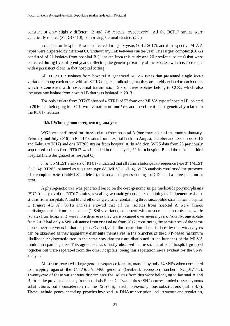
Focus on toxin A-negative/toxin B-positive strains isolated in Portugal
21
constant or only slightly different (2 and 7-8 repeats, respectively). All the R0T17 strains were
genetically related (STDR ≤ 10), comprising 5 clonal clusters (CC).
Isolates from hospital B were collected during six-years (2012-2017), and the respective MLVA
types were dispersed by different CC without any link between cluster/year. The largest complex (CC-2)
consisted of 21 isolates from hospital B (1 isolate from this study and 20 previous isolates) that were
collected during five different years, reflecting the genetic proximity of the isolates, which is consistent
with a persistent clone in that hospital setting.
All 11 RT017 isolates from hospital A generated MLVA types that presented single locus
variation among each other, with an STRD of ≤ 10, indicating that they are highly related to each other,
which is consistent with nosocomial transmission. Six of these isolates belong to CC-3, which also
includes one isolate from hospital B that was isolated in 2013.
The only isolate from RT265 showed a STRD of 53 from one MLVA type of hospital B isolated
in 2016 and belonging to CC-1, with variation in four loci, and therefore it is not genetically related to
the RT017 isolates.
4.5.1. Whole genome sequencing analysis
WGS was performed for three isolates from hospital A (one from each of the months January,
February and July 2016), 5 RT017 strains from hospital B (from August, October and December 2016
and February 2017) and one RT265 strains from hospital A. In addition, WGS data from 25 previously
sequenced isolates from RT017 was included in the analysis, 22 from hospital B and three from a third
hospital (here designated as hospital C).
In silico MLST analysis of RT017 indicated that all strains belonged to sequence type 37 (MLST
clade 4); RT265 assigned as sequence type 88 (MLST clade 4). WGS analysis confirmed the presence
of a complete tcdB (PubMLST allele 9), the absent of genes coding for CDT and a large deletion in
tcdA.
A phylogenetic tree was generated based on the core-genome single nucleotide polymorphisms
(SNPs) analyses of the RT017 strains, revealing two main groups, one containing the imipenem-resistant
strains from hospitals A and B and other single cluster containing three susceptible strains from hospital
C (Figure 4.3 A). SNPs analysis showed that all the isolates from hospital A were almost
undistinguishable from each other (1 SNPs variant), consistent with nosocomial transmission, while
isolates from hospital B were more diverse as they were obtained over several years. Notably, one isolate
from 2017 had only 4 SNPs distance from one isolate from 2012, confirming the persistence of the same
clones over the years in that hospital. Overall, a similar separation of the isolates by the two analyses
can be observed as they apparently distribute themselves in the branches of the SNP-based maximum
likelihood phylogenetic tree in the same way that they are distributed in the branches of the MLVA
minimum spanning tree. This agreement was firstly observed as the strains of each hospital grouped
together but were separated from the other hospitals, being this separation more evident for the SNPs
analysis.
All strains revealed a large genome sequence identity, marked by only 74 SNPs when compared
to mapping against the C. difficile M68 genome (GenBank accession number: NC_017175).
Twenty-two of these variant sites discriminate the isolates from this work belonging to hospital A and
B, from the previous isolates, from hospitals B and C. Two of these SNPs corresponded to synonymous
substitutions, but a considerable number (20) originated, non-synonymous substitutions (Table 4.7).
These include genes encoding proteins involved in DNA transcription, cell structure and regulation,
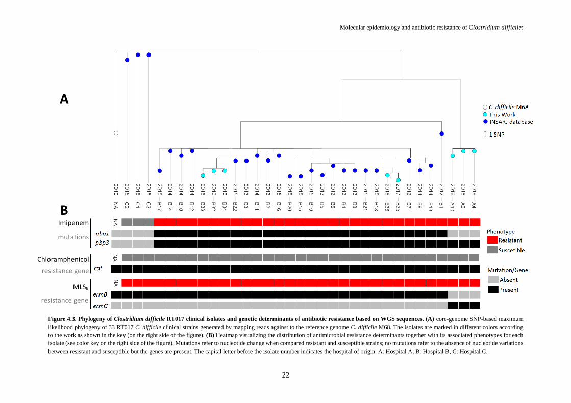
Molecular epidemiology and antibiotic resistance of Clostridium difficile:
22
Figure 4.3. Phylogeny of Clostridium difficile RT017 clinical isolates and genetic determinants of antibiotic resistance based on WGS sequences. (A) core-genome SNP-based maximum
likelihood phylogeny of 33 RT017 C. difficile clinical strains generated by mapping reads against to the reference genome C. difficile M68. The isolates are marked in different colors according
to the work as shown in the key (on the right side of the figure). (B) Heatmap visualizing the distribution of antimicrobial resistance determinants together with its associated phenotypes for each
isolate (see color key on the right side of the figure). Mutations refer to nucleotide change when compared resistant and susceptible strains; no mutations refer to the absence of nucleotide variations
between resistant and susceptible but the genes are present. The capital letter before the isolate number indicates the hospital of origin. A: Hospital A; B: Hospital B, C: Hospital C.
MLSB
resistance gene
Imipenem
mutations
Chloramphenicol
resistance gene
A
B
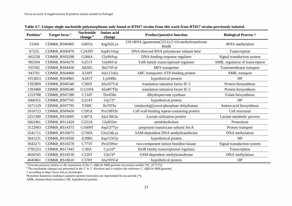
Focus on toxin A-negative/toxin B-positive strains isolated in Portugal
23
Table 4.7. Unique single nucleotide polymorphisms only found at RT017 strains from this work from RT017 strains previously isolated.
Position a Target locus a Nucleotide
change b
Amino acid
change Product/putative function Biological Process †
53164 CDM68_RS00405 G605A Arg202Lys 23S rRNA (guanosine(2251)-2'-O)-methyltransferase
RlmB rRNA methylation
67225 CDM68_RS00470 C2439T Asp813Asp DNA-directed RNA polymerase subunit beta' Transcription
662258 CDM68_RS03290 G266A Gly89Asp DNA-binding response regulator Signal transduction system
902504 CDM68_RS04270 G251T Gly84Val TetR family transcriptional regulator AMR, regulation of transcription
935302 CDM68_RS04430 A820G Ile274Val MFS transporter Transmembrane transport
943701 CDM68_RS04460 A339T Ala113Ala ABC transporter ATP-binding protein AMR, transport
1053823 CDM68_RS04965 A281T Lys94Ile hypothetical protein HP
1392809 CDM68_RS06540 C560T Ala187Val translation initiation factor IF-2 Protein biosynthesis
1393468 CDM68_RS06540 G1219A Ala407Thr translation initiation factor IF-2 Protein biosynthesis
1553788 CDM68_RS07280 C134T Thr45Ile dihydropteroate synthase Folate biosynthesis
1666351 CDM68_RS07765 G214T Gly72* hypothetical protein HP
1671129 CDM68_RS07795 T209C Ile70Thr imidazoleglycerol-phosphate dehydratase Amino-acid biosynthesis
2010723 CDM68_RS09450 C595A Pro199Thr Cell wall-binding repeat-containing protein Cell structural
2251300 CDM68_RS10605 C407A Ala136Glu Lactate utilization protein Lactate metabolic process
2662461 CDM68_RS12420 G253A Gly85Ser amidohydrolase Proteolysis
3122003 CDM68_RS14375 G1609T Asp537Tyr preprotein translocase subunit SecA Protein transport
3541711 CDM68_RS16075 G700A Glu234Lys SAM-dependent DNA methyltransferase DNA methylation
3641231 CDM68_RS16560 A398G Asp133Gly hypothetical protein HP
3643271 CDM68_RS16570 C775T Pro259Ser two-component sensor histidine kinase Signal transduction system
3795253 CDM68_RS17445 C30A Cys10* HxlR family transcriptional regulator Transcription
4026545 CDM68_RS18530 C220T Gln74* SAM-dependent methyltransferase DNA methylation
4045861 CDM68_RS18610 C578T Ala193Val hypothetical protein HP a Genome position relative to the annotation of the C. difficile M68 genome (accession number NC_017175); b The nucleotide changes are presented in the 5’ to 3’ direction and is relative the reference C. difficile M68 genome;
† according to http://www.ebi.ac.uk/interpro
Nonsense mutations leading to putative protein truncation are represented by an asterisk (*);
AMR, antimicrobial resistance; HP, hypothetical protein.
Molecular epidemiology and antibiotic resistance of Clostridium difficile:
24
response to environmental changes and metabolic reactions. It also includes hypothetical proteins and
possible genes implicated in antimicrobial resistance as well. Nonsense mutations resulting in premature
truncation of the encoding protein was observed in genes encoding for a S-adenosyl-methionine
(SAM)-dependent methyltransferase, for a transcriptional regulator (HxlR family transcriptional
regulator) and for a hypothetical protein.
C. difficile usually contains four HMW PBP, except the strains from RT017, which contain a
fifth pbp gene, here designated as pbp5 (corresponding to CDM68_RS02615 locus tag in C. difficile
M68 strain). This gene was also found in the RT265 strain, in the same genomic context as the RT017
strains, with both PBP5 sharing 99% homology.
All the imipenem-resistant strains, from hospitals A and B, harboured mutations in HMW PBP
genes, located near conserved motifs, when compared to imipenem-susceptible strains, but notably with
different profiles: the resistant strains from hospital B have the two mutations previously reported (Isidro
et al. 2017), one (G1663A) in pbp1 (corresponding to CDM68_RS04280 locus) leading to an amino
acid substitution Ala555Thr, and the other (A2162C) in pbp3 (corresponding to CDM68_RS05670
locus) resulting in the amino acid substitution Tyr721Ser; isolates from hospital A harbour only the
mutation (A2162C) in pbp3 (Figure 4.3 B). For this clone, no mutations were found in other PBPs
encoding genes (pbp1, pbp2, pbp4 and pbp5).
In silico screening of antimicrobial resistance genes based on WGS analysis using CARD and
ResFinder databases, revealed the presence of several genes in this category. First, a gene encoding a
putative chloramphenicol acetyltransferase CAT (CDM68_RS02605) was found in all the strains,
although they were susceptible (Figure 4.3 B).
The isolates from hospital B were positive for ermB gene, previously associated with resistance
to MLSB antibiotics in C. difficile, confirming the results of the PCR screening, while all strains from
RT017 from hospital A were positive for ermG gene. Both genes encode a rRNA adenine
N-6-methyltransferase. Homology search of ermG using nucleotide Blast showed that this gene shared
100% identity to ermG gene found in several other Gram-positive species, such as Enterococcus
cecorum (RA45_RS03490 locus) and Lysinibacillus sphaericus (A7J11_00653 locus). Transformation
of C. difficile strain 630∆erm with a plasmid containing ermG resulted in a resistance phenotype for
clindamycin and erythromycin, with MIC > 256 mg/L, showing that this gene is functional in C. difficile
and is associated with high level of resistance to both these antibiotics.
The ermG gene was located inside a large region of 63.4 kb that displays traces of horizontal
gene transfer (encompassing recombinases, phage-related proteins and flanked by transposases), as
shown in Figure 4.4. Nucleotide Blast was conduct for the entire region, showing high degree of
homology with an identical region present in a C. difficile non-toxigenic strain (Peptoclostridium
difficile strain Z31, GenBank accession number: CP013196.1). Table 4.8 lists the suggested annotation
and homologues locus tag (PubMed) for all the ORFs found at this putative mobile element containing
the ermG gene. A streptogramin A acetyltransferase was also detected in this putative mobile element.
Screening for resistance determinants at ResFinder also showed homology to other two genes
possible involved in MLSB resistance, the mefA and the msrD genes, located near the ermG on the
putative mobile element (Figure 4.4); blast of the nucleotide sequence gave 96% for a mefA gene
(A6J31_02365) and 98% of identity for msrD (A6J31_02370) in Streptococcus sp. Those genes encode
for a macrolide efflux MFS transporter Mef(A) and a ABC-F type ribosomal protection protein Msr(D),
respectively (Table 4.8).
Regarding the other classes of antibiotics, WGS analysis confirmed the results of the PCR
screening.
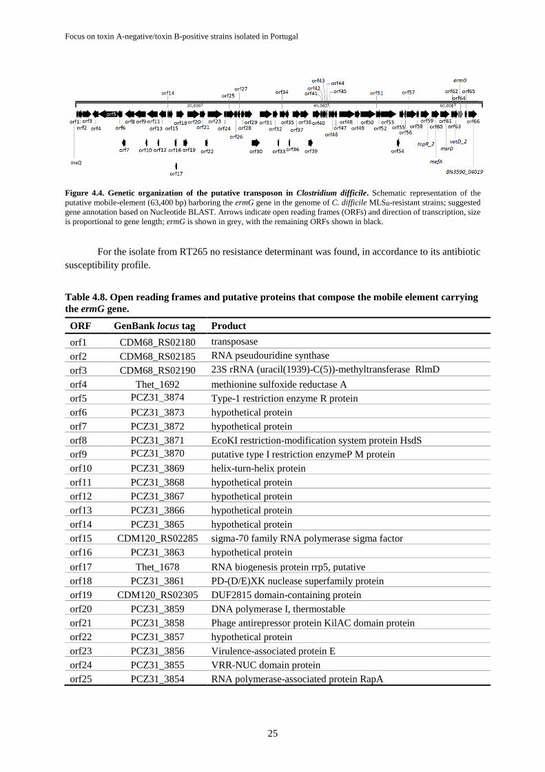
Focus on toxin A-negative/toxin B-positive strains isolated in Portugal
25
Figure 4.4. Genetic organization of the putative transposon in Clostridium difficile. Schematic representation of the
putative mobile-element (63,400 bp) harboring the ermG gene in the genome of C. difficile MLSB-resistant strains; suggested
gene annotation based on Nucleotide BLAST. Arrows indicate open reading frames (ORFs) and direction of transcription, size
is proportional to gene length; ermG is shown in grey, with the remaining ORFs shown in black.
For the isolate from RT265 no resistance determinant was found, in accordance to its antibiotic
susceptibility profile.
Table 4.8. Open reading frames and putative proteins that compose the mobile element carrying
the ermG gene. ORF GenBank locus tag Product
orf1 CDM68_RS02180 transposase
orf2 CDM68_RS02185 RNA pseudouridine synthase
orf3 CDM68_RS02190 23S rRNA (uracil(1939)-C(5))-methyltransferase RlmD
orf4 Thet_1692 methionine sulfoxide reductase A
orf5 PCZ31_3874 Type-1 restriction enzyme R protein
orf6 PCZ31_3873 hypothetical protein
orf7 PCZ31_3872 hypothetical protein
orf8 PCZ31_3871 EcoKI restriction-modification system protein HsdS
orf9 PCZ31_3870 putative type I restriction enzymeP M protein
orf10 PCZ31_3869 helix-turn-helix protein
orf11 PCZ31_3868 hypothetical protein
orf12 PCZ31_3867 hypothetical protein
orf13 PCZ31_3866 hypothetical protein
orf14 PCZ31_3865 hypothetical protein
orf15 CDM120_RS02285 sigma-70 family RNA polymerase sigma factor
orf16 PCZ31_3863 hypothetical protein
orf17 Thet_1678 RNA biogenesis protein rrp5, putative
orf18 PCZ31_3861 PD-(D/E)XK nuclease superfamily protein
orf19 CDM120_RS02305 DUF2815 domain-containing protein
orf20 PCZ31_3859 DNA polymerase I, thermostable
orf21 PCZ31_3858 Phage antirepressor protein KilAC domain protein
orf22 PCZ31_3857 hypothetical protein
orf23 PCZ31_3856 Virulence-associated protein E
orf24 PCZ31_3855 VRR-NUC domain protein
orf25 PCZ31_3854 RNA polymerase-associated protein RapA
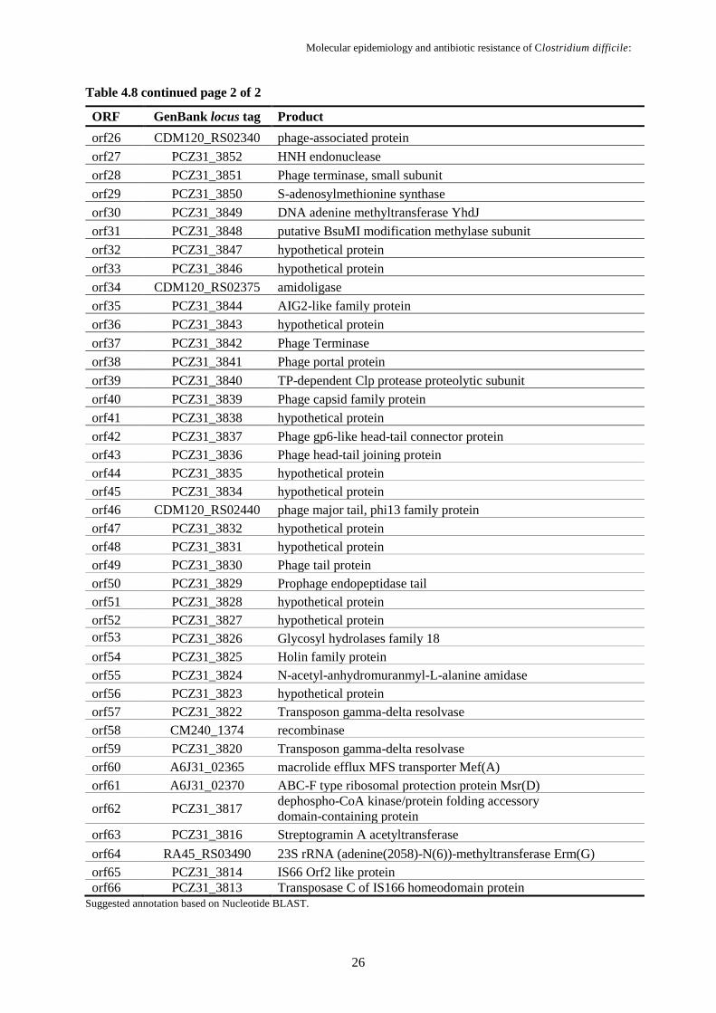
Molecular epidemiology and antibiotic resistance of Clostridium difficile:
26
Table 4.8 continued page 2 of 2
ORF GenBank locus tag Product
orf26 CDM120_RS02340 phage-associated protein
orf27 PCZ31_3852 HNH endonuclease
orf28 PCZ31_3851 Phage terminase, small subunit
orf29 PCZ31_3850 S-adenosylmethionine synthase
orf30 PCZ31_3849 DNA adenine methyltransferase YhdJ
orf31 PCZ31_3848 putative BsuMI modification methylase subunit
orf32 PCZ31_3847 hypothetical protein
orf33 PCZ31_3846 hypothetical protein
orf34 CDM120_RS02375 amidoligase
orf35 PCZ31_3844 AIG2-like family protein
orf36 PCZ31_3843 hypothetical protein
orf37 PCZ31_3842 Phage Terminase
orf38 PCZ31_3841 Phage portal protein
orf39 PCZ31_3840 TP-dependent Clp protease proteolytic subunit
orf40 PCZ31_3839 Phage capsid family protein
orf41 PCZ31_3838 hypothetical protein
orf42 PCZ31_3837 Phage gp6-like head-tail connector protein
orf43 PCZ31_3836 Phage head-tail joining protein
orf44 PCZ31_3835 hypothetical protein
orf45 PCZ31_3834 hypothetical protein
orf46 CDM120_RS02440 phage major tail, phi13 family protein
orf47 PCZ31_3832 hypothetical protein
orf48 PCZ31_3831 hypothetical protein
orf49 PCZ31_3830 Phage tail protein
orf50 PCZ31_3829 Prophage endopeptidase tail
orf51 PCZ31_3828 hypothetical protein
orf52 PCZ31_3827 hypothetical protein
orf53 PCZ31_3826 Glycosyl hydrolases family 18
orf54 PCZ31_3825 Holin family protein
orf55 PCZ31_3824 N-acetyl-anhydromuranmyl-L-alanine amidase
orf56 PCZ31_3823 hypothetical protein
orf57 PCZ31_3822 Transposon gamma-delta resolvase
orf58 CM240_1374 recombinase
orf59 PCZ31_3820 Transposon gamma-delta resolvase
orf60 A6J31_02365 macrolide efflux MFS transporter Mef(A)
orf61 A6J31_02370 ABC-F type ribosomal protection protein Msr(D)
orf62 PCZ31_3817 dephospho-CoA kinase/protein folding accessory
domain-containing protein
orf63 PCZ31_3816 Streptogramin A acetyltransferase
orf64 RA45_RS03490 23S rRNA (adenine(2058)-N(6))-methyltransferase Erm(G)
orf65 PCZ31_3814 IS66 Orf2 like protein
orf66 PCZ31_3813 Transposase C of IS166 homeodomain protein Suggested annotation based on Nucleotide BLAST.
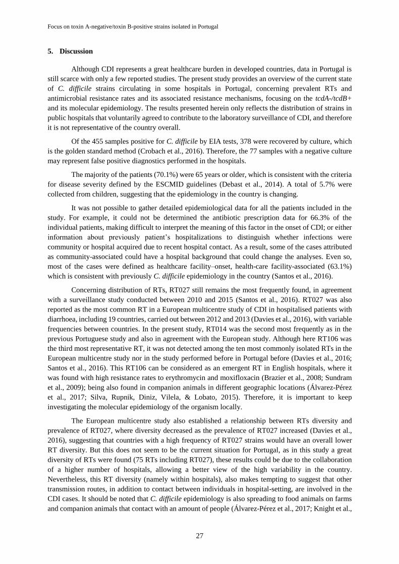
Focus on toxin A-negative/toxin B-positive strains isolated in Portugal
27
5. Discussion
Although CDI represents a great healthcare burden in developed countries, data in Portugal is
still scarce with only a few reported studies. The present study provides an overview of the current state
of C. difficile strains circulating in some hospitals in Portugal, concerning prevalent RTs and
antimicrobial resistance rates and its associated resistance mechanisms, focusing on the tcdA-/tcdB+
and its molecular epidemiology. The results presented herein only reflects the distribution of strains in
public hospitals that voluntarily agreed to contribute to the laboratory surveillance of CDI, and therefore
it is not representative of the country overall.
Of the 455 samples positive for C. difficile by EIA tests, 378 were recovered by culture, which
is the golden standard method (Crobach et al., 2016). Therefore, the 77 samples with a negative culture
may represent false positive diagnostics performed in the hospitals.
The majority of the patients (70.1%) were 65 years or older, which is consistent with the criteria
for disease severity defined by the ESCMID guidelines (Debast et al., 2014). A total of 5.7% were
collected from children, suggesting that the epidemiology in the country is changing.
It was not possible to gather detailed epidemiological data for all the patients included in the
study. For example, it could not be determined the antibiotic prescription data for 66.3% of the
individual patients, making difficult to interpret the meaning of this factor in the onset of CDI; or either
information about previously patient’s hospitalizations to distinguish whether infections were
community or hospital acquired due to recent hospital contact. As a result, some of the cases attributed
as community-associated could have a hospital background that could change the analyses. Even so,
most of the cases were defined as healthcare facility–onset, health-care facility-associated (63.1%)
which is consistent with previously C. difficile epidemiology in the country (Santos et al., 2016).
Concerning distribution of RTs, RT027 still remains the most frequently found, in agreement
with a surveillance study conducted between 2010 and 2015 (Santos et al., 2016). RT027 was also
reported as the most common RT in a European multicentre study of CDI in hospitalised patients with
diarrhoea, including 19 countries, carried out between 2012 and 2013 (Davies et al., 2016), with variable
frequencies between countries. In the present study, RT014 was the second most frequently as in the
previous Portuguese study and also in agreement with the European study. Although here RT106 was
the third most representative RT, it was not detected among the ten most commonly isolated RTs in the
European multicentre study nor in the study performed before in Portugal before (Davies et al., 2016;
Santos et al., 2016). This RT106 can be considered as an emergent RT in English hospitals, where it
was found with high resistance rates to erythromycin and moxifloxacin (Brazier et al., 2008; Sundram
et al., 2009); being also found in companion animals in different geographic locations (Álvarez-Pérez
et al., 2017; Silva, Rupnik, Diniz, Vilela, & Lobato, 2015). Therefore, it is important to keep
investigating the molecular epidemiology of the organism locally.
The European multicentre study also established a relationship between RTs diversity and
prevalence of RT027, where diversity decreased as the prevalence of RT027 increased (Davies et al.,
2016), suggesting that countries with a high frequency of RT027 strains would have an overall lower
RT diversity. But this does not seem to be the current situation for Portugal, as in this study a great
diversity of RTs were found (75 RTs including RT027), these results could be due to the collaboration
of a higher number of hospitals, allowing a better view of the high variability in the country.
Nevertheless, this RT diversity (namely within hospitals), also makes tempting to suggest that other
transmission routes, in addition to contact between individuals in hospital-setting, are involved in the
CDI cases. It should be noted that C. difficile epidemiology is also spreading to food animals on farms
and companion animals that contact with an amount of people (Álvarez-Pérez et al., 2017; Knight et al.,
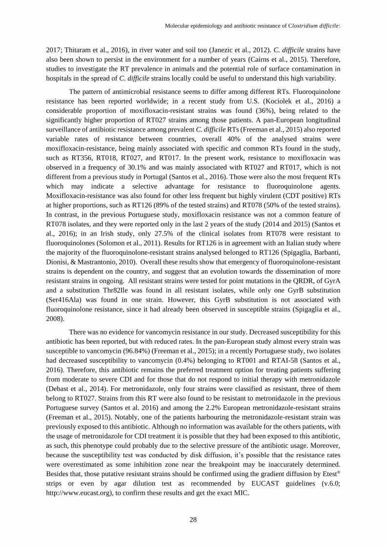
Molecular epidemiology and antibiotic resistance of Clostridium difficile:
28
2017; Thitaram et al., 2016), in river water and soil too (Janezic et al., 2012). C. difficile strains have
also been shown to persist in the environment for a number of years (Cairns et al., 2015). Therefore,
studies to investigate the RT prevalence in animals and the potential role of surface contamination in
hospitals in the spread of C. difficile strains locally could be useful to understand this high variability.
The pattern of antimicrobial resistance seems to differ among different RTs. Fluoroquinolone
resistance has been reported worldwide; in a recent study from U.S. (Kociolek et al., 2016) a
considerable proportion of moxifloxacin-resistant strains was found (36%), being related to the
significantly higher proportion of RT027 strains among those patients. A pan-European longitudinal
surveillance of antibiotic resistance among prevalent C. difficile RTs (Freeman et al., 2015) also reported
variable rates of resistance between countries, overall 40% of the analysed strains were
moxifloxacin-resistance, being mainly associated with specific and common RTs found in the study,
such as RT356, RT018, RT027, and RT017. In the present work, resistance to moxifloxacin was
observed in a frequency of 30.1% and was mainly associated with RT027 and RT017, which is not
different from a previous study in Portugal (Santos et al., 2016). Those were also the most frequent RTs
which may indicate a selective advantage for resistance to fluoroquinolone agents.
Moxifloxacin-resistance was also found for other less frequent but highly virulent (CDT positive) RTs
at higher proportions, such as RT126 (89% of the tested strains) and RT078 (50% of the tested strains).
In contrast, in the previous Portuguese study, moxifloxacin resistance was not a common feature of
RT078 isolates, and they were reported only in the last 2 years of the study (2014 and 2015) (Santos et
al., 2016); in an Irish study, only 27.5% of the clinical isolates from RT078 were resistant to
fluoroquinolones (Solomon et al., 2011). Results for RT126 is in agreement with an Italian study where
the majority of the fluoroquinolone-resistant strains analysed belonged to RT126 (Spigaglia, Barbanti,
Dionisi, & Mastrantonio, 2010). Overall these results show that emergency of fluoroquinolone-resistant
strains is dependent on the country, and suggest that an evolution towards the dissemination of more
resistant strains in ongoing. All resistant strains were tested for point mutations in the QRDR, of GyrA
and a substitution Thr82Ile was found in all resistant isolates, while only one GyrB substitution
(Ser416Ala) was found in one strain. However, this GyrB substitution is not associated with
fluoroquinolone resistance, since it had already been observed in susceptible strains (Spigaglia et al.,
2008).
There was no evidence for vancomycin resistance in our study. Decreased susceptibility for this
antibiotic has been reported, but with reduced rates. In the pan-European study almost every strain was
susceptible to vancomycin (96.84%) (Freeman et al., 2015); in a recently Portuguese study, two isolates
had decreased susceptibility to vancomycin (0.4%) belonging to RT001 and RTAI-58 (Santos et al.,
2016). Therefore, this antibiotic remains the preferred treatment option for treating patients suffering
from moderate to severe CDI and for those that do not respond to initial therapy with metronidazole
(Debast et al., 2014). For metronidazole, only four strains were classified as resistant, three of them
belong to RT027. Strains from this RT were also found to be resistant to metronidazole in the previous
Portuguese survey (Santos et al. 2016) and among the 2.2% European metronidazole-resistant strains
(Freeman et al., 2015). Notably, one of the patients harbouring the metronidazole-resistant strain was
previously exposed to this antibiotic. Although no information was available for the others patients, with
the usage of metronidazole for CDI treatment it is possible that they had been exposed to this antibiotic,
as such, this phenotype could probably due to the selective pressure of the antibiotic usage. Moreover,
because the susceptibility test was conducted by disk diffusion, it’s possible that the resistance rates
were overestimated as some inhibition zone near the breakpoint may be inaccurately determined.
Besides that, those putative resistant strains should be confirmed using the gradient diffusion by Etest®
strips or even by agar dilution test as recommended by EUCAST guidelines (v.6.0;
http://www.eucast.org), to confirm these results and get the exact MIC.
Focus on toxin A-negative/toxin B-positive strains isolated in Portugal
29
Rifamycin’s resistance was reported in a United States study with rates of 1.6% for paediatric
isolates and 6.7% for adults isolates (Kociolek et al., 2016). In the pan-European study resistance
frequency for rifampicin was 13.4%, however, with variable rates between countries (from 0 to 62.4%)
(Freeman et al., 2015). In the current study, 11.5% of the tested strains were rifampicin-resistance,
although lower than the overall European rate, was higher to those observed in the majority of the
countries. Rifampicin resistance was detected in all RT017 isolates, as well as in a RT241 (1/2, 50%):
two already know amino acid substitutions in RpoB were detect, His502Asn and Arg505Lys (Curry et
al., 2009). However, a novel amino acid substitution (Ser507Leu) was observed in the only RT043
resistant-strain found, which didn’t harbour any of the know mutations. Being therefore likely associated
with this phenotype. Previous studies with Staphylococcus aureus and Mycobacterium tuberculosis have
shown that independently derived mutations in these residues are frequently detected, being that an
equivalent Serine to Leucine substitution was already describe for S. aureus resistant strains (Murphy
et al., 2006; O’Connor et al., 2008). Resistance to rifampicin may compromise the use of rifaximin,
considered an alternative therapy for relapsing CDI. (Curry et al., 2009; Debast et al., 2014).
Imipenem resistance is not well documented in C. difficile. In the present study only RT017
strains were resistant to imipenem. A Korean study also reported resistant for this antibiotic in RT017
(12%), simultaneously with the introduction of the drug at the hospitals (Lee, Lee, Lee, Riley, & Kim,
2014). In the pan-European study it was reported a 7.4% imipenem-resistance rate, but no distribution
per RTs was described. Nonetheless, the geometric mean imipenem MICs were highest among RT027
and RT106.
In the present study, the distribution of resistance determinants for MSLB antibiotics (ermB
gene) and for tetracycline (tetM gene) were highly variable between and within RTs. Overall, ermB gene
was detected in only 10.1% of the strains tested; in a study conducted by Wasels, Spigaglia, Barbanti
and Mastrantonio (2013) they reported a burden associated with the in vitro fitness in strains harbouring
mobile elements containing the ermB-gene. This could be the explanation behind the low frequency of
this gene in our study.
Concerning the tetM gene, used to estimate tetracycline resistance strains, a low rate was
observed (20.1%) as compared to a study conducted in China where tetM frequency reached 41.7% in
RTs such as RT017, RT065, RT014, RT046 and RT002 (Dong et al. 2013). In our study, tetM was
ubiquitous among RT017, RT126 and RT078, showing that it is associated with specific RTs, which
may explain differences among studies. Most of the strains harboured the Tn916 transposon while a
small fraction carried the Tn5397 element which is in agreement with other reports (Dong et al., 2013;
Spigaglia et al., 2005). Of relevance, four tetM-positive strains were negative for those two transposons
markers. Further studies should be carried on these strains using WGS in order to characterize putative
novel tetM-carrying elements. Forty percent of the non-toxigenic strains tested harboured at least one
resistance determinant (ermB or tetM), as such, it should be noted that these generally unnoticed strains
could act as a pool of resistance determinants transmissible to toxigenic strains.
The catD gene, which confers resistance to chloramphenicol, was not found in any of the strains
tested; despite the small sample (n = 338), it can be speculated that this gene does not circulate in
Portugal. The causes of the relatively low incidence of chloramphenicol-resistant C. difficile in the
country are not known, although it’s conceivable that would be related to the local drug prescription
habits. Accordingly, in the pan-European study, most prevalent RTs were largely susceptible (95–100%
of isolates) to chloramphenicol. Clusters of chloramphenicol-resistant isolates were found only in
Germany, The Netherlands and Latvia (Freeman et al., 2015).
Aside from an European study were a set of multidrug resistant isolates were characterized
(particularly belonging to RT001, RT012 and RT017) (Spigaglia et al., 2011), in the current study, the
Molecular epidemiology and antibiotic resistance of Clostridium difficile:
30
most prominent multidrug resistant RT was RT017. Only one strain belonging to RT241 was also
associated with multidrug resistance, harbouring the ermB resistance gene and mutations conferring
resistance to moxifloxacin and rifampicin. In addition, when tested for further antibiotics, all RT017
strains showed simultaneous resistance to six different classes of antibiotics. According to Coia (2009)
the use of a particular antimicrobial agent could result in selective advantage for the spread of C. difficile
if the organism is resistant to that particular drug. Therefore, it is tempting to suggest that multidrug
resistance would be a good driver of CDI epidemics. Having that in mind, in the currently study, three
different techniques (phenotypic characterization, MLVA and WGS) were used to characterize toxin
A-negative/toxin-B-positive group of strains to which the multidrug resistant RT017 belongs.
Twenty strains were tcdA-/tcdB+ of which, 19 belonging to RT017 and one to RT265. To date,
there were no reports of RT265 strains in Portugal. According to van Dorp et al. (2017) this RT appears
to be very uncommon or absent in North American and European countries, except for Belgium and the
Netherlands being primarily isolated from children aged < 2 years. In our study, this RT was isolated in
an elderly patient. WGS analysis showed that RT265 shares many features with RT017, such as: an
intact tcdB gene, a 1.8 kb deletion in tcdA, and no genes of CDT genes, according to previous data (van
Dorp et al., 2017). This strain also belonged to MLST clade 4 and was assigned toST88. Also, similarly
to RT017, this RT contains a gene coding for a fifth HMW PBP. Overall, these indicates that these two
RTs are close, although belong to different clusters by MLVA and SNPs analysis.
Regarding C. difficile RT017, despite the loss of the capability to produce both toxins, this RT
is increasingly becoming a potential epidemic strain, being isolated over the past decade in Asian as
well as emerging in a few European countries (Arvand et al., 2009; Dobreva et al., 2013; Lee et al.,
2014; Yang et al., 2017). In the currently study, RT017 make up 5.1% of the isolates, which is pretty
much similar to the five years study previously conduct in Portugal (Santos et al., 2016). It is worth
noting that all the isolates found are either endemic, such as the ones found in hospital B, or associated
with an outbreak, such as in hospital A (since all the isolates from that hospital remote from a short
period of time, sharing the same molecular characteristics), with no single cases found, showing the
virulence and epidemics of these strains.
As already mentioned above, resistance was found for moxifloxacin, rifampicin and imipenem,
plus the presence of tetM gene for all the strains and ermB in hospital B isolates. Those RT017 strains
were also tested for antibiotic susceptibility for other drugs; as result, all of the isolates in the present
study were susceptible to chloramphenicol and tigecycline and resistant to tetracycline. Interestingly the
presence of tetM gene in all strains seems to have an effect on tetracycline susceptibility but not to
tigecycline (a tetracycline derivative antibiotic), probably because this third generation drug display
enhanced antimicrobial activity overcoming ribosome protection mechanisms as the one promoted by
tetM (Dönhöfer et al., 2012).
All RT017 isolates also had high-level erythromycin and clindamycin-resistance, including the
hospital A strains classified as ermB-negative; for those strains the ermG gene was found by WGS
analyses. This ermG gene encodes for an rRNA adenine N-6-methyltransferase, which can methylate
adenine at position 2058 of 23S rRNA, conferring resistance to MLSB antibiotics. This gene has
appeared in human colonic Bacteroides species where it was found on conjugative transposons
(Shoemaker, Vlamakis, Hayes, & Salyers, 2001; Wang et al., 2003). Although, the results presented
here do not prove that DNA was transferred directly from these species to C. difficile, the data indicates
that this gene was transferred by a mobile element; Wasels et al. (2014) have already provides evidence
of the possible transmission of antibiotic resistance determinants among pathogenic bacteria occupying
the same human intestinal niche, which may have happened in that case. In C. difficile the presence of
Focus on toxin A-negative/toxin B-positive strains isolated in Portugal
31
ermG gene was previously reported in one non-toxigenic strains (RT009), but without any phenotype
associated (Pereira et al., 2016).
The ermG gene was shown to be associated with high level of resistance to clindamycin and
erythromycin, through transformation of C. difficile 630Δerm with a plasmid containing ermG. Despite
its previous occurrence in a non-toxigenic strain, this is the first time it is described in toxigenic strains,
and associated with a resistance phenotype. Its presence only in the strains belonging to hospital A may
be indicative of recent gene transfer, highlighting the importance of an active surveillance of resistance
determinants.
Since ermG is located in a mobile element, there is the possibility to spread via horizontal gene
transfer. The ermB-negativity in high-level MSLB-resistant isolates is a well-known phenomenon (Nyc
et al., 2016; Spigaglia et al., 2011), and therefore it can be speculated if some of these reported strains
harboured the ermG gene. Further studies should be also carried to understand if the carriage of the
element containing the gene would have fitness cost associated.
The presence of mefA and the msrD genes were also observed in this mobile element; those two
genes are known to promote low or moderate levels to macrolide resistance by active efflux of the
antibiotic, due to an efflux pump that belongs to the major facilitator superfamily encoded by the mefA
gene, or by an efflux protein belonging to the ABC transporter superfamily encoded by msrD. The msr
group differs from the mef genes because it confer resistance to both macrolide and streptogramin B
antibiotics, although for both, clindamycin is neither an inducer or a substrate for the pump, and thus
the strains caring those genes are fully susceptible to this antibiotic (Leclercq, 2002; Metcalf et al.,
2017). Since the RT017 strains had high-level resistance to the later antibiotic, we confirm that these
genes don’t have a role in resistance. In addition, the presence of mefA and the msrD genes could only
explain low or moderate resistance levels to macrolides or streptogramin B, again emphasizing the role
of ermG.
These results indicate that our real MLSB-resistance rate must be higher than the one deduced
from the frequency of the ermB gene, since other mechanisms can be involved; therefore, all strains
should be tested for antibiotic susceptibility to identify the resistant strains, and then the associated
genetic determinants should be characterized. As reported by the European Centre for Disease
Prevention and Control (2016), MLSB antibiotics are one of the most common drugs used in both
community and hospital sector in Europe, what makes really important to keep track on the resistance
patterns for these group of antibiotics.
The putative mobile element also contained a gene encoding a streptogramin A
acetyltransferase. This protein belongs to the large family of virginiamycin O-acetyltransferase (Vat)
enzymes, known to cause resistance to the streptogramin A antibiotics (Stogios et al., 2014), however
this group was not tested here, hence, its potential impact in antibiotic susceptibility of those C. difficile
strains is unknown.
Concerning imipenem resistance, mutations in HMW PBP genes were found in all RT017
resistant strains, as in an earlier study where RT017 collected between 2012–2015 carried mutations in
PBPs and showed statistically higher imipenem MIC than the isolates with no mutations (Isidro et al.,
2017). In that study two mutations (at pbp1 and pbp3) were thought to be causative of resistance,
although, in the current study all the resistant strains from hospital A harboured only mutations in pbp3
(Figure 3.3 B). So, it’s legitimate to speculate if this single mutation is the one crucial for resistance.
Yet those results show clearly that the interpretation of such data is not going to be straightforward, and
more studies need to be done. Portugal has been one the major consumers of carbapenems in the hospital
sector in Europe (European Centre for Disease Prevention and Control, 2016), as Dingle et al. (2017)
Molecular epidemiology and antibiotic resistance of Clostridium difficile:
32
associated RT027 fluoroquinolone-resistant emergency with fluoroquinolone use, it’s conceivable to
conclude that the emergence of carbapenems resistance in C. difficile is likely due to the high usage of
this class of antibiotic in the recent years, highlighting the importance of a responsible use of antibiotics.
MLVA was performed for RT265 and RT017 strains. Minimum spanning tree analysis clearly
demonstrate the genetic relationships among the RT017 Portuguese isolates being possible to identify
five clonal clusters. Furthermore, the fact that a group of isolates were collected from the same hospital
within a 6-years period, suggests that this degree of similarity among MLVA types is highly indicative
of nosocomial transmission and persistence of the same clones over the years.
The VNTR loci used in this study were previously demonstrated to be useful for epidemiologic
investigation of C. difficile outbreaks and nosocomial transmission (Marsh et al., 2006); their utility for
detection of outbreaks was demonstrated by analysis of a group of RT017 from hospital A which
clustered on the same branch of the minimum-spanning tree indicating a genetic relatedness among
these isolates.
Previous studies using MLVA have demonstrated genetic relatedness among a large collection
of tcdA-/tcdB+ strains from seven countries (van den Berg et al., 2004). In this study, RT265 revel to be
distance from the others analysed strains, consistence with the fact it belongs to a different RT.
In summary, the MLVA results indicate that INSARJ’s isolates collection consists primarily of
three major complexes of highly related isolates, suggestive of hospital-acquired infections.
A phylogenetic tree of SNPs identified through WGS analyse revealed two main related groups
comprising 33 RT017 clinical isolates that distinguish the imipenem-resistant strains from the
susceptible ones. Nevertheless, it was possible to distinguish the local RT017 isolates from the
international control strain (C. difficile M68). Overall, 74 SNPs were found which includes variants
between the clinical strains and the reference in sequences that have an ortholog to a known gene
sequences, and 22 of these variant sites were able to discriminate the isolates from this work belonging
to hospitals A and B, from the previous isolates, collected from hospitals B and C.
Among this 22 SNPs variations there was a series of non-synonymous mutations in important
genes, including genes involved in the metabolism and antibiotic resistance, mutations leading to
truncated proteins were observed for a hypothetical protein, a transcriptional regulator and
SAM-dependent methyltransferase, the last one associated to diverse metabolic functions in biocatalysis
and biosynthesis (Struck, Thompson, Wong, & Micklefield, 2012); the meaning of these findings has to
be studied in future works. The high number of SNPs found in hypothetical proteins is also very
interesting, and this group should be further studied to predict their function and the effect of the point-
mutations found in some of them.
Remarkably, the most recently strain and one of the most ancient strain from hospital B,
separated by a period of 6-years, had only 4 SNPs distance, what is consistent with the evolutionary
mutation rate estimated by Eyre et al. (2013) of 0.74 SNPs per year, suggesting that the isolates share a
common ancestor. This result supports the idea that a clonal strain from RT017 is endemic in hospital
B, persisting for at least 6-years leading to nosocomial transmission. Further studies are needed to
investigate the persistence of these strains in the hospital setting.
WGS results also suggest that the clonal strains persisting in hospital B is different from the
hospital A strains. Additionally, all the isolates from hospital A were tight closely, reinforcing that an
outbreak has occurred. How this outbreak clonal strain was inserted in the hospital remains unclear, but
it could be due a patient that was infected in the community and spread the strain during its hospital
Focus on toxin A-negative/toxin B-positive strains isolated in Portugal
33
stay, which raises many concerns especially on the spread of this multidrug resistant strain in the
community.
In conclusion, the current work gives a contribution to the knowledge of the molecular
epidemiology and resistance patterns of C. difficile in Portugal, especially of the emergent strains of
RT017, which are multiresistant and associated with endemic or epidemic cases of CDI. A new
resistance determinant, found in a mobile element, was characterized for the first time in C. difficile
toxigenic strains. The results also suggest that the lineage of strains from RT017 appears to be constantly
evolving, acquiring new resistance determinants, which highlights the need for continued
epidemiological and antimicrobial surveillance. Finally, this work provides the foundation for future
studies, namely the role in RT017 evolution of SNPs found in several proteins, including many
hypothetical ones; to study the spread and impact on the fitness of the new mobile element, as well as
to assess whether this element can be easily transferred among clinical strains, from the same or different
RTs, by horizontal gene transfer.
Molecular epidemiology and antibiotic resistance of Clostridium difficile:
34
6. References
Alfa, M. J., Kabani, A., Lyerly, D., Moncrief, S., Neville, L. M., Al-Barrak, A., Harding, G. K.
H., Dyck, B., Olekson, K., & Embil, J. M. (2000). Characterization of a toxin A-negative, toxin
B-positive strain of Clostridium difficile responsible for a nosocomial outbreak of Clostridium
difficile-associated diarrhea. Journal of Clinical Microbiology, 38(7), 2706–2714.
Álvarez-Pérez, S., Blanco, J. L., Harmanus, C., Kuijper, E. J., & García, M. E. (2017). Data
from a survey of Clostridium perfringens and Clostridium difficile shedding by dogs and cats in the
Madrid region (Spain), including phenotypic and genetic characteristics of recovered isolates. Data in
Brief, 14, 88–100. https://doi.org/10.1016/j.dib.2017.07.029
Arvand, M., Hauri, A. M., Zaiss, N. H., Witte, W., & Bettge-Weller, G. (2009). Clostridium
difficile ribotypes 001, 017, and 027 are associated with lethal C. difficile infection in Hesse, Germany.
Euro Surveillance, 14(45). Retrieved from
http://www.eurosurveillance.org/ViewArticle.aspx?ArticleId=19403
Barbut, F., Mastrantonio, P., Delmée, M., Brazier, J., Kuijper, E., Poxton, I., Ackermann, G.,
Bouza, E., Balmelli, C., Drudy, D., Ladas, H., Nagy, E., Pituch, H., Wullt, M., Yücesoy, M., & Rupnik,
M. (2007). Prospective study of Clostridium difficile infections in Europe with phenotypic and genotypic
characterisation of the isolates. Clinical Microbiology and Infection, 13(11), 1048–1057.
https://doi.org/10.1111/j.1469-0691.2007.01824.x
Bartlett, J. G., Chang, T. W., Gurwith, M., Gorbach, S. L., & Onderdonk, A. B. (1978).
Antibiotic-associated pseudomembranous colitis due to toxin-producing clostridia. New England
Journal of Medicine, 283(10), 531–534. https://doi.org/10.1056/NEJM198603273141302
Bidet, P., Frédéric, B., Valérie, L., & Petit, J. C. (1999). Development of a new PCR-ribotyping
method for Clostridium difficile based on ribosomal RNA gene sequencing. FEMS Microbiology
Letters, 175(2), 261–266. https://doi.org/10.1111/j.1574-6968.1999.tb13629.x
Brazier, J. S., Raybould, R., Patel, B., Duckworth, G., Pearson, A., Charlett, A., & Duerden, B.
I. (2008). Distribution and antimicrobial susceptibility patterns of Clostridium difficile PCR ribotypes
in English hospitals. Euro Surveillance, 13(41), 541–544. Retrieved from
http://www.eurosurveillance.org/ViewArticle.aspx?ArticleId=19000
Cairns, M. D., Preston, M. D., Lawley, T. D., Clark, T. G., Stabler, R. A., & Wren, B. W. (2015).
Genomic epidemiology of a protracted hospital outbreak caused by a toxin A-negative Clostridium
difficile sublineage PCR ribotype 017. Journal of Biological Chemistry, 53(10), 3141–3147.
https://doi.org/10.1128/JCM.00648-15
Candela, T., Marvaud, J. C., Nguyen, T. K., & Lambert, T. (2017). A cfr-like gene cfr(C)
conferring linezolid resistance is common in Clostridium difficile. International Journal of
Antimicrobial Agents. https://doi.org/10.1016/j.ijantimicag.2017.03.013
Carter, G. P., Awad, M. M., Kelly, M. L., Rood, J. I., & Lyras, D. (2011). TcdB or not TcdB: a
tale of two Clostridium difficile toxins. Future Microbiology, 6(2), 121–123.
https://doi.org/10.2217/fmb.10.169
Centers for Disease Control and Prevention. (2005). Severe Clostridium difficile-associated
disease in populations previously at low risk — four states, 2005. Morbidity and Mortality Weekly
Focus on toxin A-negative/toxin B-positive strains isolated in Portugal
35
Report, 54(47), 1201–1205.
Centers for Disease Control and Prevention. (2016). 2014 National and State Associated
Infections Progress Report. Centers for Disease Control and Prevention. Retrieved from
www.cdc.gov/hai/progress-report/index.htm
Chaves-Olarte, E., Löw, P., Freer, E., Norlin, T., Weidmann, M., Von Eichel-Streiber, C., &
Thelestam, M. (1999). A novel cytotoxin from Clostridium difficile serogroup F is a functional hybrid
between two other large clostridial cytotoxins. Journal of Biological Chemistry, 274(16), 11046–11052.
https://doi.org/10.1074/jbc.274.16.11046
CLSI. (2012). Methods for antimicrobial susceptibility testing of anaerobic bacteria; Approved
Standard-Eighth Edition. CLSI document M11-A8. Wayne, PA: Clinical and Laboratory Standards
Institute.
Cohen, S. H., Gerding, D. N., Johnson, S., Kelly, C. P., Loo, V. G., McDonald, L. C., Pepin, J.,
& Wilcox, M. H. (2010). Clinical practice guidelines for Clostridium difficile infection in adults: 2010
update by the society for healthcare epidemiology of america (SHEA) and the infectious diseases society
of America (IDSA). Inection Control & Hospital Epidemiologyfection Control & Hospital
Epidemiology, 31(5), 431–455. https://doi.org/10.1086/651706
Coia, J. E. (2009). What is the role of antimicrobial resistance in the new epidemic of
Clostridium difficile? International Journal of Antimicrobial Agents, 33, S9–S12.
https://doi.org/10.1016/S0924-8579(09)70009-3
Cowardin, C. A., Buonomo, E. L., Saleh, M. M., Wilson, M. G., Burgess, S. L., Kuehne, S. A.,
Schwan, C., Eichhoff, A. M, Koch-Nolte, F., Lyras, D., Aktories, K., Minton, N. P., & Petri Jr, W. A.
(2016). The binary toxin CDT enhances Clostridium difficile virulence by suppressing protective colonic
eosinophilia. Nature Microbiology, 1(8), 1–21. https://doi.org/10.1038/nmicrobiol.2016.108
Crobach, M. J. T., Planche, T., Eckert, C., Barbut, F., Terveer, E. M., Dekkers, O. M., Wilcox,
M., & Kuijper, E. J. (2016). European Society of Clinical Microbiology and Infectious Diseases: update
of the diagnostic guidance document for Clostridium difficile infection. Clinical Microbiology and
Infection, 20(s2), 1–26. https://doi.org/10.1016/j.cmi.2016.03.010
Curry, S. R., Marsh, J. W., Shutt, K. A., Muto, C. A., O’Leary, M. M., Saul, M. I., Pasculle, A.
W., & Harrison, L. H. (2009). High frequency of rifampin resistance identified in an epidemic
Clostridium difficile clone from a large teaching hospital. Clinical Infectious Diseases, 48(4), 425–429.
https://doi.org/10.1086/596315
Davies, K. A., Ashwin, H., Longshaw, C. M., Burns, D. A., Davis, G. L., & Wilcox, M. H.
(2016). Diversity of Clostridium difficile PCR ribotypes in Europe: results from the European,
multicentre, prospective, biannual, point-prevalence study of Clostridium difficile infection in
hospitalised patients with diarrhoea (EUCLID), 2012 and 2. Euro Surveillance, 21(29).
https://doi.org/10.2807/1560-7917.ES.2016.21.29.30294
Davies, K. A., Longshaw, C. M., Davis, G. L., Bouza, E., Barbut, F., Barna, Z., Delmée, M.,
Fitzpatrick, F., Ivanova, K., Kuijper, E., Macovei, I. S., Mentula, S., Mastrantonio, P., von Müller, L.,
Oleastro, M., Petinaki, E., Pituch, H., Norén, T., Nováková, E., Nyč, O., Rupnik, M., Schmid, D., &
Wilcox, M. H. (2014). Underdiagnosis of Clostridium difficile across Europe: the European, multicentre,
prospective, biannual, point-prevalence study of Clostridium difficile infection in hospitalised patients
Molecular epidemiology and antibiotic resistance of Clostridium difficile:
36
with diarrhoea (EUCLID). The Lancet Infectious Diseases, 14(12), 1208–1219.
https://doi.org/10.1016/S1473-3099(14)70991-0
Dawson, L. F., Valiente, E., Donahue, E. H., Birchenough, G., & Wren, B. W. (2011).
Hypervirulent Clostridium difficile pcr-ribotypes exhibit resistance to widely used disinfectants. PLoS
ONE, 6(10), e25754. https://doi.org/10.1371/journal.pone.0025754
Debast, S. B., Bauer, M. P., Kuijper, E. J., Allerberger, F., Bouza, E., Coia, J. E., Cornely, O.
A., Fitzpatrick, F., Guery, B., Wilcox, M., Nathwani, D., Norén, T., Olesen, B., Rakoczi, E., Welte, T.,
& Widmer, A. F. (2014). European society of clinical microbiology and infectious diseases: Update of
the treatment guidance document for Clostridium difficile infection. Clinical Microbiology and
Infection, 20(S2), 1–26. https://doi.org/10.1111/1469-0691.12418
Dharmarajan, T. S., Sipalay, M., Shyamsundar, R., Norkus, E. P., & Pitchumoni, C. S. (2000).
Co-morbidity, not age predicts adverse outcome in Clostridium difficile colitis. World Journal of
Gastroenterology, 6(2), 198–201. https://doi.org/10.3748/wjg.v6.i2.198
Dingle, K. E., Didelot, X., Quan, T. P., Eyre, D. W., Stoesser, N., Golubchik, T., Harding,
Rosalind M., Wilson, D. J. Griffiths, D., Vaughan, A., Finney, J. M., Wyllie, D. H., Oakley, S. J.,
Fawley, W. N., Freeman, J., Morris, K., Martin, J., Howard, P., Gorbach, S., Goldstein, E. J.C., Citron,
D. M., Hopkins, S., Hope, R., Johnson, A. P., Wilcox, M. H., Peto, T. E.A., Walker, A. S., Crook, D.W.,
Del Ojo Elias, C. Crichton, C., Kostiou, V., Giess, A., & Davies, J., (2017). Effects of control
interventions on Clostridium difficile infection in England: an observational study. The Lancet Infectious
Diseases, 17(4), 411–421. https://doi.org/10.1016/S1473-3099(16)30514-X
Dobreva, E. G., Ivanov, I. N., Vathcheva-Dobrevska, R. S., Ivanova, K. I., Asseva, G. D.,
Petrov, P. K., & Kantardjiev, T. V. (2013). Advances in molecular surveillance of Clostridium difficile
in Bulgaria. Journal of Medical Microbiology, 62(9), 1428–1434.
https://doi.org/10.1099/jmm.0.058149-0
Dong, D., Zhang, L., Chen, X., Jiang, C., Yu, B., Wang, X., & Peng, Y. (2013). Antimicrobial
susceptibility and resistance mechanisms of clinical Clostridium difficile from a Chinese tertiary
hospital. International Journal of Antimicrobial Agents, 41(1), 80–84.
https://doi.org/10.1016/j.ijantimicag.2012.08.011
Dönhöfer, A., Franckenberg, S., Wickles, S., Berninghausen, O., Beckmann, R., & Wilson, D.
N. (2012). Structural basis for TetM-mediated tetracycline resistance. Proceedings of the National
Academy of Sciences of the United States of America, 109(42), 16900–5.
https://doi.org/10.1073/pnas.1208037109
Dridi, L., Tankovic, J., Burghoffer, B., Barbut, F., & Petit, J. C. (2002). gyrA and gyrB Mutations
Are Implicated in Cross-Resistance to Ciprofloxacin and Moxifloxacin in Clostridium difficile.
Antimicrobial Agents and Chemotherapy, 46(11), 3418–3421. https://doi.org/10.1128/AAC.46.11.3418
Drudy, D., Harnedy, N., Fanning, S., Hannan, M., & Kyne, L. (2007). Emergence and control
of fluoroquinolone-resistant, toxin A–negative, toxin B–positive Clostridium difficile. Infection Control
& Hospital Epidemiology, 28(8), 932–940. https://doi.org/10.1086/519181
Erikstrup, L. T., Danielsen, T. K. L., Hall, V., Olsen, K. E. P., Kristensen, B., Kahlmeter, G.,
Fuursted, K., & Justesen, U. S. (2012). Antimicrobial susceptibility testing of Clostridium difficile using
EUCAST epidemiological cut-off values and disk diffusion correlates. Clinical Microbiology and
Focus on toxin A-negative/toxin B-positive strains isolated in Portugal
37
Infection, 18(8), E266–E272. https://doi.org/10.1111/j.1469-0691.2012.03907.x
European Centre for Disease Prevention and Control. (2016). Summary of the latest data on
antibiotic consumption in the European Union Antibiotic consumption in Europe. Stockholm: ECDC.
Eyre, D. W., Cule, M. L., Wilson, D. J., Griffiths, D., Vaughan, A., O’Connor, L., Ip, C. L. C.,
Golubchik, T., Batty, E. M., Finney, J. M., Wyllie, D. H., Didelot, X., Piazza, P., Bowden, R., Dingle,
K. E., Harding, R. M., Crook, D. W., Wilcox, M. H., Peto, T. E. A., & Walker, A. S. (2013). Diverse
sources of C. difficile infection identified on whole-genome sequencing. The New England Journal of
Medicine, 369(13), 1195–205. https://doi.org/10.1056/NEJMoa1216064
Fagan, R. P., & Fairweather, N. F. (2011). Clostridium difficile has two parallel and essential
sec secretion systems. Journal of Biological Chemistry, 286(31), 27483–27493.
https://doi.org/10.1074/jbc.M111.263889
Freeman, J., Vernon, J., Morris, K., Nicholson, S., Todhunter, S., Longshaw, C., & Wilcox, M.
H. (2015). Pan-European longitudinal surveillance of antibiotic resistance among prevalent Clostridium
difficile ribotypes. Clinical Microbiology and Infection, 21(3), 248.e9-248.e16.
https://doi.org/10.1016/j.cmi.2014.09.017
Gerding, D. N. (2004). Clindamycin, Cephalosporins, Fluoroquinolones and Clostridium
difficile – Associated Diarrhea : This is an Antimicrobial Resistance Problem. Clinical Infectious
Diseases, 38(5), 646–648.
Govind, R., & Dupuy, B. (2012). Secretion of Clostridium difficile toxins A and B requires the
holin-like protein TcdE. PLoS Pathogens, 8(6). https://doi.org/10.1371/journal.ppat.1002727
Hall, I. C., & O’Toole, E. (1935). Intestinal flora in new-born infants: with a description of a
new pathogenic anaerobe, Bacillus difficilis. American Journal of Diseases of Children, 49(2), 390–402.
He, M., Sebaihia, M., Lawley, T. D., Stabler, R. A., Dawson, L. F., Martin, M. J., Holt, K. E..,
Seth-Smith, H. M. B., Quail, M. A., Rance, R., Brooks, K., Churcher, C., Harris, D., Bentley, S. D.,
Burrows, C., Clark, L., Corton, C., Murray, V., Rose, G., Thurston, S., van Tonder, A., Walker, D.,
Wren, B. W., Dougan, G., & Parkhill, J. (2010). Evolutionary dynamics of Clostridium difficile over
short and long time scales. Proceedings of the National Academy of Sciences, 107(16), 7527–7532.
https://doi.org/10.1073/pnas.0914322107
Howerton, A., Ramirez, N., & Abel-Santos, E. (2011). Mapping interactions between
germinants and Clostridium difficile spores. Journal of Bacteriology, 193(1), 274–282.
https://doi.org/10.1128/JB.00980-10
Isidro, J., Santos, A., Nunes, A., Borges, V., Silva, C., Chaves C., Boaventura, L., Sancho. L.,
Vieira, L., Gomes, J.P., & Oleastro, M. (2017). Imipenem resistance in Clostridium difficile ribotype
017. Emerging Infectious Diseases. (submitted).
Janarthanan, S., Ditah, I., Adler, D. G., & Ehrinpreis, M. N. (2012). Clostridium
difficile-Associated Diarrhea and Proton Pump Inhibitor Therapy: A Meta-Analysis. The American
Journal of Gastroenterology, 107(7), 1001–1010. https://doi.org/10.1038/ajg.2012.179
Janezic, S., Ocepek, M., Zidaric, V. & Rupnik, M. (2012). Clostridium difficile genotypes other
than ribotype 078 that are prevalent among human, animal and environmental isolates. BMC
Microbiology, 12(1), 48. https://doi.org/10.1186/1471-2180-12-48
Molecular epidemiology and antibiotic resistance of Clostridium difficile:
38
Janoir, C., Denève, C., Bouttier, S., Barbut, F., Hoys, S., Caleechum, L., Chapetón-Montes, D.,
Pereira, F. C., Henriques, A. O., Collignon, A., Monot, M., & Dupuy, B. (2013). Adaptive strategies
and pathogenesis of Clostridium difficile from In vivo transcriptomics. Infection and Immunity, 81(10),
3757–3769. https://doi.org/10.1128/IAI.00515-13
Just, I., & Gerhard, R. (2004). Large clostridial cytotoxins. Reviews of Physiology, Biochemistry
and Pharmacology, 152, 23–47. https://doi.org/10.1007/s10254-004-0033-5
Kato, H., Kato, N., Katow, S., Maegawa, T., Nakamura, S., & Lyerly, D. M. (1999). Deletions
in the repeating sequences of the toxin A gene of toxin A-negative, toxin B-positive Clostridium difficile
strains. FEMS Microbiology Letters, 175(2), 197–203. https://doi.org/10.1016/S0378-1097(99)00196-2
Knight, D. R., Squire, M. M., Collins, D. A., & Riley, T. V. (2017). Genome analysis of
Clostridium difficile PCR ribotype 014 lineage in australian pigs and humans reveals a diverse genetic
repertoire and signatures of long-range interspecies transmission. Frontiers in Microbiology, 7.
https://doi.org/10.3389/fmicb.2016.02138
Kociolek, L. K., Gerding, D. N., Osmolski, J. R., Patel, S. J., Snydman, D. R., McDermott, L.
A., & Hecht, D. W. (2016). Differences in the molecular epidemiology and antibiotic susceptibility of
Clostridium difficile in pediatric and adult patients. Antimicrobial Agents and Chemotherapy, 60(8),
4896–4900. https://doi.org/10.1128/AAC.00714-16
Kuehne, S. A., Cartman, S. T., Heap, J. T., Kelly, M. L., Cockayne, A., & Minton, N. P. (2010).
Both, toxin A and toxin B, are important in Clostridium difficile infection. Nature, 467(7316), 711–713.
https://doi.org/10.1038/nature09397
Lawson, P. A., Citron, D. M., Tyrrell, K. L., & Finegold, S. M. (2016). Reclassification of
Clostridium difficile as Clostridioides difficile (Hall and O’Toole 1935) Prévot 1938. Anaerobe, 40, 95–
99. https://doi.org/10.1016/j.anaerobe.2016.06.008
Leclercq, R. (2002). Mechanisms of resistance to macrolides and lincosamides: nature of the
resistance elements and their clinical implications. Clinical Infectious Diseases, 34(4), 482–492.
https://doi.org/10.1086/324626
Lee, J. H., Lee, Y., Lee, K., Riley, T. V., & Kim, H. (2014). The changes of PCR ribotype and
antimicrobial resistance of Clostridium difficile in a tertiary care hospital over 10 years. Journal of
Medical Microbiology, 63(6), 819–823. https://doi.org/10.1099/jmm.0.072082-0
Leeds, J. A., Sachdeva, M., Mullin, S., Whitney Barnes, S., & Ruzin, A. (2014). In vitro
selection, via serial passage, of Clostridium difficile mutants with reduced susceptibility to fidaxomicin
or vancomycin. Journal of Antimicrobial Chemotherapy, 69(1), 41–44.
https://doi.org/10.1093/jac/dkt302
Lessa, F. C., Mu, Y., Bamberg, W. M., Beldavs, Z. G., Dumyati, G. K., Dunn, J. R., Farley, M.
M., Holzbauer, S. M., Meek, J. I., Phipps, E. C., Wilson, L. E., Winston, L. G., Cohen, J. A., Limbago,
B. M., Fridkin, S. K., Gerding, D. N., & McDonald, L. C. (2015). Burden of Clostridium difficile
Infection in the United States. New England Journal of Medicine, 9(26), 825–834.
https://doi.org/10.1056/NEJMoa1408913
Lynch, T., Chong, P., Zhang, J., Hizon, R., Du, T., Graham, M. R., Beniac, D. R., Booth, T. F.,
Kibsey, P., Miller, M., Gravel, D., & Mulvey, M. R. (2013). Characterization of a stable, metronidazole-
Focus on toxin A-negative/toxin B-positive strains isolated in Portugal
39
resistant Clostridium difficile clinical isolate. PLoS ONE, 8(1), e53757.
https://doi.org/10.1371/journal.pone.0053757
Lyras, D., O’Connor, J. R., Howarth, P. M., Sambol, S. P., Carter, G. P., Phumoonna, T., Poon,
R., Adams, V., Vedantam, G., Johnson, S., Gerding, D. N., & Rood, J. I. (2009). Toxin B is essential
for virulence of Clostridium difficile. Nature, 458(7242), 1176–9. https://doi.org/10.1038/nature07822
Manges, A. R., Labbe, A., Loo, V. G., Atherton, J. K., Behr, M. A., Masson, L., Tellis, P. A., &
Brousseau, R. (2010). Comparative metagenomic study of alterations to the intestinal microbiota and
risk of nosocomial Clostridum difficile-associated disease. The Journal of Infectious Diseases, 202(12),
1877–84. https://doi.org/10.1086/657319
Mani, N., & Dupuy, B. (2001). Regulation of toxin synthesis in Clostridium difficile by an
alternative RNA polymerase sigma factor. Proceedings of the National Academy of Sciences of the
United States of America, 98(10), 5844–5849. https://doi.org/10.1073/pnas.101126598
Marchese, A., Ramirez, M., Schito, G. C., & Tomasz, A. (1998). Molecular epidemiology of
penicillin-resistant Streptococcus pneumoniae isolates recovered in Italy from 1993 to 1996. Journal of
Clinical Microbiology, 36(10), 2944–2949.
Marín, M., Martín, A., Alcalá, L., Cercenado, E., Iglesias, C., Reigadas, E., & Bouza, E. (2015).
Clostridium difficile isolates with high linezolid MICs harbor the multiresistance gene cfr. Antimicrobial
Agents and Chemotherapy, 59(1), 586–589. https://doi.org/10.1128/AAC.04082-14
Marsh, J. W., O’Leary, M. M., Shutt, K. A., Pasculle, A. W., Johnson, S., Gerding, D. N., Muto,
C. A., & Harrison, L. H. (2006). Multilocus variable-number tandem-repeat analysis for investigation
of Clostridium difficile transmission in hospitals. Journal of Clinical Microbiology, 44(7), 2558–2566.
https://doi.org/10.1128/JCM.02364-05
Martin, J. S. H., Monaghan, T. M., & Wilcox, M. H. (2016). Clostridium difficile infection:
epidemiology, diagnosis and understanding transmission. Nature Reviews. Gastroenterology &
Hepatology, 13(4), 206–16. https://doi.org/10.1038/nrgastro.2016.25
Matamouros, S., England, P., & Dupuy, B. (2007). Clostridium difficile toxin expression is
inhibited by the novel regulator TcdC. Molecular Microbiology, 64(5), 1274–1288.
https://doi.org/10.1111/j.1365-2958.2007.05739.x
McDonald, L. C., Killgore, G. E., Thompson, A., Owens, R. C., Kazakova, S. V., Sambol, S.
P., Johnson, S., & Gerding, D. N. (2005). An Epidemic, Toxin Gene–Variant Strain of Clostridium
difficile. New England Journal of Medicine, 353(23), 2433–2441.
https://doi.org/10.1056/NEJMoa051590
Metcalf, B. J., Chochua, S., Jr., R. E. G., Hawkins, P. A., Ricaldi, J., Li, Z., Walker, H., Tran,
T., Rivers, J., Mathis, S., Jackson, D., Glennen, A., Lynfield, R., McGee, L., & Beall, B. (2017).
Short-read whole genome sequencing for determination of antimicrobial resistance mechanisms and
capsular serotypes of current invasive Streptococcus agalactiae recovered in the USA. Clinical
Microbiology and Infection, 23(8), 574.e7-574.e14. https://doi.org/10.1016/j.cmi.2017.02.021
Moura, I., Monot, M., Tani, C., Spigaglia, P., Barbanti, F., Norais, N., Dupuy, B., Bouza, E. &
Mastrantonio, P. (2014). Multidisciplinary analysis of a nontoxigenic Clostridium difficile strain with
stable resistance to metronidazole. Antimicrobial Agents and Chemotherapy, 58(8), 4957–4960.
Molecular epidemiology and antibiotic resistance of Clostridium difficile:
40
https://doi.org/10.1128/AAC.02350-14
Murphy, C. K., Mullin, S., Osburne, M. S., Van Duzer, J., Siedlecki, J., Yu, X., Kerstein, K.,
Cynamon, M., & Rothstein, D. M. (2006). In vitro activity of novel rifamycins against rifamycin-
resistant Staphylococcus aureus . Antimicrobial Agents and Chemotherapy, 50(3), 827–834.
https://doi.org/10.1128/AAC.50.3.827-834.2006
Muto, C. A., Pokrywka, M., Shutt, K., Mendelsohn, A. B., Nouri, K., Posey, K., Roberts, T.,
Croyle, K., Krystofiak, S., Brown, P., Pasculle, A. W., Paterson, D. L. Saul, M., & Harrison, L. H.
(2005). A large outbreak of Clostridium difficile–associated disease with an unexpected proportion of
deaths and colectomies at a teaching hospital following increased fluoroquinolone use. Infection Control
& Hospital Epidemiology, 26(3), 273–280.
Nelson, R. L., Suda, K. J., & Evans, C. T. (2017). Antibiotic treatment for Clostridium difficile
-associated diarrhoea in adults. Cochrane Database of Systematic Reviews.
https://doi.org/10.1002/14651858.CD004610.pub5
Nyc, O., Tejkalova, R., Kriz, Z., Ruzicka, F., Kubicek, L., Matejkova, J., Kuijper, E., & Krutova,
M. (2016). Two Clusters of Fluoroquinolone and Clindamycin-Resistant Clostridium difficile PCR
Ribotype 001 Strain Recognized by Capillary Electrophoresis Ribotyping and Multilocus Variable
Tandem Repeat Analysis. Microbial Drug Resistance, 23(5), 609–615.
https://doi.org/10.1089/mdr.2016.0159
O’Connor, J. R., Galang, M. A., Sambol, S. P., Hecht, D. W., Vedantam, G., Gerding, D. N., &
Johnson, S. (2008). Rifampin and rifaximin resistance in clinical isolates of Clostridium difficile.
Antimicrobial Agents and Chemotherapy, 52(8), 2813–2817. https://doi.org/10.1128/AAC.00342-08
Oleastro, M., Coelho, M., Gião, M., Coutinho, S., Mota, S., Santos, A., Rodrigues, J., & Faria,
D. (2014). Outbreak of Clostridium difficile PCR ribotype 027 - the recent experience of a regional
hospital. BMC Infectious Diseases, 14(1), 209. https://doi.org/10.1186/1471-2334-14-209
Paltansing, S., van den Berg, R. J., Guseinova, R. A., Visser, C. E., van der Vorm, E. R., &
Kuijper, E. J. (2007). Characteristics and incidence of clostridium difficile-associated disease in The
Netherlands, 2005. Clinical Microbiology and Infection, 13(11), 1058–1064.
https://doi.org/10.1111/j.1469-0691.2007.01793.x
Pépin, J., Valiquette, L., & Cossette, B. (2005). Mortality attributable to nosocomial Clostridium
difficile-associated disease during an epidemic caused by a hypervirulent strain in Quebec. Canadian
Medical Association Journal, 173(9), 1037–1041. https://doi.org/10.1503/cmaj.050978
Pereira, F. L., Oliveira Júnior, C. A., Silva, R. O. S., Dorella, F. A., Carvalho, A. F., Almeida,
G. M. F., Leal, C. A. G., Lobato, F. C. F., & Figueiredo, H. C. P. (2016). Complete genome sequence
of Peptoclostridium difficile strain Z31. Gut Pathogens, 8(1), 11. https://doi.org/10.1186/s13099-016-
0095-3
Persson, S., Torpdahl, M., & Olsen, K. E. P. (2008). New multiplex PCR method for the
detection of Clostridium difficile toxin A (tcdA) and toxin B (tcdB) and the binary toxin (cdtA/cdtB)
genes applied to a Danish strain collection. Clinical Microbiology and Infection, 14(11), 1057–1064.
https://doi.org/10.1111/j.1469-0691.2008.02092.x
Pituch, H., Obuch-Woszczatyński, P., Wultańska, D., Nurzyńska, G., Harmanus, C.,
Focus on toxin A-negative/toxin B-positive strains isolated in Portugal
41
Banaszkiewicz, A., Radzikowski, A., Łuczak, M., van Belkum, A., & Kuijper, E. (2011).
Characterization and antimicrobial susceptibility of Clostridium difficile strains isolated from adult
patients with diarrhoea hospitalized in two university hospitals in Poland, 2004-2006. Journal of
Medical Microbiology, 60(8), 1200–1205. https://doi.org/10.1099/jmm.0.029801-0
Planche, T. D., Davies, K. A., Coen, P. G., Finney, J. M., Monahan, I. M., Morris, K. A.,
O'Connor, L., Oakley, S. J., Pope, C. F., Wren, M. W., Shetty, N. P., Crook, D. W., & Wilcox, M. H.
(2013). Differences in outcome according to Clostridium difficile testing method: A prospective
multicentre diagnostic validation study of C. difficile infection. The Lancet Infectious Diseases, 13(11),
936–945. https://doi.org/10.1016/S1473-3099(13)70200-7
Pultz, N. J., & Donskey, C. J. (2005). Effect of Antibiotic Treatment on Growth of and Toxin
Production by Clostridium difficile in the Cecal Contents of Mice. Antimicrobial Agents and
Chemotherapy, 49(8), 3529–3532. https://doi.org/10.1128/AAC.49.8.3529
Reineke, J., Tenzer, S., Rupnik, M., Koschinski, A., Hasselmayer, O., Schrattenholz, A., Schild,
H., & von Eichel-Streiber, C. (2007). Autocatalytic cleavage of Clostridium difficile toxin B. Nature,
446(7134), 415–419. https://doi.org/10.1038/nature05622
Rupnik, M., Kato, N., Grabnar, M., & Kato, H. (2003). New types of toxin A-negative, toxin
B-positive strains among Clostridium difficile isolates from Asia. Journal of Clinical Microbiology,
41(3), 1118–1125. https://doi.org/10.1128/JCM.41.3.1118
Santos, A., Isidro, J., Silva, C., Boaventura, L., Diogo, J., Faustino, A., Toscano, C. & Oleastro,
M. (2016). Molecular and epidemiologic study of Clostridium difficile reveals unusual heterogeneity in
clinical strains circulating in different regions in Portugal. Clinical Microbiology and Infection, 22(8),
695–700. https://doi.org/10.1016/j.cmi.2016.04.002
Schwan, C., Stecher, B., Tzivelekidis, T., Van Ham, M., Rohde, M., Hardt, W. D., Wehland, J.,
& Aktories, K. (2009). Clostridium difficile toxin CDT induces formation of microtubule-based
protrusions and increases adherence of bacteria. PLoS Pathogens, 5(10), e1000626.
https://doi.org/10.1371/journal.ppat.1000626
Shoemaker, N. B., Vlamakis, H., Hayes, K., & Salyers, A. A. (2001). Evidence for extensive
resistance gene transfer among Bacteroides spp. and among Bacteroides and other genera in the buman
colon. Applied and Environmental Microbiolicrobiology, 67(2), 561–568.
https://doi.org/10.1128/AEM.67.2.561
Silva, R. O. S., Rupnik, M., Diniz, A. N., Vilela, E. G., & Lobato, F. C. F. (2015). Clostridium
difficile ribotypes in humans and animals in Brazil. Memorias Do Instituto Oswaldo Cruz, 110(8), 1062–
1065. https://doi.org/10.1590/0074-02760150294
Solomon, K., Fanning, S., McDermott, S., Murray, S., Scott, L., Martin, A., Skally, M., Burns,
K. Kuijper, E., Fitzpatrick, F., Fenelon, L., & Kyne, L. (2011). PCR ribotype prevalence and molecular
basis of macrolide-lincosamide-streptogramin B (MLSB) and fluoroquinolone resistance in Irish clinical
Clostridium difficile isolates. Journal of Antimicrobial Chemotherapy, 66(9), 1976–1982.
https://doi.org/10.1093/jac/dkr275
Spigaglia, P., Barbanti, F., Dionisi, A. M., & Mastrantonio, P. (2010). Clostridium difficile
isolates resistant to fluoroquinolones in Italy: Emergence of PCR ribotype 018. Journal of Clinical
Microbiology, 48(8), 2892–2896. https://doi.org/10.1128/JCM.02482-09
Molecular epidemiology and antibiotic resistance of Clostridium difficile:
42
Spigaglia, P., Barbanti, F., Mastrantonio, P., European Study Group on Clostridium difficile,
(ESGCD), Ackermann, G., Balmelli, C., Barbut, F., Bouza, E., Brazier, J., Delmée, M., & Drudy, D.
(2011). Multidrug resistance in European Clostridium difficile clinical isolates. Journal of Antimicrobial
Chemotherapy, 66(10), 2227–2234. https://doi.org/10.1093/jac/dkr292
Spigaglia, P., Barbanti, F., Mastrantonio, P., Brazier, J. S., Barbut, F., Delmée, M., Kuijper, E.,
& Poxton, I. R. (2008). Fluoroquinolone resistance in Clostridium difficile isolates from a prospective
study of C. difficile infections in Europe. Journal of Medical Microbiology, 57(6), 784–789.
https://doi.org/10.1099/jmm.0.47738-0
Spigaglia, P., Carucci, V., Barbanti, F., & Mastrantonio, P. (2005). ErmB determinants and
Tn916-like elements in clinical isolates of Clostridium difficile. Antimicrobial Agents and
Chemotherapy, 49(6), 2550–2553. https://doi.org/10.1128/AAC.49.6.2550
Spigaglia, P., & Mastrantonio, P. (2004). Comparative analysis of Clostridium difficile clinical
isolates belonging to different genetic lineages and time periods. Journal of Medical Microbiology,
53(11), 1129–1136. https://doi.org/10.1099/jmm.0.45682-0
Stogios, P. J., Kuhn, M. L., Evdokimova, E., Courvalin, P., Anderson, W. F., & Savchenko, A.
(2014). Potential for reduction of streptogramin a resistance revealed by structural analysis of
acetyltransferase VatA. Antimicrobial Agents and Chemotherapy, 58(12), 7083–7092.
https://doi.org/10.1128/AAC.03743-14
Struck, A. W., Thompson, M. L., Wong, L. S., & Micklefield, J. (2012). S-Adenosyl-
Methionine-Dependent Methyltransferases: Highly Versatile Enzymes in Biocatalysis, Biosynthesis and
Other Biotechnological Applications. ChemBioChem, 13(18), 2642–2655.
https://doi.org/10.1002/cbic.201200556
Sundram, F., Guyot, A., Carboo, I., Green, S., Lilaonitkul, M., & Scourfield, A. (2009).
Clostridium difficile ribotypes 027 and 106: clinical outcomes and risk factors. Journal of Hospital
Infection, 72(2), 111–118. https://doi.org/10.1016/j.jhin.2009.02.020
Tanner, H. E., Hardy, K. J., & Hawkey, P. M. (2010). Coexistence of multiple multilocus
variable-number tandem-repeat analysis subtypes of Clostridium difficile PCR ribotype 027 strains
within fecal specimens. Journal of Clinical Microbiology, 48(3), 985–987.
https://doi.org/10.1128/JCM.02012-09
Tedesco, F. J., Barton, R. W., & Alpers, D. H. (1974). Clindamycin-associated colitis. Annals
of Internal Medicine, 81(4), 429–2333.
Tenover, F. C., Tickler, I. A., & Persing, D. H. (2012). Antimicrobial-resistant strains of
Clostridium difficile from North America. Antimicrobial Agents and Chemotherapy, 56(6), 2929–2932.
https://doi.org/10.1128/AAC.00220-12
Thitaram, S. N., Frank, J. F., Siragusa, G. R., Bailey, J. S., Dargatz, D. A., Lombard, J. E.,
Haley, C. A., Lyon, S. A. & Fedorka-Cray, P. J. (2016). Antimicrobial susceptibility of Clostridium
difficile isolated from food animals on farms. International Journal of Food Microbiology, 227, 1–5.
https://doi.org/10.1016/j.ijfoodmicro.2016.03.017
van den Berg, R. J., Claas, E. C. J., Oyib, D. H., Klaassen, C. H. W., Dijkshoorn, L., Brazier, J.
S., & Kuijper, E. J. (2004). Characterization of toxin A-negative, toxin B-positive Clostridium difficile
Focus on toxin A-negative/toxin B-positive strains isolated in Portugal
43
isolates from outbreaks in different countries by amplified fragment length polymorphism and PCR
ribotyping. Journal of Clinical Microbiology, 42(3), 1035–1041.
https://doi.org/10.1128/JCM.42.3.1035-1041.2004
van den Berg, R. J., Schaap, I., Templeton, K. E., Klaassen, C. H. W., & Kuijper, E. J. (2007).
Typing and subtyping of Clostridium difficile isolates by using multiple-locus variable-number tandem-
repeat analysis. Journal of Clinical Microbiology, 45(3), 1024–1028.
https://doi.org/10.1128/JCM.02023-06
van Dorp, S. M., Smajlovic, E., Knetsch, C. W., Notermans, D. W., de Greeff, S. C., & Kuijper,
E. J. (2017). Clinical and microbiological characteristics of Clostridium difficile infection among
hospitalized children in the Netherlands. Clinical Infectious Diseases, 64(2), 192–198.
https://doi.org/10.1093/cid/ciw699
Vonberg, R. P., Kuijper, E. J., Wilcox, M. H., Barbut, F., Tüll, P., Gastmeier, P., van den Broek,
P. J., Colville, A., Coignard, B., Daha, T., Debast, S., Duerden, B. I., van den Hof, S., van der Kooi, T.,
Maarleveld, H. J. H., Nagy, E., Notermans, D. W., O'Driscoll, J., Patel, B., Stone, S., & Wiuff, C. (2008).
Infection control measures to limit the spread of Clostridium difficile. Clinical Microbiology and
Infection, 14(s5), 2–20. https://doi.org/10.1111/j.1469-0691.2008.01992.x
Wang, H., Roberts, A. P., Lyras, D., Rood, J. I., Wilks, M., & Mullany, P. (2000).
Characterization of the ends and target sites of the novel conjugative transposon Tn5397 from
Clostridium difficile: Excision and circularization is mediated by the large resolvase, TndX. Journal of
Bacteriology, 182(13), 3775–3783. https://doi.org/10.1128/JB.182.13.3775-3783.2000
Wang, Y., Wang, G., Shelby, A., Shoemaker, N. B., Salyers, A. A., & Al, W. E. T. (2003). A
newly discovered Bacteroides conjugative transposon, CTnGERM1, contains genes also found in
gram-positive bacteria. Applied and Environmental Microbiolicrobiology, 69(8), 4595–4603.
https://doi.org/10.1128/AEM.69.8.4595
Wasels, F., Monot, M., Spigaglia, P., Barbanti, F., Ma, L., Bouchier, C., Dupuy, B., &
Mastrantonio, P. (2014). Inter- and intraspecies tansfer of a Clostridium difficile conjugative transposon
conferring resistance to MLSB. Microbial Drug Resistance, 20(6), 555–560.
https://doi.org/10.1089/mdr.2014.0015
Wasels, F., Spigaglia, P., Barbanti, F., & Mastrantonio, P. (2013). Clostridium difficile
erm(B)-containing elements and the burden on the in vitro fitness. Journal of Medical Microbiology,
62, 1461–1467. https://doi.org/10.1099/jmm.0.057117-0
Yang, J., Zhang, X., Liu, X., Cai, L., Feng, P., Wang, X., & Zong, Z. (2017). Antimicrobial
susceptibility of Clostridium difficile isolates from ICU colonized patients revealed alert to ST-37 (RT
017) isolates. Diagnostic Microbiology and Infectious Disease.
https://doi.org/10.1016/j.diagmicrobio.2017.06.021
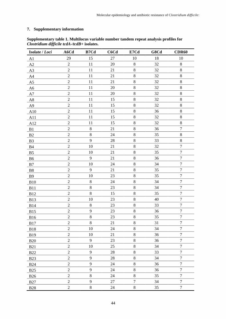
Molecular epidemiology and antibiotic resistance of Clostridium difficile:
44
7. Supplementary information
Supplementary table 1. Multilocus variable number tandem repeat analysis profiles for
Clostridium difficile tcdA-/tcdB+ isolates.
Isolate / Loci A6Cd B7Cd C6Cd E7Cd G8Cd CDR60
A1 29 15 27 10 18 10
A2 2 11 20 8 32 8
A3 2 11 21 8 32 8
A4 2 11 21 8 32 8
A5 2 11 21 8 32 8
A6 2 11 20 8 32 8
A7 2 11 20 8 32 8
A8 2 11 15 8 32 8
A9 2 11 15 8 32 8
A10 2 11 15 8 36 8
A11 2 11 15 8 32 8
A12 2 11 15 8 32 8
B1 2 8 21 8 36 7
B2 2 8 24 8 35 8
B3 2 9 28 8 33 8
B4 2 10 21 8 32 7
B5 2 10 21 8 35 7
B6 2 9 21 8 36 7
B7 2 10 24 8 34 7
B8 2 9 21 8 35 7
B9 2 10 23 8 35 7
B10 2 8 24 8 34 7
B11 2 8 23 8 34 7
B12 2 8 15 8 35 7
B13 2 10 23 8 40 7
B14 2 8 23 8 33 7
B15 2 9 23 8 36 7
B16 2 8 23 8 35 7
B17 2 8 21 8 31 7
B18 2 10 24 8 34 7
B19 2 10 21 8 36 7
B20 2 9 23 8 36 7
B21 2 10 25 8 34 7
B22 2 9 28 8 33 7
B23 2 9 28 8 34 7
B24 2 9 24 8 36 7
B25 2 9 24 8 36 7
B26 2 8 24 8 35 7
B27 2 9 27 7 34 7
B28 2 8 24 8 35 7
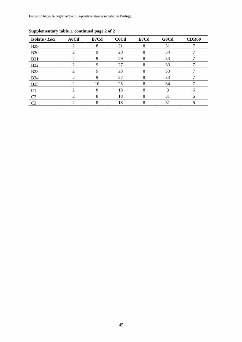
Focus on toxin A-negative/toxin B-positive strains isolated in Portugal
45
Supplementary table 1. continued page 2 of 2
Isolate / Loci A6Cd B7Cd C6Cd E7Cd G8Cd CDR60
B29 2 8 21 8 31 7
B30 2 9 28 8 34 7
B31 2 9 29 8 33 7
B32 2 9 27 8 33 7
B33 2 9 28 8 33 7
B34 2 9 27 8 33 7
B35 2 10 25 8 34 7
C1 2 8 18 8 3 6
C2 2 8 18 8 31 6
C3 2 8 18 8 31 6


![White Paper Antibiotic Use and Resistance: Moving Forward ... - Symp... · White Paper: Antibiotic Use & Resistance [3] BACKGROUND The symposium Antibiotic Use and Resistance: Moving](https://img.pdfslide.us/doc/110x75/5f0aa9c27e708231d42cb9b0/white-paper-antibiotic-use-and-resistance-moving-forward-symp-white.jpg)