Embed Size (px)
Citation preview
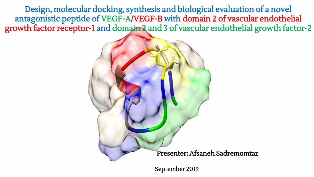
Design, molecular docking, synthesis and biological evaluation of a novel antagonistic peptide of VEGF-A/VEGF-B with domain 2 of vascular endothelial
growth factor receptor-1 and domain 2 and 3 of vascular endothelial growth factor-2
Presenter: Afsaneh Sadremomtaz
September 2019
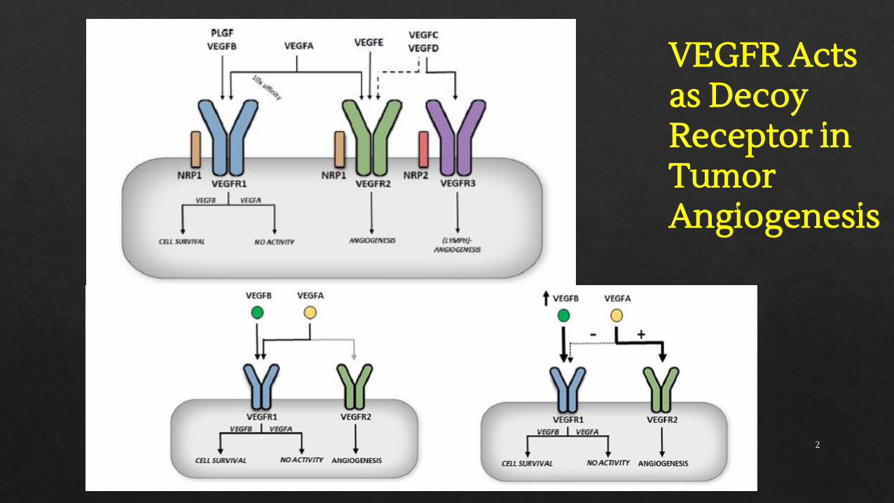
VEGFR Acts as DecoyReceptor in TumorAngiogenesis
2
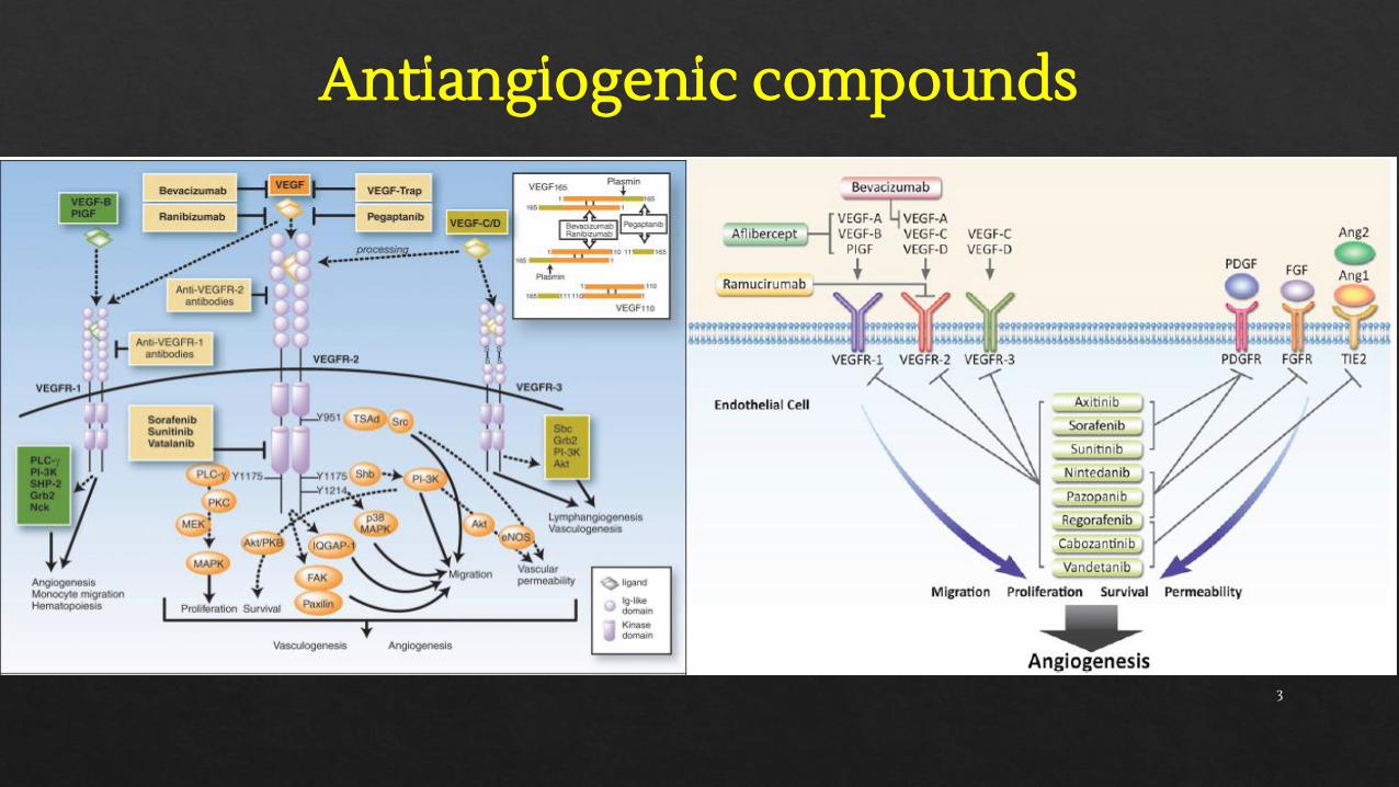
Antiangiogenic compounds
3
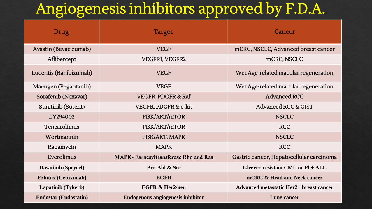
Drug Target Cancer
Avastin (Bevacizumab) VEGF mCRC, NSCLC, Advanced breast cancer
Aflibercept VEGFR1, VEGFR2 mCRC, NSCLC
Lucentis (Ranibizumab) VEGF Wet Age-related macular regeneration
Macugen (Pegaptanib) VEGF Wet Age-related macular regeneration
Sorafenib (Nexavar) VEGFR, PDGFR & Raf Advanced RCC
Sunitinib (Sutent) VEGFR, PDGFR & c-kit Advanced RCC & GIST
LY294002 PI3K/AKT/mTOR NSCLC
Temsirolimus PI3K/AKT/mTOR RCC
Wortmannin PI3K/AKT, MAPK NSCLC
Rapamycin MAPK RCC
Everolimus MAPK- Farnesyltransferase Rho and Ras Gastric cancer, Hepatocellular carcinoma
Dasatinib (Sprycel) Bcr-Abl & Src Gleevec-resistant CML or Ph+ ALL
Erbitux (Cetuximab) EGFR mCRC & Head and Neck cancer
Lapatinib (Tykerb) EGFR & Her2/neu Advanced metastatic Her2+ breast cancer
Endostar (Endostatin) Endogenous angiogenesis inhibitor Lung cancer
Angiogenesis inhibitors approved by F.D.A.
4
Advantages of peptides in antiangiogenic therapy
⮚ High cost of recombinant proteins⮚ Small size of peptides⮚ Ease of synthesis and modification⮚ Penetrate further into tissues, tumor-penetrating ability,
less immunogenic, easy to produce, lower manufacturing costs, greater efficacy, selectivity and specificityand good biocompatibility
⮚ Structure-function relationship and active epitopes of proteins
5

Aim of study
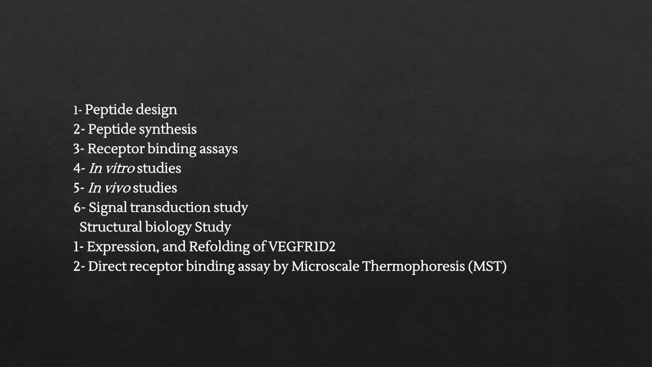
1- Peptide design2- Peptide synthesis3- Receptor binding assays4- In vitro studies5- In vivo studies6- Signal transduction study
Structural biology Study1- Expression, and Refolding of VEGFR1D2
2- Direct receptor binding assay by Microscale Thermophoresis (MST)
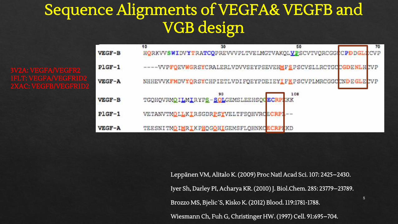
Sequence Alignments of VEGFA& VEGFB and VGB design
Leppänen VM, Alitalo K. (2009) Proc Natl Acad Sci. 107: 2425–2430.
Iyer Sh, Darley PI, Acharya KR. (2010) J. Biol.Chem. 285: 23779–23789.
Brozzo MS, Bjelic´S, Kisko K. (2012) Blood. 119:1781-1788.
Wiesmann Ch, Fuh G, Christinger HW. (1997) Cell. 91:695–704.
3V2A: VEGFA/VEGFR21FLT: VEGFA/VEGFR1D22XAC: VEGFB/VEGFR1D2
8
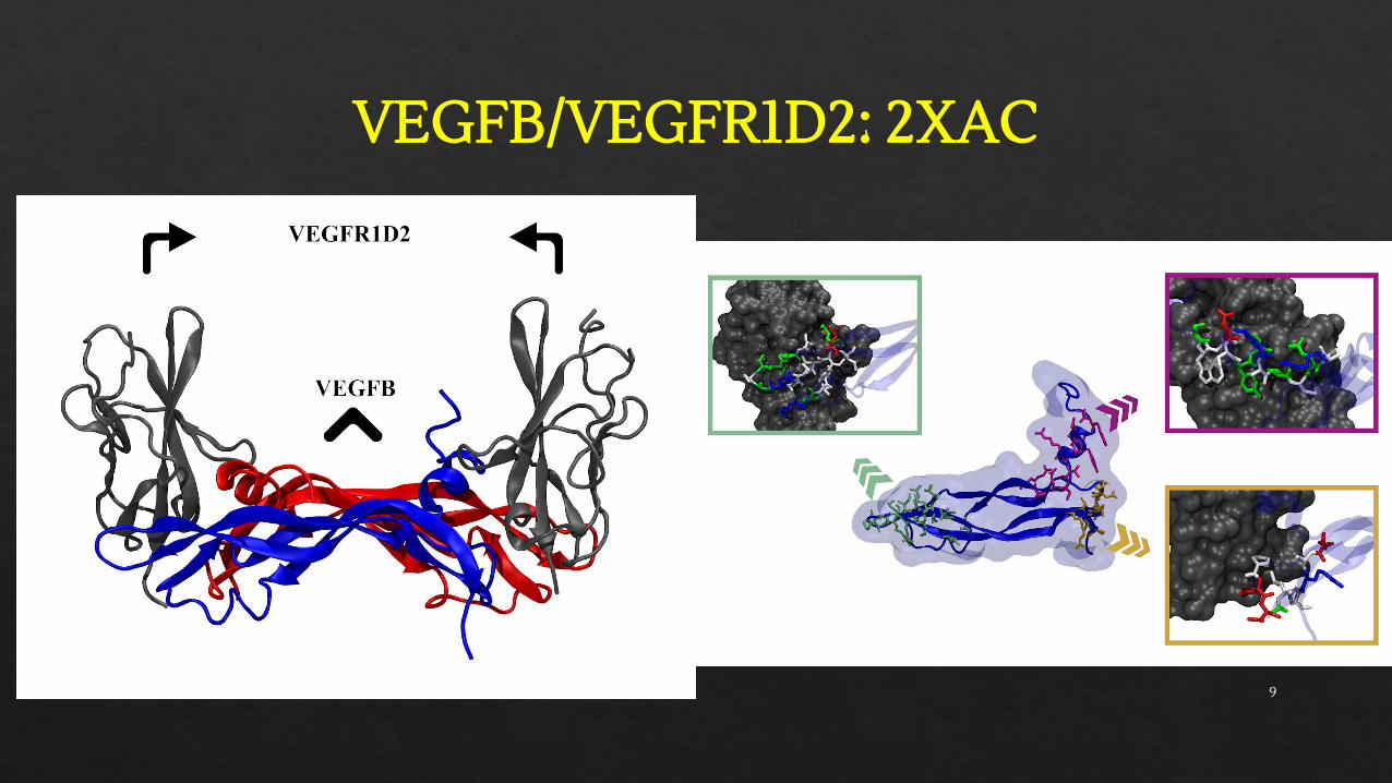
VEGFB/VEGFR1D2: 2XAC
9
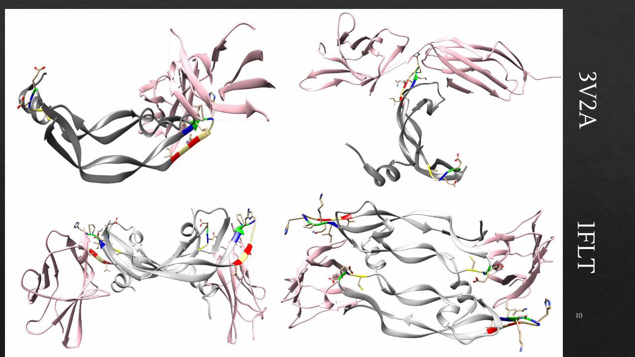
3V2A
1FLT
10
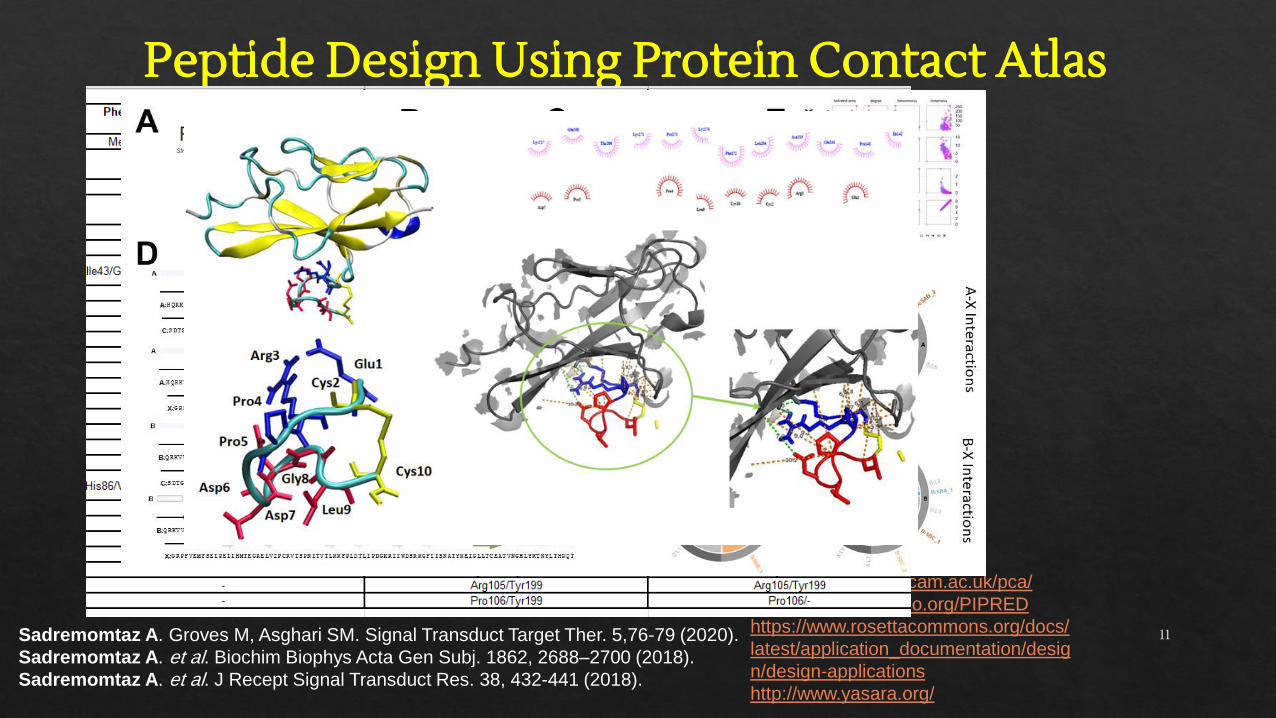
Peptide Design Using Protein Contact Atlas
Sadremomtaz A. Groves M, Asghari SM. Signal Transduct Target Ther. 5,76-79 (2020).
Sadremomtaz A. et al. Biochim Biophys Acta Gen Subj. 1862, 2688–2700 (2018).
Sadremomtaz A. et al. J Recept Signal Transduct Res. 38, 432-441 (2018).
http://www.mrclmb.cam.ac.uk/pca/
http://www.bioinsilico.org/PIPRED
https://www.rosettacommons.org/docs/
latest/application_documentation/desig
n/design-applications
http://www.yasara.org/
11
1621.
8
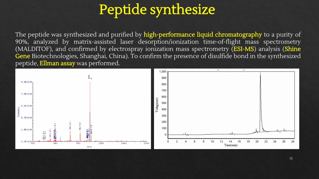
The peptide was synthesized and purified by high-performance liquid chromatography to a purity of90%, analyzed by matrix-assisted laser desorption/ionization time-of-flight mass spectrometry(MALDITOF), and confirmed by electrospray ionization mass spectrometry (ESI-MS) analysis (ShineGene Biotechnologies, Shanghai, China). To confirm the presence of disulfide bond in the synthesizedpeptide, Ellman assay was performed.
Peptide synthesize
12
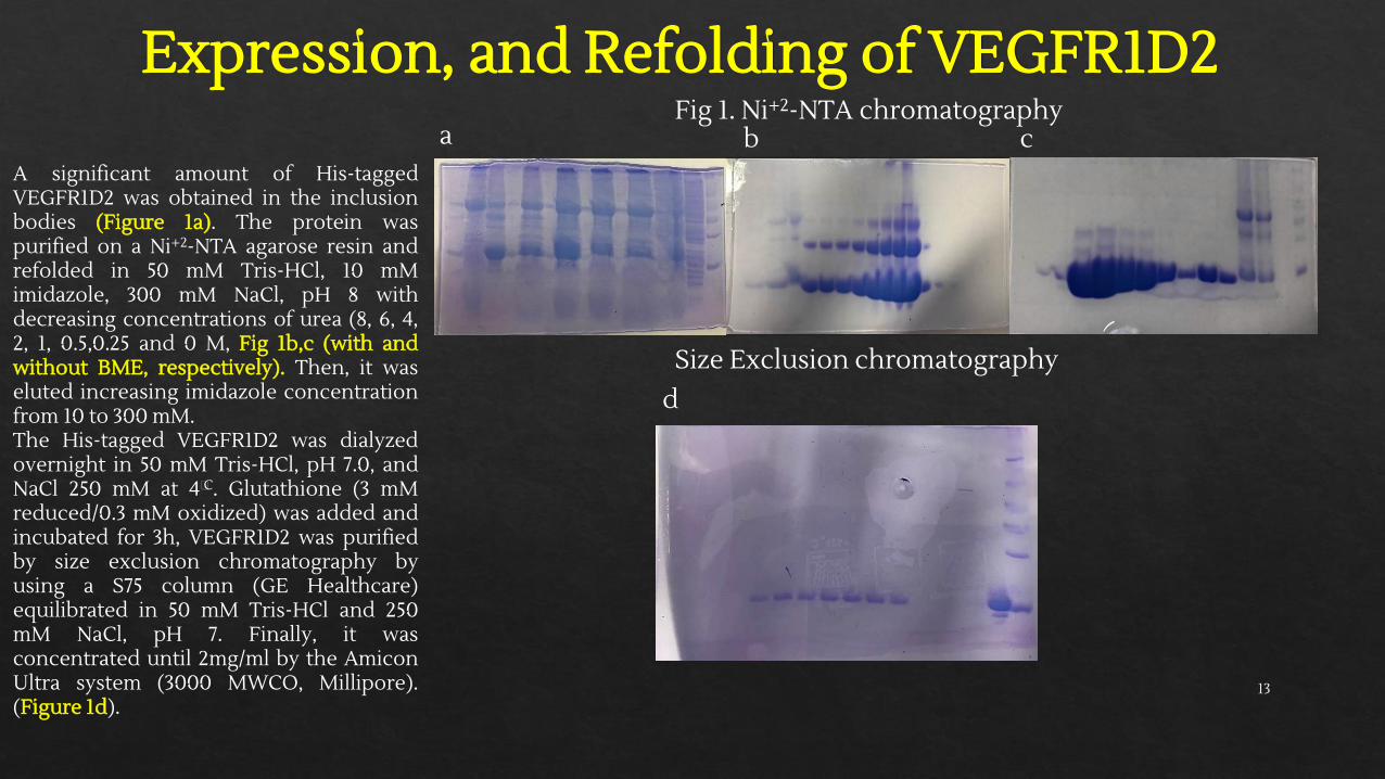
Expression, and Refolding of VEGFR1D2
A significant amount of His-taggedVEGFR1D2 was obtained in the inclusionbodies (Figure 1a). The protein waspurified on a Ni+2-NTA agarose resin andrefolded in 50 mM Tris-HCl, 10 mMimidazole, 300 mM NaCl, pH 8 withdecreasing concentrations of urea (8, 6, 4,2, 1, 0.5,0.25 and 0 M, Fig 1b,c (with andwithout BME, respectively). Then, it waseluted increasing imidazole concentrationfrom 10 to 300 mM.The His-tagged VEGFR1D2 was dialyzedovernight in 50 mM Tris-HCl, pH 7.0, andNaCl 250 mM at 4⸰C. Glutathione (3 mMreduced/0.3 mM oxidized) was added andincubated for 3h, VEGFR1D2 was purifiedby size exclusion chromatography byusing a S75 column (GE Healthcare)equilibrated in 50 mM Tris-HCl and 250mM NaCl, pH 7. Finally, it wasconcentrated until 2mg/ml by the AmiconUltra system (3000 MWCO, Millipore).(Figure 1d).
Fig 1. Ni+2-NTA chromatography
Size Exclusion chromatography
a b c
d
13
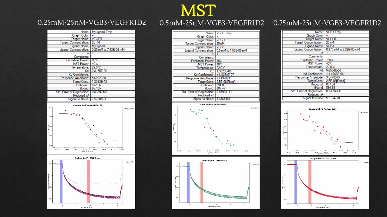
MST0.5mM-25nM-VGB3-VEGFR1D20.25mM-25nM-VGB3-VEGFR1D2 0.75mM-25nM-VGB3-VEGFR1D2
14
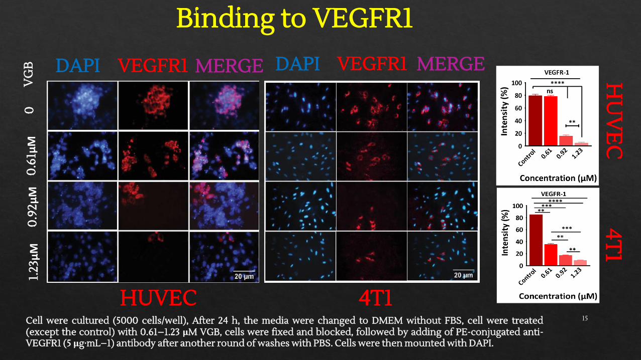
Binding to VEGFR1
HUVEC 4T1
DAPI VEGFR1 MERGE DAPI VEGFR1 MERGE
0.9
2μM
00
.61μM
1.23
μM
Cell were cultured (5000 cells/well), After 24 h, the media were changed to DMEM without FBS, cell were treated(except the control) with 0.61–1.23 μM VGB, cells were fixed and blocked, followed by adding of PE-conjugated anti-VEGFR1 (5 μg·mL−1) antibody after another round of washes with PBS. Cells were then mounted with DAPI.
HU
VE
C4T
1V
GB
15
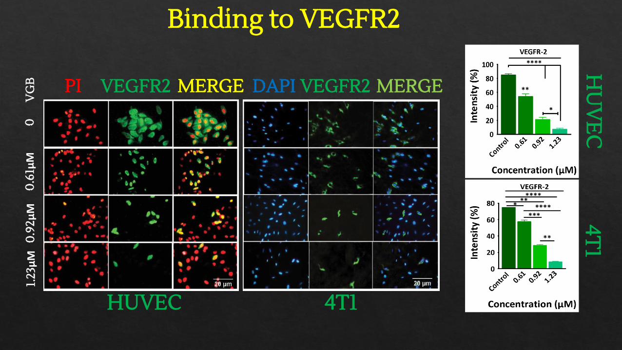
Binding to VEGFR2
HUVEC 4T1
PI VEGFR2 MERGE DAPI VEGFR2 MERGE
00
.61μM
0.9
2μM
1.23
μM
HU
VE
C4T
1V
GB
16

Receptor binding specificity
PI bFGFR1 MERGE0
1.23
μM
VG
B
17
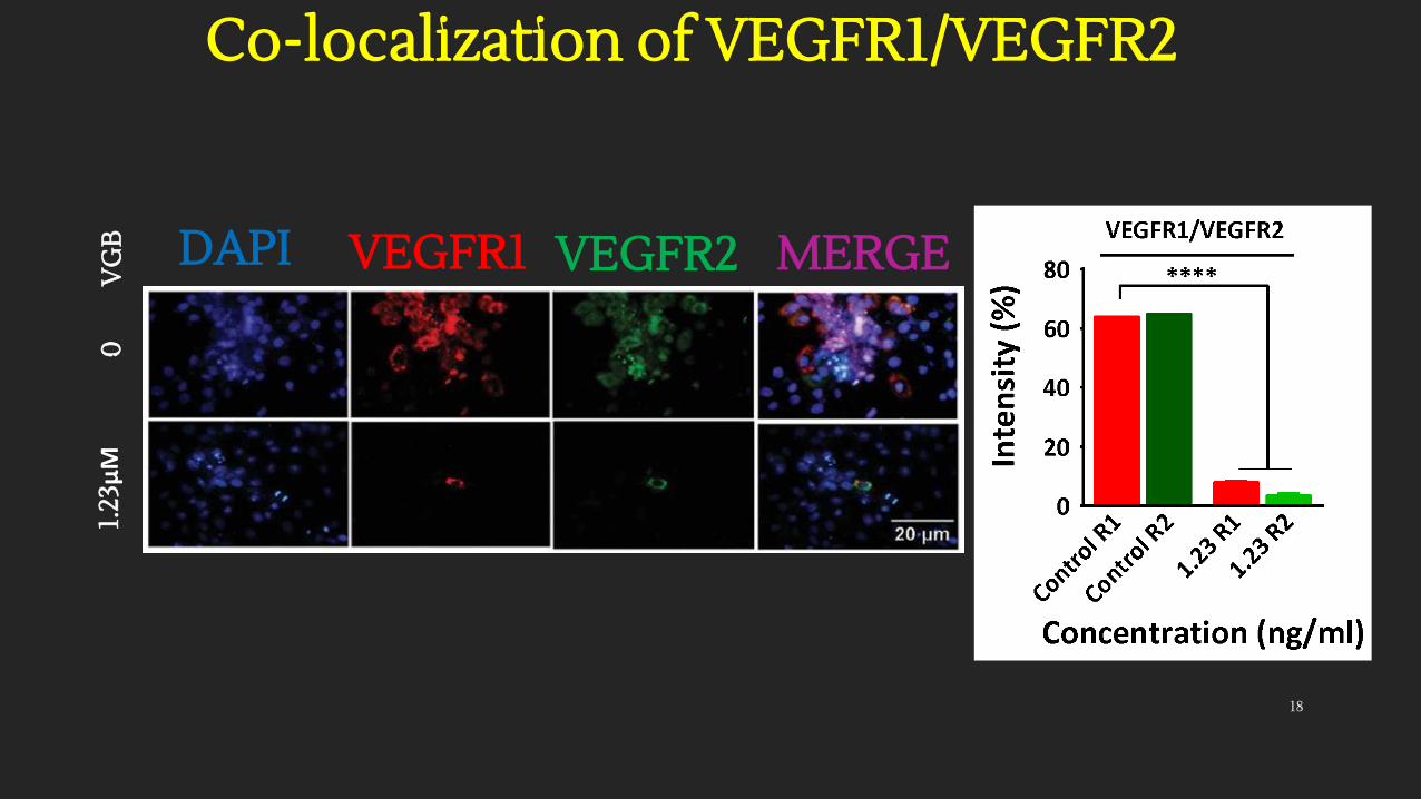
Co-localization of VEGFR1/VEGFR2
DAPI VEGFR1 VEGFR2 MERGE
01.
23μM
VG
B
18

Studies on difference hallmarks of angiogenesis

Antagonistic peptide (VGB) of VEGFA & VEGFB can able to block downstream signaling pathways of VEGFR1 & VEGFR2
Proliferation: Inhibiting activation of MAPK/ERK1/2, PI3K/AKT, NF-kB, c-Myc
Migration: Downregulation on expression of; FAK/Paxilin, PAK/Cofilin, PI3K/AKT, MAPK/ERK1/2, EMT/E-cadherin
Metastasis: Downregulating on expression of; PI3K/AKT, EMT/E-cadherin, MMP-9, c-Myc, NF-kB
Apoptosis: Downregulation of PI3K/AKT/p53 (proapoptotic), Bcl-2 (antiapoptotic)
Do
wn
stre
am s
ign
alin
g p
ath
way
s
Cell survival: Downregulation of PI3K/AKT/NF-kB
20
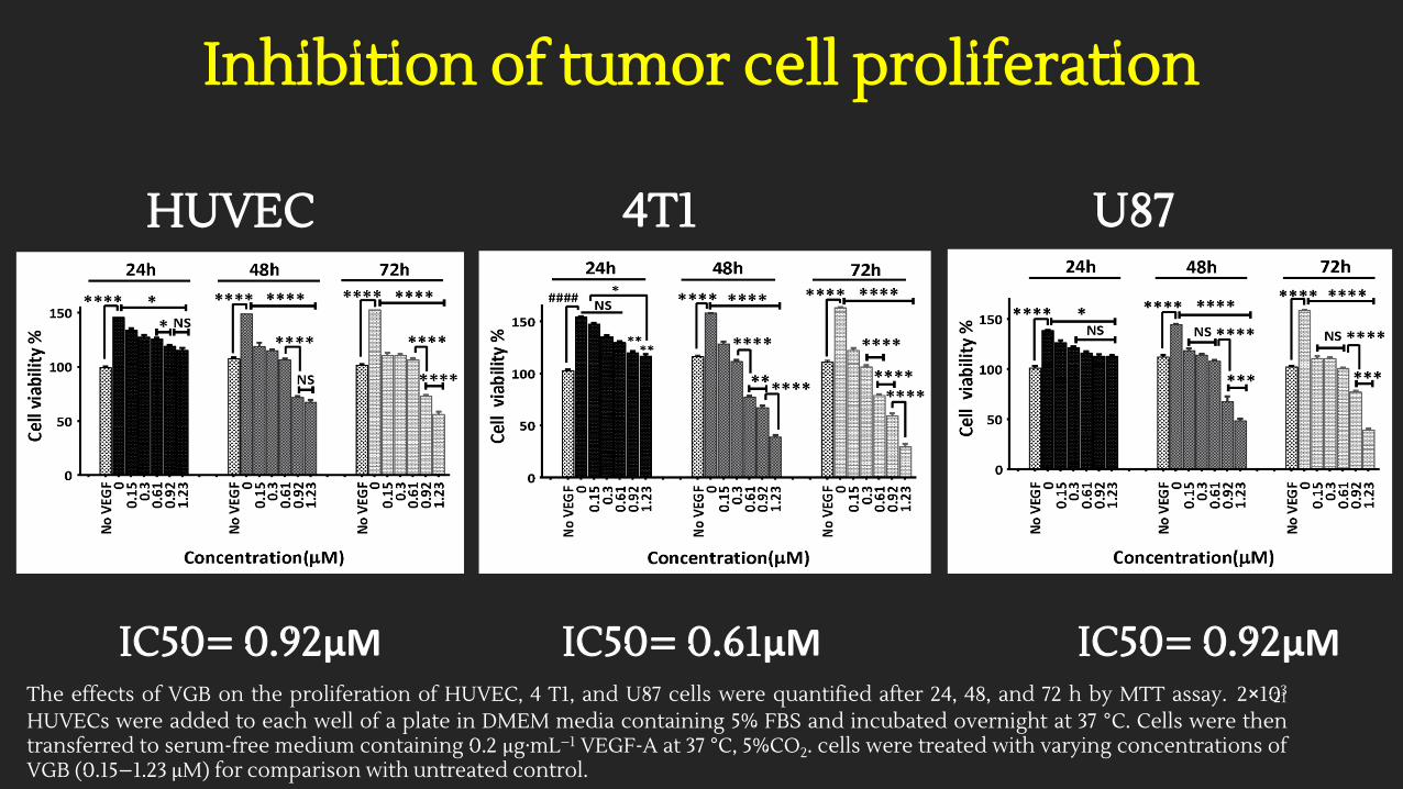
Inhibition of tumor cell proliferation
HUVEC 4T1 U87
IC50= 0.92μM IC50= 0.61μM IC50= 0.92μMThe effects of VGB on the proliferation of HUVEC, 4 T1, and U87 cells were quantified after 24, 48, and 72 h by MTT assay. 2×10³
HUVECs were added to each well of a plate in DMEM media containing 5% FBS and incubated overnight at 37 °C. Cells were thentransferred to serum-free medium containing 0.2 μg·mL−1 VEGF-A at 37 °C, 5%CO2. cells were treated with varying concentrations ofVGB (0.15–1.23 μM) for comparison with untreated control.
21
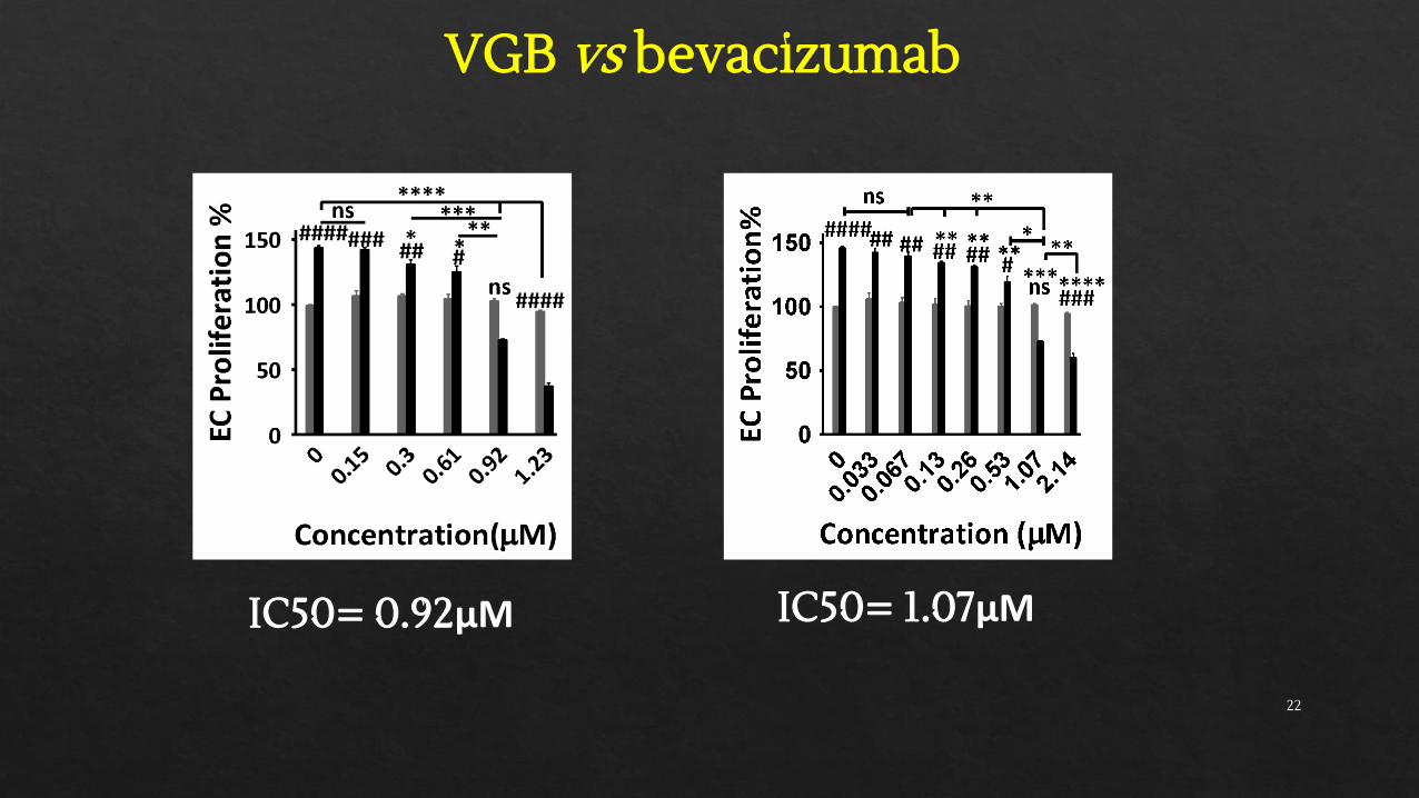
VGB vs bevacizumab
IC50= 0.92μM IC50= 1.07μM
22
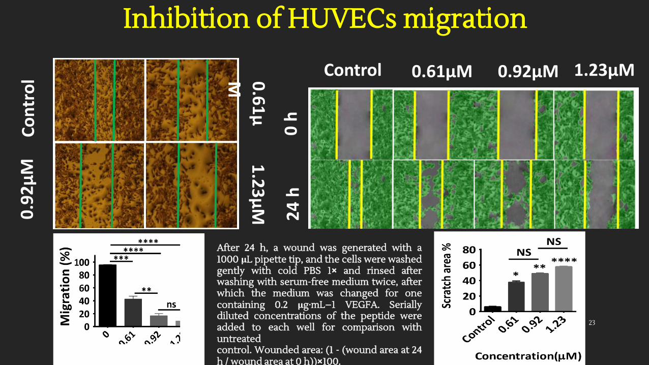
Inhibition of HUVECs migrationC
on
tro
l0
.92
µM
0.6
1µ
M
1.2
3µ
M
0 h
24
h
Control 0.61µM 0.92µM 1.23µM
After 24 h, a wound was generated with a1000 μL pipette tip, and the cells were washedgently with cold PBS 1× and rinsed afterwashing with serum-free medium twice, afterwhich the medium was changed for onecontaining 0.2 μg·mL−1 VEGFA. Seriallydiluted concentrations of the peptide wereadded to each well for comparison withuntreatedcontrol. Wounded area: (1 - (wound area at 24h / wound area at 0 h))×100.
23
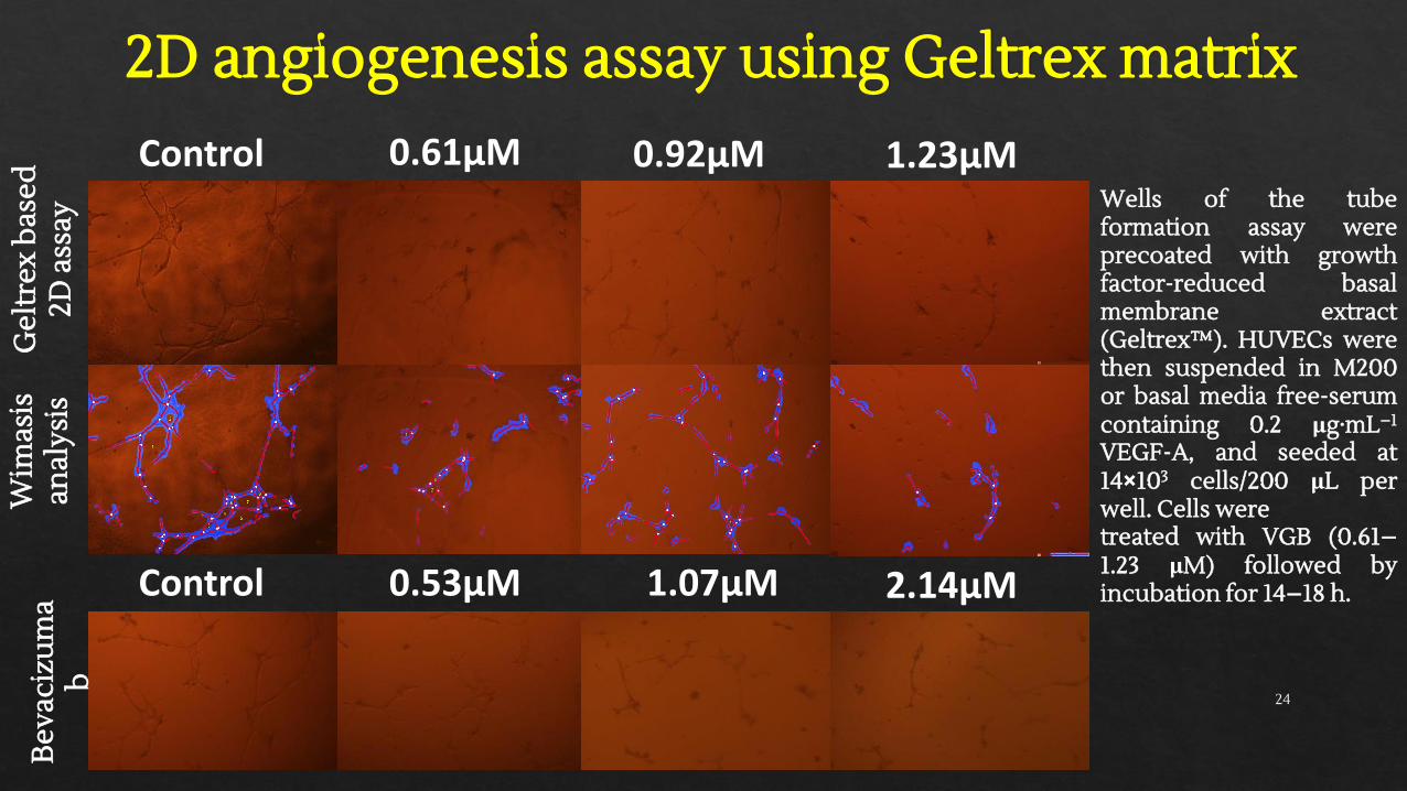
2D angiogenesis assay using Geltrex matrixG
eltr
ex b
ased
2D
ass
ayW
imas
is
anal
ysis
Control 0.61µM 0.92µM 1.23µM
Bev
aciz
um
ab
0.53µM 1.07µM 2.14µMControl
Wells of the tubeformation assay wereprecoated with growthfactor-reduced basalmembrane extract(Geltrex™). HUVECs werethen suspended in M200or basal media free-serumcontaining 0.2 μg·mL−1
VEGF-A, and seeded at14×103 cells/200 μL perwell. Cells weretreated with VGB (0.61–1.23 μM) followed byincubation for 14–18 h.
24
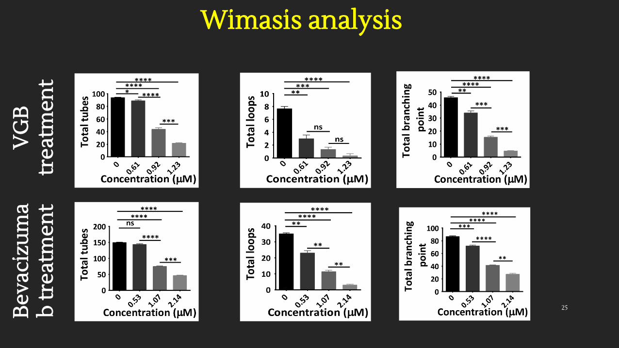
Wimasis analysisV
GB
tr
eatm
ent
Bev
aciz
um
ab
trea
tmen
t
25
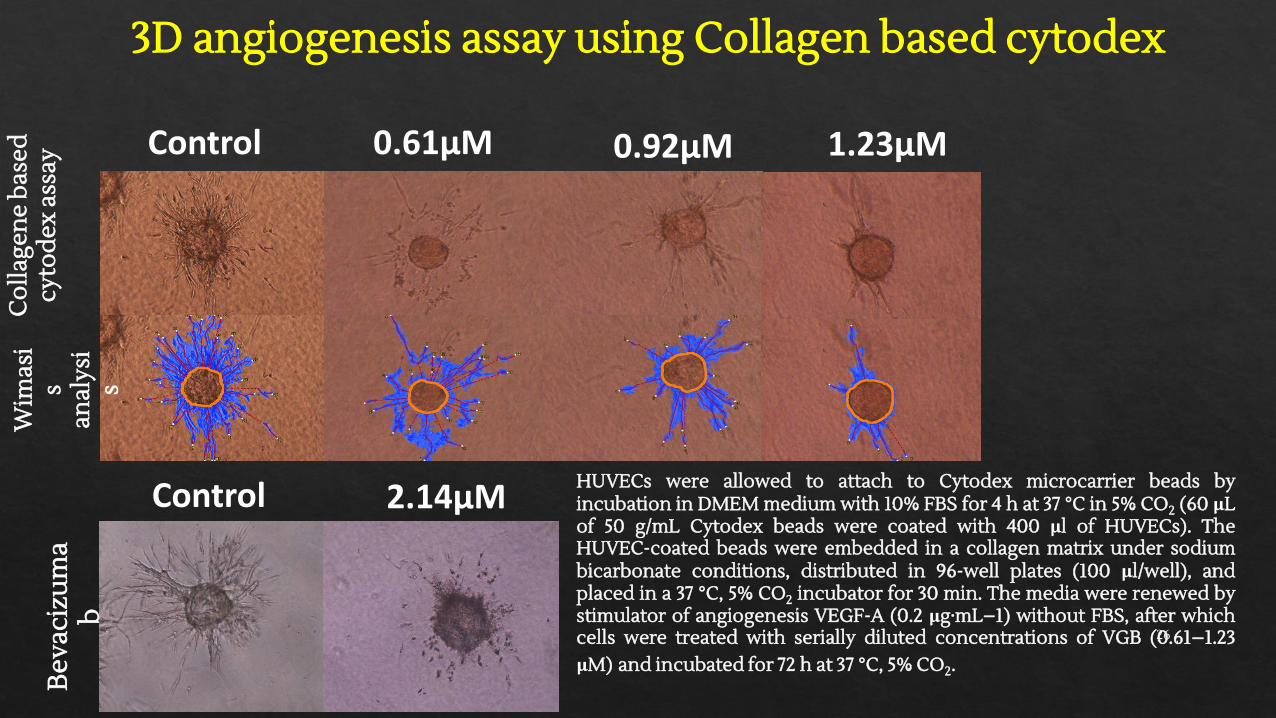
3D angiogenesis assay using Collagen based cytodex
Control 0.61µM 0.92µM 1.23µM
Co
llage
ne
bas
ed
cyto
dex
ass
ayW
imas
is
anal
ysi
s
Control 2.14µM
Bev
aciz
um
ab
HUVECs were allowed to attach to Cytodex microcarrier beads byincubation in DMEM medium with 10% FBS for 4 h at 37 °C in 5% CO2 (60 μLof 50 g/mL Cytodex beads were coated with 400 μl of HUVECs). TheHUVEC-coated beads were embedded in a collagen matrix under sodiumbicarbonate conditions, distributed in 96-well plates (100 μl/well), andplaced in a 37 °C, 5% CO2 incubator for 30 min. The media were renewed bystimulator of angiogenesis VEGF-A (0.2 μg·mL−1) without FBS, after whichcells were treated with serially diluted concentrations of VGB (0.61–1.23μM) and incubated for 72 h at 37 °C, 5% CO2.
26
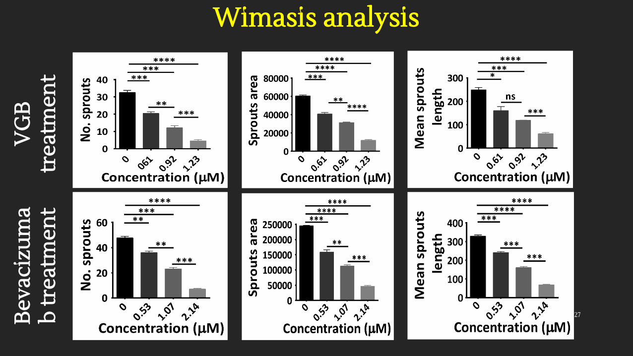
Wimasis analysisV
GB
tr
eatm
ent
Bev
aciz
um
ab
trea
tmen
t
27

In vivo and Signal transduction studies

Regression of 4T1 murine mammary carcinoma tumor
Tumor cells (4 T1; 1×10⁶ cells/500 μl or 1×10⁵ cells/50 μl)were injected subcutaneously into the right flanks ofmice (n=3–5). To generate the metastatic model, 4 T1tumor models were sterilized, excised from the breastcancer bearing BALB/c mice, cut into pieces of<0.3cm3, and subcutaneously implanted into the animals'right flanks under ketamine (100 mg/kg, i.p.) andxylazine (10 mg/kg, i.p.) anesthesia.Animals carrying tumors of size ~400mm3 wererandomized to groups (n=6). The treatment groupsreceived 1, 2.5, and 10 mg/kg i.p. of the peptide dailyand control group received PBS i.p for two weeks. Thetumor volume was measured every two days by adigital Vernier caliper (Mitutoyo, Japan), using thefollowing formula: v=a2×b×0.52.
a b c d e f
29
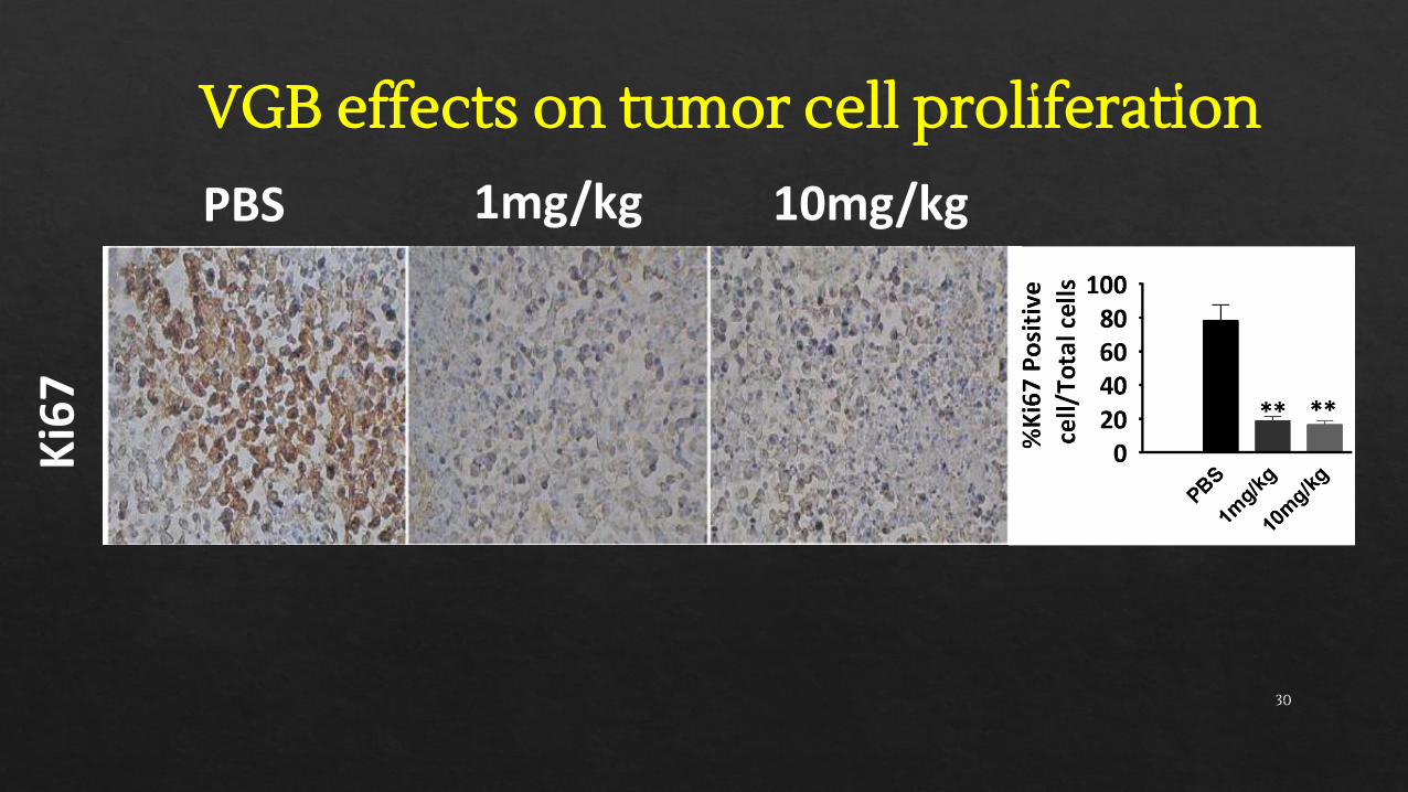
VGB effects on tumor cell proliferationK
i67
PBS 1mg/kg 10mg/kg
30
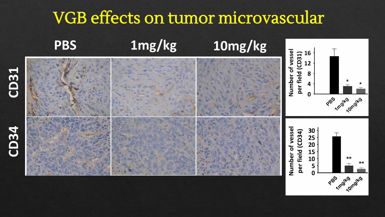
VGB effects on tumor microvascular
PBS 1mg/kg 10mg/kg
CD
31
CD
34
31

VGB effects on apoptosis inductionPBS 1mg/kg 10mg/kgB
cl-2
P5
3TU
NEL
Ear
ly a
po
pto
sis
Lat
e ap
op
tosi
s
32
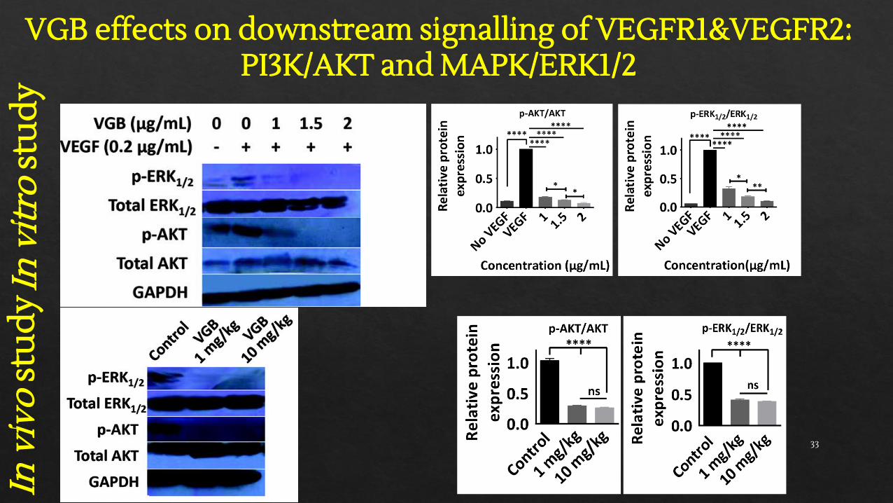
VGB effects on downstream signalling of VEGFR1&VEGFR2: PI3K/AKT and MAPK/ERK1/2
In v
ivo
stu
dy
In v
itro
stu
dy
33
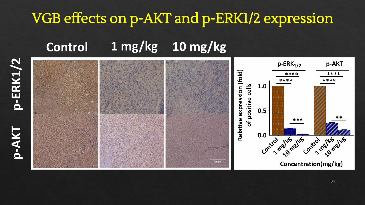
VGB effects on p-AKT and p-ERK1/2 expressionp
-AK
Tp
-ER
K1
/2
Control 1 mg/kg 10 mg/kg
34
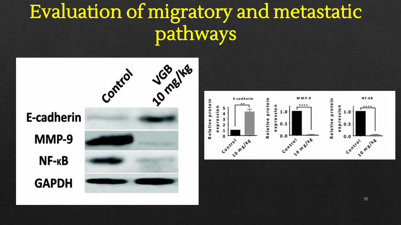
Evaluation of migratory and metastatic pathways
35
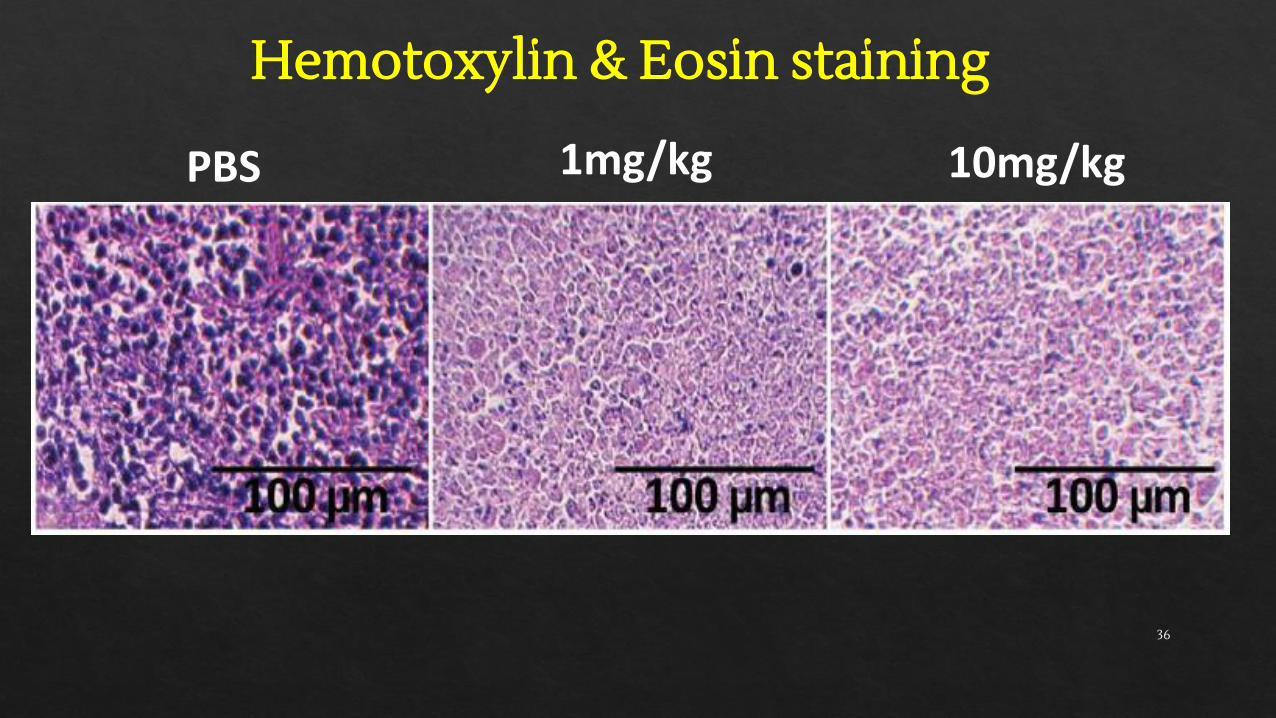
Hemotoxylin & Eosin staining
PBS 1mg/kg 10mg/kg
36
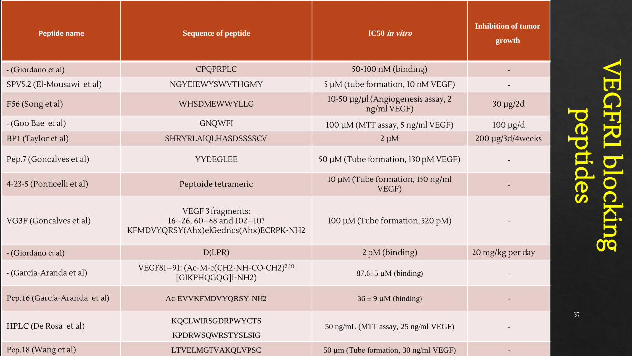
Peptide name Sequence of peptide IC50 in vitroInhibition of tumor
growth
- (Giordano et al) CPQPRPLC 50-100 nM (binding) -
SPV5.2 (El-Mousawi et al) NGYEIEWYSWVTHGMY 5 µM (tube formation, 10 nM VEGF) -
F56 (Song et al) WHSDMEWWYLLG10-50 µg/µl (Angiogenesis assay, 2
ng/ml VEGF)30 µg/2d
- (Goo Bae et al) GNQWFI 100 µM (MTT assay, 5 ng/ml VEGF) 100 µg/d
BP1 (Taylor et al) SHRYRLAIQLHASDSSSSCV 2 µM 200 µg/3d/4weeks
Pep.7 (Goncalves et al) YYDEGLEE 50 µM (Tube formation, 130 pM VEGF) -
4-23-5 (Ponticelli et al) Peptoide tetrameric10 µM (Tube formation, 150 ng/ml
VEGF) -
VG3F (Goncalves et al)VEGF 3 fragments:
16–26, 60–68 and 102–107KFMDVYQRSY(Ahx)elGedncs(Ahx)ECRPK-NH2
100 µM (Tube formation, 520 pM) -
- (Giordano et al) D(LPR) 2 pM (binding) 20 mg/kg per day
- (García-Aranda et al)VEGF81–91: (Ac-M-c(CH2-NH-CO-CH2)2,10
[GIKPHQGQG]I-NH2) 87.6±5 µM (binding) -
Pep.16 (García-Aranda et al) Ac-EVVKFMDVYQRSY-NH2 36 ± 9 µM (binding) -
HPLC (De Rosa et al)KQCLWIRSGDRPWYCTS
KPDRWSQWRSTYSLSIG50 ng/mL (MTT assay, 25 ng/ml VEGF) -
Pep.18 (Wang et al) LTVELMGTVAKQLVPSC 50 µm (Tube formation, 30 ng/ml VEGF) -
VE
GFR
1 blo
cking
pep
tides
37
VE
GFR
2 blo
cking
pep
tides
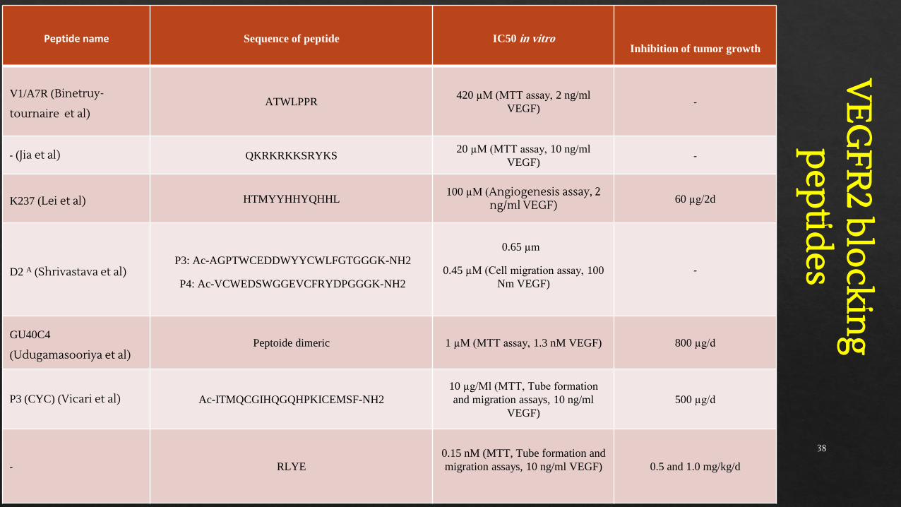
Peptide name Sequence of peptide IC50 in vitroInhibition of tumor growth
V1/A7R (Binetruy-
tournaire et al)ATWLPPR
420 µM (MTT assay, 2 ng/ml
VEGF)-
- (Jia et al) QKRKRKKSRYKS20 µM (MTT assay, 10 ng/ml
VEGF)-
K237 (Lei et al) HTMYYHHYQHHL100 µM (Angiogenesis assay, 2
ng/ml VEGF) 60 µg/2d
D2 A (Shrivastava et al)P3: Ac-AGPTWCEDDWYYCWLFGTGGGK-NH2
P4: Ac-VCWEDSWGGEVCFRYDPGGGK-NH2
0.65 µm
0.45 µM (Cell migration assay, 100
Nm VEGF)
-
GU40C4
(Udugamasooriya et al)Peptoide dimeric 1 µM (MTT assay, 1.3 nM VEGF) 800 µg/d
P3 (CYC) (Vicari et al) Ac-ITMQCGIHQGQHPKICEMSF-NH2
10 µg/Ml (MTT, Tube formation
and migration assays, 10 ng/ml
VEGF)
500 µg/d
- RLYE
0.15 nM (MTT, Tube formation and
migration assays, 10 ng/ml VEGF) 0.5 and 1.0 mg/kg/d
38
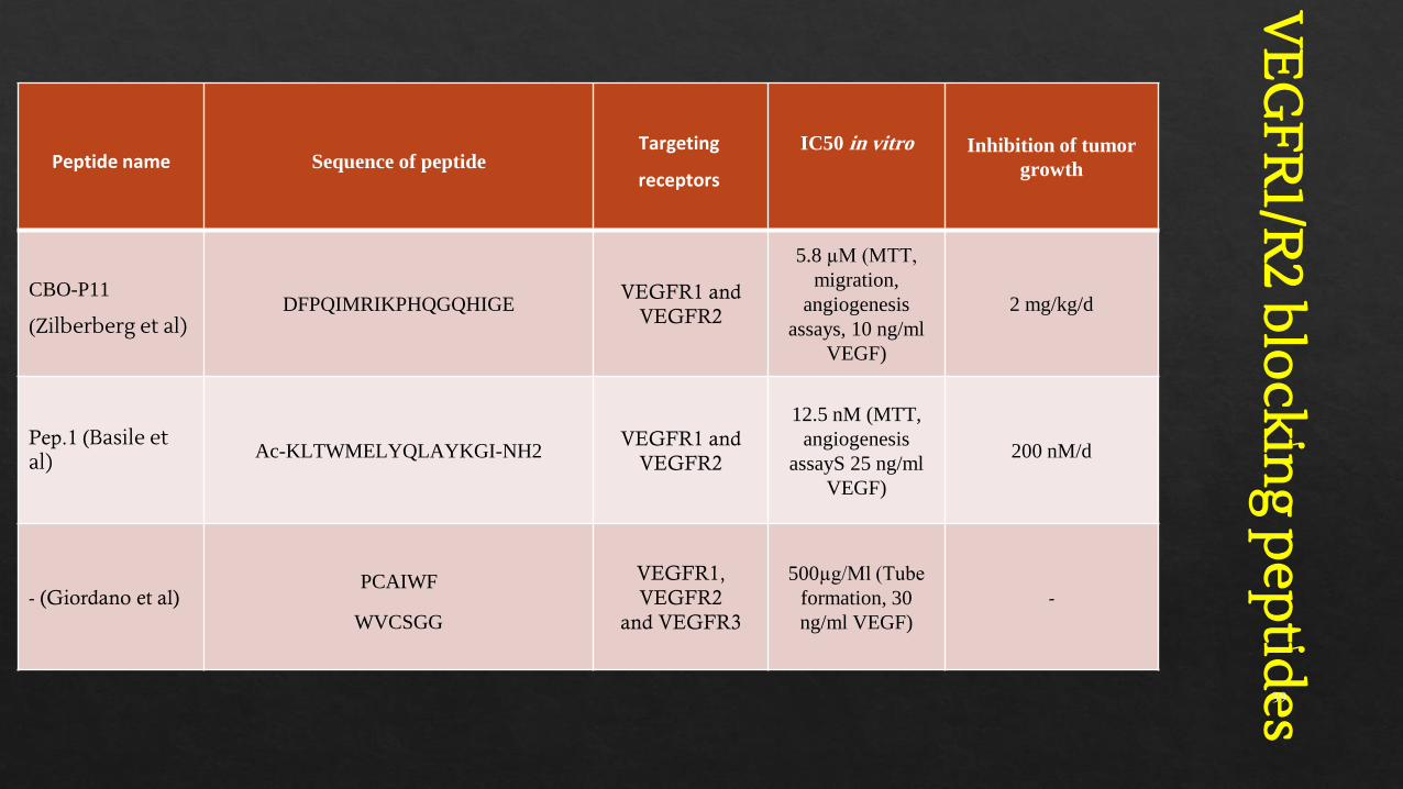
Peptide name Sequence of peptideTargeting
receptors
IC50 in vitro Inhibition of tumor
growth
CBO-P11
(Zilberberg et al)DFPQIMRIKPHQGQHIGE
VEGFR1 and
VEGFR2
5.8 µM (MTT,
migration,
angiogenesis
assays, 10 ng/ml
VEGF)
2 mg/kg/d
Pep.1 (Basile et al) Ac-KLTWMELYQLAYKGI-NH2
VEGFR1 and
VEGFR2
12.5 nM (MTT,
angiogenesis
assayS 25 ng/ml
VEGF)
200 nM/d
- (Giordano et al)PCAIWF
WVCSGG
VEGFR1,
VEGFR2
and VEGFR3
500µg/Ml (Tube
formation, 30
ng/ml VEGF)
-
VE
GFR
1/R2 b
lockin
g pep
tides 39
Conclusion:1.VGB bound to both VEGFR1 and VEGFR2 and blocked their homo- and heterodimerization in human umbilicalvein endothelial cells (HUVECs) as well as 4T1 mammary carcinoma tumor cells.
2.Dual specificity of VGB was confirmed by its dose-dependent inhibitory effect on the VEGF (200 ng/ml)-stimulated proliferation of 4T1 mammary carcinoma tumor cells (that express VEGFR1more than VEGFR-2) andU87 glioblastoma cells (that highly express VEGFR-2). In good agreement with our previous study, MST resultsreveals that The Kd for the VGB3 (0.5mM)-VEGFR1D2 (25nM) complex (1:1 binding stoichiometry) was 1.96µM.
3.The anti-angiogenic potency of VGB was shown by the observation that, through abrogation of AKT and ERK1/2phosphorylation, VEGFA-stimulated proliferation, migration, and two- and three-dimensional tube formation inHUVECs were inhibited more potently by VGB than by bevacizumab.
4.In a murine 4T1 MCT model, VGB strongly inhibited tumor growth without causing weight loss.
5.Blocking tumor growth and tumor angiogenesis in VGB-treatment against PBS-treated one accompanied byinhibition of AKT and ERK1/2 phosphorylation, a significant decrease in tumor cell proliferation (Ki-67expression), migration (FAK/Paxilin, PAK2/Cofilin expression), angiogenesis (CD31 and CD34 expression), anincrease in apoptosis index (increased TUNEL staining and p53 expression and decreased Bcl-2 expression) andthe expression level of a hallmark of EMT axis (E-cadherin expression), and the suppression of systematicspreading of the tumor (reduced NF-κB and MMP-9 and increased E-cadherin expression).
Our results demonstrate the dual specificity of VGB for VEGFR1 and VEGFR2, through which the PI3K/AKT and MAPK/ERK1/2 signaling pathways can be abrogated and, subsequently,
angiogenesis, tumor growth, and metastasis are inhibited and apoptosis is induced.
40
Publications
◈ 1- Sadremomtaz A. et al. Dual blockade of VEGFR1 and VEGFR2 by a novel peptide abrogates VEGFdriven angiogenesis, tumor growth, and metastasis through PI3K/ AKT and MAPK/ERK1/2 pathway. Biochim Biophys Acta Gen Subj. 1862, 2688–2700 (2018).
◈ 2- Sadremomtaz A. et al. Suppression of migratory and metastatic pathways via blocking VEGFR1 and VEGFR2. J Recept Signal Transduct Res. 38, 432-441 (2018).
◈ 3- Sadremomtaz A. Groves M, Asghari SM. Molecular docking, synthesis and biological evaluation of Vascular Endothelial Growth Factor (VEGF) B based peptide as anti-angiogenic agent targeting the second domain of the Vascular Endothelial Growth Factor Receptor 1 (VEGFR1D2) for anticancer application. Signal Transduct Target Ther. 5,76-79 (2020); https://doi.org/10.1038/s41392-020-0177-z.
41

Thank you for your attention
42