Embed Size (px)
Citation preview
Page 368 www.ijiras.com | Email: [email protected]
International Journal of Innovative Research and Advanced Studies (IJIRAS)
Volume 4 Issue 1, January 2017
ISSN: 2394-4404
Molecular Docking Studies; Dietary Compounds As HIV 1 Protease
Inhibitors
Jisha Undarathi
Research Scholar, Department of Chemistry,
Kannur University
I. INTRODUCTION
A. BIOINFORMATICS
The term bioinformatics was coined by Paulien Hogeweg
and Ben Hesper in 1978 for the study of informatic processes
in biotic systems. Bioinformatics is a rapidly developing
branch of biology that derives knowledge from computer
analysis of biological information. Bioinformatics is the
science of developing computer databases and algorithms for
the purpose of speeding up and enhancing biological research.
It is a scientific discipline that comprises all aspects of the
gathering, storing, handling, analyzing, interpreting, and
spreading of biological information.
It involve the use of techniques from applied
mathematics, information, statistics and computer science to
solve biological problems. It has many practical applications
in different areas of biology and medicine [1][2].
B. BRANCHES OF BIOINFORMATICS
Bioinformatics has mainly three branches: 1) Molecular
bioinformatics, 2) Cellular and sub cellular bioinformatics and
3) Organic and community bioinformatics.
Subsidiary branches of these are:
Molecular bioinformatics:
Genomics
Proteomics
Drug design
Cellular and sub-cellular bioinformatics:
Metabolic pathway
Epigenetic
Neuro bioinformatics
Organic and community bioinformatics:
Bioinformatics of species diversity
Behaviour, evolution and effects of
Pollutanats
Abstract: The study indicates the importance of diet in the prevention and treatment of diseases. Five molecules that
“Docked” well into the active site of the target, HIV 1 Protease are selected. And they are considered to be as the “Hit
Molecules”. Glibenclamide, Lectin, Resveratrol, Oleuropein, Silymarin are considered as the hit molecules. As they are
docked into the active site of the target with a minimum energy (found to be stable). So these molecules can be suggested
to be the interesting candidates for further testing in the laboratory. Finally, this study strongly underscores the
importance of computational approaches in drug discovery, supplementing classical methods, thus saving enormous
amount of time and money.
Index Terms: Acquired Immunodeficiency Syndrome, ArgusLab, Bioinformatics, Computer-Aided Drug Design, Hit
Molecules, HIV 1 Protease, Inhibitor, Molecular docking, Pharmacophores, QSAR, Virtual Screening.
Page 369 www.ijiras.com | Email: [email protected]
International Journal of Innovative Research and Advanced Studies (IJIRAS)
Volume 4 Issue 1, January 2017
ISSN: 2394-4404
C. AIMS AND TASKS OF BIOINFORMATICS
Bioinformatics has three components: 1) the creation of
databases allowing the storage and management of large
biological data sets, 2) the development of algorithms and
statistics to determine relationships among members of large
datasets and 3) the use of these tools for the analysis and
interpretation of various types of biological data [3][4].
AIMS
To organize data in a way that follows researches to
access existing information and to submit new entries as
they are produced.
To develop tools and resources that aid in the analysis and
management of data.
To use these tools to analyse the data and interpret the
results in a biologically meaningful manner.
TASKS
The tasks in bioinformatics involve the analysis of
sequence information. This involves the following:
Identifying the genes in the DNA sequence from various
organism.
Developing methods to study the structure and function of
newly identified sequences and corresponding structural
RNA sequences.
Identifying families of related sequences and the
development of models.
Aligning similar sequences and generating phylogenetic
trees to examine evolutionary relationships.
Besides these, one of the important dimensions of
bioinformatics is identifying drug targets and pointing out lead
compounds.
D. APPLICATIONS OF BIOINFORMATICS
Biocomputing has found its application in many areas,
These include the following areas:
Genome and sequence analysis
Protein modeling
Molecular medicine
Functional genomics
Proteomics
Personalized medicine and Gene therapy
Antibiotic resistance
Crop improvement
Bioweapons
Drug designing [5][6]
E. DRUG DESIGNING
Drug design is the approach of finding drugs, based on
their biological targets. Drug discovery can be arrived by two
methods. The empirical and rational. The empirical method is
a bind and hit or lose method. Thousands of chemical
compounds are tested on the disease without even knowing the
target on which the drug acts and the mechanism of action.
Advent of bioinformatics has changed this paradigm to
rational approach. Rational approach starts from the clear
knowledge of the target as well as the mechanism by which it
is to be attacked [7].
F. BIOINFORMATICS AND DRUG DESIGNING
Drug designing is one of the important area in
bioinformatics. The processes of designing a new drug using
bioinformatics tools have open a new area of research.
Computers are used to gather, store, analyse and integrate
biological and genetic information which can then be applied
to gene-based drug discovery and development.
Bioinformatics helps to identify and validate drug targets, to
find and design molecules that uniquely interact with a
specific protein target and to optimize molecule [8][9].
G. COMPUTER-AIDED DRUG DESIGN
Computer-aided drug design is a theoretical tool that can
be used to identify novel potential drugs. The aim of using the
computer for drug design is to analyze the interactions
between the drug and its receptor site and to “design”
molecules that can give an optimal fit. The principle is that a
good fit results from structural and chemical complementary
to the target receptor. The techniques provided by
computational methods include computer graphics for
visualization and the methadology of theoretical chemistry. By
means of quantum mechanics the structure of small molecules
can be predicted to experimental accuaracy. Statistical
mechanics permits molecular motion and solvent effects to be
incorporated.
CADD, Computer Aided Drug Design is a discipline that
uses computational methods to stimulate drug-receptor
interactions. CADD methods are heavily depend on
bioinformatics tools, applications and databases. The CADD
helps to reduce the time and expense of the drug discovery
than the conventional drug discovery methods which used the
trail and error method for the discovery of new drugs [10].
BENEFITS OF CADD
CADD methods and bioinformatics tools offer significant
benefits for drug discovery programs.
COST SAVINGS: It suggests that the cost of drug
discovery and development has reached 800 million for each
drug successfully brought to market. Many biopharmaceutical
companies now use computational methods [11] and
bioinformatics tools to reduce this cost burden. Virtual
screening, lead optimization and predictions of bioavailability
and bioactivity can help guide experimental research. Only the
most promising experimental lines of inquiry can be followed
and experimental dead-ends can be avoided early based on the
results of CADD simulations [12].
TIME-TO-MARKET: The predictive power of CADD can
help drug research program to choose only the most promising
drug candidates. By focusing drug research on specific lead
candidates and avoiding potential “dead-end” compounds,
biopharmaceutical companies can get drugs to market more
quickly.
Page 370 www.ijiras.com | Email: [email protected]
International Journal of Innovative Research and Advanced Studies (IJIRAS)
Volume 4 Issue 1, January 2017
ISSN: 2394-4404
INSIGHT: one of the non-quantifiable benefits of CADD
and the use of bioinformatics tools is the deep insight that
researches acquire about drug-receptor interactions. Molecular
models of drug compounds can reveal intricate, atomic scale
binding properties that are difficult to envision in any other
way. This is an intangible benefit that can help design research
programs. Therefore CADD and bioinformatics together is a
powerful combination in drug research and development.
H. CONCEPT OF DRUG AND TARGETS
TARGETS
The first step and a challenging task in drug discovery is
to identify the target. Drug targets are the macromolecular
structures like proteins and nucleic acids, which play crucial
roles in the physiology and metabolism of an organism.
Targets are molecules that are strictly involved in affecting
process that is crucial for the existance of an organism. It is
critical to a disease that may be targeted with potential
therapeutic agent. Target classification can be done by using
several bioinformatics approaches [13]. It may be single
molecular entities where inputs and outputs of the system may
be visible, but the actual processes taking place in the system
may be not be discernible. Membrane protein is an important
class of drug targets that can be analyzed and validated by
proteomic technologies.
LIGAND
Ligand is any chemical molecule that acts on a biological
molecule to bring some effect, which could be positive or
negative [14]. It is a molecule that binds with another through
non covalent forces that is usually does not involve chemical
bond formation. It should have the ability to inhibit the
activity of the specific biological molecule. Ligands can be
downloaded from various small molecule databases.
ACTIVE SITE
The active site of an enzyme is the binding site where
catalysis occurs. The active site of an enzyme is the region
that binds the substrate and contributes the amino acid
residues that directly participate in the making and breaking of
chemical bonds. The structure and chemical properties of the
active site allow the recognition and binding of the substrate.
The active site is usually a small pocket at the surface of the
enzyme that contains residues responsible for the substrate
specificity (charge, hydrophobicity, steric hindrance) and
catalytic residues which often act as proton donors or
acceptors or are responsible for binding a cofactor. The active
site is often the site of inhibition of enzyme.
These are several models of how enzyme work: The lock-
and-key model [15] and the induced fit model. Substrates bind
to the active site of the enzyme or a specificity pocket by
hydrogen bonds, hydrophobic interactions, temporary covalent
bond or a combination. Residues of the active site will act as
donors or acceptors protons or other groups on the substrate to
facilitate the reaction.
An enzyme inhibitor is a molecule that binds to an
enzyme and decreases its rate of reaction. Many drugs are
enzyme inhibitors.
I. STRUCTURE BASED DRUG DESIGN
The majority of drugs on the market today to treat
disorders in humans, animals and plants were discovered
either by chance observation or by systematic screening of
large series of synthetic and natural substances. The traditional
method of drug discovery is now supplemented by methods
exploiting the increasing knowledge of the molecular targets
assumed to participate in some disorder, computer technology
and the physical principle underlying drug-target interactions.
Rational drug design- traditional method were or are not
irrational-or-better “structure based ligand” continue to
increase in importance in the endeavor of promoting a
biologically active ligand towards the status of a drug useful in
human and veterinary medicine and the phytopharmaceutical
world.
Structure based drug design is a very powerful approach
in drug design and is most effective, when 3D structure of an
existing inhibitor complex with its target is known. This
technique has played a major role in designing a number of
drug candidates that have progressed to clinical trials. A
requirement for this approach is an understanding of the
principles of molecular recognition in the protein ligand
complexes. It involves proposing or evaluating novel ligand to
biologal receptors prior to synthesis, based on structure
information and provides a rapid and controlled exploration of
the geometric intricacies of the target sites.
“Structure-based drug design represents an idea that we
can see exactly how our molecules interact with its target
protein”. The structural information can be obtained with X-
ray crystallography (NMR). Ideally, these two techniques
complement one another.
Originally, structure-based drug design was equated with
De novo design or building a molecule from ground up [16].
The active site of the protein was a space to be filled with a
molecule that complemented in terms of shape, charge and
other binding components. One of the driving forces behind
structure based drug design is lead optimization. Structure is
really good way of quickly getting a handle on how the lead
compound bind to the target and what one able to do with
chemistry to modify the molecule to get desired properties
(Milburn). A major development that structure based method
of a place of prominence in drug discovery has been increased
speed.
J. LIGAND BASED DRUG DESIGN
Ligand based (analog based) drug design is a computer
aided drug designing. It is based on a set of known ligands and
there will not be any structural information about the receptor.
This mainly uses pharmacophore maps and quantitative
structure-activity relationship (QSAR) to identify or modify a
lead in the absence of a known 3D structure of a receptor or
target.
Page 371 www.ijiras.com | Email: [email protected]
International Journal of Innovative Research and Advanced Studies (IJIRAS)
Volume 4 Issue 1, January 2017
ISSN: 2394-4404
K. PHARMACOPHORES
It is a hypothesis of the critical features of a ligand. The
features include Hydrogen-bond donors and acceptors,
charged groups and hydrophobic patterns. These can be used
to screen databases for compounds and to refine existing
leads.
L. QSAR
The goal of this is to predict the activity of new
compounds based on their chemical structure. Information
about the strength of interactions of each compound can be
calculated by steric, electronic, and hydrophobic descriptors. It
is a mathematical relationship used to determine how the
structural features of a molecule are related to biological
activity [17].
M. VIRTUAL SCREENING
In rational drug design, protein structural data is used to
predict the type of ligands that will interact with a given
target. The identification of lead compounds is based on the
high throughput screening. Virtual screening is a drug
discovery method for the identification of new lead
compounds against a drug target. Virtual screening is to
computationally screen large libraries of chemical compounds
that are complement to large structure and experimentally test
those that are bind well with target. It involves protein
structure based compound screening or docking and chemical
similarity search based on small molecules. Virtual screening
technologies are used in high throughput docking, homology
searching and pharmacophore searches of 3D databases [18].
N. MOLECULAR DOCKING
In the field of molecular modeling, docking is a method
which predicts the preferred orientation of one molecule to a
second, when bound to each other to form a stable complex
[19]. Knowledge of the preferred orientation in turn may be
used to predict the strength of association or binding affinity
between two molecules.
The association between biologically relevant molecules
such as proteins, nucleic acids, carbohydrates, and lipids play
a central role in signal transduction. Furthermore, the relative
orientation of the two interacting partners may affect the type
of signal produced (e.g.,agonism vs antagonism). Therefore
docking is useful for predicting both the strength and type of
signal produced.
Docking is frequently used to predict the binding
orientation of small molecule drug candidates to their protein
targets in order to predict the affinity and activity of the small
molecule. Hence docking plays an important role in the
rational design of drugs [20]. Giving the biological and
pharmaceutical significance of molecular docking,
considerable efforts have been directed towards improving the
methods used to predict docking.
DOCKING GLOSSARY
RECEPTOR OR HOST OR LOCK – The "receiving"
molecule, most commonly a protein or other biopolymer.
LIGAND OR GUEST OR KEY – The complementary
partner molecule which binds to the receptor. Ligands are
most often small molecules but could also be another
biopolymer.
DOCKING – Computational simulation of a candidate
ligand binding to a receptor.
BINDING MODE – The orientation of the ligand relative
to the receptor as well as the conformation of the ligand and
receptor when bound to each other.
POSE – A candidate binding mode.
SCORING – The process of evaluating a particular pose
by counting the number of favorable intermolecular
interactions such as hydrogen bonds and hydrophobic
contacts.
RANKING – The process of classifying which ligands are
most likely to interact favorably to a particular receptor based
on the predicted free energy of binding.
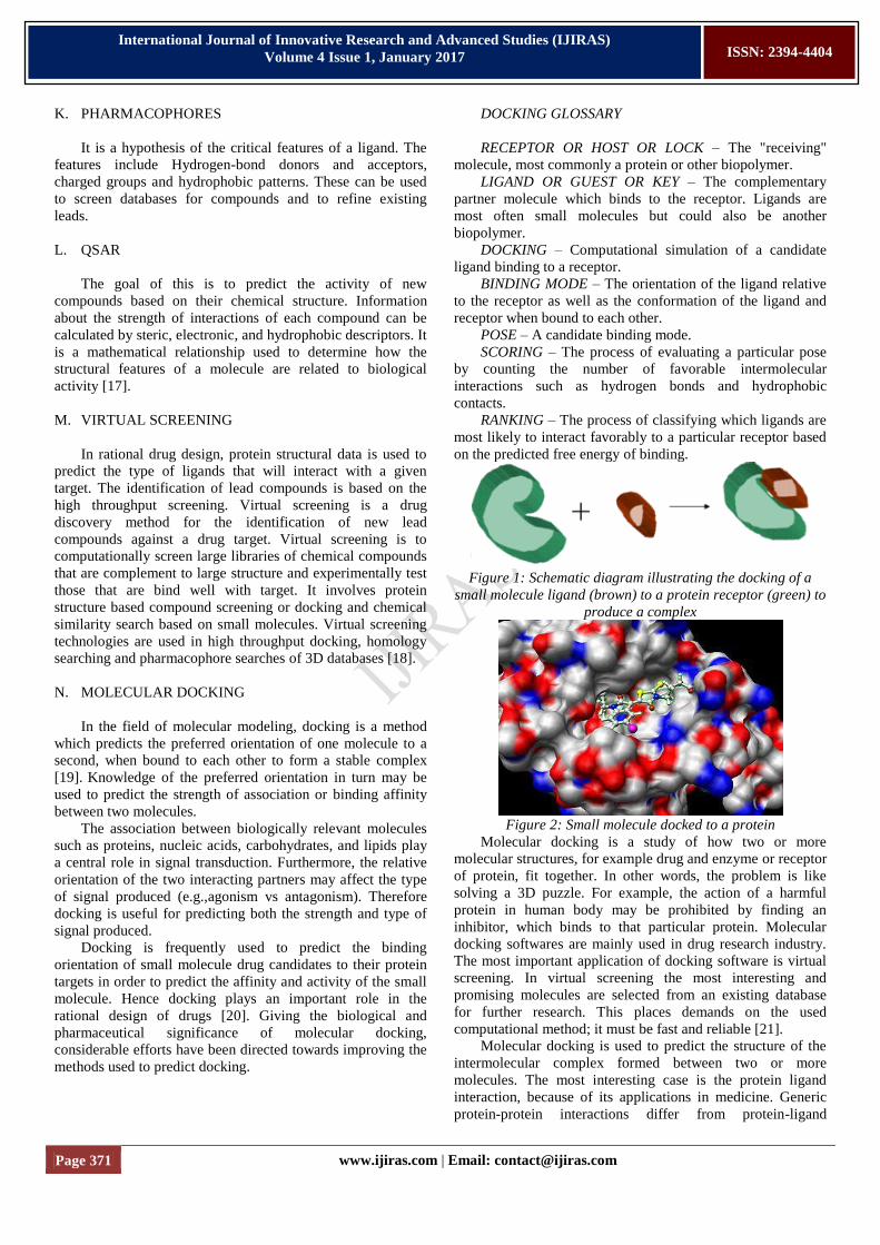
Figure 1: Schematic diagram illustrating the docking of a
small molecule ligand (brown) to a protein receptor (green) to
produce a complex
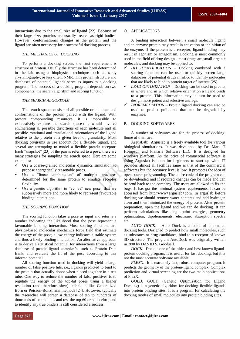
Figure 2: Small molecule docked to a protein
Molecular docking is a study of how two or more
molecular structures, for example drug and enzyme or receptor
of protein, fit together. In other words, the problem is like
solving a 3D puzzle. For example, the action of a harmful
protein in human body may be prohibited by finding an
inhibitor, which binds to that particular protein. Molecular
docking softwares are mainly used in drug research industry.
The most important application of docking software is virtual
screening. In virtual screening the most interesting and
promising molecules are selected from an existing database
for further research. This places demands on the used
computational method; it must be fast and reliable [21].
Molecular docking is used to predict the structure of the
intermolecular complex formed between two or more
molecules. The most interesting case is the protein ligand
interaction, because of its applications in medicine. Generic
protein-protein interactions differ from protein-ligand
Page 372 www.ijiras.com | Email: [email protected]
International Journal of Innovative Research and Advanced Studies (IJIRAS)
Volume 4 Issue 1, January 2017
ISSN: 2394-4404
interactions due to the small size of ligand [22]. Because of
their large size, proteins are usually treated as rigid bodies.
However, conformational changes in the protein and the
ligand are often necessary for a successful docking process.
THE MECHANICS OF DOCKING
To perform a docking screen, the first requirement is
structure of protein. Usually the structure has been determined
in the lab using a biophysical technique such as x-ray
crystallography, or less often, NMR. This protein structure and
databases of potential ligands serve as inputs to a docking
program. The success of a docking program depends on two
components: the search algorithm and scoring function.
THE SEARCH ALGORITHM
The search space consists of all possible orientations and
conformations of the protein paired with the ligand. With
present compounding resources, it is impossible to
exhaustively explore the search space-this would involve
enumerating all possible distortions of each molecule and all
possible rotational and translational orientations of the ligand
relative to the protein at a given level of granularity. Most
docking programs in use account for a flexible ligand, and
several are attempting to model a flexible protein receptor.
Each “snapshot” [23] of the pair is referred to a pose, there are
many strategies for sampling the search space. Here are some
examples:
Use a coarse-grained molecular dynamics simulation to
propose energetically reasonable poses.
Use a “linear combination” of multiple structures
determined for the same protein to emulate receptor
flexibility.
Use a genetic algorithm to “evolve” new poses that are
successively more and more likely to represent favourable
binding interactions.
THE SCORING FUNCTION
The scoring function takes a pose as input and returns a
number indicating the likelihood that the pose represent a
favourable binding interaction. Most scoring functions are
physics-based molecular mechanics force field that estimate
the energy of the pose; a low energy indicates a stable system
and thus a likely binding interaction. An alternative approach
is to derive a statistical potential for interactions from a large
database of protein-ligand complex‟s, such as Protein Data
Bank, and evaluate the fit of the pose according to this
inferred potential.
All scoring function used in docking will yield a large
number of false positive hits, i.e., ligands predicted to bind to
the protein that actually donot when placed together in a test
tube. One way to reduce the number of false positives is to
regulate the energy of the top-hit poses using a higher
resolution (and therefore slow) technique like Generalized
Born or Poisson-Boltzmann methods [24]. However, typically
the researcher will screen a database of ten to hundreds of
thousands of compounds and test the top 60 or so in vitro, and
to identify any true binders is still considered a success.
O. APPLICATIONS
A binding interaction between a small molecule ligand
and an enzyme protein may result in activation or inhibition of
the enzyme. If the protein is a receptor, ligand binding may
result in agonism or antagonism. Docking is most commonly
used in the field of drug design - most drugs are small organic
molecules, and docking may be applied to:
HIT IDENTIFICATION – Docking combined with a
scoring function can be used to quickly screen large
databases of potential drugs in silico to identify molecules
that are likely to bind to protein target of interest [25].
LEAD OPTIMIZATION – Docking can be used to predict
in where and in which relative orientation a ligand binds
to a protein. This information may in turn be used to
design more potent and selective analogs.
BIOREMEDIATION – Protein ligand docking can also be
used to predict pollutants that can be degraded by
enzymes.
P. DOCKING SOFTWARES
A number of softwares are for the process of docking.
Some of them are:
ArgusLab: Arguslab is a freely available tool for various
biological simultations. It was developed by Dr. Mark T
Hompson and Planaria Software LLC. It is designed for
windows platform. As the price of commercial software is
rising Arguslab is boon for beginners to start up with. IT
provides almost all facilities same as that of the commercial
softwares but the accuracy level is low. It promotes the idea of
open source programming. The entire code of the program can
be downloaded and if required changes can be made and can
be send back to the company. The users are allowed to fix the
bugs. It has got the minimal system requirements. It can be
accessed from http//www>arguslab>com. In arguslab before
docking we should remove water contents and add hydrogen
atom and then minimized the energy of protein. After protein
preparation, open the ligand and we can do docking. It can
perform calculations like single-point energies, geometry
optimization, dipolemoments, electronic absorption spectra
etc.
AUTO DOCK: Auto Dock is a suite of automated
docking tools. Designed to predict how small molecules, such
as substrates or drug candidates, bind to a receptor of known
3D structure. The program AutoDock was originally written
in1990 by DAVID S. Goodsell.
DOCK: Dock is one of the oldest and best known ligand-
protein docking program. It is useful for fast docking, but it is
not the most accurate software available.
FLEXX: It is extremely fast, robust computer program. It
predicts the geometry of the protein-ligand complex. Complex
prediction and virtual screening are the two main applications
of FlexX.
GOLD: GOLD (Genetic Optimization for Ligand
Docking) is a genetic algorithm for docking flexible ligands
into protein binding sites. It is a program for calculating the
docking modes of small molecules into protein binding sites.
Page 373 www.ijiras.com | Email: [email protected]
International Journal of Innovative Research and Advanced Studies (IJIRAS)
Volume 4 Issue 1, January 2017
ISSN: 2394-4404
FRED: It is a highly accurate, multiconformer docking
program. It examines all possible poses within a protein active
site.
II. AIM AND OBJECTIVES
To apply computational techniques in drug design against
Human Immunodeficiency Virus.
To validate the anti-viral potential of dietary compounds.
To identify dietary compounds that can structurally bind
to the HIV 1 Protease.
III. SCOPE
The primary goal of bioinformatics is to increase the
understanding of biological processes. It focus on developing
and applying computationally intensive techniques (e.g.,
pattern recognition, data mining, machine learning algorithms,
and visualization) to achieve this goal. Major research efforts
in the field include sequence alignment, gene finding, genome
assembly, drug design, drug discovery, protein structure
alignment, protein structure prediction, prediction of gene
expression and protein–protein interactions, genome-wide
association studies and the modeling of evolution.
A. ACQUIRED IMMUNODEFICIENCY SYNDROME
(AIDS)
In June 5,1981, scientists in the United States and France
first recognized the Acquired Immuno Deficiency Syndrome
(AIDS). The emergence and pandemic spread of AIDS have
posed the greatest challenge to public health in modern times.
HIV; the Human Immunodeficiency Virus is the etiological
agent of AIDS. AIDS is a medical condition where the
immune system cannot function properly and protect the body
from disease. It is an infectious disease caused by human
immunodeficiency virus that causes immune system failure
and debilitation. The virus can lie hidden in the body for up to
10 years without producing any obvious symptoms or before
developing into the AIDS disease, and in the meantime the
person can unknowingly infect others. Currently, an estimated
40 million people worldwide are HIV carriers, and three
million a year are dying of AIDS.
HIV is spread through direct contact with the bodily
fluids of an infected person. It is a fatal. AIDS is a measure of
how much damage HIV has done to person‟s immune system.
It is not a disease. AIDS can develop after someone gets HIV.
After HIV has been inside someone‟s body for a long time it
can weaken or destroy their immune system. The immune
system can‟t fight germs any more. They get different disease
or illnesses, also called opportunistic infections [26].
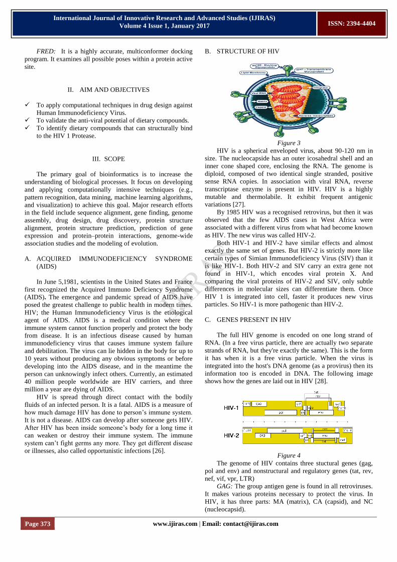
B. STRUCTURE OF HIV
Figure 3
HIV is a spherical enveloped virus, about 90-120 nm in
size. The nucleocapside has an outer icosahedral shell and an
inner cone shaped core, enclosing the RNA. The genome is
diploid, composed of two identical single stranded, positive
sense RNA copies. In association with viral RNA, reverse
transcriptase enzyme is present in HIV. HIV is a highly
mutable and thermolabile. It exhibit frequent antigenic
variations [27].
By 1985 HIV was a recognised retrovirus, but then it was
observed that the few AIDS cases in West Africa were
associated with a different virus from what had become known
as HIV. The new virus was called HIV-2.
Both HIV-1 and HIV-2 have similar effects and almost
exactly the same set of genes. But HIV-2 is strictly more like
certain types of Simian Immunodeficiency Virus (SIV) than it
is like HIV-1. Both HIV-2 and SIV carry an extra gene not
found in HIV-1, which encodes viral protein X. And
comparing the viral proteins of HIV-2 and SIV, only subtle
differences in molecular sizes can differentiate them. Once
HIV 1 is integrated into cell, faster it produces new virus
particles. So HIV-1 is more pathogenic than HIV-2.
C. GENES PRESENT IN HIV
The full HIV genome is encoded on one long strand of
RNA. (In a free virus particle, there are actually two separate
strands of RNA, but they're exactly the same). This is the form
it has when it is a free virus particle. When the virus is
integrated into the host's DNA genome (as a provirus) then its
information too is encoded in DNA. The following image
shows how the genes are laid out in HIV [28].
Figure 4
The genome of HIV contains three stuctural genes (gag,
pol and env) and nonstructural and regulatory genes (tat, rev,
nef, vif, vpr, LTR)
GAG: The group antigen gene is found in all retroviruses.
It makes various proteins necessary to protect the virus. In
HIV, it has three parts: MA (matrix), CA (capsid), and NC
(nucleocapsid).
Page 374 www.ijiras.com | Email: [email protected]
International Journal of Innovative Research and Advanced Studies (IJIRAS)
Volume 4 Issue 1, January 2017
ISSN: 2394-4404
POL: The polymerase gene is also found in all
retroviruses. It makes enzymes necessary for virus replication.
In HIV, it also has three parts: PR (protease), IN
(endonuclease), and RT (reverse transcriptase).
ENV: The envelope gene is also found in all retroviruses.
It makes proteins for the envelope to the virus. In HIV, it has
two parts. SU (surface envelope, gp120) and TM
(transmembrane envelop, gp41).
TAT: The transactivator gene influences the function of
genes some distance away. It controls transactivation of all
HIV proteins.
REV: The differential regulator of expression of virus
protein genes.
VIF: The virus infectivity factor gene is required for
infectivity as cell-free virus.
NEF: The negative regulator factor retards HIV
replication.
VPR: The virus protein R gene has an undetermined
function.
VPU: The virus protein U gene is required for efficient
viral replication and release. It is found only in HIV-1.
VPX: The virus protein X gene has an undetermined
function. It is found only in HIV-2 and SIV.
The HIV genome also has a "Long Terminal Repeat"
(LTR) at each end of its genome-not quite a gene, but a
sequence of RNA/DNA which is the same at either end and
which serves some structural and regulatory purposes.
D. HIV 1 PROTEASE AS DRUG TARGET
HIV-1 protease (HIV PR) is a retroviral aspartyl protease
that is essential for the lifecycle of HIV, the retrovirus that
causes AIDS. HIV PR cleaves newly synthesized polyproteins
at appropriate places to create the mature protein components
of an infectious HIV virion. Without effective HIV PR, HIV
virions remain uninfectious. Thus, mutation of HIV PR‟s
active site or inhibition of its activity disrupts HIV‟s ability to
replicate and infect additional cells, making HIV PR inhibition
the subject of much pharmaceutical research.
Due to its time-sensitivity and crucial role, HIV protease
is an effective target for drug therapy [29][30].
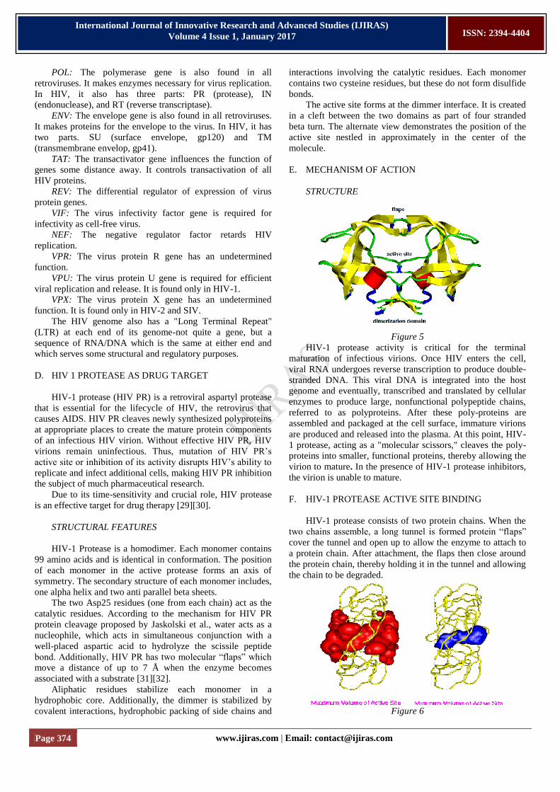
STRUCTURAL FEATURES
HIV-1 Protease is a homodimer. Each monomer contains
99 amino acids and is identical in conformation. The position
of each monomer in the active protease forms an axis of
symmetry. The secondary structure of each monomer includes,
one alpha helix and two anti parallel beta sheets.
The two Asp25 residues (one from each chain) act as the
catalytic residues. According to the mechanism for HIV PR
protein cleavage proposed by Jaskolski et al., water acts as a
nucleophile, which acts in simultaneous conjunction with a
well-placed aspartic acid to hydrolyze the scissile peptide
bond. Additionally, HIV PR has two molecular “flaps” which
move a distance of up to 7 Å when the enzyme becomes
associated with a substrate [31][32].
Aliphatic residues stabilize each monomer in a
hydrophobic core. Additionally, the dimmer is stabilized by
covalent interactions, hydrophobic packing of side chains and
interactions involving the catalytic residues. Each monomer
contains two cysteine residues, but these do not form disulfide
bonds.
The active site forms at the dimmer interface. It is created
in a cleft between the two domains as part of four stranded
beta turn. The alternate view demonstrates the position of the
active site nestled in approximately in the center of the
molecule.
E. MECHANISM OF ACTION
STRUCTURE
Figure 5
HIV-1 protease activity is critical for the terminal
maturation of infectious virions. Once HIV enters the cell,
viral RNA undergoes reverse transcription to produce double-
stranded DNA. This viral DNA is integrated into the host
genome and eventually, transcribed and translated by cellular
enzymes to produce large, nonfunctional polypeptide chains,
referred to as polyproteins. After these poly-proteins are
assembled and packaged at the cell surface, immature virions
are produced and released into the plasma. At this point, HIV-
1 protease, acting as a "molecular scissors," cleaves the poly-
proteins into smaller, functional proteins, thereby allowing the
virion to mature. In the presence of HIV-1 protease inhibitors,
the virion is unable to mature.
F. HIV-1 PROTEASE ACTIVE SITE BINDING
HIV-1 protease consists of two protein chains. When the
two chains assemble, a long tunnel is formed protein “flaps”
cover the tunnel and open up to allow the enzyme to attach to
a protein chain. After attachment, the flaps then close around
the protein chain, thereby holding it in the tunnel and allowing
the chain to be degraded.
Figure 6
Page 375 www.ijiras.com | Email: [email protected]
International Journal of Innovative Research and Advanced Studies (IJIRAS)
Volume 4 Issue 1, January 2017
ISSN: 2394-4404
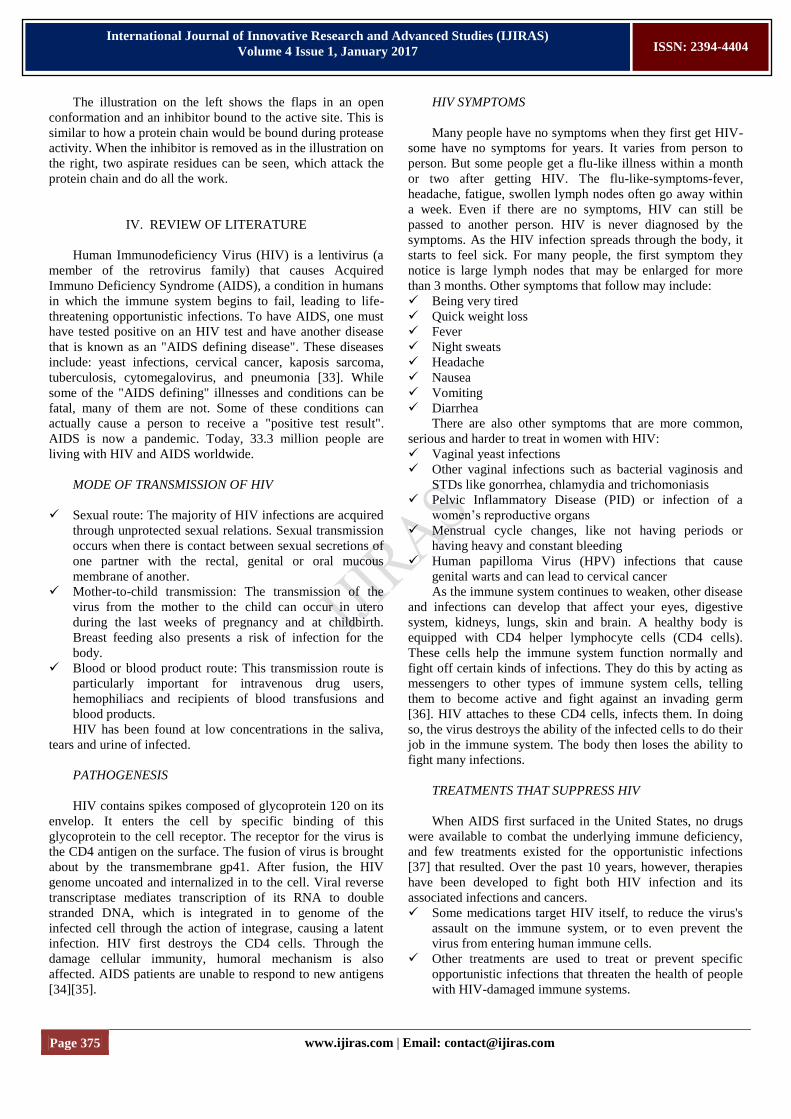
The illustration on the left shows the flaps in an open
conformation and an inhibitor bound to the active site. This is
similar to how a protein chain would be bound during protease
activity. When the inhibitor is removed as in the illustration on
the right, two aspirate residues can be seen, which attack the
protein chain and do all the work.
IV. REVIEW OF LITERATURE
Human Immunodeficiency Virus (HIV) is a lentivirus (a
member of the retrovirus family) that causes Acquired
Immuno Deficiency Syndrome (AIDS), a condition in humans
in which the immune system begins to fail, leading to life-
threatening opportunistic infections. To have AIDS, one must
have tested positive on an HIV test and have another disease
that is known as an "AIDS defining disease". These diseases
include: yeast infections, cervical cancer, kaposis sarcoma,
tuberculosis, cytomegalovirus, and pneumonia [33]. While
some of the "AIDS defining" illnesses and conditions can be
fatal, many of them are not. Some of these conditions can
actually cause a person to receive a "positive test result".
AIDS is now a pandemic. Today, 33.3 million people are
living with HIV and AIDS worldwide.
MODE OF TRANSMISSION OF HIV
Sexual route: The majority of HIV infections are acquired
through unprotected sexual relations. Sexual transmission
occurs when there is contact between sexual secretions of
one partner with the rectal, genital or oral mucous
membrane of another.
Mother-to-child transmission: The transmission of the
virus from the mother to the child can occur in utero
during the last weeks of pregnancy and at childbirth.
Breast feeding also presents a risk of infection for the
body.
Blood or blood product route: This transmission route is
particularly important for intravenous drug users,
hemophiliacs and recipients of blood transfusions and
blood products.
HIV has been found at low concentrations in the saliva,
tears and urine of infected.
PATHOGENESIS
HIV contains spikes composed of glycoprotein 120 on its
envelop. It enters the cell by specific binding of this
glycoprotein to the cell receptor. The receptor for the virus is
the CD4 antigen on the surface. The fusion of virus is brought
about by the transmembrane gp41. After fusion, the HIV
genome uncoated and internalized in to the cell. Viral reverse
transcriptase mediates transcription of its RNA to double
stranded DNA, which is integrated in to genome of the
infected cell through the action of integrase, causing a latent
infection. HIV first destroys the CD4 cells. Through the
damage cellular immunity, humoral mechanism is also
affected. AIDS patients are unable to respond to new antigens
[34][35].
HIV SYMPTOMS
Many people have no symptoms when they first get HIV-
some have no symptoms for years. It varies from person to
person. But some people get a flu-like illness within a month
or two after getting HIV. The flu-like-symptoms-fever,
headache, fatigue, swollen lymph nodes often go away within
a week. Even if there are no symptoms, HIV can still be
passed to another person. HIV is never diagnosed by the
symptoms. As the HIV infection spreads through the body, it
starts to feel sick. For many people, the first symptom they
notice is large lymph nodes that may be enlarged for more
than 3 months. Other symptoms that follow may include:
Being very tired
Quick weight loss
Fever
Night sweats
Headache
Nausea
Vomiting
Diarrhea
There are also other symptoms that are more common,
serious and harder to treat in women with HIV:
Vaginal yeast infections
Other vaginal infections such as bacterial vaginosis and
STDs like gonorrhea, chlamydia and trichomoniasis
Pelvic Inflammatory Disease (PID) or infection of a
women‟s reproductive organs
Menstrual cycle changes, like not having periods or
having heavy and constant bleeding
Human papilloma Virus (HPV) infections that cause
genital warts and can lead to cervical cancer
As the immune system continues to weaken, other disease
and infections can develop that affect your eyes, digestive
system, kidneys, lungs, skin and brain. A healthy body is
equipped with CD4 helper lymphocyte cells (CD4 cells).
These cells help the immune system function normally and
fight off certain kinds of infections. They do this by acting as
messengers to other types of immune system cells, telling
them to become active and fight against an invading germ
[36]. HIV attaches to these CD4 cells, infects them. In doing
so, the virus destroys the ability of the infected cells to do their
job in the immune system. The body then loses the ability to
fight many infections.
TREATMENTS THAT SUPPRESS HIV
When AIDS first surfaced in the United States, no drugs
were available to combat the underlying immune deficiency,
and few treatments existed for the opportunistic infections
[37] that resulted. Over the past 10 years, however, therapies
have been developed to fight both HIV infection and its
associated infections and cancers.
Some medications target HIV itself, to reduce the virus's
assault on the immune system, or to even prevent the
virus from entering human immune cells.
Other treatments are used to treat or prevent specific
opportunistic infections that threaten the health of people
with HIV-damaged immune systems.
Page 376 www.ijiras.com | Email: [email protected]
International Journal of Innovative Research and Advanced Studies (IJIRAS)
Volume 4 Issue 1, January 2017
ISSN: 2394-4404
HIV-1 protease (HIV PR) is a homodimeric enzyme, that
comprise the human immunodeficiency virus. Competitive
inhibitors are used to bind to the protease and block its
function, thereby suppressing the virus, which cannot
transform to its mature, infectious form. HIV 1 Protease
inhibitors are one class of drugs that target the viral enzyme
HIV-1 protease. They are drugs used to treat or prevent
infection by HIV. It prevent protease from splitting proteins
into peptides. That is, they prevent the cleavage of HIV
precursor proteins into active proteins. Drugs that interfere
with the activity of retrovirus HIV are generally known as
antiretrovirals [38]. There is no cure for HIV/AIDS, but a
variety of drugs can be used in combination to control the
virus. Each of the classes of anti-HIV drugs blocks the virus in
different ways. It's best to combine at least three drugs from
two different classes to avoid creating strains of HIV that are
immune to single drugs. The classes of anti-HIV drugs include
[39]:
Nucleoside analog reverse transcriptase inhibitors
(NRTIs): NRTIs interrupt an early stage of HIV
replication. AZT (zidovudine), the first drug approved for
treating HIV infection, is an NRTI.
Non-nucleoside reverse transcriptase inhibitors
(NNRTIs): This class of drugs includes delavirdine,
nevirapine, and efavirenz.
Protease inhibitors: Protease inhibitors interrupt a later
stage of viral replication. This class of drugs includes
saquinavir, indinavir, ritonavir, nelfinavir, and
amprenavir.
Fusion inhibitors: Fusion inhibitors prevent HIV from
entering human immune cells. The only fusion inhibitor
approved to date is enfuvirtide.
Some, Dietary compounds that are used as drugs; like
silymarin, lectin, glibenclamide, resveratrol, oleuropein etc;
are used as HIV 1 protease inhibitors. They greatly inhibit the
function of the enzyme. Also they have no adverse effect as
carcinogenicity and mutagenicity. Various bioactivities of
dietary compounds are responsible for their chemopreventive
properties and also contribute to their inducing apoptosis by
arresting cell cycle, regulating carcinogen metabolism and
ontogenesis expression, inhibiting DNA binding and cell
adhesion, migration, proliferation or differentiation, and
blocking signaling pathways.
A. INHIBITORS OF HIV 1 PROTEASE
HIV protease inhibitors was first invented between 1989
and 1994 by researchers working for the pharmaceutical
companies of Hoffmann- La Roche Inc. (of Nutley, New
Jersey), Abbott Laboratories and Merck & Co., Inc. HIV
protease inhibitors are used in the treatment of patients with
AIDS and were considered the first breakthrough in over a
decade of AIDS research. HIV protease inhibitors can lower
the viral load carried by AIDS patients.
We can design a drug (inhibitor) to inhibit the activity of
gene. As a gene produces protein/enzyme, so to avoid the
formation of any disease causing proteins, we have to stop the
activity of that gene. With the help of different bioinformatics
tools we can do this.
HIV 1 Protease inhibitors are one class of drugs that
target the viral enzyme HIV-1 protease. They are drugs used
to treat or prevent infection by HIV. It prevent protease from
splitting proteins into peptides. That is, they prevent the
cleavage of HIV precursor proteins into active proteins, a
process that normally occurs when HIV replicates.
B. THE DIETARY COMPOUNDS USED AS HIV 1
PROTEASE INHIBITORS
It is well known that combination of foods and dietary
constituents plays an important role in prevention and
treatment of many diseases. Several types of natural products
such as carotenoids, phenolics, dietary fibers and fish oils are
successful drugs. Epidemiologic evidence suggests that
regular consumption of fruits and vegetables may
reduce susceptibility to many diseases.
Various bioactivities of dietary compounds are
responsible for their chemopreventive properties (e.g.,
antioxidant, anticarcinogenic, or antimutagenic and anti-
inflammatory effects).
BETA CAROTENE
β-Carotene (from Carrot) is a strongly-coloured red-
orange pigment abundant in plants and fruits. It is an organic
compound and chemically is classified as a hydrocarbon and
specifically as a terpenoid (isoprenoid), reflecting its
derivation from isoprene units. β-Carotene is biosynthesized
from geranylpyrophosphate. It is a member of the carotenes,
which are tetraterpenes, synthesized biochemically from eight
isoprene units and thus having 40 carbons. Among this general
class of carotenes, β-Carotene is distinguished by having beta-
rings at both ends of the molecule.
CAFFEINE
Caffeine (from Coffee) is a bitter, white crystalline
xanthine alkaloid and psychoactive stimulant. Caffeine was
first isolated from coffee in 1820 by the German chemist
Friedlieb Ferdinand Runge and again in 1821 by French
chemists Robiquet, Pelletier, and Caventou. Pelletier first
coined the word "cafeine", which became the English word
"caffeine".
Caffeine is found in varying quantities in the seeds,
leaves, and fruit of some plants, where it acts as a natural
pesticide that paralyzes and kills certain insects feeding on the
plants. In humans, caffeine acts as a central nervous system
stimulant, temporarily warding off drowsiness and restoring
alertness. Caffeine is the world's most widely consumed
psychoactive substance, but, unlike many other psychoactive
substances, is legal and unregulated in nearly all jurisdictions.
D-LIMONENE
D-limonene (from Lemon) is a colourless liquid
hydrocarbon classified as a cyclic terpene. The more common
D isomer posesses a strong smell of oranges. It is used in
chemical synthesis as a precursor to carvone and as a
renewably-based solvent in cleaning products. Limonene is a
Page 377 www.ijiras.com | Email: [email protected]
International Journal of Innovative Research and Advanced Studies (IJIRAS)
Volume 4 Issue 1, January 2017
ISSN: 2394-4404
chiral molecule, and biological sources produce one
enantiomer: the principal industrial source, citrus fruit,
contains D-limonene ((+)-limonene), which is the (R)-
enantiomer. Racemic limonene is known as dipentene. D-
Limonene is obtained commercially from citrus fruits through
two primary methods: centrifugal separation or steam
distillation.
ALLIXIN
Allixin (from Garlic), first isolated and characterized in
1989. When garlic is stored for long periods of time, it can
form visible accumulations of crystalline allixin on its surface,
particularly in areas where tissue has become necrotic. After 2
years of storage, the amount of allixin accumulated can
approach 1% of the dry weight of the cloves.
ELLAGIC ACID
Ellagic acid (from Black berries) is a natural phenol
antioxidant. The antiproliferative and antioxidant properties of
ellagic acid have spurred preliminary research into the
potential health benefits of ellagic acid consumption.
EMODIN
Emodin (from Rhubarb) is a purgative resin. It shows
promise as an agent that could reduce the impact of type 2
diabetes. It effectively limits the effect of the glucocorticoids,
and ameliorates diabetes and insulin resistance.
Pharmacological studies have demonstrated that emodin from
rhubarb exhibits anti-cancer effects on several human cancers,
including human pancreatic cancer.
PHENETHYL ISOTHIOCYANATE
Phenethyl isothiocyanate, PEITC (from Cabbage) inhibit
carcinogenesis and tumorigenesis. It is also used for amino
acid sequencing in the Edman degradation.
OLEUROPEIN
Oleuropein is a chemical compound found in olive leaf
from the olive tree. Oleuropein has powerful antioxidant
activity both in vivo and in vitro and give extra-virgin olive oil
its bitter, pungent taste. Oleuropein preparations have been
claimed for several pharmacological effects among them
strengthening of the immune system.
ANTHOCYANIN
Anthocyanins (from Egg plant) are water soluble vacuolar
pigments that may appear red, purple, or blue according to pH.
They belong to a parent class of molecules called flavonoids
synthesized via the phenylpropanoid pathway; they are
odourless and nearly flavourless, contributing to taste as a
moderately astringent sensation. Anthocyanins are derivatives
of anthocyanidins which include pendant sugars.
QUERCETIN
Quercetin (from Onion) is a flavonoid widely distributed
in nature. The name has been used since 1857, and is derived
from quercetum, It is a naturally occurring polar auxin
transport inhibitor.
LECTIN
Lectins (from Banana) are sugar binding proteins that are
highly specific for their sugar moieties. They play a role in
biological recognition phenomena involving cells and
proteins. For example, some viruses use lectins to attach
themselves to the cells of the host organism during infection.
Lectins may be disabled by specific mono- and
oligosaccharides, which bind to them and prevent their
attachment to cell membranes.
APIGENIN
Apigenin (from Apple) is a flavone that is the aglycone of
several glycosides. It is a yellow crystalline solid that has been
used to dye wool. Apigenin is commonly recognised as to
mediate at least part of the chemopreventive action of
vegetables and fruits in the cancerous process. Recently it was
shown that Apigenin induces a process called autophagia (a
kind of cellular dormancy) which may well explain it
chemopreventive properties. Apigenin acts as a monoamine
transporter activator, and is one of the few chemicals
demonstrated to possess this property.
COUMARIN
Coumarin (from Tonka bean) is a pleasantly fragrant
chemical compound. The name comes from a French word,
coumarou. It has a sweet odour, readily recognised as the
scent of newly-mown hay, and has been used in perfumes
since 1882. It is used in the pharmaceutical industry as a
precursor molecule in the synthesis of a number of synthetic
anticoagulant pharmaceuticals. Coumarin has clinical medical
value by itself, as an edema modifier. It is also used as a gain
medium in some dye lasers, and as a sensitizer in older
photovoltaic technologies.
SILYMARIN
It is a flavanoid extracted from milk thistle. Both in vitro
and animal research suggest that silymarin has
hepatoprotective properties that protect liver cells against
toxins. Chemically modified form is used in treatment of
severe intoxications with hepatotoxic substances, such as
death cap.
INDOMETHACIN
Indomethacin (from Pumpkin) uncouples oxidative
phosphorylation in cartilaginous (and hepatic) mitochondria,
like salicylates.

Page 378 www.ijiras.com | Email: [email protected]
International Journal of Innovative Research and Advanced Studies (IJIRAS)
Volume 4 Issue 1, January 2017
ISSN: 2394-4404
GLIBENCLAMIDE
Glibenclamide (from Bitter melon) also known as
glyburide, is drug in a class of medications known as
sulfonylureas, closely related to sulfa drugs.
FISETIN
Fisetin (from Strawberry) is a flavonol, a structurally
distinct chemical substance that belongs to the flavonoid
group of polyphenols. Its chemical formula was first described
by Austrian chemist Josef Herzig in 1891. Fisetin is a potent
sirtuin activating compound, an agent that modulates sirtuins.
It is therefore a caloric restriction mimetic candidate, a drug
that has been shown to be able to alleviate ageing effects in
certain model organisms such as the yeast s cerevisiae. It is a
potent antioxidant. Its antioxidative activity may be due to its
structural properties as well as to its ability to modulate certain
cellular signaling pathways.
RESVERATROL
Resveratrol (from Grapes) is a stilbenoid, a type of natural
phenol, and a phytoalexin produced naturally by several plants
when under attack by pathogens such as bacteria or fungi. It is
also produced by chemical synthesis
http://en.wikipedia.org/wiki/Resveratrol - cite_note-
pmid16401555-6 or by biotechnological synthesis and is sold
as a nutritional supplement.
INULIN
Inulins (from Asparagus) are a group of naturally
occurring polysaccharides. Inulin is increasingly used in
processed foods because it has unusually adaptable
characteristics. Its flavour ranges from bland to subtly sweet.
It can be used to replace sugar, fat, and flour. This is
particularly advantageous because inulin contains a quarter to
a third of the food energy of sugar or other carbohydrates and
a ninth to a sixth of the food energy of fat. While inulin is a
versatile ingredient, it also has health benefits. Inulin increases
calcium absorption and possibly magnesium absorption, while
promoting the growth of intestinal bacteria. In terms of
nutrition, it is considered a form of soluble fiber and is
sometimes categorized as a prebiotic. Due to the body's
limited ability to process polysaccharides, inulin has minimal
increasing impact on blood sugar, and unlike fructose is not
insulemic and does not raise triglycerides, making it
considered suitable for diabetics and potentially helpful in
managing blood sugar related illnesses.
CAPSAICIN
It is the active component of chili peppers, which are
plants belonging to the genus Capsicum. It is an irritant for
mammals, including humans, and produces a sensation of
burning in any tissue with which it comes into contact.
Capsaicin and several related compounds are called
capsaicinoids and are produced as a secondary metabolite by
chili peppers, probably as deterrents against certain herbivores
and fungi. Pure capsaicin is a hydrophobic, colourless,
odourless, crystalline to waxy compound.
V. METHODOLOGY
Using ArgusLab software we can prepare and run
molecular docking calculations. Before docking a molecule,
define the atoms that make up the Ligand (drug, inhibitor, etc.)
and the Binding Site of the protein where the drug binds. The
3D structure of HIV 1 Protease (PDB ID - 3OXC) can be
downloaded from the Brookhaven Protein Databank (PDB).
Steps taken for molecular docking using Arguslab is
shown below.
Open the PDB file, contains the active site residues
attached to the inhibitor
Open a ligand from the molecule database
Select the active site residues and right click the mouse
Click “Make a group from the residues”. Select „Binding
sit‟ and click OK on the dialogue box appeared
Now select the original ligand and right click the mouse
Click “make a ligand group from the residue”
Select the ligand from the small molecule database
Right click on it and click “Make a ligand group from the
residue”
Select calculation->Dock a ligand
Select the ligand and active site in the dialogue box
appeared. Click the button Calculate size and then click
OK. Then docking will starts and number of poses with
different energies will be displayed.
Explanation of steps-
DOWNLOAD AND OPEN THE PDB STRUCTURE
OF 3OXC
Open the web browser and type in the Bookhaven Protein
Databank (www.rcsb.org) web address and in the search field
type 3OXC. Download the displayed structure as PDB file
preferably to Desktop. Open the 3OXC.pdb located in the
folder designated during download in ArgusLab program
(Arguslab icon is located on the desktop). This is a PDB file
so make sure to select the correct file type in the file open
dialog (pdb type). Expand the Molecule Tree View tool of
3OXC (located on the left side on the screen).
We can see something like this:
Figure 7
Page 379 www.ijiras.com | Email: [email protected]
International Journal of Innovative Research and Advanced Studies (IJIRAS)
Volume 4 Issue 1, January 2017
ISSN: 2394-4404
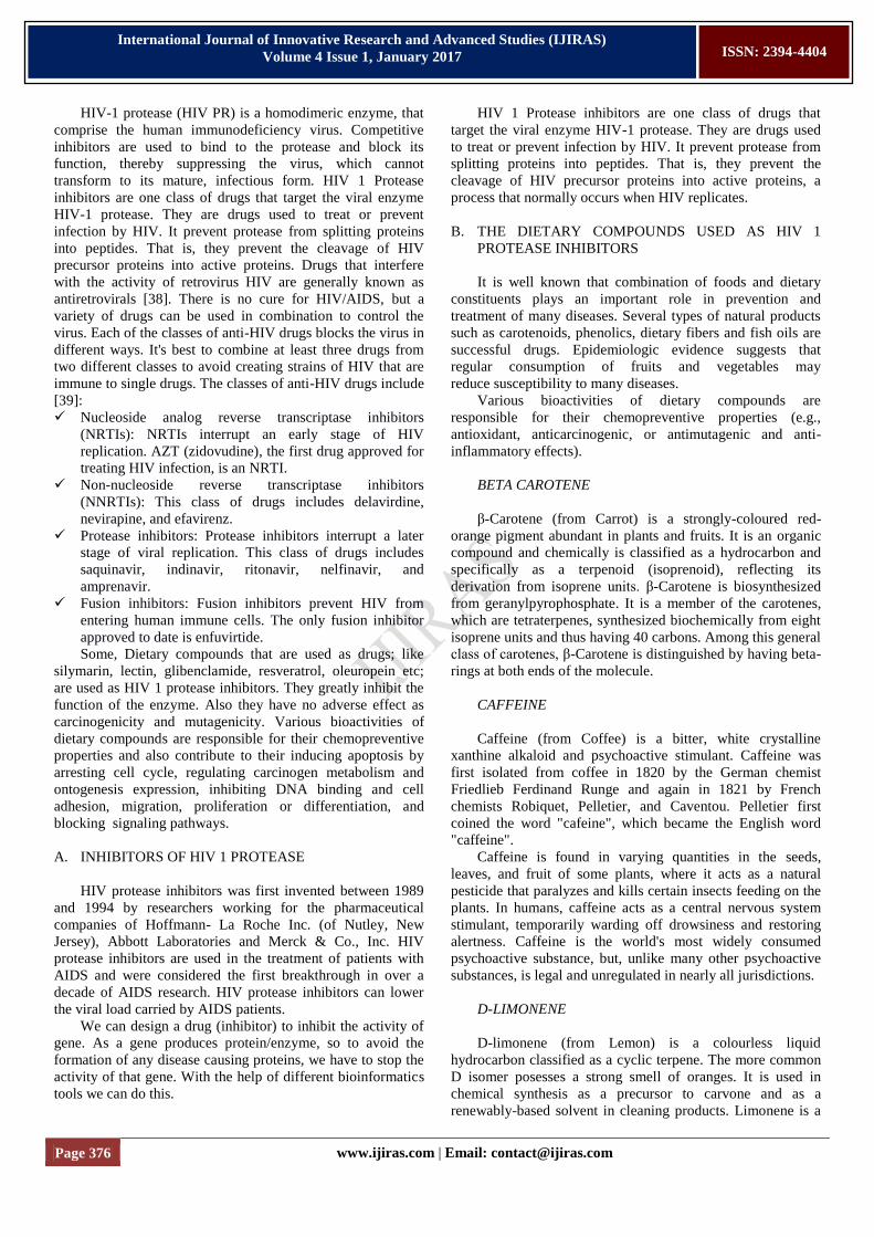
CREATE "LIGAND AND BINDING SITE GROUPS"
Left-click on 30XC in the Tree View. It contains ROC,
which is already defined as a ligand and the binding site has
been made.
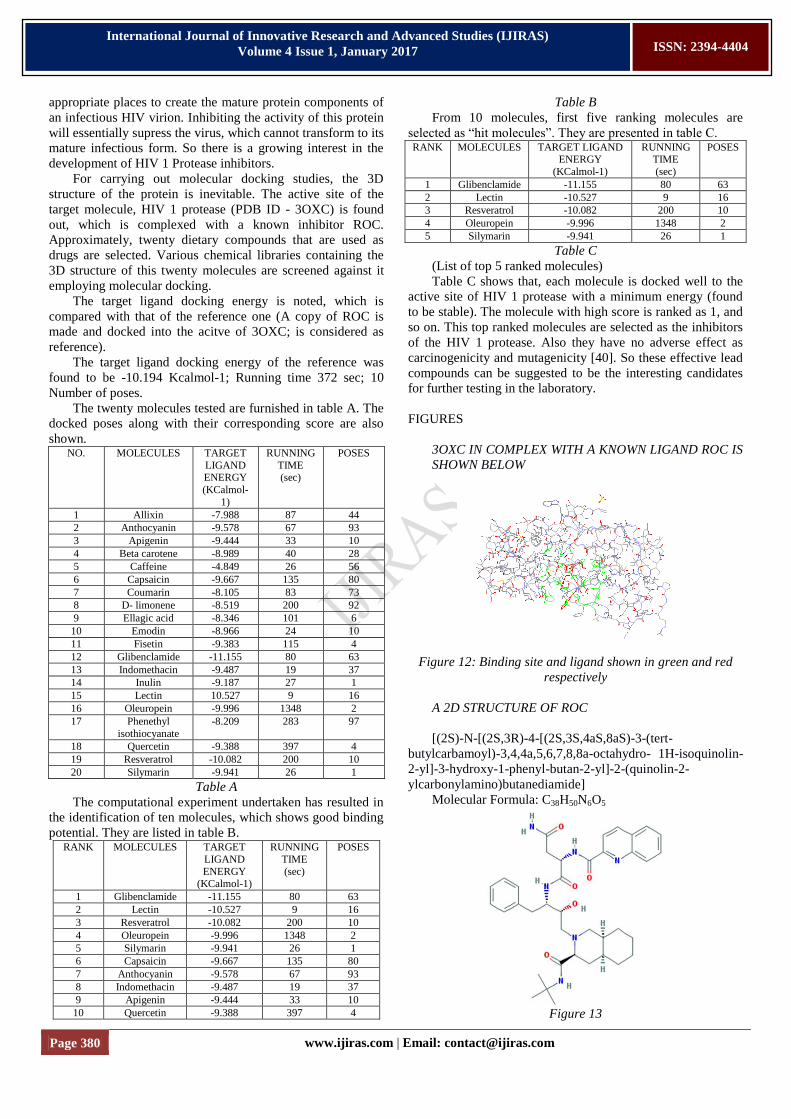
Figure 8: Binding site and ligand shown in green and red
respectively
Select the Edit/Hide Unselected menu option to hide all
atoms that are not selected.
We can see something like this in the screen:
Figure 9
DOCK THE LIGAND INTO DEFINED BINDING SITE
Dietary compounds used as drugs are selected as HIV 1
protease inhibitor. The 3D structure of this compounds are
downloaded from PubChem in SDF (Structure Data File)
format, which is then converted to pdb using Vega ZZ
software (www.vegazz.net). And, finally they are docked into
the binding site of 3OXC. For this, bring up the dock settings
dialog box by selecting the Calculation/Dock a Ligand menu
option or clicking on the button on the toolbar.
The Dock Settings dialog box looks like:
Figure 10
Select the ligand to dock in the "ligand" drop-box. Make
sure to select the group named "ligand" group and not the
"ligand-xray" group. Click on the "Calculate Size" button and
a docking box tailored to the binding site will be made and
shown on the screen. Make sure "ArgusDock" is the selected
docking engine, the Calculation type = Dock, and the Ligand
is Flexible. Click the "Start" button and the docking
calculation will begin. Notice the messages that appear in the
status line at the bottom of the molecule window. ArgusLab
first generate the scoring grids used during the docking, then
the various search phases will occur, and finally the candidate
poses should be processed until the calculation is done.
When 3OXC is docked with silymarin, we can see
something like this:
Figure 11: Silymarin shown in violet colour
DOCK ANOTHER COPY OF ROC INTO THE BINDING
SITE
Open the file 3OXC.pdb. A ligand group, ROC is already
created for this structure and verify this by looking in the
groups folder under the 3OXC molecule in the Tree View.
Make a copy of this ligand, and is docked into the binding site
of 3OXC. This is made as the REFERENCE..
ANALYZE AND SAVE THE RESULTS
There are two sources of information about the performed
docking calculation: The separate log file and information
located in the Tree View tool. Expand the ArgusDock
calculation in the Calculations folder in the Tree View. There
will be listing of docking settings and several "poses" starting
with the most stable as Pose 1 according to the calculated
energy in kcal/mol. (A term "pose" is usually used to
designate the specific set of coordinates of a docked structure).
Right click on pose 2 and select the "Display" option. The
coordinates of ligand will change to this pose and its score will
be displayed on the screen.
VI. RESULT AND DISCUSSION
Human Immunodeficiency Virus (HIV) is the retrovirus
that causes AIDS. The HIV virus undergoes continuous
mutation. As a result new genotypes are evolved. Hence it is
relevant that identifying the genotype is important for drug
development and disease management. The better way of
developing a drug against HIV is to inhibit the activity of HIV
1 Protease, which cleaves newly synthesized polyproteins at
Page 380 www.ijiras.com | Email: [email protected]
International Journal of Innovative Research and Advanced Studies (IJIRAS)
Volume 4 Issue 1, January 2017
ISSN: 2394-4404
appropriate places to create the mature protein components of
an infectious HIV virion. Inhibiting the activity of this protein
will essentially supress the virus, which cannot transform to its
mature infectious form. So there is a growing interest in the
development of HIV 1 Protease inhibitors.
For carrying out molecular docking studies, the 3D
structure of the protein is inevitable. The active site of the
target molecule, HIV 1 protease (PDB ID - 3OXC) is found
out, which is complexed with a known inhibitor ROC.
Approximately, twenty dietary compounds that are used as
drugs are selected. Various chemical libraries containing the
3D structure of this twenty molecules are screened against it
employing molecular docking.
The target ligand docking energy is noted, which is
compared with that of the reference one (A copy of ROC is
made and docked into the acitve of 3OXC; is considered as
reference).
The target ligand docking energy of the reference was
found to be -10.194 Kcalmol-1; Running time 372 sec; 10
Number of poses.
The twenty molecules tested are furnished in table A. The
docked poses along with their corresponding score are also
shown. NO. MOLECULES TARGET
LIGAND ENERGY
(KCalmol-
1)
RUNNING
TIME (sec)
POSES
1 Allixin -7.988 87 44
2 Anthocyanin -9.578 67 93
3 Apigenin -9.444 33 10
4 Beta carotene -8.989 40 28
5 Caffeine -4.849 26 56
6 Capsaicin -9.667 135 80
7 Coumarin -8.105 83 73
8 D- limonene -8.519 200 92
9 Ellagic acid -8.346 101 6
10 Emodin -8.966 24 10
11 Fisetin -9.383 115 4
12 Glibenclamide -11.155 80 63
13 Indomethacin -9.487 19 37
14 Inulin -9.187 27 1
15 Lectin 10.527 9 16
16 Oleuropein -9.996 1348 2
17 Phenethyl
isothiocyanate
-8.209 283 97
18 Quercetin -9.388 397 4
19 Resveratrol -10.082 200 10
20 Silymarin -9.941 26 1
Table A
The computational experiment undertaken has resulted in
the identification of ten molecules, which shows good binding
potential. They are listed in table B. RANK MOLECULES TARGET
LIGAND
ENERGY (KCalmol-1)
RUNNING
TIME
(sec)
POSES
1 Glibenclamide -11.155 80 63
2 Lectin -10.527 9 16
3 Resveratrol -10.082 200 10
4 Oleuropein -9.996 1348 2
5 Silymarin -9.941 26 1
6 Capsaicin -9.667 135 80
7 Anthocyanin -9.578 67 93
8 Indomethacin -9.487 19 37
9 Apigenin -9.444 33 10
10 Quercetin -9.388 397 4
Table B
From 10 molecules, first five ranking molecules are
selected as “hit molecules”. They are presented in table C. RANK MOLECULES TARGET LIGAND
ENERGY
(KCalmol-1)
RUNNING TIME
(sec)
POSES
1 Glibenclamide -11.155 80 63
2 Lectin -10.527 9 16
3 Resveratrol -10.082 200 10
4 Oleuropein -9.996 1348 2
5 Silymarin -9.941 26 1
Table C
(List of top 5 ranked molecules)
Table C shows that, each molecule is docked well to the
active site of HIV 1 protease with a minimum energy (found
to be stable). The molecule with high score is ranked as 1, and
so on. This top ranked molecules are selected as the inhibitors
of the HIV 1 protease. Also they have no adverse effect as
carcinogenicity and mutagenicity [40]. So these effective lead
compounds can be suggested to be the interesting candidates
for further testing in the laboratory.
FIGURES
3OXC IN COMPLEX WITH A KNOWN LIGAND ROC IS
SHOWN BELOW
Figure 12: Binding site and ligand shown in green and red
respectively
A 2D STRUCTURE OF ROC
[(2S)-N-[(2S,3R)-4-[(2S,3S,4aS,8aS)-3-(tert-
butylcarbamoyl)-3,4,4a,5,6,7,8,8a-octahydro- 1H-isoquinolin-
2-yl]-3-hydroxy-1-phenyl-butan-2-yl]-2-(quinolin-2-
ylcarbonylamino)butanediamide]
Molecular Formula: C38H50N6O5
Figure 13
Page 381 www.ijiras.com | Email: [email protected]
International Journal of Innovative Research and Advanced Studies (IJIRAS)
Volume 4 Issue 1, January 2017
ISSN: 2394-4404
REFERENCE
Figure 14: Copy of known ligand, ROC shown in yellow
STRUCTURE OF TOP 5 RANKED MOLECULES
Glibenclamide
[5-chloro-N-[2-[4-
(cyclohexylcarbamoylsulfamoyl)phenyl]ethyl]-
2-methoxybenzamide]
Molecular formula: C23H28ClN3O5S
Molecular weight: 494.003 g/mol
Compound ID: 3488
H bond donor: 3
H bond acceptor: 5
Figure 15
Lectin
[9-benzyl-3-methylidene-1,5-bis-(4-
methylphenyl)sulfonyl-1,5,9-triazacyclododecane]
Molecular formula: C31H39N3O4S2
Molecular weight: 581.789 g/mol
Compound ID: 466371
H bond donor: 0
H bond acceptor: 7
Figure 16
Resveratrol
[5-[(E)-2-(4-hydroxyphenyl)ethenyl]benzene-1,3-diol]
Molecular formula: C14H12O3
Molecular weight: 228.243 g/mol
Compound ID: 445154
H bond donor: 3
H bond acceptor: 3
Figure 17
Oleuropein
[methyl(4S,5E,6S)-4-[2-[2-(3,4-
dihydroxyphenyl)ethoxy]-2-oxoethyl]-5-ethylidene-6-
[(2S,3R,4S,5S,6R)-3,4,5- trihydroxy-6-(hydroxymethyl)oxan-
2-yl]oxy-4H-pyran-3-carboxylate]
Molecular formula: C25H32O13
Molecular weight: 540.513 g/mol
Compound ID: 5281544
H bond donor: 6
H bond acceptor: 13
Page 382 www.ijiras.com | Email: [email protected]
International Journal of Innovative Research and Advanced Studies (IJIRAS)
Volume 4 Issue 1, January 2017
ISSN: 2394-4404
Figure 18
Silymarin
[(2R,3R)-3,5,7-trihydroxy-2-[(2R,3R)-3-(4-hydroxy-3-
methoxyphenyl)-2-(hydroxymethyl)-2,3-dihydro-1,4-
benzodioxin- 6-yl]-2,3-dihydrochromen-4-one]
Molecular formula: C25H22O10
Molecular weight: 482.436 g/mol
Compound ID: 31553
H bond donor: 5
H bond acceptor: 10
Figure 19
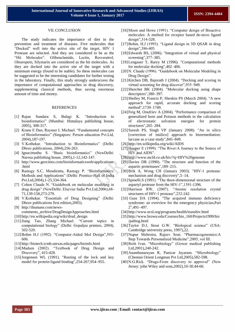
DOCKING OF TOP 5 RANKED MOLECULES INTO THE
ACTIVE SITE OF 3OXC
Docking of Glibenclamide
Figure 20: Glibenclamide shown in violet colour
Docking of Lectin
Figure 21: Lectin shown in violet colour
Docking of Resveratrol
Figure 22: Resveratrol shown in violet colour
Docking of Oleuropein
Figure 23: Oleuropein shown in violet colour
Docking of Silymarin
Figure 24: Silymarin shown in violet colour
Page 383 www.ijiras.com | Email: [email protected]
International Journal of Innovative Research and Advanced Studies (IJIRAS)
Volume 4 Issue 1, January 2017
ISSN: 2394-4404
VII. CONCLUSION
The study indicates the importance of diet in the
prevention and treatment of diseases. Five molecules that
“Docked” well into the active site of the target, HIV 1
Protease are selected. And they are considered to be as the
“Hit Molecules”. Glibenclamide, Lectin, Resveratrol,
Oleuropein, Silymarin are considered as the hit molecules. As
they are docked into the active site of the target with a
minimum energy (found to be stable). So these molecules can
be suggested to be the interesting candidates for further testing
in the laboratory. Finally, this study strongly underscores the
importance of computational approaches in drug discovery,
supplementing classical methods, thus saving enormous
amount of time and money.
REFERENCES
[1] Rajan Sundara S, Balagi K. “Introduction to
bioinformatics” (Mumbai: Himalaya publishing house,
2005), 308-317.
[2] Krane E Dan, Raymer L Michael. “Fundamental concepts
of Bioinformatics” (Singapore: Person education Pvt.Ltd,
2004),187-197.
[3] V.Kothekar. “Introduction to Bioinformatics” (Delhi:
Dhruv publications, 2004),256-263.
[4] Ignacimuthu S. “Basic bioinformatics” (NewDelhi:
Narosa publishing house, 2005),1-12,142-147.
[5] http://www.geocities.com/bioinformaticsweb/applications
.html
[6] Rastogy S.C, Mendiratta, Rastogy P. “Bioinformatics:
Methods and Applications” (Delhi: Prentice-Hall of India
Pvt.Ltd,2004),1-25,334-364.
[7] Cohen Claude N. “Guidebook on molecular modeling in
drug design” (NewDelhi: Elsevier India Pvt.Ltd,2006),68-
71,139-158,273-279.
[8] V.Kothekar. “Essentials of Drug Designing” (Delhi:
Dhruv publications first edition,2005).
[9] http://ihumans.com/news-
comments_archive/DrugDesignApproaches.html
[10] http://en.wilkipedia.org/wiki/drud_design
[11] Jiang Tao, Zhang Michael. “Current topics in
computational biology” (Delhi: Gopaljee printers, 2004),
502-520.
[12] Bohm H.J (1992). “Computer-Aided Mol Design”,593-
606.
[13] http://biotech.icmb.utexas.edu/pages/bioinfo.html
[14] Madsen (2002). “Textbook of Drug Design and
Discovery”, 415-428.
[15] Jorgensen WL (1991). "Rusting of the lock and key
model for protein-ligand binding",254-267,954–955.
[16] Moon and Howe (1991). “Computer design of Bioactive
molecules: A method for receptor based de-novo ligand
design”,314-328.
[17] Bohm, H.J (1993). “Ligand design in 3D QSAR in drug
design”,386-405
[18] Edwards BS, (2006). "Integration of virtual and physical
screening",377–385.
[19] Lengauer T, Rarey M (1996). "Computational methods
for molecular docking",402–406.
[20] N. Claude (1996). “Guidebook on Molecular Modeling in
Drug Design”.
[21] Kitchen DB, Bajorath J (2004). "Docking and scoring in
virtual screening for drug discover”,935–949.
[22] Shoichet BK (2004). "Molecular docking using shape
descriptors",380–397.
[23] Shelley M, Francis P, Shenkin PS (March 2004). "A new
approach for rapid, accurate docking and scoring
method",1739–1749.
[24] Feig M, Onufriev A (2004). "Performance comparison of
generalized born and Poisson methods in the calculation
of electrostatic solvation energies for protein
structures",265–284.
[25] Suresh PS, Singh VP (January 2008). "An in silico
[correction of insilico] approach to bioremediation:
laccase as a case study",845–849.
[26] http://en.wilkipedia.org/wiki/AIDS
[27] Hooper E (1999). “The River:A Journey to the Source of
HIV and AIDS”.
[28] http://www.mcld.co.uk/hiv/?q=HIV%20genome
[29] Davies DR (1990). "The structure and function of the
aspartic proteinases",189–215.
[30] Brik A, Wong CH (January 2003). "HIV-1 protease:
mechanism and drug discovery",5–14.
[31] Spinelli.S (1991). “The three-dimensional structure of the
aspartyl protease from the HIV-1”,1391-1396.
[32] Harrison RW, (2007). “Atomic resolution crystal
structures of HIV-1 protease”,232-242.
[33] Guss DA (1994). "The acquired immuno deficiency
syndrome: an overview for the emergency physician,Part
2",491–497.
[34] http://www.ncsl.org/programs/health/transhiv.html
[35] http://www.brown.edu/Courses/bio_160/Projects1999/hiv
/pathog.html
[36] Taylor D.J, Stout G.W. “Biological science” (USA:
Cambridge university press, 1997),22.
[37] Nupur Mehrotra, Rajeev Soni. “Pharmacogenomics:A
Step Towards Personalized Medicine”,2005, vol III.
[38] Roitt Ivan. “Microbiology” (Gower medical publishing
Ltd,2001),240-242.
[39] Ananthanarayan R, Panicar Jayaram. “Microbiology”
(Chennai Orient Longman Pvt Ltd,2005),582-598.
[40] N.G.Rick. “Drugs-From discovery to approval” (New
Jersey: john Wiley and sons,2002),10-30,44-66.