Embed Size (px)
Citation preview
Molecular Diagnosis
In Oncology &
Genetics
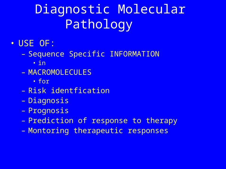
Diagnostic Molecular Pathology
• USE OF:– Sequence Specific INFORMATION
• in
– MACROMOLECULES• for
– Risk identfication– Diagnosis– Prognosis– Prediction of response to therapy– Montoring therapeutic responses
Macromolecules
• Peptides/proteins• Polysaccharides• Polynucleotides/nucleic acids
“Nucleic Acid Diagnosis”
• Use of specific sequence information in
• nucleic acids – DNA and RNA
• for clinical diagnosis
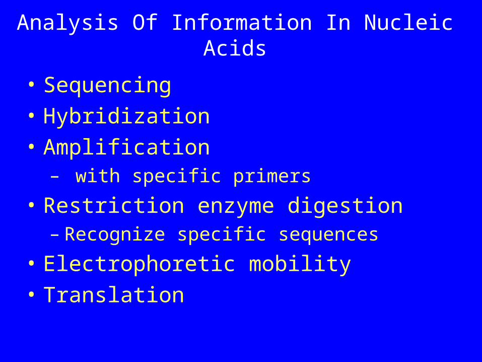
Analysis Of Information In Nucleic Acids
• Sequencing• Hybridization• Amplification
– with specific primers
• Restriction enzyme digestion– Recognize specific sequences
• Electrophoretic mobility• Translation
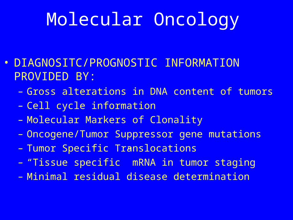
Molecular Oncology
• DIAGNOSITC/PROGNOSTIC INFORMATION PROVIDED BY:– Gross alterations in DNA content of tumors – Cell cycle information – Molecular Markers of Clonality– Oncogene/Tumor Suppressor gene mutations– Tumor Specific Translocations– “Tissue specific” mRNA in tumor staging– Minimal residual disease determination
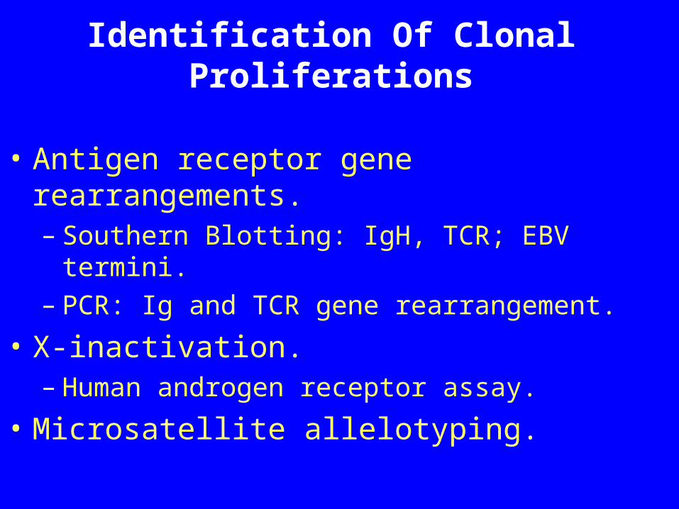
Identification Of Clonal Proliferations
• Antigen receptor gene rearrangements.– Southern Blotting: IgH, TCR; EBV termini.– PCR: Ig and TCR gene rearrangement.
• X-inactivation.– Human androgen receptor assay.
• Microsatellite allelotyping.
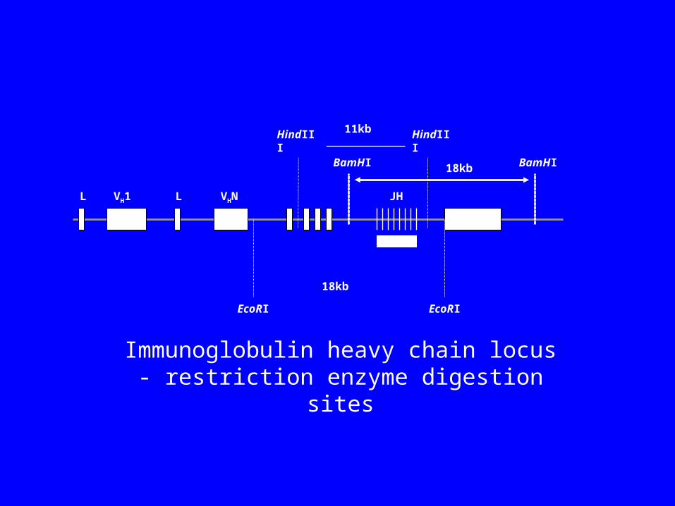
L LVH1 VHN
HindIII HindIII11kb
EcoRI EcoRI
18kb
18kbBamHI BamHI
Immunoglobulin heavy chain locus - restriction enzyme digestion sites
JH
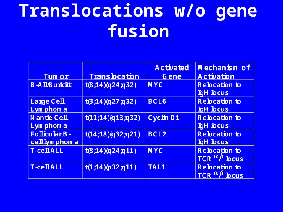
Translocations w/o gene fusion
Tumor
Translocation
Activated Gene
Mechanism of Activation
B-All/Burkitt t(8;14)(q24;q32) MYC Relocation to IgH locus
Large Cell Lymphoma
t(3;14)(q27;q32) BCL6 Relocation to IgH locus
Mantle Cell Lymphoma
t(11;14)(q13;q32) Cyclin D1 Relocation to IgH locus
Follicular B-cell lymphoma
t(14;18)(q32;q21) BCL2 Relocation to IgH locus
T-cell ALL t(8;14)(q24;q11) MYC Relocation to TCR / locus
T-cell ALL t(1;14)(p32;q11) TAL1 Relocation to TCR / locus
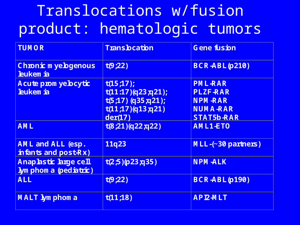
Translocations w/fusion product: hematologic tumors
TUMOR Translocation Gene fusion
Chronic myelogenous leukemia
t(9;22) BCR-ABL(p210)
Acute promyelocytic leukemia
t(15;17); t(11:17)(q23;q21); t(5;17) (q35;q21); t(11;17)(q13;q21) der(17)
PML-RAR PLZF-RAR NPM-RAR NUMA-RAR STAT5b-RAR
AML t(8;21)(q22;q22) AML1-ETO
AML and ALL (esp. infants and post-Rx)
11q23 MLL-(~30 partners)
Anaplastic large cell lymphoma (pediatric)
t(2;5)(p23;q35) NPM-ALK
ALL t(9;22) BCR-ABL(p190)
MALT lymphoma t(11;18) API2-MLT
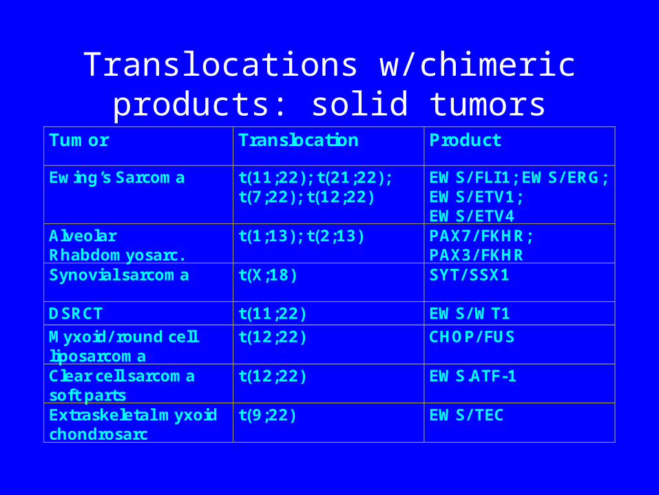
Translocations w/chimeric products: solid tumors
Tumor Translocation Product
Ewing’s Sarcoma t(11;22); t(21;22);t(7;22); t(12;22)
EWS/ FLI1; EWS/ ERG;EWS/ ETV1;EWS/ ETV4
AlveolarRhabdomyosarc.
t(1;13); t(2;13) PAX7/ FKHR;PAX3/ FKHR
Synovial sarcoma t(X;18) SYT/ SSX1
DSRCT t(11;22) EWS/ WT1Myxoid/ round cellliposarcoma
t(12;22) CHOP/ FUS
Clear cell sarcomasoft parts
t(12;22) EWS.ATF-1
Extraskeletal myxoidchondrosarc
t(9;22) EWS/ TEC
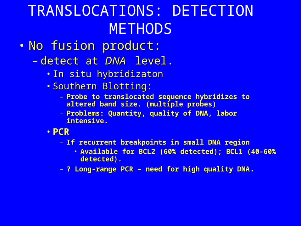
TRANSLOCATIONS: DETECTION METHODS
• No fusion product:No fusion product:– detect at detect at DNA DNA level. level.
• In situ hybridizatonIn situ hybridizaton• Southern Blotting: Southern Blotting:
– Probe to translocated sequence hybridizes to Probe to translocated sequence hybridizes to altered band size. (multiple probes)altered band size. (multiple probes)
– Problems: Quantity, quality of DNA, labor intensive. Problems: Quantity, quality of DNA, labor intensive.
• PCRPCR– If recurrent breakpoints in small DNA regionIf recurrent breakpoints in small DNA region
• Available for BCL2 (60% detected); BCL1 (40-Available for BCL2 (60% detected); BCL1 (40-60% detected). 60% detected).
– ? Long-range PCR – need for high quality DNA? Long-range PCR – need for high quality DNA..
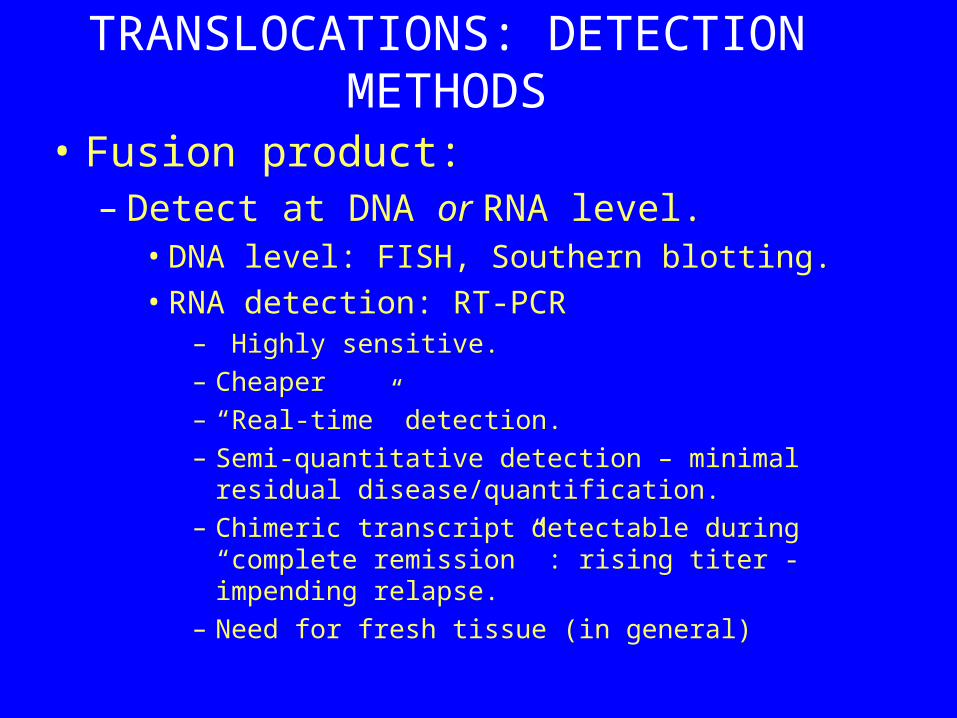
TRANSLOCATIONS: DETECTION METHODS
• Fusion product:– Detect at DNA or RNA level.
• DNA level: FISH, Southern blotting. • RNA detection: RT-PCR
– Highly sensitive. – Cheaper– “Real-time” detection.– Semi-quantitative detection – minimal residual
disease/quantification. – Chimeric transcript detectable during “complete
remission” : rising titer - impending relapse. – Need for fresh tissue (in general)
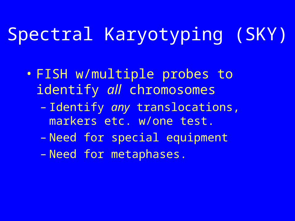
Spectral Karyotyping (SKY)
• FISH w/multiple probes to identify all chromosomes – Identify any translocations, markers
etc. w/one test. – Need for special equipment– Need for metaphases.
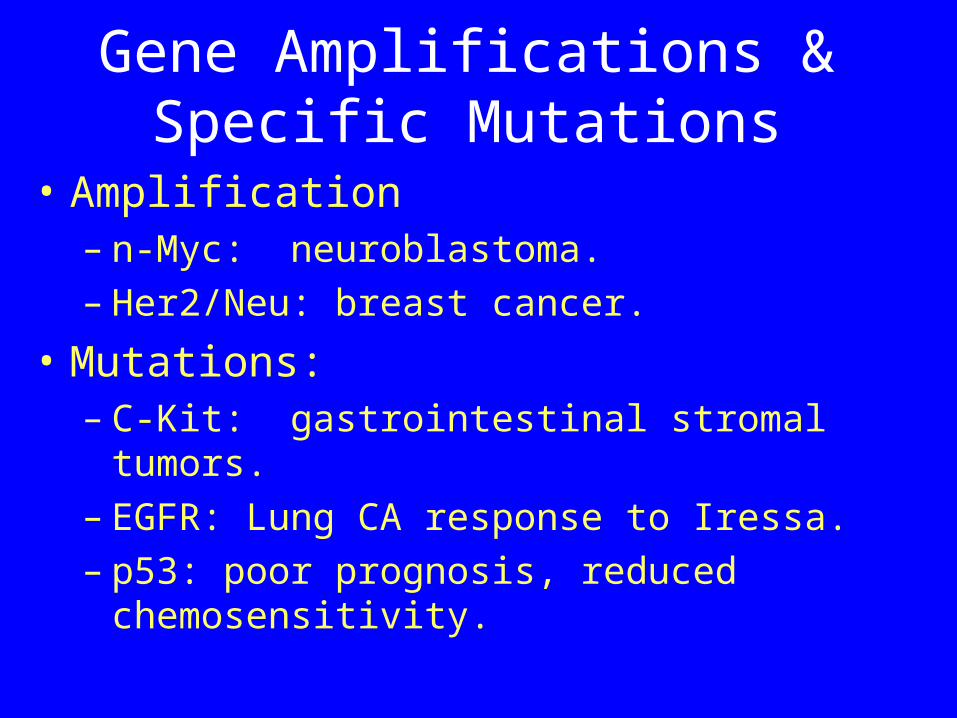
Gene Amplifications & Specific Mutations
• Amplification– n-Myc: neuroblastoma.– Her2/Neu: breast cancer.
• Mutations: – C-Kit: gastrointestinal stromal tumors. – EGFR: Lung CA response to Iressa. – p53: poor prognosis, reduced
chemosensitivity.
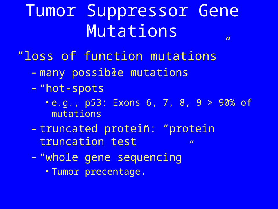
Tumor Suppressor Gene Mutations
“loss of function mutations”– many possible mutations– “hot-spots”
• e.g., p53: Exons 6, 7, 8, 9 > 90% of mutations
– truncated protein: “protein truncation test”
– “whole gene sequencing”• Tumor precentage.
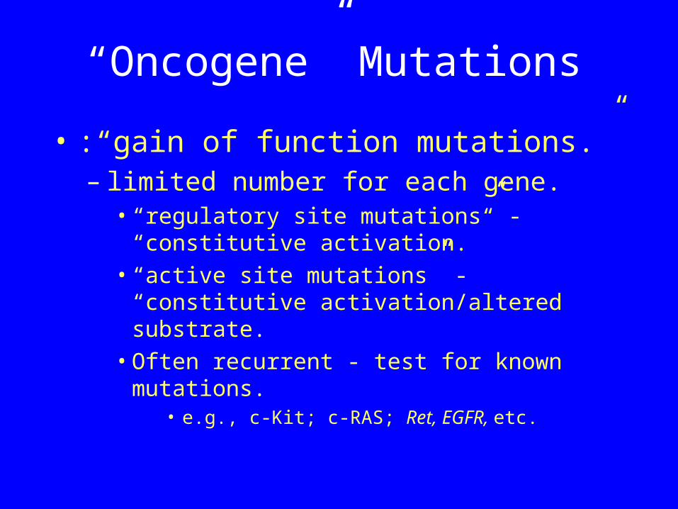
“Oncogene” Mutations
• :“gain of function mutations.”– limited number for each gene.
• “regulatory site mutations” - “constitutive activation.”
• “active site mutations” - “constitutive activation/altered substrate.
• Often recurrent - test for known mutations.
• e.g., c-Kit; c-RAS; Ret, EGFR, etc.
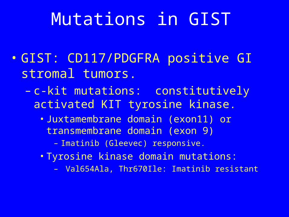
Mutations in GIST
• GIST: CD117/PDGFRA positive GI stromal tumors. – c-kit mutations: constitutively activated
KIT tyrosine kinase.• Juxtamembrane domain (exon11) or
transmembrane domain (exon 9)– Imatinib (Gleevec) responsive.
• Tyrosine kinase domain mutations:– Val654Ala, Thr670Ile: Imatinib resistant
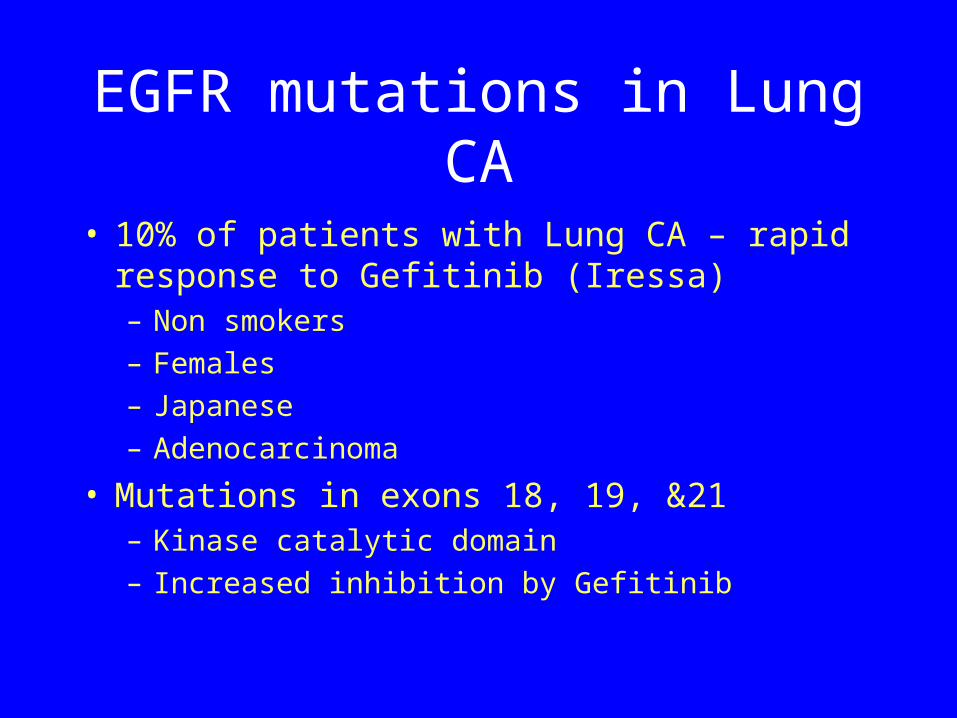
EGFR mutations in Lung CA
• 10% of patients with Lung CA – rapid response to Gefitinib (Iressa)– Non smokers– Females– Japanese– Adenocarcinoma
• Mutations in exons 18, 19, &21– Kinase catalytic domain– Increased inhibition by Gefitinib
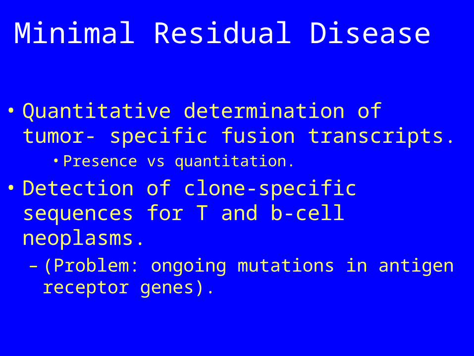
Minimal Residual Disease
• Quantitative determination of tumor- specific fusion transcripts.
• Presence vs quantitation.
• Detection of clone-specific sequences for T and b-cell neoplasms.– (Problem: ongoing mutations in antigen
receptor genes).
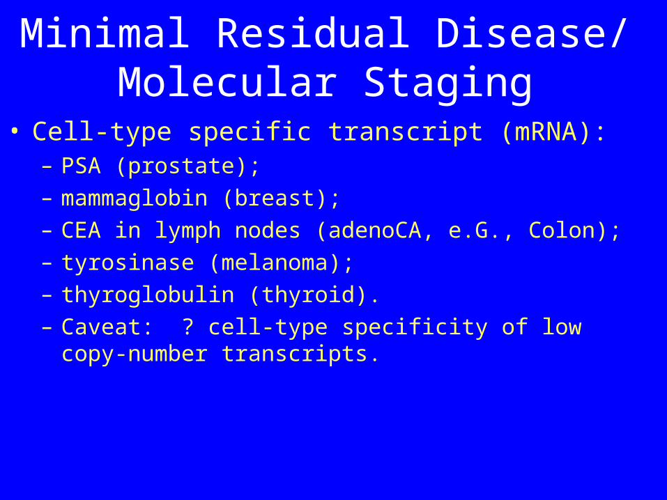
Minimal Residual Disease/ Molecular Staging
• Cell-type specific transcript (mRNA):– PSA (prostate);– mammaglobin (breast);– CEA in lymph nodes (adenoCA, e.G., Colon);– tyrosinase (melanoma);– thyroglobulin (thyroid).– Caveat: ? cell-type specificity of low copy-number
transcripts.
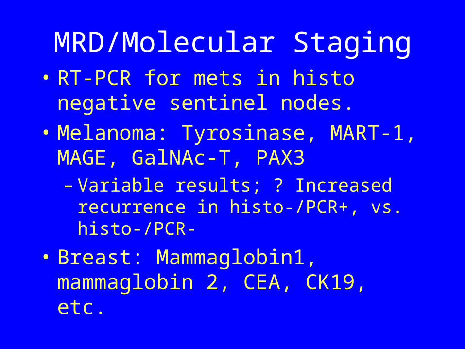
MRD/Molecular Staging• RT-PCR for mets in histo negative
sentinel nodes. • Melanoma: Tyrosinase, MART-1,
MAGE, GalNAc-T, PAX3– Variable results; ? Increased
recurrence in histo-/PCR+, vs. histo-/PCR-
• Breast: Mammaglobin1, mammaglobin 2, CEA, CK19, etc.
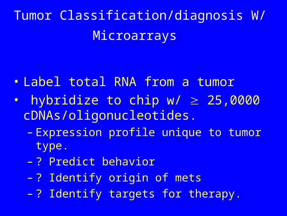
Tumor Classification/diagnosis W/
Microarrays
• Label total RNA from a tumor• hybridize to chip w/ 25,0000
cDNAs/oligonucleotides.– Expression profile unique to tumor type. – ? Predict behavior– ? Identify origin of mets– ? Identify targets for therapy.
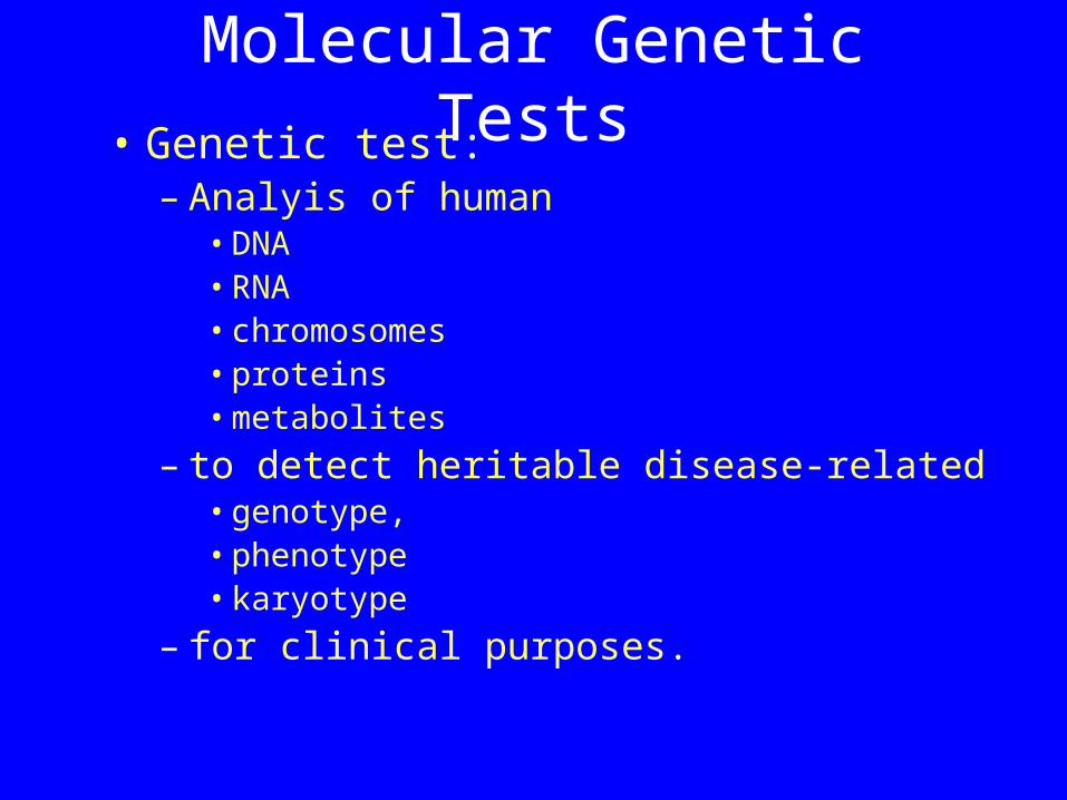
Molecular Genetic Tests• Genetic test:
– Analyis of human• DNA• RNA• chromosomes• proteins• metabolites
– to detect heritable disease-related• genotype, • phenotype• karyotype
– for clinical purposes.
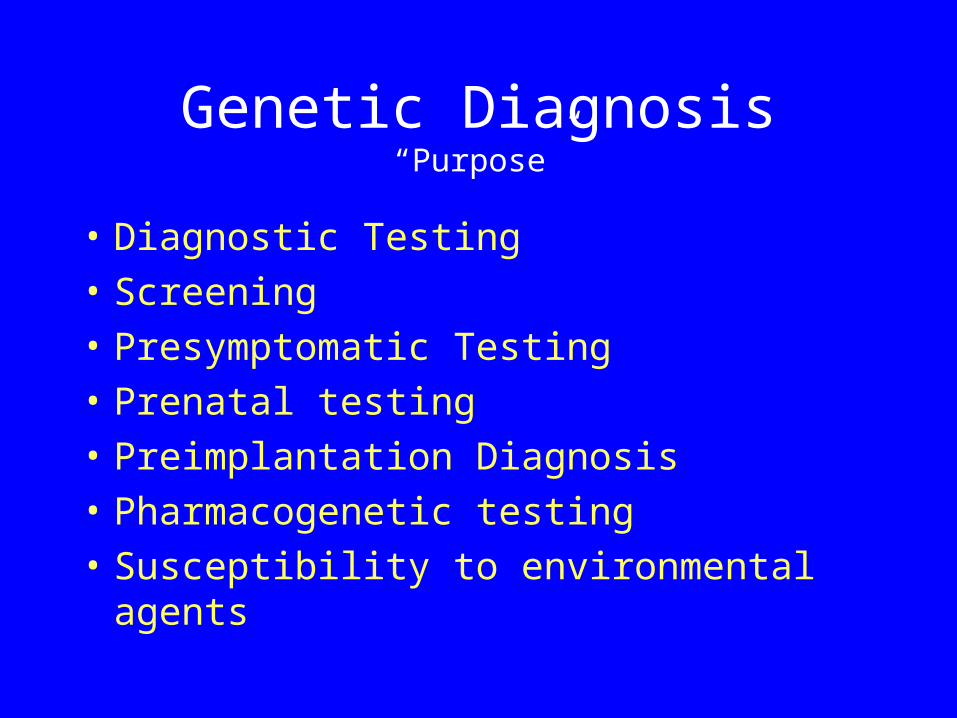
Genetic Diagnosis“Purpose”
• Diagnostic Testing• Screening • Presymptomatic Testing• Prenatal testing• Preimplantation Diagnosis• Pharmacogenetic testing• Susceptibility to environmental
agents
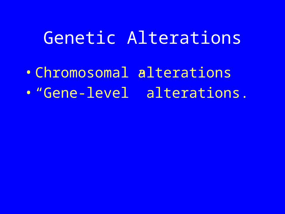
Genetic Alterations
• Chromosomal alterations• “Gene-level” alterations.
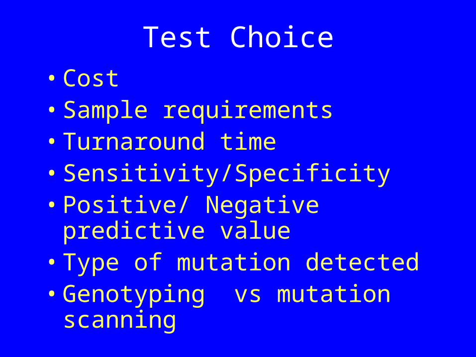
Test Choice• Cost• Sample requirements• Turnaround time• Sensitivity/Specificity• Positive/ Negative predictive
value• Type of mutation detected• Genotyping vs mutation
scanning

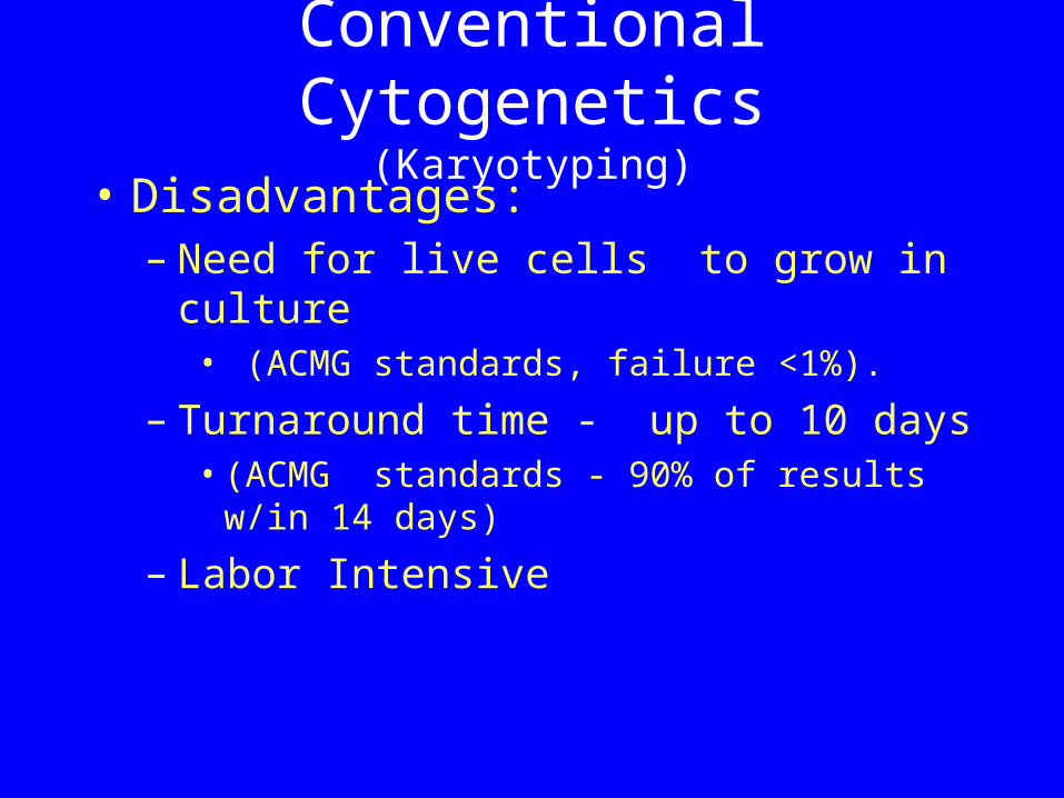
Conventional Cytogenetics(Karyotyping)
• Detect numerical structural chromosomal alterations– trisomy– monosomy– duplications– translocations, etc.
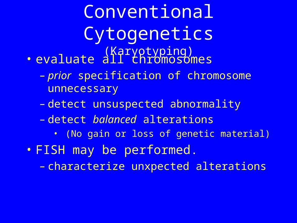
Conventional Cytogenetics(Karyotyping)
• evaluate all chromosomes – prior specification of chromosome
unnecessary– detect unsuspected abnormality – detect balanced alterations
• (No gain or loss of genetic material)
• FISH may be performed. – characterize unxpected alterations
Conventional Cytogenetics(Karyotyping)
• Disadvantages:– Need for live cells to grow in culture
• (ACMG standards, failure <1%).
– Turnaround time - up to 10 days • (ACMG standards - 90% of results w/in
14 days)
– Labor Intensive
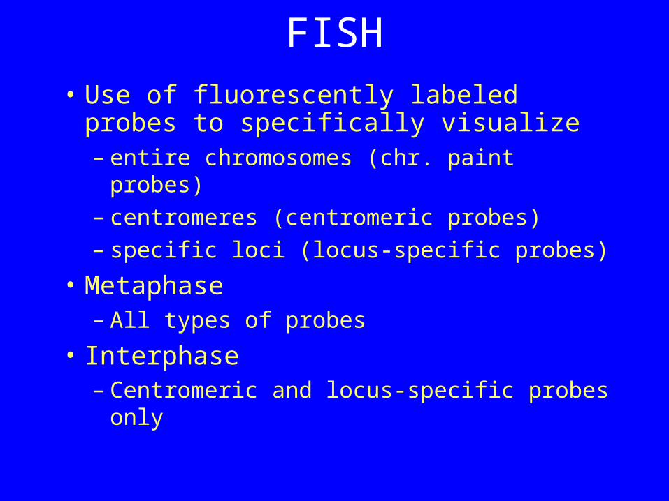
FISH• Use of fluorescently labeled probes
to specifically visualize – entire chromosomes (chr. paint probes) – centromeres (centromeric probes)– specific loci (locus-specific probes)
• Metaphase – All types of probes
• Interphase – Centromeric and locus-specific probes
only
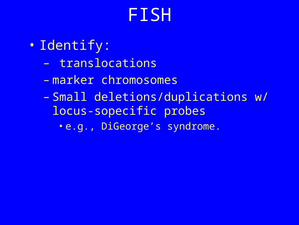
FISH
• Identify:– translocations– marker chromosomes – Small deletions/duplications w/ locus-
sopecific probes • e.g., DiGeorge’s syndrome.
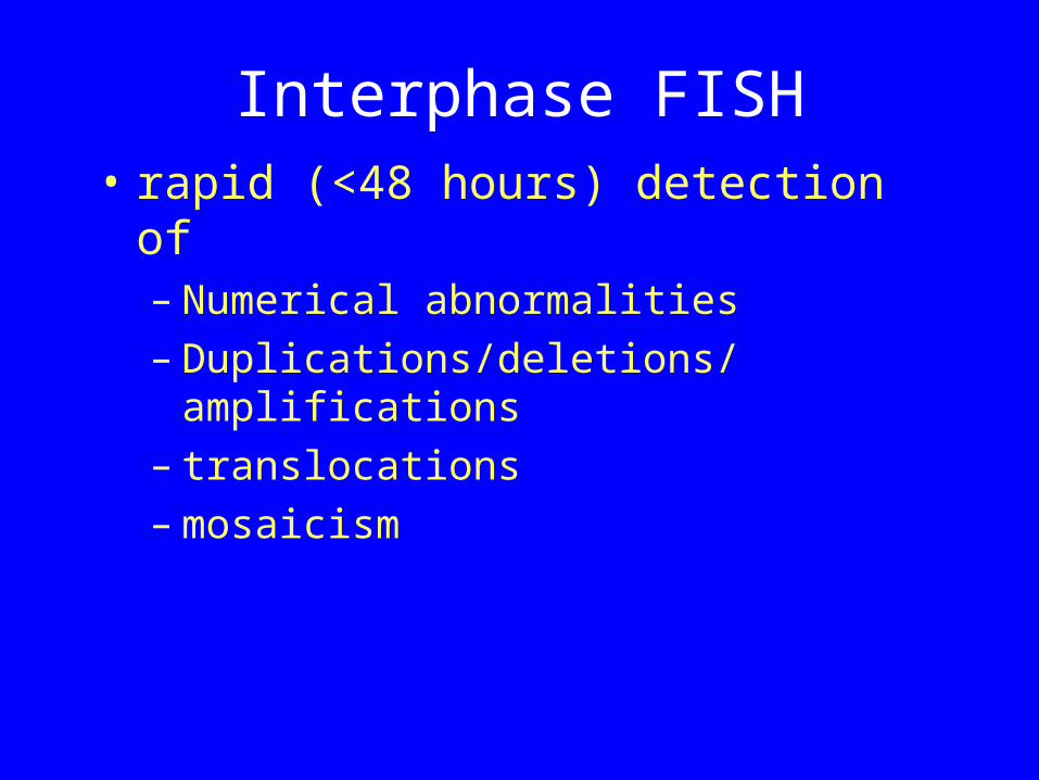
Interphase FISH• rapid (<48 hours) detection of
– Numerical abnormalities– Duplications/deletions/amplifications– translocations– mosaicism
Interphase FISH• Prenatal Chr.13, 18, 21, X + Y
– approx. 75-85% of all clinically relevant abnormalities.
• Dual color FISH w/ subtelomeric probes:– Prenatal dx of chromosomal
translocations
Interphase FISH
• Need for confirmatory conventional cytogenetic testing.
• Need to specify chromosome – Information only about specific
chromosome/locus tested.
Metaphase FISH• Supplement conventional
cytogenetics – Identify marker chromosomes– extra unknown material attached to
chromosome/loss of segment– detect/identify rearrangements (incl.
cryptic translocations), – identify/quantify mosaicism
Metaphase FISH• Need to specify Chromosome/locus
– Multiple tests to identify marker chromosome.
– Multiprobe FISH.
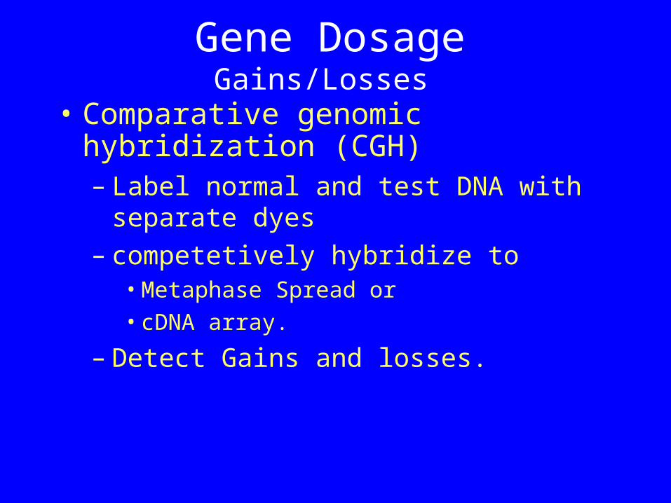
Gene DosageGains/Losses
• Comparative genomic hybridization (CGH)– Label normal and test DNA with
separate dyes– competetively hybridize to
• Metaphase Spread or • cDNA array.
– Detect Gains and losses.
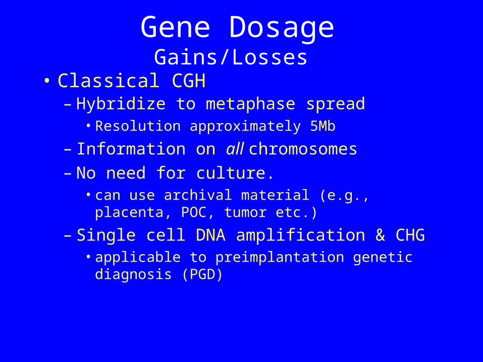
Gene DosageGains/Losses
• Classical CGH– Hybridize to metaphase spread
• Resolution approximately 5Mb
– Information on all chromosomes– No need for culture.
• can use archival material (e.g., placenta, POC, tumor etc.)
– Single cell DNA amplification & CHG• applicable to preimplantation genetic
diagnosis (PGD)
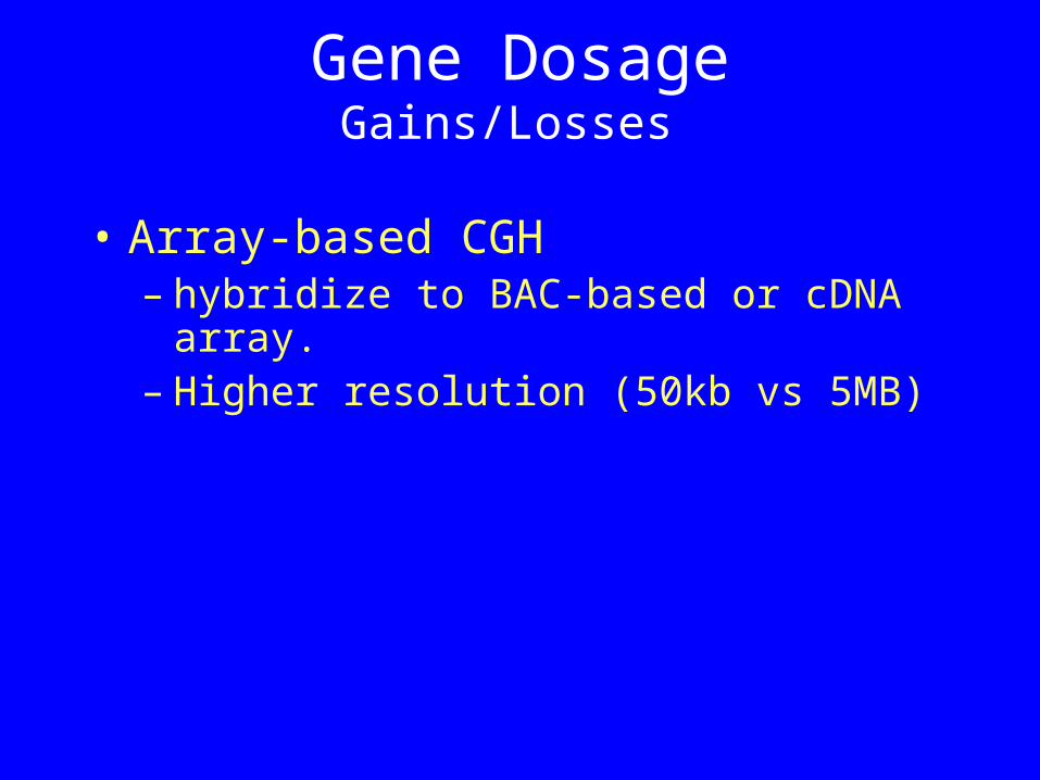
Gene DosageGains/Losses
• Array-based CGH– hybridize to BAC-based or cDNA
array.– Higher resolution (50kb vs 5MB)
Gene DosageGains/Losses
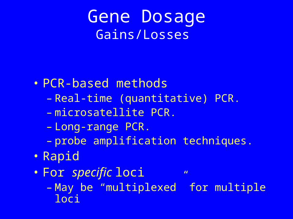
• PCR-based methods– Real-time (quantitative) PCR.– microsatellite PCR.– Long-range PCR.– probe amplification techniques.
• Rapid• For specific loci
– May be “multiplexed” for multiple loci
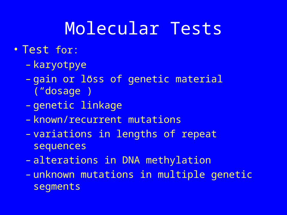
Molecular Tests• Test for:
– karyotpye– gain or loss of genetic material (“dosage”)– genetic linkage– known/recurrent mutations– variations in lengths of repeat sequences– alterations in DNA methylation– unknown mutations in multiple genetic
segments
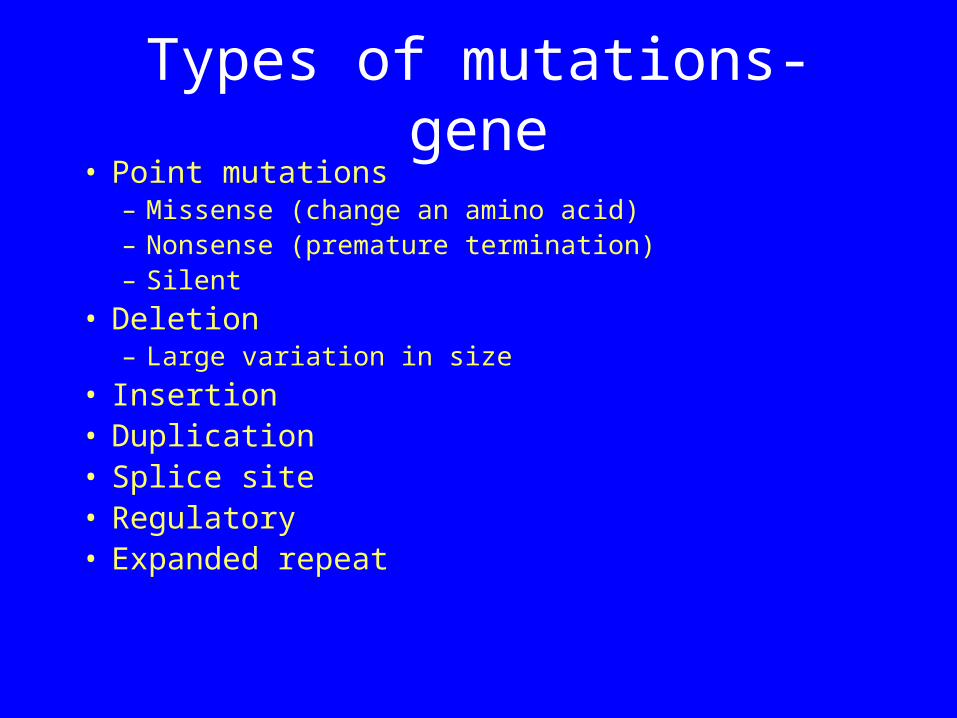
Types of mutations-gene• Point mutations
– Missense (change an amino acid)– Nonsense (premature termination)– Silent
• Deletion– Large variation in size
• Insertion• Duplication• Splice site• Regulatory• Expanded repeat
0 2000 4000 6000 8000
gross deletions
complex rearrangements
Gross insertion and duplications
repeat expansions
small ins/del
small insertions
small deletions
regulatory
splicing
Nonsense
Missense
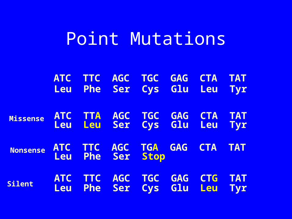
Point Mutations
ATC TTC AGC TGC GAG CTA TAT
ATC TTA AGC TGC GAG CTA TAT
ATC TTC AGC TGA GAG CTA TAT
ATC TTC AGC TGC GAG CTG TAT
Leu Phe Ser Cys Glu Leu Tyr
Leu Phe Ser Stop
Leu Leu Ser Cys Glu Leu Tyr
Leu Phe Ser Cys Glu Leu Tyr
Missense
Nonsense
Silent
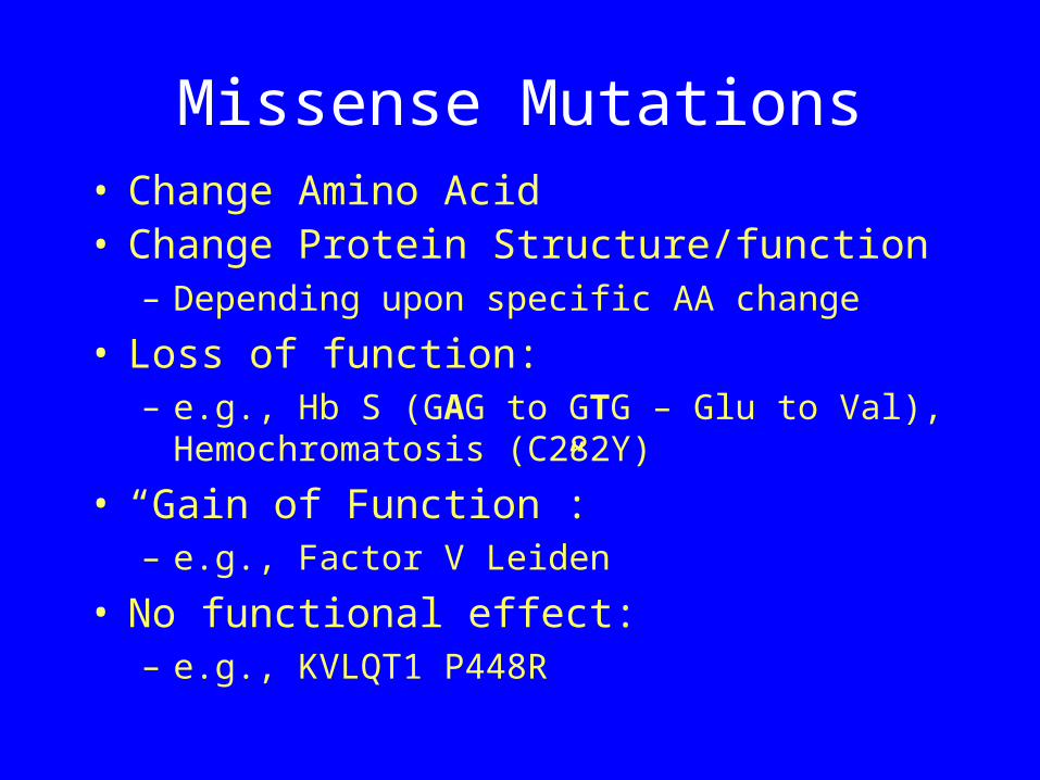
Missense Mutations• Change Amino Acid• Change Protein Structure/function
– Depending upon specific AA change
• Loss of function: – e.g., Hb S (GAG to GTG – Glu to Val),
Hemochromatosis (C282Y)
• “Gain of Function”:– e.g., Factor V Leiden
• No functional effect:– e.g., KVLQT1 P448R

Missense mutations
• When is a missense mutation significant?
• known structural and functional domain• evolutionarily conserved residue• independent occurrence in unrelated patients• absent in large control sample• novel appearance & cosegregation w/disease
phenotype in pedigreee• In vitro loss of function• restoration of function by WT protein.
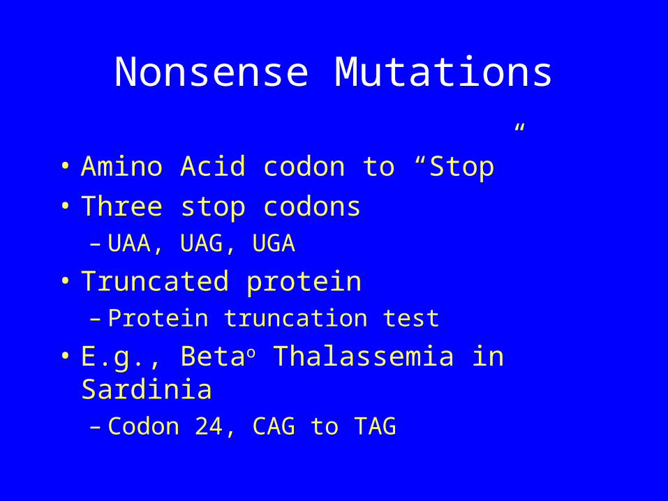
Nonsense Mutations
• Amino Acid codon to “Stop”• Three stop codons
– UAA, UAG, UGA
• Truncated protein– Protein truncation test
• E.g., Betao Thalassemia in Sardinia– Codon 24, CAG to TAG
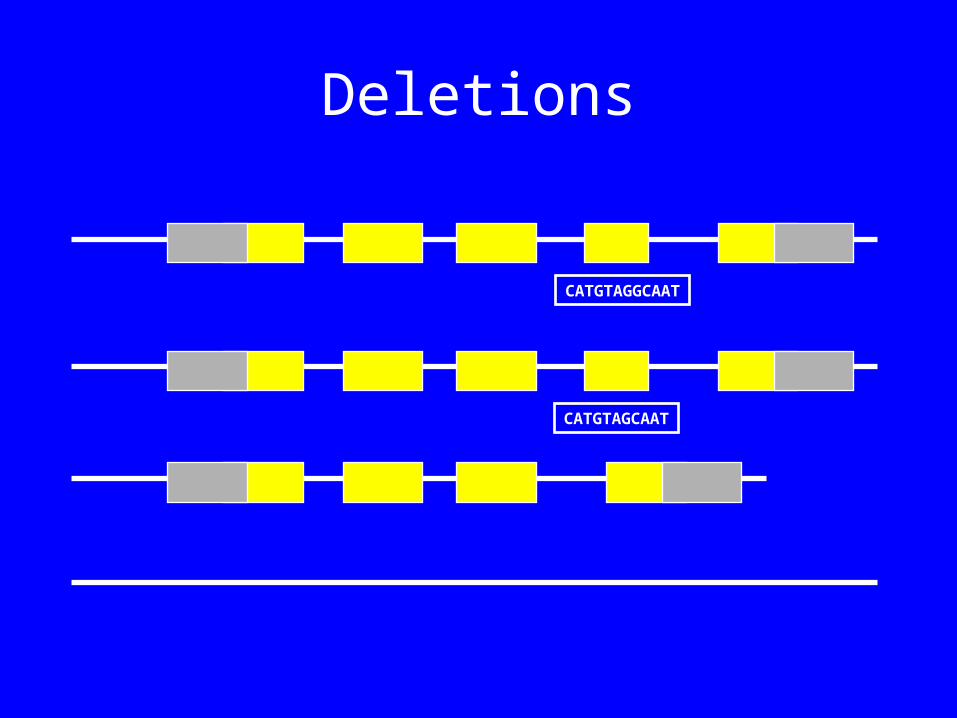
Deletions
CATGTAGGCAAT
CATGTAGCAAT
Deletions
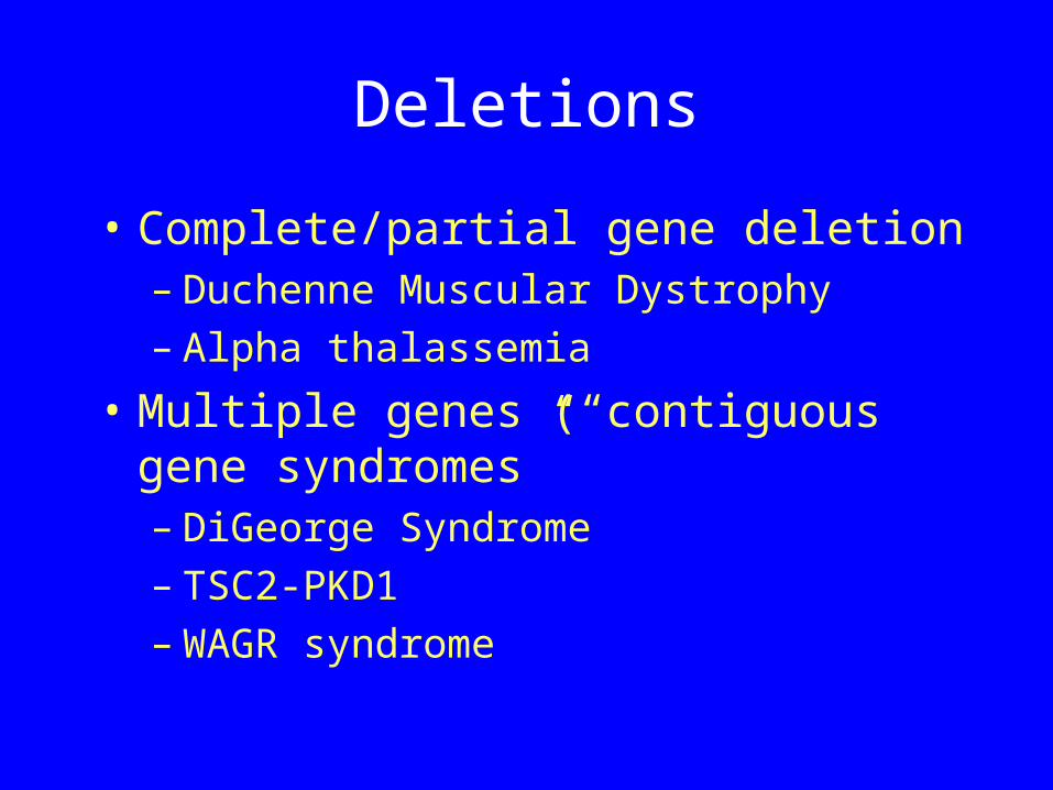
• Complete/partial gene deletion– Duchenne Muscular Dystrophy– Alpha thalassemia
• Multiple genes (“contiguous gene syndromes”– DiGeorge Syndrome– TSC2-PKD1 – WAGR syndrome
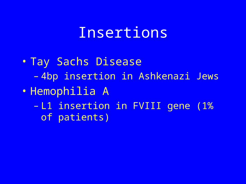
Insertions
• Tay Sachs Disease– 4bp insertion in Ashkenazi Jews
• Hemophilia A– L1 insertion in FVIII gene (1% of
patients)
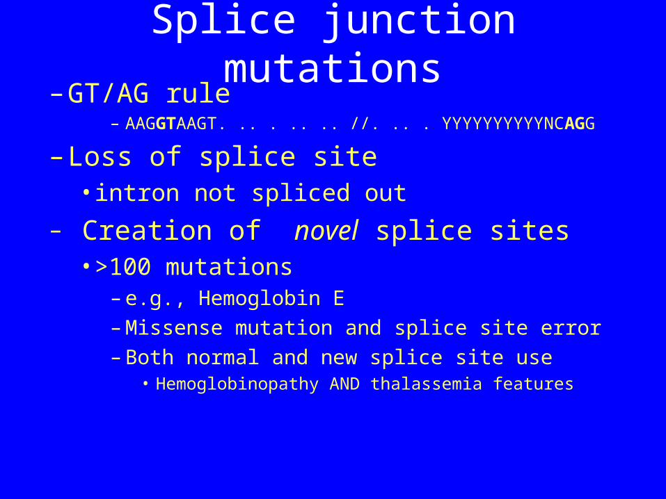
Splice junction mutations– GT/AG rule
– AAGGTAAGT. .. . .. .. //. .. . YYYYYYYYYYNCAGG
– Loss of splice site •intron not spliced out
– Creation of novel splice sites•>100 mutations
– e.g., Hemoglobin E – Missense mutation and splice site error– Both normal and new splice site use
• Hemoglobinopathy AND thalassemia features
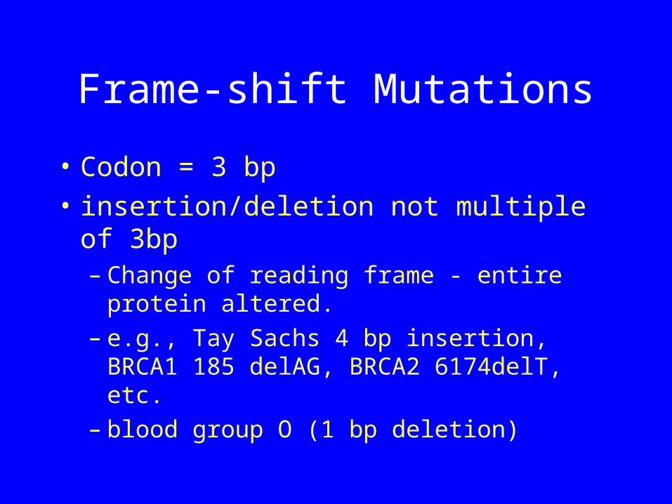
Frame-shift Mutations
• Codon = 3 bp• insertion/deletion not multiple of
3bp– Change of reading frame - entire
protein altered. – e.g., Tay Sachs 4 bp insertion, BRCA1
185 delAG, BRCA2 6174delT, etc.– blood group O (1 bp deletion)
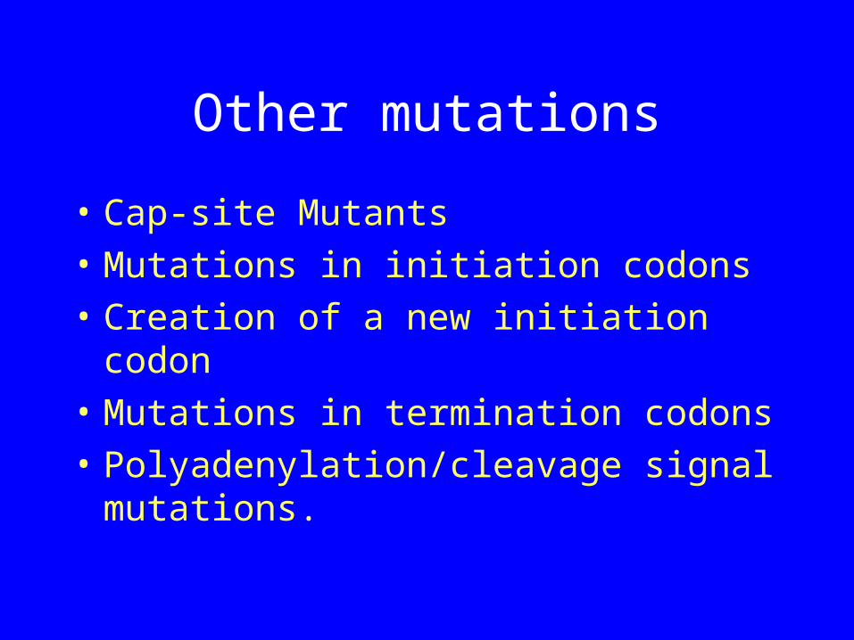
Other mutations
• Cap-site Mutants• Mutations in initiation codons• Creation of a new initiation codon• Mutations in termination codons• Polyadenylation/cleavage signal
mutations.
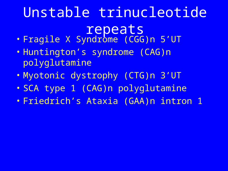
Unstable trinucleotide repeats• Fragile X Syndrome (CGG)n 5’UT• Huntington’s syndrome (CAG)n
polyglutamine• Myotonic dystrophy (CTG)n 3’UT• SCA type 1 (CAG)n polyglutamine• Friedrich’s Ataxia (GAA)n intron 1
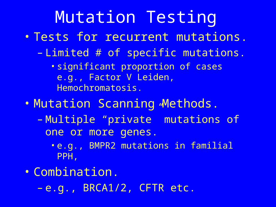
Mutation Testing• Tests for recurrent mutations.
– Limited # of specific mutations.• significant proportion of cases e.g., Factor
V Leiden, Hemochromatosis.
• Mutation Scanning Methods.– Multiple “private” mutations of one or
more genes.• e.g., BMPR2 mutations in familial PPH,
• Combination.– e.g., BRCA1/2, CFTR etc.
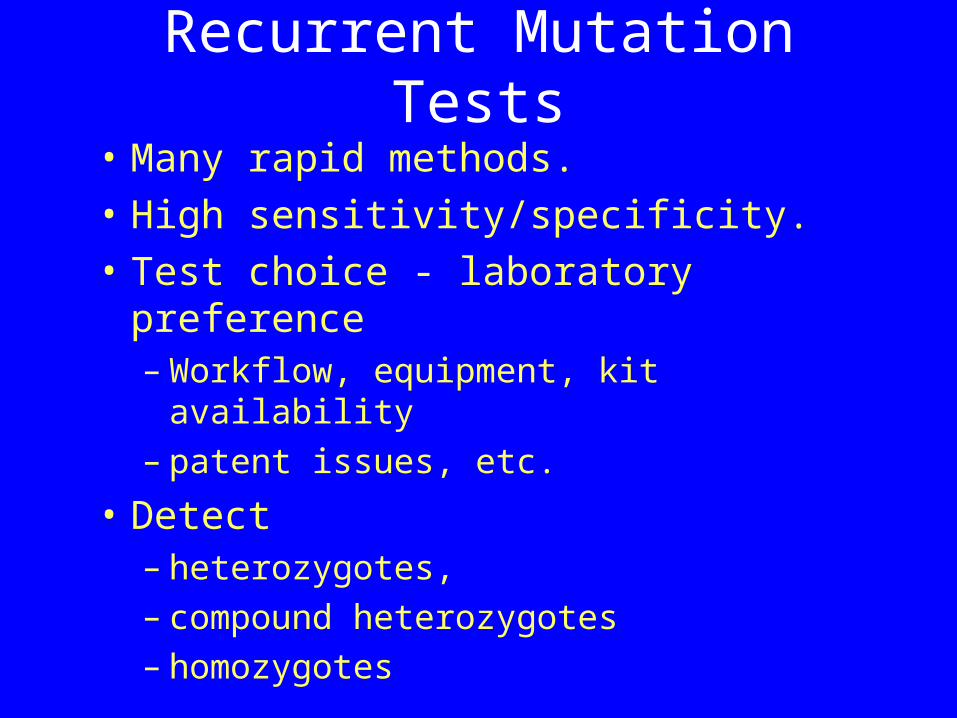
Recurrent Mutation Tests
• Many rapid methods.• High sensitivity/specificity. • Test choice - laboratory preference
– Workflow, equipment, kit availability– patent issues, etc.
• Detect – heterozygotes, – compound heterozygotes– homozygotes
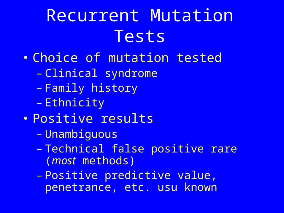
Recurrent Mutation Tests
• Choice of mutation tested– Clinical syndrome– Family history– Ethnicity
• Positive results– Unambiguous– Technical false positive rare (most
methods)– Positive predictive value, penetrance,
etc. usu known
Recurrent Mutation Tests
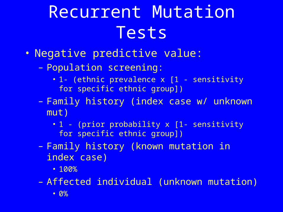
• Negative predictive value:– Population screening:
• 1- (ethnic prevalence x [1 - sensitivity for specific ethnic group])
– Family history (index case w/ unknown mut)• 1 - (prior probability x [1- sensitivity for specific
ethnic group])
– Family history (known mutation in index case)
• 100%
– Affected individual (unknown mutation)• 0%
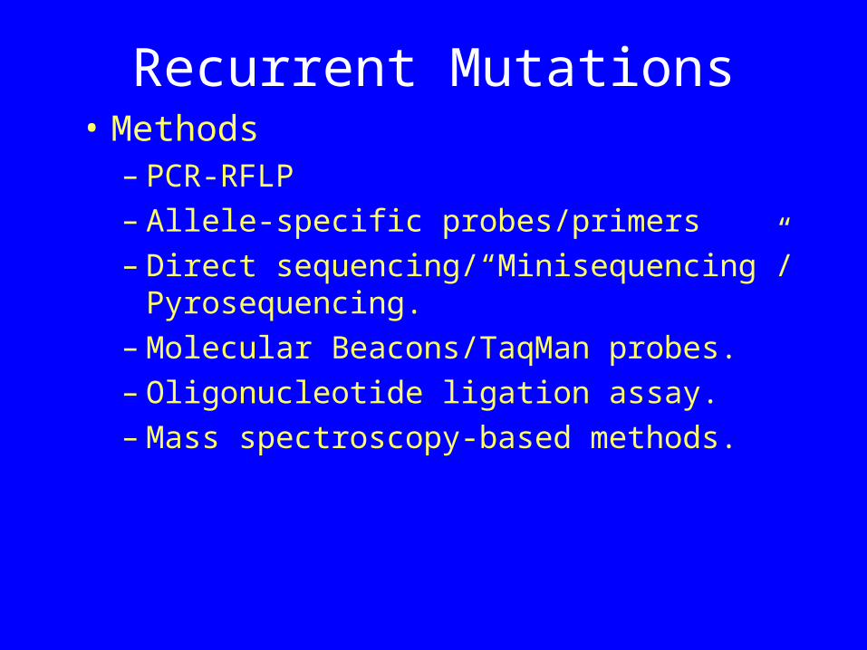
Recurrent Mutations• Methods
– PCR-RFLP– Allele-specific probes/primers– Direct sequencing/“Minisequencing”/
Pyrosequencing. – Molecular Beacons/TaqMan probes. – Oligonucleotide ligation assay.– Mass spectroscopy-based methods.
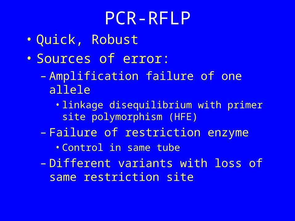
PCR-RFLP• Quick, Robust• Sources of error:
– Amplification failure of one allele • linkage disequilibrium with primer site
polymorphism (HFE)
– Failure of restriction enzyme• Control in same tube
– Different variants with loss of same restriction site
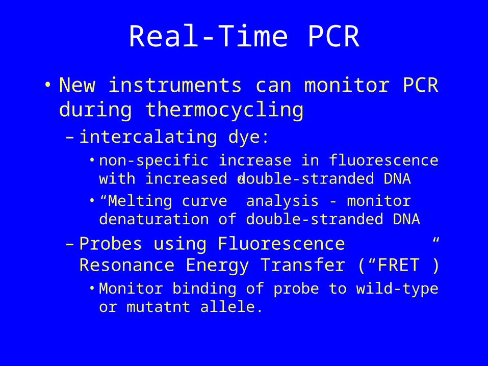
Real-Time PCR
• New instruments can monitor PCR during thermocycling– intercalating dye:
• non-specific increase in fluorescence with increased double-stranded DNA
• “Melting curve” analysis - monitor denaturation of double-stranded DNA
– Probes using Fluorescence Resonance Energy Transfer (“FRET”)• Monitor binding of probe to wild-type or
mutatnt allele.
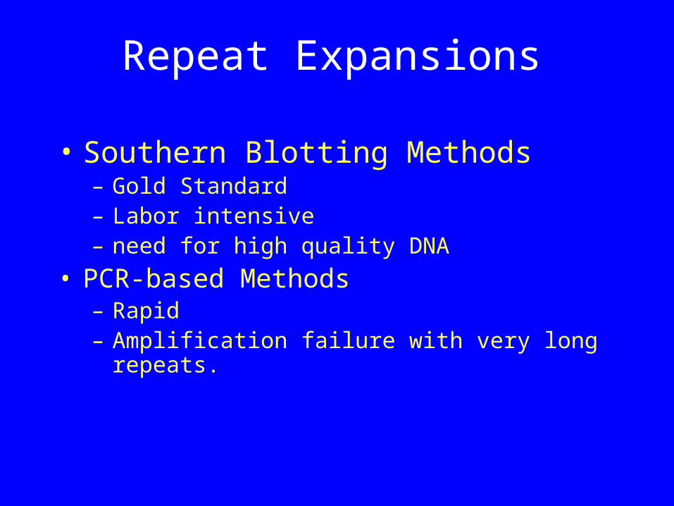
Repeat Expansions
• Southern Blotting Methods– Gold Standard– Labor intensive– need for high quality DNA
• PCR-based Methods– Rapid– Amplification failure with very long repeats.
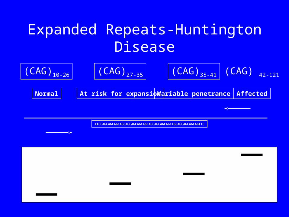
Expanded Repeats-Huntington Disease
(CAG)10-26 (CAG)35-41 (CAG) 42-121(CAG)27-35
Normal At risk for expansion Variable penetrance Affected
ATCCAGCAGCAGCAGCAGCAGCAGCAGCAGCAGCAGCAGCAGCAGCAGCAGCAGTTC
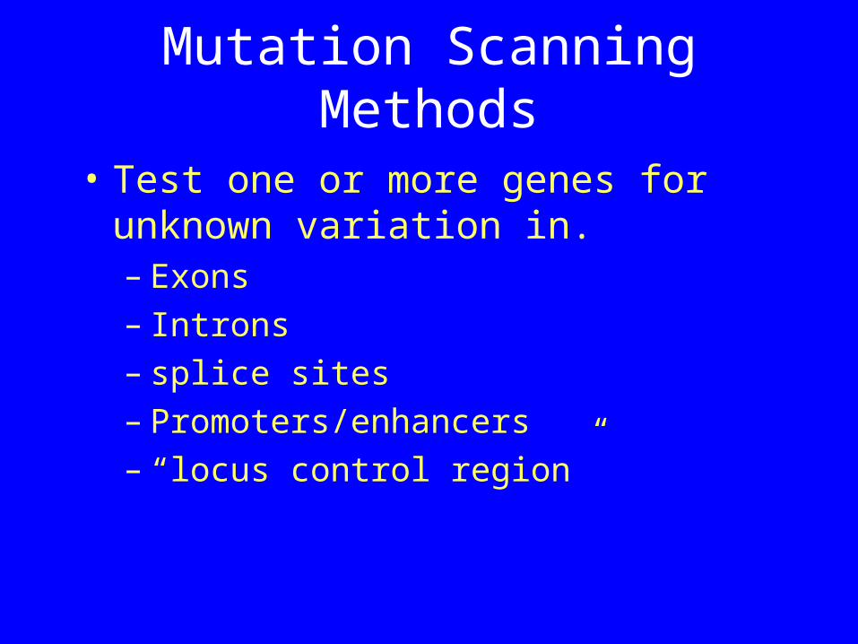
Mutation Scanning Methods
• Test one or more genes for unknown variation in. – Exons – Introns – splice sites– Promoters/enhancers – “locus control region”
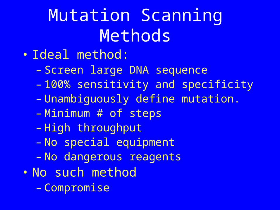
Mutation Scanning Methods
• Ideal method:– Screen large DNA sequence– 100% sensitivity and specificity– Unambiguously define mutation.– Minimum # of steps– High throughput– No special equipment – No dangerous reagents
• No such method– Compromise
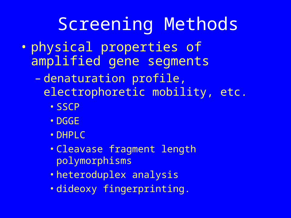
Screening Methods• physical properties of amplified
gene segments
– denaturation profile, electrophoretic mobility, etc. • SSCP• DGGE• DHPLC• Cleavase fragment length polymorphisms• heteroduplex analysis• dideoxy fingerprinting.
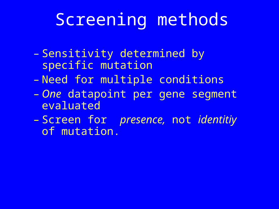
– Sensitivity determined by specific mutation
– Need for multiple conditions– One datapoint per gene segment
evaluated – Screen for presence, not identitiy of
mutation.
Screening methods
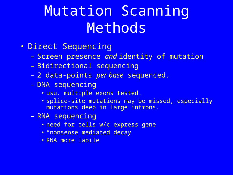
Mutation Scanning Methods
• Direct Sequencing– Screen presence and identity of mutation– Bidirectional sequencing – 2 data-points per base sequenced. – DNA sequencing
• usu. multiple exons tested. • splice-site mutations may be missed, especially
mutations deep in large introns.
– RNA sequencing• need for cells w/c express gene• “nonsense mediated decay” • RNA more labile
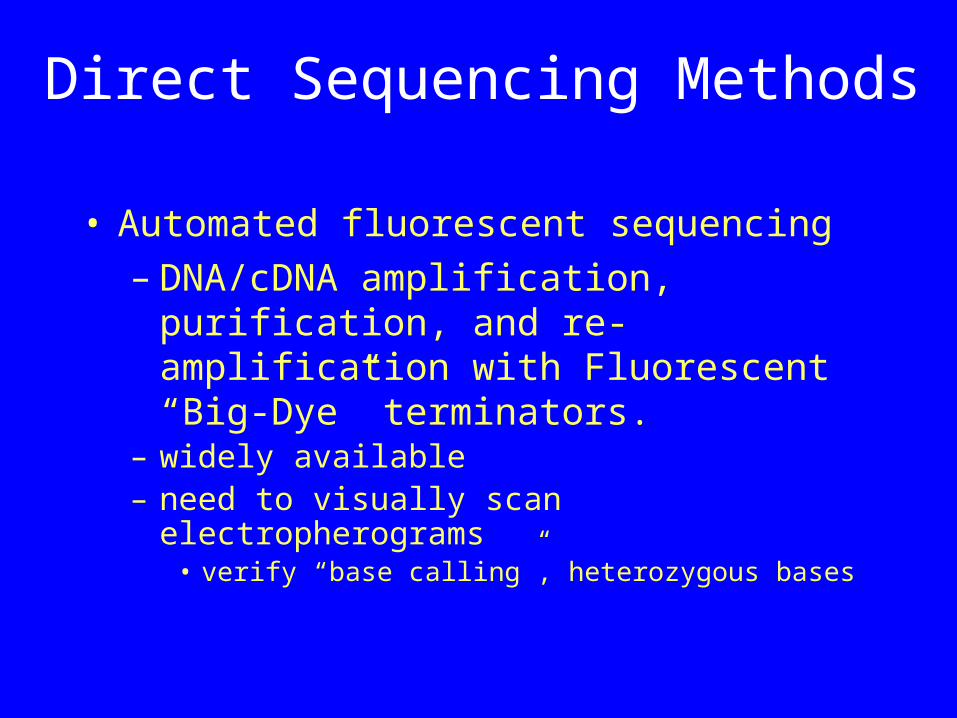
Direct Sequencing Methods
• Automated fluorescent sequencing– DNA/cDNA amplification, purification,
and re-amplification with Fluorescent “Big-Dye” terminators.
– widely available– need to visually scan electropherograms
• verify “base calling”, heterozygous bases
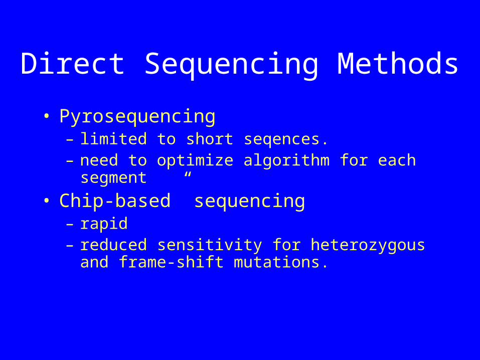
Direct Sequencing Methods
• Pyrosequencing– limited to short seqences.– need to optimize algorithm for each
segment
• Chip-based” sequencing – rapid– reduced sensitivity for heterozygous and
frame-shift mutations.
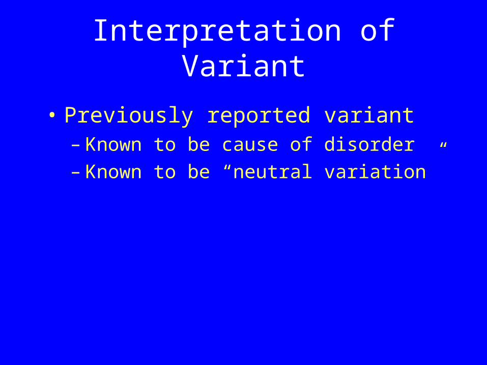
Interpretation of Variant
• Previously reported variant– Known to be cause of disorder– Known to be “neutral variation”
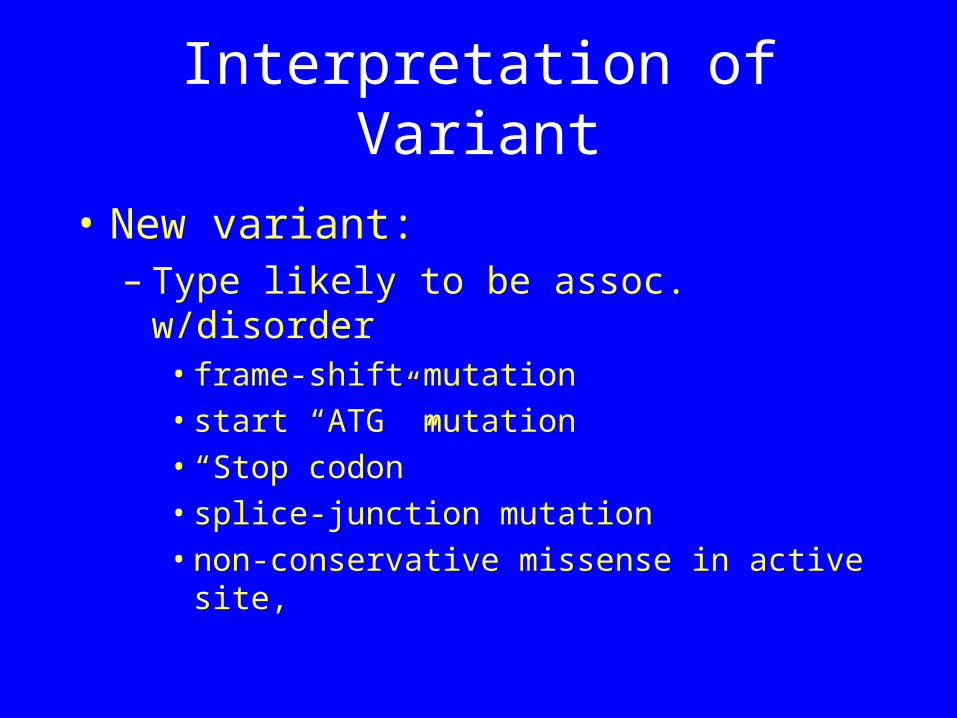
Interpretation of Variant
• New variant: – Type likely to be assoc. w/disorder
• frame-shift mutation • start “ATG” mutation• “Stop codon” • splice-junction mutation• non-conservative missense in active site,
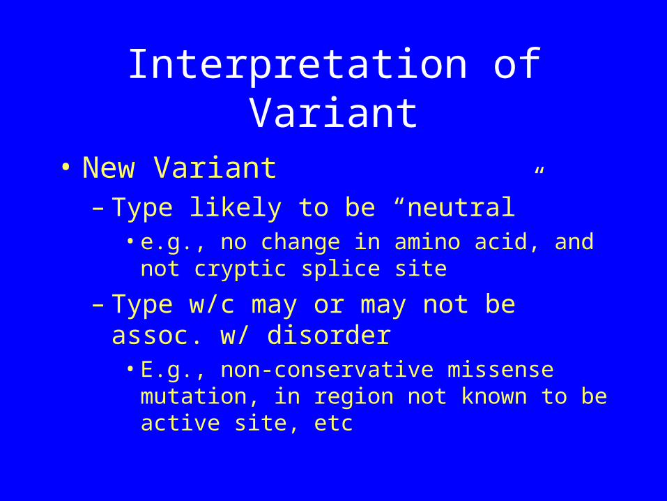
Interpretation of Variant
• New Variant– Type likely to be “neutral”
• e.g., no change in amino acid, and not cryptic splice site
– Type w/c may or may not be assoc. w/ disorder• E.g., non-conservative missense
mutation, in region not known to be active site, etc
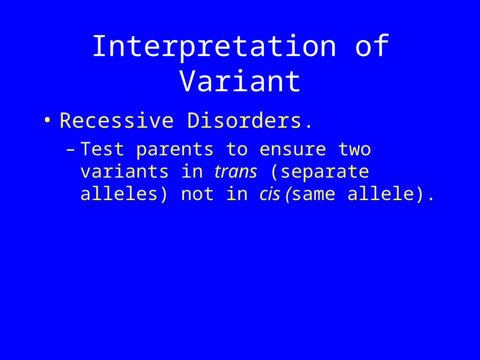
Interpretation of Variant
• Recessive Disorders.– Test parents to ensure two variants in
trans (separate alleles) not in cis (same allele).
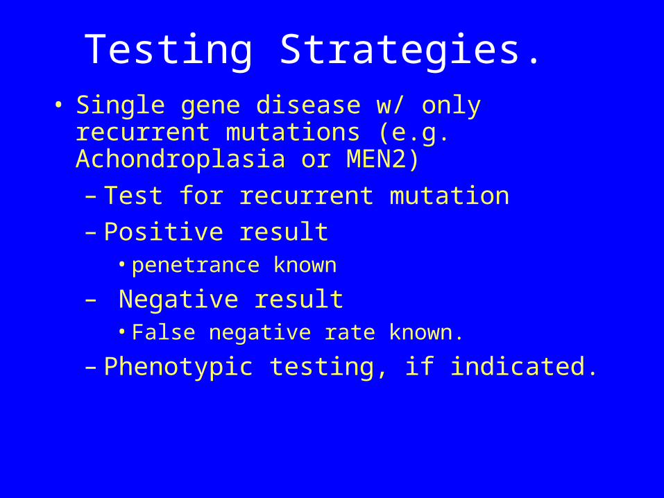
Testing Strategies. • Single gene disease w/ only recurrent
mutations (e.g. Achondroplasia or MEN2)– Test for recurrent mutation– Positive result
• penetrance known
– Negative result• False negative rate known.
– Phenotypic testing, if indicated.
Testing Strategies.
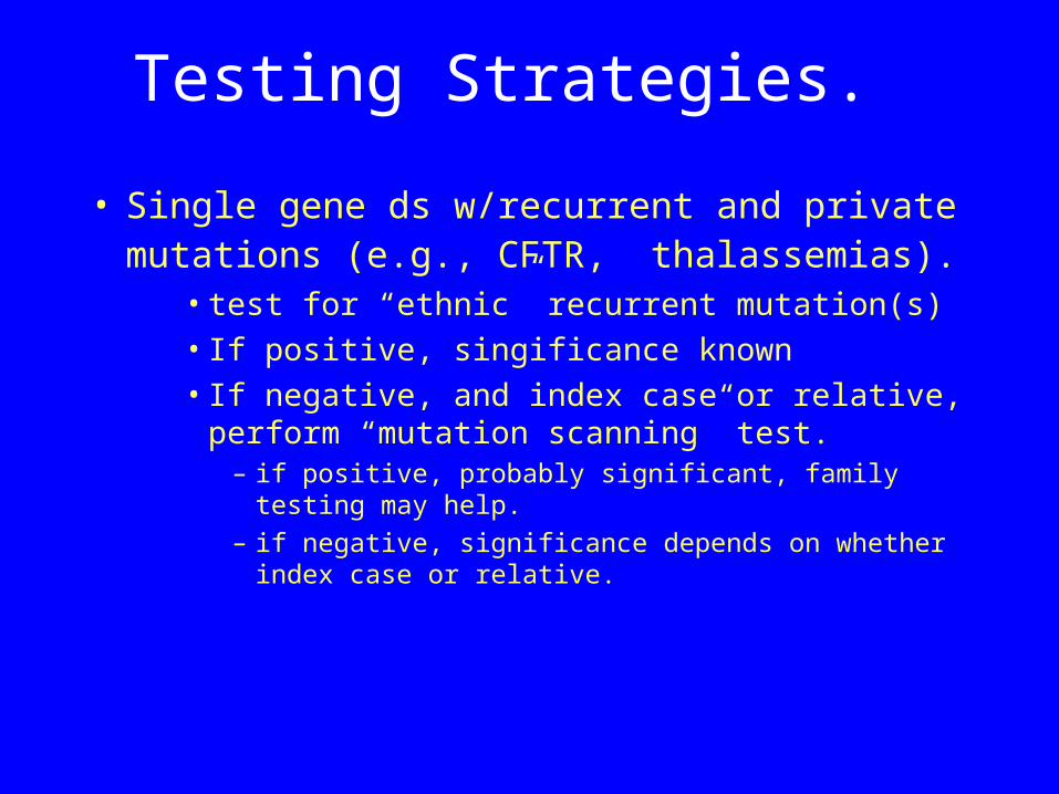
• Single gene ds w/recurrent and private mutations (e.g., CFTR, thalassemias).
• test for “ethnic” recurrent mutation(s)• If positive, singificance known• If negative, and index case or relative,
perform “mutation scanning” test. – if positive, probably significant, family testing
may help. – if negative, significance depends on whether
index case or relative.
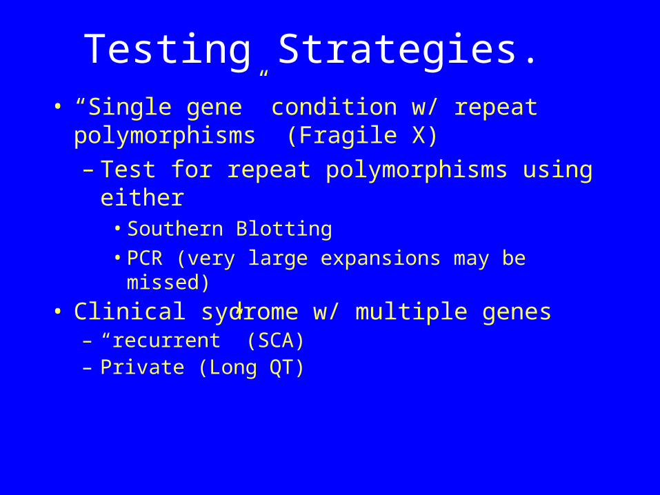
Testing Strategies. • “Single gene” condition w/ repeat
polymorphisms (Fragile X)– Test for repeat polymorphisms using
either • Southern Blotting• PCR (very large expansions may be
missed)• Clinical sydrome w/ multiple genes
– “recurrent” (SCA)– Private (Long QT)
Testing Strategies
Cystic Fibrosis
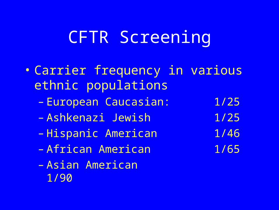
CFTR Screening
• Carrier frequency in various ethnic populations– European Caucasian: 1/25– Ashkenazi Jewish 1/25– Hispanic American 1/46– African American 1/65– Asian American
1/90
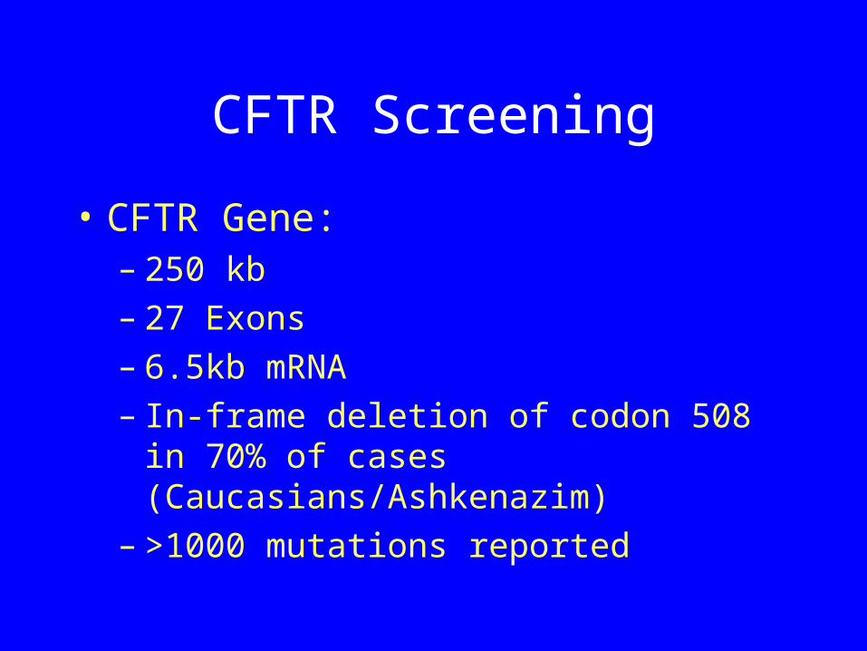
CFTR Screening
• CFTR Gene:– 250 kb– 27 Exons– 6.5kb mRNA– In-frame deletion of codon 508 in 70%
of cases (Caucasians/Ashkenazim)– >1000 mutations reported
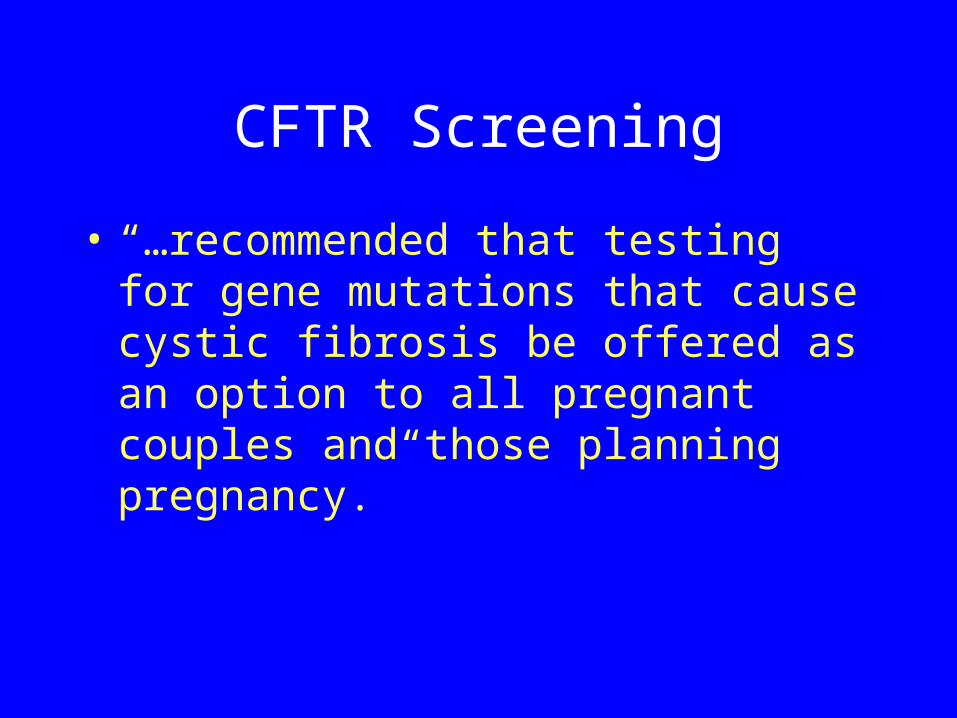
• “…recommended that testing for gene mutations that cause cystic fibrosis be offered as an option to all pregnant couples and those planning pregnancy.”
CFTR Screening
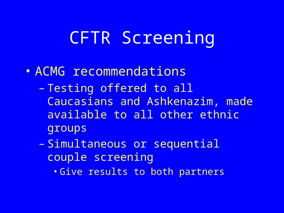
• ACMG recommendations– Testing offered to all Caucasians and
Ashkenazim, made available to all other ethnic groups
– Simultaneous or sequential couple screening • Give results to both partners
CFTR Screening
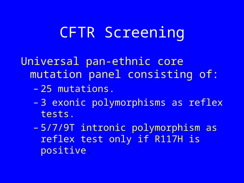
Universal pan-ethnic core mutation panel consisting of:– 25 mutations.– 3 exonic polymorphisms as reflex
tests.– 5/7/9T intronic polymorphism as
reflex test only if R117H is positive.
CFTR Screening
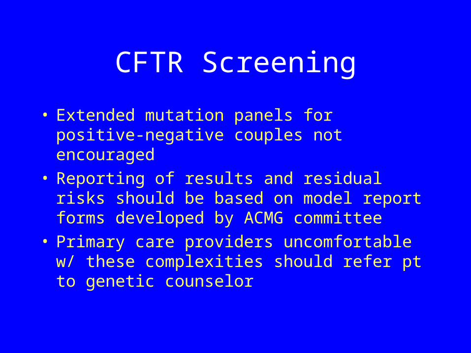
• Extended mutation panels for positive-negative couples not encouraged
• Reporting of results and residual risks should be based on model report forms developed by ACMG committee
• Primary care providers uncomfortable w/ these complexities should refer pt to genetic counselor
CFTR Screening
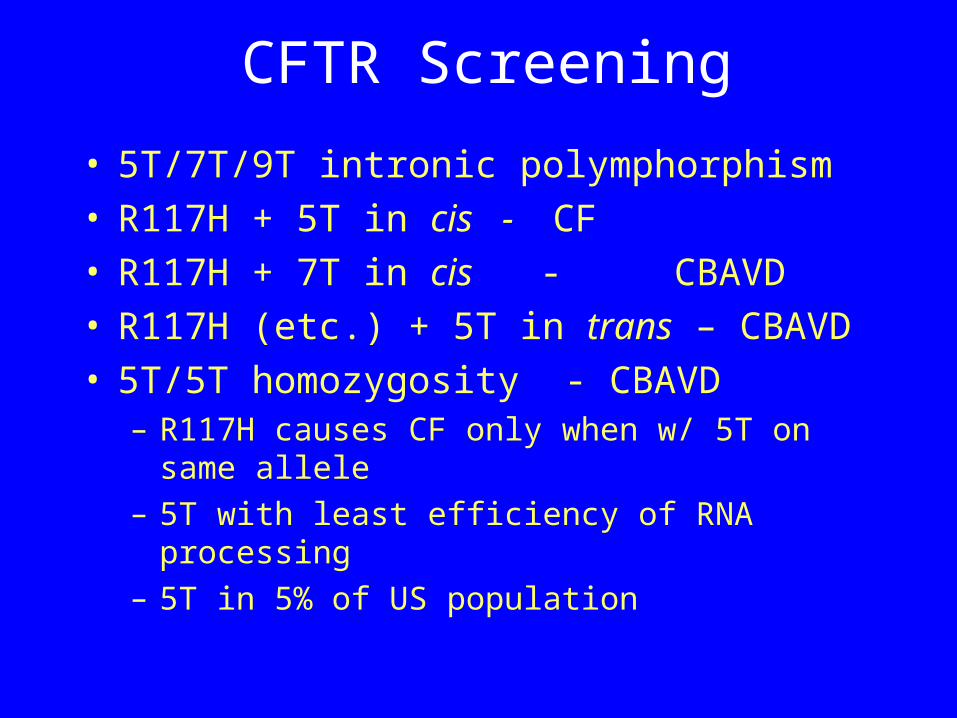
CFTR Screening
• 5T/7T/9T intronic polymphorphism• R117H + 5T in cis - CF• R117H + 7T in cis - CBAVD• R117H (etc.) + 5T in trans – CBAVD• 5T/5T homozygosity - CBAVD
– R117H causes CF only when w/ 5T on same allele
– 5T with least efficiency of RNA processing– 5T in 5% of US population
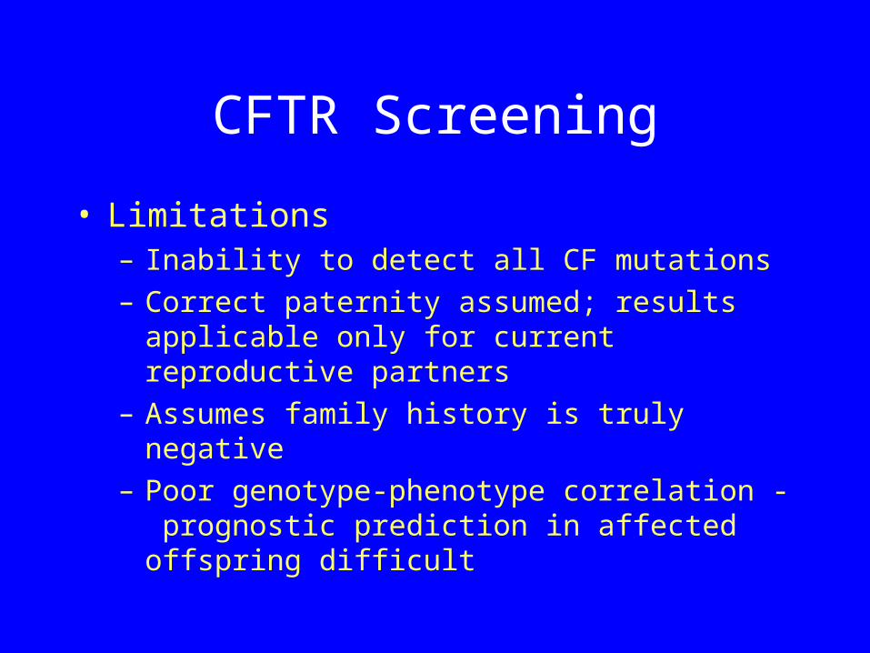
CFTR Screening
• Limitations– Inability to detect all CF mutations– Correct paternity assumed; results
applicable only for current reproductive partners
– Assumes family history is truly negative– Poor genotype-phenotype correlation -
prognostic prediction in affected offspring difficult
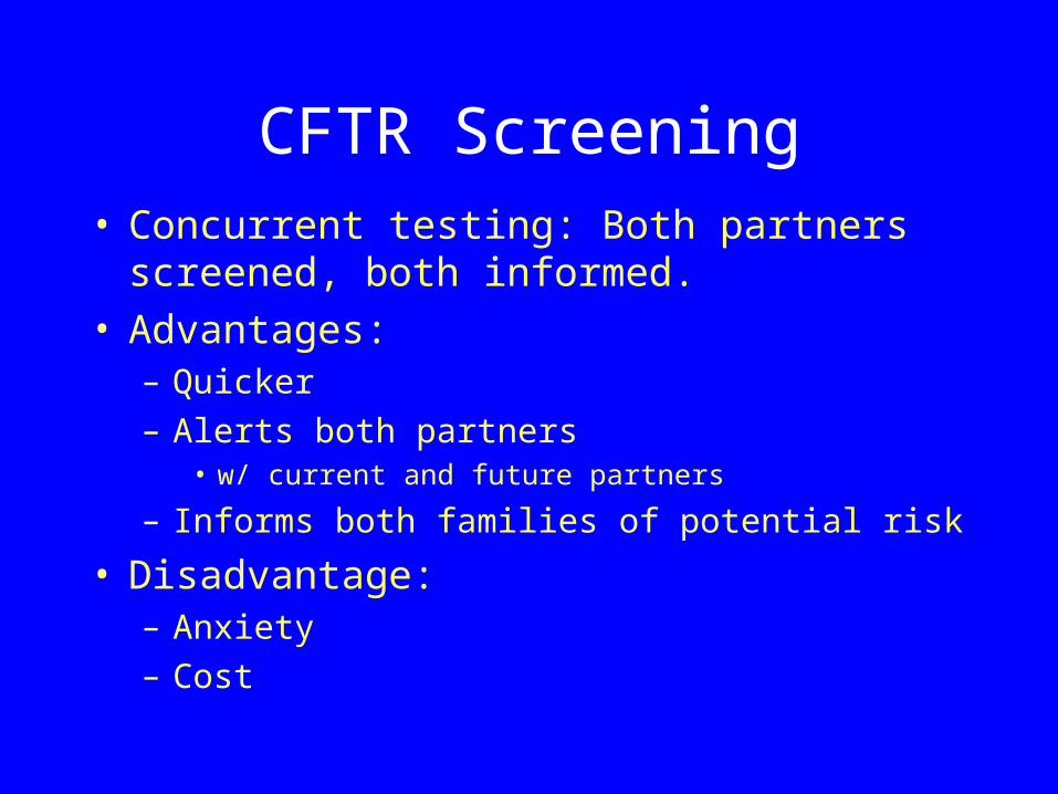
CFTR Screening• Concurrent testing: Both partners
screened, both informed.• Advantages:
– Quicker– Alerts both partners
• w/ current and future partners
– Informs both families of potential risk
• Disadvantage:– Anxiety– Cost
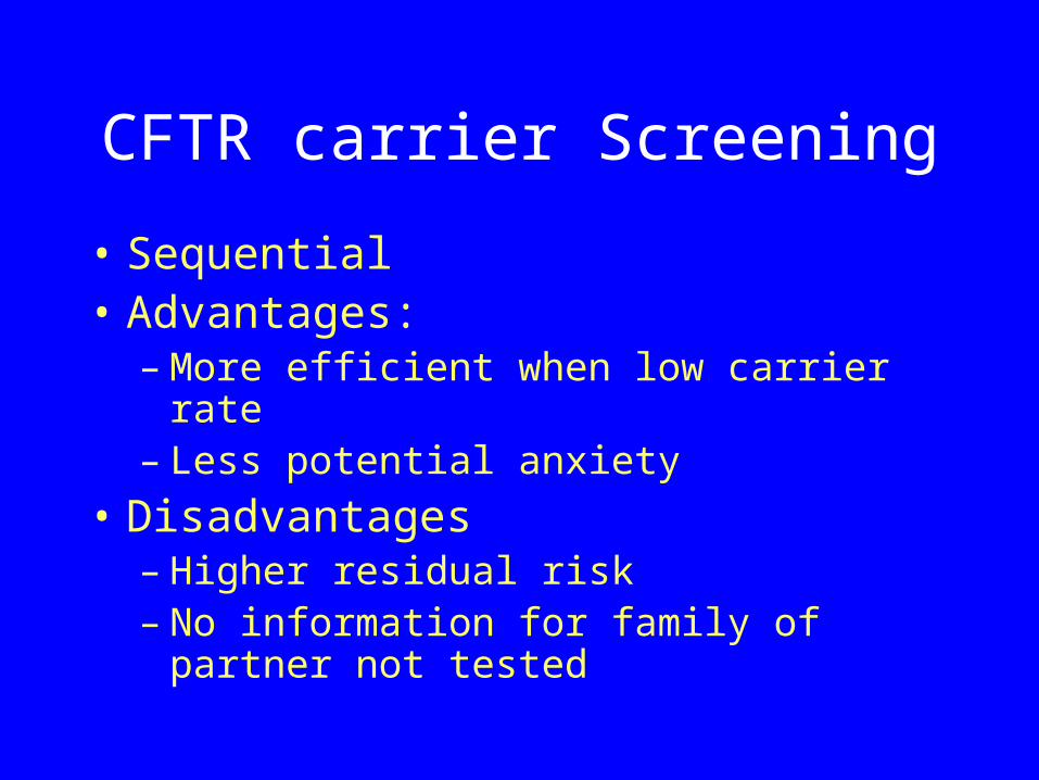
CFTR carrier Screening
• Sequential• Advantages:
– More efficient when low carrier rate– Less potential anxiety
• Disadvantages– Higher residual risk– No information for family of partner
not tested
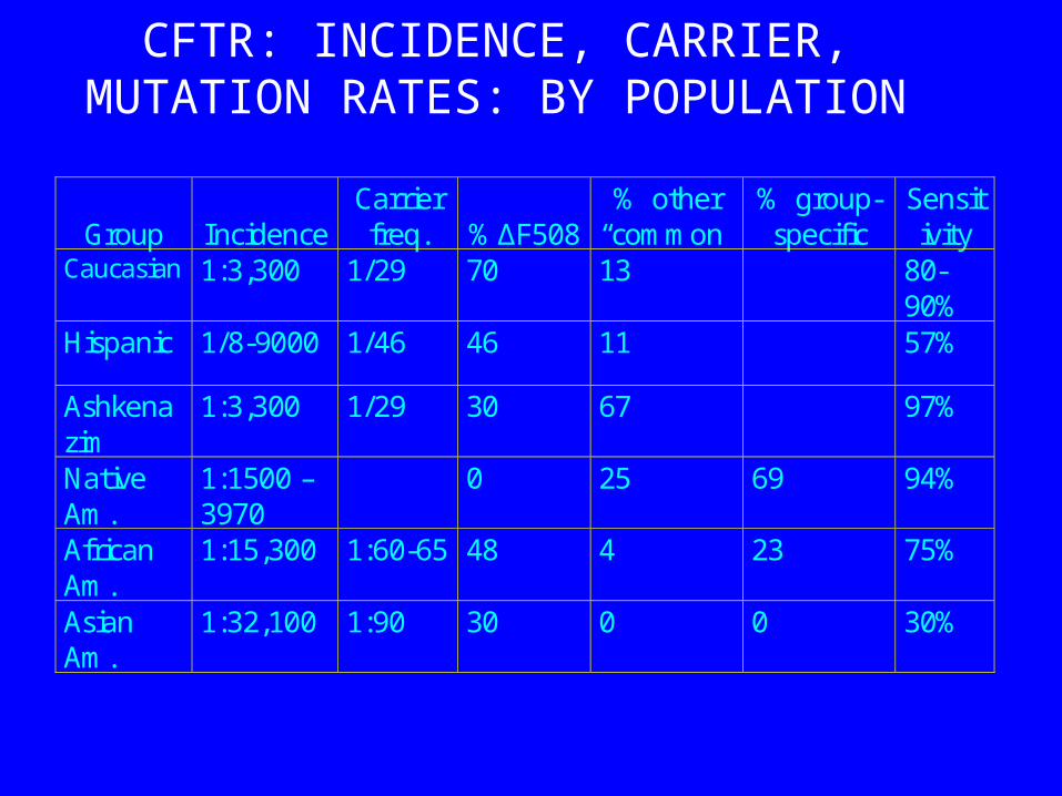
CFTR: INCIDENCE, CARRIER, MUTATION RATES: BY POPULATION
Group
Incidence
Carrier freq.
%ΔF508
% other “common”
% group- specific
Sensitivity
Caucasian 1:3,300 1/29 70 13 80-90%
Hispanic 1/8-9000 1/46 46 11 57%
Ashkenazim
1:3,300 1/29 30 67 97%
Native Am.
1:1500 – 3970
0 25 69 94%
African Am.
1:15,300 1:60-65 48 4 23 75%
Asian Am.
1:32,100 1:90 30 0 0 30%
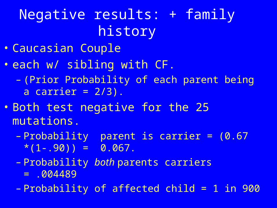
Negative results: + family history
• Caucasian Couple• each w/ sibling with CF.
– (Prior Probability of each parent being a carrier = 2/3).
• Both test negative for the 25 mutations.– Probability parent is carrier = (0.67
*(1-.90)) = 0.067. – Probability both parents carriers = .004489– Probability of affected child = 1 in 900
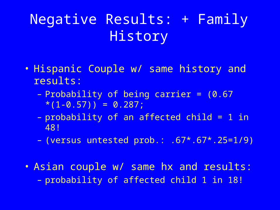
• Hispanic Couple w/ same history and results: – Probability of being carrier = (0.67 *(1-
0.57)) = 0.287; – probability of an affected child = 1 in 48!– (versus untested prob.: .67*.67*.25=1/9)
• Asian couple w/ same hx and results:– probability of affected child 1 in 18!
Negative Results: + Family History
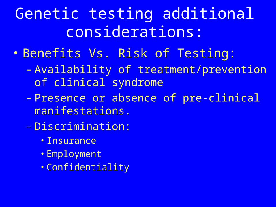
Genetic testing additional considerations:
• Benefits Vs. Risk of Testing:– Availability of treatment/prevention of
clinical syndrome– Presence or absence of pre-clinical
manifestations.– Discrimination:
• Insurance• Employment• Confidentiality
Additional Considerations
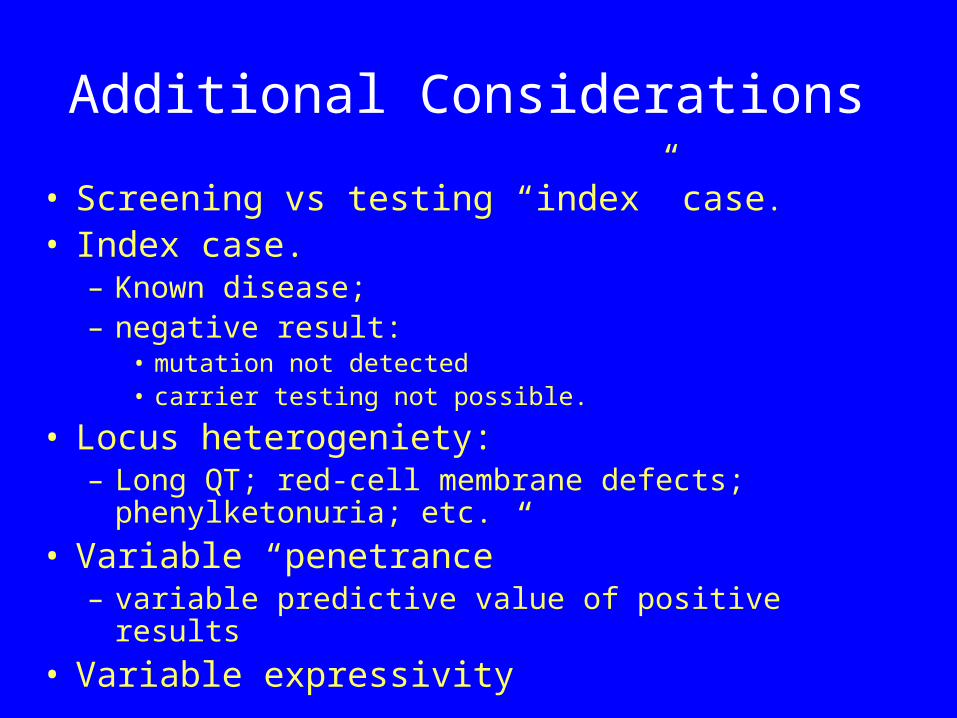
• Screening vs testing “index” case.
• Index case. – Known disease; – negative result:
• mutation not detected• carrier testing not possible.
• Locus heterogeniety:– Long QT; red-cell membrane defects;
phenylketonuria; etc.
• Variable “penetrance”– variable predictive value of positive results
• Variable expressivity
Additional Considerations
• Potential interventions:– Behavioral
• lung cancer-risk - smoking cessation; • heart disease risk - diet/exercise; • risk of breast/colon cancer - screening
accepatnce.
– Medical• e.g., prophylactic mastectomy/thyroidectomy; • blood-letting/blood donation for HFE; • anti-arrhythmics for Long QT, etc.
Additional Considerations
• Pre-morbid/clinical syndrome– Is there a clinically identifiable syndrome ?– ? Need for intervention prior to clinical
manifestations
• Technical considerations– e.g., Fragile X-syndrome.
• Patent issues– affect availability/cost of testing
Additional Considerations• Ethics
– implications for patients and relatives.• e.g., identical twins; siblings; • paternity issues -
• Legal issues– New York State Civil Right Law:
• Need for nformed consent – Genetic testing only (not phenotypic testing)– Standards for informed consent in civil rights law,
section 79-l [http://assembly.state.ny.us/leg/?cl=17&a=12].
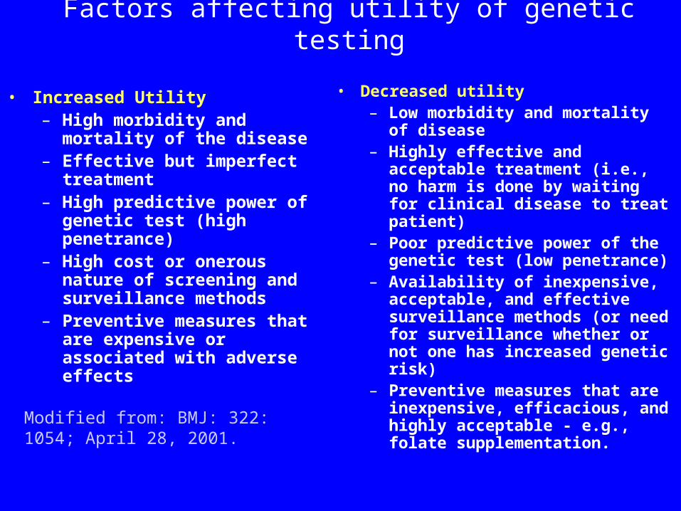
Factors affecting utility of genetic testing
• Increased Utility– High morbidity and
mortality of the disease– Effective but imperfect
treatment– High predictive power of
genetic test (high penetrance)
– High cost or onerous nature of screening and surveillance methods
– Preventive measures that are expensive or associated with adverse effects
• Decreased utility– Low morbidity and mortality
of disease– Highly effective and
acceptable treatment (i.e., no harm is done by waiting for clinical disease to treat patient)
– Poor predictive power of the genetic test (low penetrance)
– Availability of inexpensive, acceptable, and effective surveillance methods (or need for surveillance whether or not one has increased genetic risk)
– Preventive measures that are inexpensive, efficacious, and highly acceptable - e.g., folate supplementation.
Modified from: BMJ: 322: 1054; April 28, 2001.
Ordering Molecular Tests• Patient preparation: None
– Avoid heparin: interferes with PCR.
• Specimens:– Fresh whole blood: EDTA/Citrate– Fresh tissues– Frozen tissues– Paraffin embeded tissues– Slides etc.
Ordering Molecular Tests
• Specimen Handling• DNA-based tests:
– Room temperature, up to 72 hours (maybe more with special buffers)
• RNA-based tests:– Deliver ASAP (4-6 hours)– Special considerations for proprietary
test.
Ordering Molecular Tests • Essential info (Molecular Genetic
Tests): – Clinical information – pedigree, if possible– Race– reason for testing.
• Informed consent:– New York State Civil Rights Law.
• Nature of test; availability of genetic counseling; implications of positive and negative tests, etc.