Embed Size (px)
Citation preview

Molecular Detection of Inherited Diseases
Chapter 13

Models of Disease Etiology
• Genetic (inherited)• Environmental (somatic)• Multifactorial (polygenic + somatic)

Transmission Patterns
• Gain of function mutations usually display a dominant phenotype. (activation on gene with overexpression or alteration of phenotype) (less common)
• Loss of function mutations usually display a recessive phenotype. (deletion of gene with loss of protein)
• Dominant negative patterns are observed with loss of function in multimeric proteins.
+
+ +
+
+
- +
+
Normal phenotype
Abnormal phenotype
+
+
+
-
Homozygous (+/+)
Heterozygous (+/-)
Family History of Phenotype Is Illustrated on a Pedigree Diagram
male affected male deceased male
female affected female deceased female
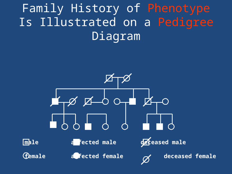
Pedigree Diagrams Reveal Transmission Patterns
Sex-linked (X-linked recessive)
Autosomal recessive (AR)Autosomal dominant (AD)

Transmission Patterns
• AR, AD, or sex-linked patterns are observed in single-gene disorders (diseases caused by one genetic mutation).
• Prediction of a transmission pattern assumes Mendelian inheritance of the mutant allele.
Single Gene Disorders
• Single gene disorders affect structural proteins, cell surface receptor proteins, growth regulators, and enzymes

Classification of Mutations
• By effect on structure (point mutations, insertions, deletions, additions, translocations)
• By effect on function

Factor V Leiden
• In the normal person, factor V functions as a cofactor to allow factor Xa to activate an enzyme called thrombin. Thrombin in turn cleaves fibrinogen to form fibrin, which form the fibrin clot.
• Activated protein C (aPC) is a natural anticoagulant that acts to limit the extent of clotting by cleaving and degrading factor V.

Factor V Leiden
• Factor V Leiden is an autosomal dominant condition that exhibits incomplete dominance and results in a factor V variant that cannot be as easily degraded by activated Protein C.
• The gene that codes the protein is referred to as F5. Mutation of this gene is a single nucleotide polymorphism located on chromosome 1 in exon 10.
• As a missense substitution it changes a protein's amino acid from arginine to glutamine.
• Depending on the chosen start the position of the nucleotide variant is either at position 1691 or 1746. It also affects the amino acid position for the variant, which is either 506 or 534.
• Since this amino acid is normally the cleavage site for aPC, the mutation prevents efficient inactivation of factor V. When factor V remains active, it facilitates overproduction of thrombin leading to generation of excess fibrin and excess clotting.

FVL
• The excessive clotting that occurs in this disorder is almost always restricted to the veins, where the clotting may cause a deep vein thrombosis (DVT).
• Studies have found that about 5 percent of Caucasians in North America have factor V Leiden

FVL-Old diagnosis
• Most laboratories screen 'at risk' patients with either a snake venom (e.g. dilute Russell's viper venom time) based test or an aPTT based test. In both methods, the time it takes for blood to clot is shortened in the presence of the factor V Leiden mutation.

FVL
• This is done by running two tests simultaneously, one test is run in the presence of activated protein C (APC) and the other, in the absence. A ratio is determined based on the two tests and the results signify to the laboratory whether APC is working or not. These are quick, three minute, automated tests that most hospital laboratories can easily perform

FVL New method
• The mutation (a 1691G→A substitution) removes a cleavage site of the restriction endonuclease MnlI, so PCR, treatment with MnlI, and then DNA electrophoresis will give a diagnosis.
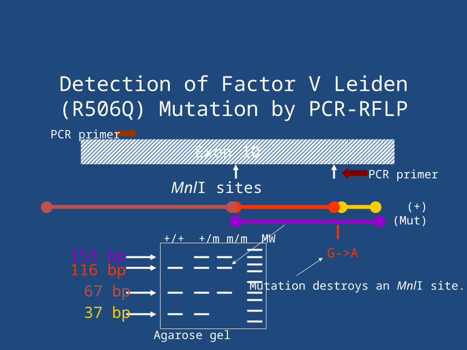
Detection of Factor V Leiden (R506Q) Mutation by PCR-RFLP
153 bp116 bp
Exon 10
G->A
67 bp 37 bp
+/+ +/m m/m MW
MnlI sites
PCR primer
PCR primer
(+)(Mut)
Mutation destroys an MnlI site.
Agarose gel

Detection of Factor V Leiden (R506Q) Mutation by SSP-PCR
148 bp123 bp
Exon 10
G->A
PCR primer
Sequence-specific PCR primers
Longer primer ends on mutated base A and makes a larger amplicon.
(Mut) (+)
Agarose gel

Sickle Cell Anemia
• Sickle-cell anemia is the name of a specific form of sickle-cell disease in which there is homozygosity for the mutation that causes HbS. Sickle-cell anemia is also referred to as "HbSS", "SS disease", "hemoglobin S" or permutations thereof.
• In heterozygous people, who have only one sickle gene and one normal adult hemoglobin gene, it is referred to as "HbAS" or "sickle cell trait". Other, rarer forms of sickle-cell disease include sickle-hemoglobin C disease (HbSC), sickle beta-plus-thalassemia (HbS/β+) and sickle beta-zero-thalassemia (HbS/β0).


• These other forms of sickle-cell disease are compound heterozygous states in which the person has only one copy of the mutation that causes HbS and one copy of another abnormal hemoglobin allele.
• Hemoglobin S results from a point mutation in HBB, changing the sixth amino acid in the β-hemoglobin chain from glutamic acid to valine (Glu6Val). Sickle cell anemia (homozygous Hb SS) accounts for 60%-70% of sickle cell disease in the US.

Solubility TestSickledex (screen) (old)
• Under conditions of low oxygen tension, the heterozygous (A/S) form can cause erythrocytes to form the characteristic sickle-shaped tactoids.
• Deoxygenated Hb-S is insoluble in the presence of a concentrated phosphate buffer solution and forms a turbid suspension that can be easily visualized. Normal Hemoglobin A and other hemoglobins remain in
solution under these conditions.

Hemoglobin electrophoresis• Advantage: detects other hemoglobinopathies• The most common types of normal hemoglobin are:• Hemoglobin F (fetal hemoglobin). This type is normally found in
fetuses and newborn babies. Hemoglobin F is replaced by hemoglobin A (adult hemoglobin) shortly after birth; only very small amounts of hemoglobin F are made after birth. Some diseases, such as sickle cell disease, aplastic anemia, and leukemia, have abnormal types of hemoglobin and higher amounts of hemoglobin F.
• Hemoglobin A. This is the most common type of hemoglobin found normally in adults. Some diseases, such as severe forms of thalassemia, may cause hemoglobin A levels to be low and hemoglobin F levels to be high.
• Hemoglobin A2. This is a normal type of hemoglobin found in small amounts in adults.

• More than 400 different types of abnormal hemoglobin have been found, but the most common are:
• Hemoglobin S. This type of hemoglobin is present in sickle cell disease.
• Hemoglobin C. This type of hemoglobin does not carry oxygen well.
• Hemoglobin E. This type of hemoglobin is found in people of Southeast Asian descent.
• Hemoglobin D. This type of hemoglobin is present in a sickle cell disorder.
• Hemoglobin H (heavy hemoglobin). This type of hemoglobin may be present in certain types of thalassemia.
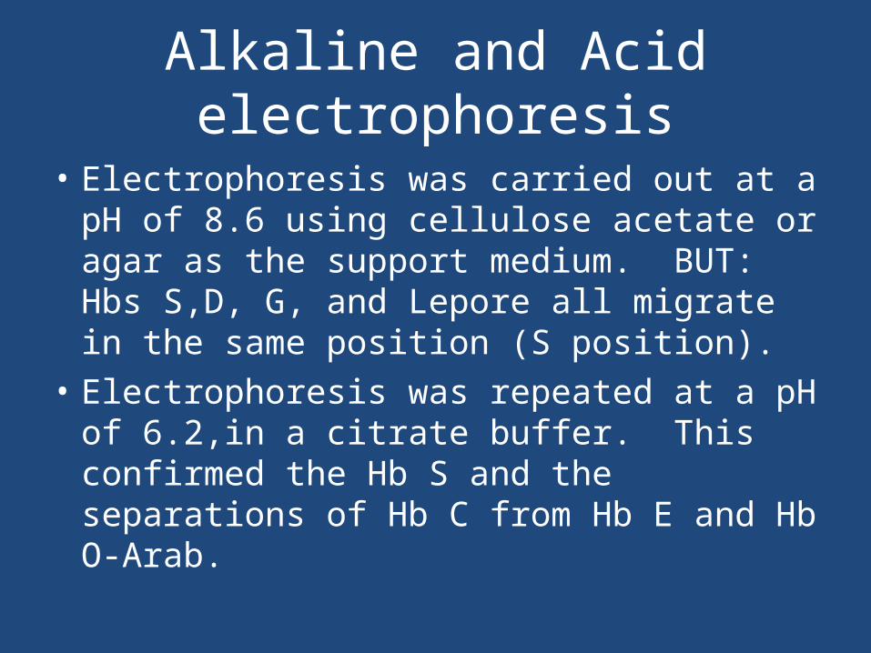
Alkaline and Acid electrophoresis
• Electrophoresis was carried out at a pH of 8.6 using cellulose acetate or agar as the support medium. BUT: Hbs S,D, G, and Lepore all migrate in the same position (S position).
• Electrophoresis was repeated at a pH of 6.2,in a citrate buffer. This confirmed the Hb S and the separations of Hb C from Hb E and Hb O-Arab.
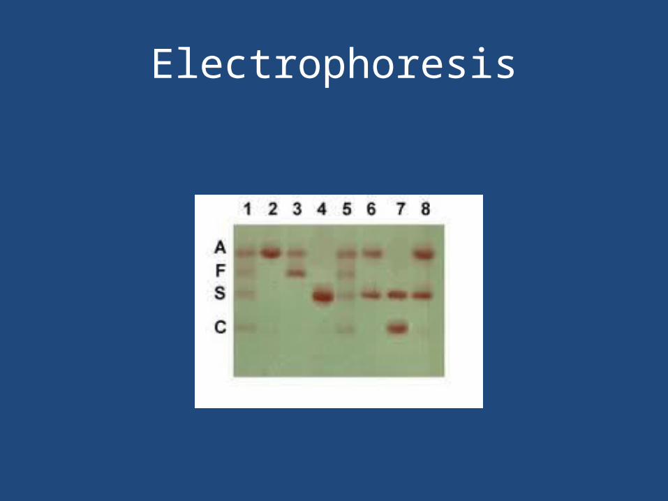
Electrophoresis

HGB by HPLC• It is rapid, automated, capable of resolving most
of the common and many uncommon variants, and provides reliable quantitative measurements of hemoglobin fractions.
• In cation-exchange HPLC, hemolysate is injected into a chromatography column containing a negatively charged resin onto which the positively charged hemoglobins are adsorbed.
• Hemoglobins are eluted by passing through a carefully calibrated developing solution containing an increasing concentration of cations.
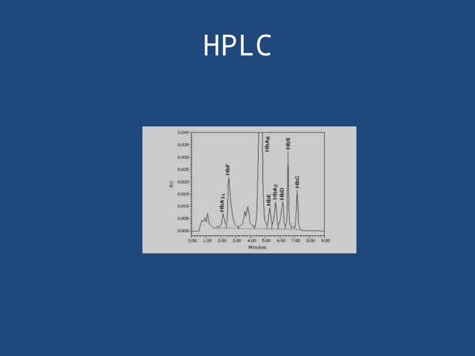
HPLC
Cystic fibrosis• Cystic fibrosis is an autosomal recessive disease, that is
characterized primarily by progressive lung disease, pancreatic insufficiency, gastrointestinal obstruction and an
excess of sodium and chloride in the sweat. The gene that, when defective, causes CF is called the cystic fibrosis transmembrane conductance regulator (CFTR, 7q31.2) gene.
• A defect in this single gene causes all the consequences of CF. There are over 1,600 known defects in the CFTR gene that can cause CF.
• However, about 70% of all people with a defective CFTR gene have a 3-bp deletion , known as delta-F508.
• In the U.S., the number of people who carry a CF gene is about:
• 1 in 29 Caucasian Americans;• 1 in 46 Hispanic Americans;• 1 in 65 African Americans; and• 1 in 90 Asian Americans.

• There are more than 1,200 known mutations of the CFTR gene that cause cystic fibrosis.
• The basic genetic test for cystic fibrosis, sometimes referred to as the ACMG/ACOG Mutation Panel or the 23-mutation panel, looks for the most commonly occurring CFTR mutations. It is about 90% effective in detecting CF mutations in the Caucasian population, but only about 70% effective in African-Americans and 60% effective in the Hispanic population.
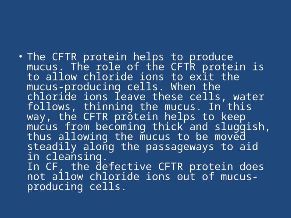
• The CFTR protein helps to produce mucus. The role of the CFTR protein is to allow chloride ions to exit the mucus-producing cells. When the chloride ions leave these cells, water follows, thinning the mucus. In this way, the CFTR protein helps to keep mucus from becoming thick and sluggish, thus allowing the mucus to be moved steadily along the passageways to aid in cleansing.In CF, the defective CFTR protein does not allow chloride ions out of mucus-producing cells.

• For normal salt reabsorption to occur, individual ions of sodium and chloride must be taken from the sweat and moved back into cells of the sweat duct via ion channels.
• In the case of sodium, there is a sodium channel; for chloride, there is a chloride channel called CFTR. For sweat to be produced with the proper concentrations of sodium and chloride, sodium channels and chloride channels (CFTRs) must work properly.
• In cystic fibrosis, the CFTR chloride channel is defective, and does not allow chloride to be reabsorbed into sweat duct cells. Consequently, more chloride stays in the duct, and more sodium remains in the sweat. The concentration of chloride in sweat is therefore elevated in individuals with cystic fibrosis.

• The screening tests for CF are done as part of the standard newborn screening.
• The screening test looks for elevated levels of a substance called immunoreactive trypsinogen (IRT), which is an enzyme created by the pancreas.
• If this is positive a sweat chloride will be done.

• The sweat chloride test, or sweat test, has been the gold standard test used to diagnose cystic fibrosis for many years. The test measures the amount of salt in a person’s sweat, which is higher than normal in people with CF. A chloride content greater than 60 mmol/liter is considered a positive result.
• If IRT levels were elevated but no CFTR mutation was detected, the primary physician may order a sweat test anyway just in case the baby has one of the less common mutations that was not included in the genetic testing panel.

• Sweating is induced by pilocarpine iontophoresis. At the test site, an electrode is placed over gauze containing pilocarpine and electrolyte solution that will not interfere with the sodium and chloride measurement.
• A second electrode (without pilocarpine) will be placed at another site and a mild electrical current will draw the pilocarpine into the skin where it stimulates the sweat glands.
• The test site is carefully cleaned and dried, then a piece of preweighed filter paper is placed over the test site and covered with parafilm to prevent evaporation.
• Sweat is collected for 30 minutes. The filter paper is retrieved and weighed to determine the weight of sweat collected. The sodium and chloride sweat concentrations are measured.
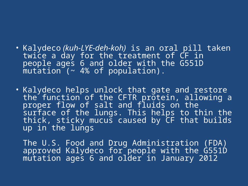
• Kalydeco (kuh-LYE-deh-koh) is an oral pill taken twice a day for the treatment of CF in people ages 6 and older with the G551D mutation (~ 4% of population).
• Kalydeco helps unlock that gate and restore the function of the CFTR protein, allowing a proper flow of salt and fluids on the surface of the lungs. This helps to thin the thick, sticky mucus caused by CF that builds up in the lungs The U.S. Food and Drug Administration (FDA) approved Kalydeco for people with the G551D mutation ages 6 and older in January 2012
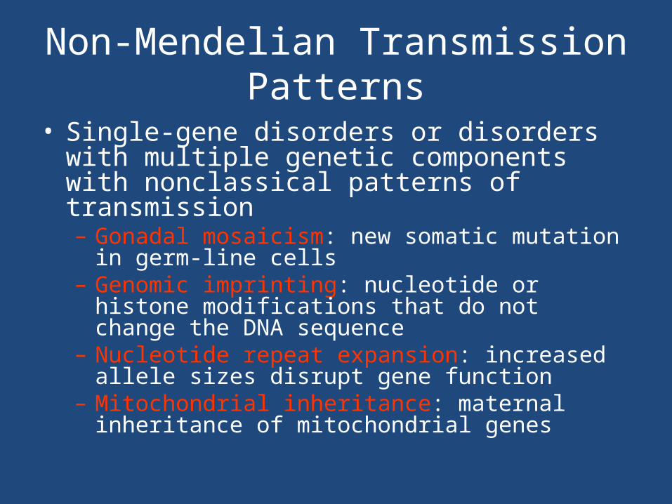
Non-Mendelian Transmission Patterns
• Single-gene disorders or disorders with multiple genetic components with nonclassical patterns of transmission– Gonadal mosaicism: new somatic mutation in germ-line
cells – Genomic imprinting: nucleotide or histone modifications
that do not change the DNA sequence– Nucleotide repeat expansion: increased allele sizes disrupt
gene function– Mitochondrial inheritance: maternal inheritance of
mitochondrial genes
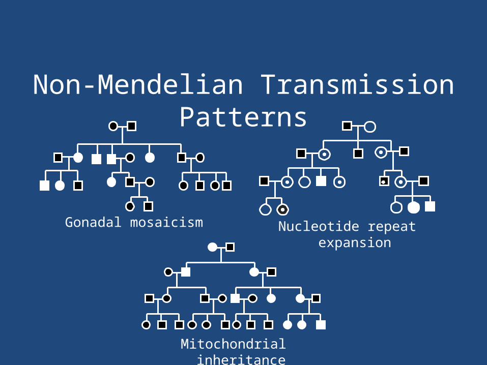
Mitochondrial inheritance
Non-Mendelian Transmission Patterns
Gonadal mosaicism Nucleotide repeat expansion

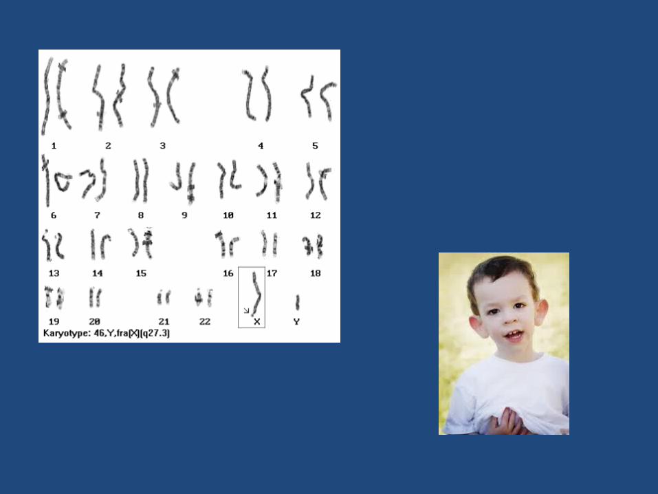
Fragile X
• Fragile X syndrome is associated with the expansion of the CGG trinucleotide repeat affecting the Fragile X mental retardation 1 (FMR1) gene on the X chromosome, resulting in a failure to express the fragile X mental retardation protein (FMRP), which is required for normal neural development

FXS
• The most common cause of inherited intellectual disability and the most common known genetic cause of autism or autism spectrum disorders.

CGG(CGG)5–55
Normal
Amplification
FMR-1
CGGCGGCGG(CGG)56–200
Premutation (Carrier) FMR-1
CGGCGGCGGCGGCGGCGG(CGG)200–2000+
Full mutation (affected)
Amplification and methylation
FMR-1
Nucleotide Repeat Expansion in Fragile X Mental Retardation Gene (FMR1)


PCR
Premutations can be detected by PCR.
20–40(normal)
50–90(premutation)
Detection of Fragile X CGG Expansion Mutations by PCR and Southern Blot
Southern blot
Due to their large size, Southern blot is required to detect full mutations.
Inactive X infemales cleaved by methylation-specific restrictionenzyme
Full mutation

Detection of Fragile X CGG Expansion Mutations by PCR and CGE
150 200 250 300 350 400
150 200 250 300 350 400
150 200 250 300 350 400
Normal
Carrier
Full Fragile X
Huntington's disease
• Huntington's disease (HD) results from genetically programmed degeneration of brain cells, called neurons, in certain areas of the brain, particularly the basal ganglia. This degeneration causes uncontrolled movements, loss of intellectual faculties, and emotional disturbance.
• All humans have two copies of the Huntingtin gene (HTT), which codes for the protein Huntingtin (Htt). The gene is also called HD and IT15, which stands for 'interesting transcript 15'. Part of this gene is a a trinucleotide repeat, which varies in length between individuals and may change length between generations.
• When the length of this repeated section reaches a certain threshold, it produces an altered form of the protein, called mutant Huntingtin protein (mHtt).

• The differing functions of these proteins are the cause of pathological changes which in turn cause the disease symptoms. The Huntington's disease mutation is genetically dominant and almost fully penetrant: mutation of either of a person's HTT genes causes the disease. It is not inherited according to sex, but the length of the repeated section of the gene, and hence its severity can be influenced by the sex of the affected parent.
• The HTT gene is located on the short arm of chromosome 4 at 4p16.3. HTT contains a sequence of CAG is the (which codes for glutamine, so a series of them results in the production of a chain of glutamine known as a polyglutamine tract (or polyQ tract), and the repeated part of the gene, the PolyQ region.
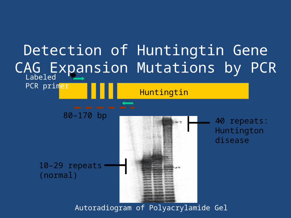
Detection of Huntingtin Gene CAG Expansion Mutations by PCR
10–29 repeats(normal)
> 40 repeats:Huntingtondisease
Huntingtin
80–170 bp
Labeled PCR primer
Autoradiogram of Polyacrylamide Gel

Genomic Imprinting
• Gene silencing due to methylation of C residues and other modifications
• Genomic imprinting occurs during production of egg and sperm.
• The phenotypic effects of imprinting are revealed in diseases in which the maternal or paternal allele is lost (uniparental disomy/deletion).

Examples of Diseases Affected by Genomic Imprinting
• Prader-Willi syndrome: caused by regional deletion or mutation in the paternally inherited chromosome 15
• Angelman syndrome: a different disease phenotype caused by regional deletion or mutation in the maternally inherited chromosome 15

Prader-Willi• PWS is caused by the deletion of the paternal
copies of the imprinted SNRPN and necdin genes along with clusters of snoRNAs: SNORD64, SNORD107, SNORD108 and two copies of SNORD109, 29 copies of SNORD116 (HBII-85) and 48 copies of SNORD115 (HBII-52).
• These are on chromosome 15 located in the region 15q11-13. This so-called PWS/AS region may be lost by one of several genetic mechanisms which, in the majority of instances occurs through chance mutation.

• Other less common mechanisms include; uniparental disomy, sporadic mutations, chromosome translocations, and gene deletions.
• Due to imprinting, the maternally inherited copies of these genes are virtually silent, only the paternal copies of the genes are expressed.
• PWS results from the loss of paternal copies of this region. Deletion of the same region on the maternal chromosome causes Angelman syndrome (AS). PWS and AS represent the first reported instances of imprinting disorders in humans.

Genetic Testing Limitations
• Intergenic mutations in splice sites or regulatory regions may be missed by analysis of gene coding regions.
• Therapeutic targets (except for gene therapy) are phenotypic. (i.e. clotting time)
• Nonsymptomatic diagnosis where disease phenotype is not (yet) expressed may raise ethical concerns.
• Most disease and normal traits are multicomponent systems.

Genetic Testing Complexities
• Variable expressivity: a single genetic mutation results in a range of phenotypes.
• Genetic heterogeneity: the same phenotype results from mutations in different genes (includes diseases with multiple genetic components).
• Penetrance: mutation is present without the predicted phenotype.

Summary
• Frequently occurring point mutations are easily detected by a variety of molecular methods, including PCR, PCR-RFLP, SSP-PCR, and Southern blot.
• Although molecular methods are ideal for detection of DNA lesions, molecular analysis may not always be the optimal strategy for laboratory testing.