Embed Size (px)
Citation preview
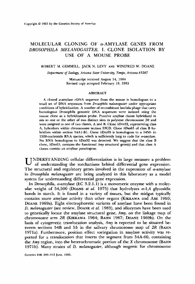
Copyright 0 1985 by the Genetics Society of America
MOLECULAR CLONING OF a-AMYLASE GENES FROM DROSOPHILA MELANOGASTER. I. CLONE ISOLATION BY
USE OF A MOUSE PROBE
ROBERT M. GEMMILL, JACK N. LEVY AND WINIFRED W. DOANE
Department of Zoology, Arizona State University, Tempe, Arizona 85287
Manuscript received August 14, 1984 Revised copy accepted February 19, 1985
ABSTRACT
A cloned a-amylase cDNA sequence from the mouse is homologous to a small set of DNA sequences from Drosophila melanogaster under appropriate conditions of hybridization. A number of recombinant lambda phage that carry homologous Drosophila genomic DNA sequences were isolated using the mouse clone as a hybridization probe. Putative amylase clones hybridized in situ to one or the other of two distinct sites in polytene chromosome 2R and were assigned to one of two classes, A and B. Clone XDm32, representing class A, hybridizes within chromosome section 53CD. Clone XDm65 of class B hy- bridizes within section 54A1-B1. Clone XDm65 is homologous to a 1450- to 1500-nucleotide RNA species, which is sufficiently long to code for a-amylase. No RNA homologous to XDm32 was detected. We suggest that the class B clone, XDm65, contains the functional Amy structural gene(s) and that class A clones contain an amylase pseudogene.
NDERSTANDING cellular differentiation is in large measure a problem U of understanding the mechanisms behind differential gene expression. The structural and regulatory genes involved in the expression of a-amylase in DrosophiEa melanoguster are being analyzed in this laboratory as a model system for understanding differential gene expression.
In Drosophila, a-amylase (EC 3.2.1.1) is a monomeric enzyme with a molec- ular weight of 54,500 (DOANE et al. 1975) that hydrolyzes a-1,4 glycosidic bonds in starch. It is found in a variety of tissues, but the midgut typically contains more amylase activity than other organs (KIKKAWA and ABE 1960; DOANE 1969a). Eight electrophoretic variants of amylase have been found in D. meLanogaster (see review, DOANE et al. 1983), and allozymes have been used to genetically locate the amylase structural gene, Amy, on the linkage map of chromosome arm 2R (KIKKAWA 1964; BAHN 1967; DOANE 1969b). On the basis of cytogenetic translocation analysis, Amy is reported to be situated be- tween sections 54B and 55 in the salivary chromosome map of 2R (BAHN 197 la). Furthermore, position effect variegation in amylase activity was re- ported for a translocation that inserts the segment from 54A-60, containing the Amy region, into the heterochromatic portion of the X chromosome (BAHN 197 1 b). Many strains of D. melanogaster, although isogenic for chromosome
Genetics 110 299-312 June, 1985.
300 R. M. GEMMILL, J. N. LEVY AND W. W. M A N E
2R, produce two amylase isozymes. This and other biochemical and genetic evidence (reviewed in DOANE et al. 1983) suggest that in these strains the structural gene for amylase is duplicated.
Amylase genes have been isolated from the mouse, Mus musculus, and anal- ysis of this gene system at the molecular level has provided important new insights into eukaryotic genetic regulatory mechanisms (HAGENB~CHLE, BOVEY and YOUNG 1980; SCHIBLER et al. 1982; SCHIBLER et al. 1983). These authors have shown that in the mouse there is more than one functional a-amylase structural gene, and that different transcripts are produced from a single struc- tural gene in a tissue-specific manner. In D. melanogaster, the tissue-specific and temporal expression of amylase in the posterior midgut of the adult is, in part, under genetic control by a separate, trans-acting locus, termed map (mid- gut activity pattern; ABRAHAM and DOANE 1978; DOANE et al. 1983). It ap- pears that the map gene regulates the level of translatable amylase mRNA, as shown by microinjection of appropriate fly RNA preparations into Xenopus oocytes and electrophoretic analysis of the Drosophila amylase isozymes sub- sequently produced by the oocytes (BUCHBERG 1983; DOANE et al. 1983). Al- though map regulates the level of amylase expressed in the adult posterior midgut, it seems to have little or no effect on amylase expression in the an- terior midgut. There appears to be at least one other controlling element located in chromosome 2R that is genetically separable from both map and Amy and that regulates amylase levels in the anterior midgut (DOANE 1980; DOANE et al. 1983). Isolation of a cloned DNA segment carrying all or part of the Amy gene from this species is the first step required for a molecular analysis of the complex genetic properties of the amylase system.
In this paper we report that a cDNA clone containing salivary a-amylase sequences from the mouse shares homology with a set of unique DNA se- quences from D. melanogaster. This homology was exploited to isolate Droso- phila DNA sequences that are in turn homologous to the Amy region in poly- tene chromosomes from D. melanogaster. One recombinant phage, XDm65, has many of the properties expected of an Amy-bearing clone. Final verification that XDm65 indeed contains Amy sequences is in our companion paper (LEVY, GEMMILL and DOANE 1985). A second group of isolated clones may represent an amylase pseudogene.
MATERIALS AND METHODS
Stocks of D. melanogaster: Fly stocks included a Canton-S strain which carries Amy',' and an Amy'.6 strain (DOANE 1969b). These strains were originally made isogenic for chromosomes I , 2 and 3 and have been maintained in this laboratory for many years. There is a ten-fold difference in total amylase activity between larval extracts of the Canton-S and Amy',6 strains, i.e., 0.176 and 1.808 nmol of maltose equivalents per microgram of protein per minute released at 2 5 " , pH 7.4, respectively (LYNDA G. TREAT-CLEMONS, unpublished data). Strains used for in situ hybridization experiments included gt' W O , FMS/y sc g?" and SM5/In(2R)G, S p BZ N-2G. The last strain was obtained from L. CRAYMER, California Institute of Technology, Pasadena; a derivative of this strain also contained FM6/y sc g?". LINDSLEY and GRELL (1968) provide explanations of other genetic symbols for fly stocks used in this study.
DNA sources nnd manipulations: The cDNA clone for mouse salivary a-amylase, pMSalO4 (HAG-
DROSOPHILA AMYLASE GENE ISOLATION 301
ENBUCHLE, BOVEY and YOUNG 1980), was obtained from U. SCHIBLER, Swiss Institute for Experi- mental Cancer Research, Epalinges, Switzerland. A library of DNA sequences from D. melanogaster Canton-S, inserted into the lambda vector Charon 4 (MANIATIS et al. 1978), was obtained from J. L. MARSH, University of California, Irvine. Plasmid DNA was purified using either a CsCl density gradient method (KUPERSZTOCH-PORTNOY, LOVETT and HELINSKY 1974; HERTZBERG et al. 1980) or an alkaline sodium dodecyl sulfate (SDS) method (BIRNBOIM and DOLY 1979). Restriction fragments were isolated (ZAIN and ROBERTS 1978) from preparative agarose gels (Wu, JAY and ROYCHOUDHURY 1976). DNA preparations (plasmids, restriction fragments or lambda phage) were labeled to high specific activity with "P (a-"P dCTP or a-"P dATP, New England Nuclear) by nick translation (RIGBY et al. 1977).
Drosophila chromosomal DNA was isolated from adult flies by gentle homogenization in ex- traction buffer containing 100 mM tris(hydroxymethy1)aminomethane (Tris)-HCI, pH 7.5, 200 mM sucrose, 50 mM ethylenediaminetetraacetic acid (EDTA) and 0.5% SDS. The homogenate was extracted three times with an equal volume of phenol equilibrated with buffer and DNA was recovered from the final supernatant by ethanol precipitation. DNA was digested with restriction endonucleases according to suppliers' specifications. Samples of digested DNA were resolved elec- trophoretically on gels containing 0.7-1.0% agarose and 90 mM Tris-borate, pH 8.3, 1 mM EDTA, Southern transfers (SOUTHERN 1975) were prepared according to the Gene Screen manual (New England Nuclear). The Drosophila Charon 4 library (MANIATIS et al. 1978) was screened by the methods of BENTON and DAVIS (1 977). Recombinant phage were isolated preparatively using polyethylene glycol precipitation followed by centrifugation on CsCl step gradients (YAMAMOTO et al. 1970). Phage DNA was prepared by lysis of phage particles in 0.5% SDS and 5 mM EDTA at 60". The solution was placed on ice and KCI was added to 0.5 M to precipitate the SDS. Following centrifugation, the supernatant was ethanol precipitated to recover the phage DNA.
Preparation of mouse amylase probe: Fifty micrograms of pMSalO4 DNA were digested with Pstl and resolved on a 1 .O% agarose gel. The inserted fragment that was recovered consisted of mouse a-amylase cDNA along with poly (dG-dC) tails at each end. Subsequent cleavage with Hinfl sepa- rated the amylase-coding sequences from the dC-dC tails. The three resulting internal Hinfl fragments constituted nearly the entire coding sequence for mouse salivary a-amylase (HAGEN- BUCHLE, BOVEY and YOUNG 1980). These fragments were purified on an agarose gel, recovered and nick translated to generate the probe referred to as the mouse amylase probe.
Hybridization to @er bound DNA: Hybridizations were carried out in the following solution: 50% formamide, 0.2% polyvinyl pyrrolidone (PVP), 0.2% Ficoll, 0.2% bovine serum albumin (BSA), 50 mM Tris-HCI, pH 7.5, 1 M NaCI, 0.1% SDS, 250 @g/ml of denatured salmon testis DNA and 10% dextran sulfate. The mouse amylase probe was hybridized to filter-bound fly DNA under conditions of reduced stringency to optimize signal and reduce background. These condi- tions were determined empirically by systematically altering the temperature of hybridization and the temperature and salt concentration of subsequent washes. Optimal conditions included hybrid- ization for 16 hr at 37" followed by two washes in 2 X SSC (1 X SSC is 0.15 M NaCI, 0.015 M Na citrate), 0.1% SDS at room temperature for 15 min each. T w o 15-min final washes were in 1 X SSC, 0.1% SDS at 50". Autoradiograms were prepared by exposing Kodak X-Omat AR-5 film to filters at -70" for 12 hr to 3 days with DuPont Cronex Lightning-Plus intensifying screens. These conditions were used both for Southern analysis of Drosophila genomic DNA and for screening the Drosophila library with the mouse amylase probe.
In situ hybridization to polytene chromosomes: Chromosome squashes were prepared from salivary glands of "giant" ( y sc gFL1/gt' w") larvae, derived from the cross FM6/y sc g f " 0 X gt' wn S. The procedure of PARDUE and GALL (1975), as modified by HAYASHI et al. (1978), was used for in situ hybridization of labeled probe to salivary polytene chromosomes. For some experiments, squashes were prepared from larvae of the genotype y sc gFl1/gt' w'; In(2R)G/+. Tritium-labeled cRNA was synthesized (WENSINK et al. 1974) using purified recombinant phage DNA as template. Hy- bridization of cRNA (lo5 cpm/slide) to chromosome preparations was performed for 16 hr at 42" in a fluid containing 50% formamide, 2 X SSC, 0.02% PVP, 0.02% Ficoll, 0.02% BSA, 200 rg/ ml of denatured salmon testis DNA, 5 mM Na phosphate, pH 6.5, and 10% dextran sulfate. Slides were washed in four changes of 2 X SSC at room temperature followed by two washes in 0.5 X SSC at 65" for 15 min each. Treatment with RNase was omitted. Slides were coated with nuclear track emulsion (Kodak) and exposed for 4-7 days at 4" .

302 R. M. GEMMILL, J. N. LEVY AND W. W. DOANE
Isolation of total and poly(A)+ RNA and analysis on Northern blots: Total RNA was isolated from third instar larvae of the Amy'!' and Amy*,6 strains using the guanidinium thiocyanate method (CHIRGWIN et al. 1979). Larvae were reared on a starch-yeast diet (DOANE 1969a). Oligo-dT cellulose chromatography (AVIV and LEDER 1972) was used to prepare a poly(A)+ RNA fraction from each RNA preparation. Three milligrams of total RNA typically yielded 50-65 pg of poly(A)+ RNA.
RNA samples were denatured with 10 mM methyl mercuric hydroxide and resolved by electro- phoresis in 1 . 1 % agarose gels containing 2 mM methyl mercuric hydroxide (BAILEY and DAVIDSON 1976). Gels were stained in 0.5 M NH+ acetate containing 0.5 pg of ethidium bromide/ml. The RNA was transferred to membrane filters and hybridized according to the method of THOMAS (1980).
RESULTS
Hybridization of a mouse a-amylase cDNA probe to spec@ sequences in Drosophila DNA: Plasmid pMSal04 contains a 1.66-kilobase (kb) cDNA fragment inserted into the PstI site of pBR322 and carries nearly all of the coding sequence for the mouse salivary a-amylase gene (HAGENB~CHLE, BOVEY and YOUNG 1980). We had conducted a number of preliminary experiments to determine whether the mouse amylase sequences present on pMSal04 could be used to detect homologous sequences in DNA from D. melanogaster. These experiments (DOANE et al. 1983) were performed using labeled pMSa104 DNA as a hy- bridization probe on Southern transfers of EcoRI-cut DNA isolated from our Oregon-R strain of D. melanogaster. We found that under appropriate condi- tions (MATERIALS AND METHODS), labeled pMSa104 does hybridize to the Dro- sophila DNA. However, the high level of background signal seen with this probe prevented its direct use. Experiments using pBR322 as probe demon- strated that much of the background could be attributed to hybridization by vector sequences. T o lower the level of background hybridization we prepared a probe consisting of mouse amylase-coding sequences purified away from the vector sequences of pMSal04 (MATERIALS AND METHODS; DOANE et al. 1983). This probe hybridized to at least two discrete EcoRI fragments in D. melano- gaster Oregon-R DNA (figure 8 in DOANE et al. 1983) with little or no back- ground. Because our genomic library was derived from a Canton-S strain (MANIATIS et al. 1978), we have here repeated these experiments using South- ern transfers of EcoRI-cut Canton-S DNA. Figure 1, lane 1, shows that the mouse amylase probe hybridized to three restriction fragments approximately 3.8, 5.8 and 20 kb in size in EcoRI-cut Canton-S DNA. These results taken together with previous findings (DOANE et al. 1983) indicate that some ho- mology exists between the mouse salivary amylase-coding sequence and a set of DNA sequences in D. melanogaster.
Drosophila genomic DNA clones homologous to the mouse a-amylase cDNA probe: The mouse a-amylase cDNA fragments (mouse amylase probe) purified from pMSa 104 were used as a hybridization probe to isolate homologous sequences from the Maniatis library (MATERIALS AND METHODS). The conditions of hy- bridization were identical with those used for the identification of homologous sequences on the Southern transfer shown in Figure 1, lane 1. A total of 12 clones was isolated in this screen from an estimated 1.6 X lo5 phage, repre-

DROSOPHILA AMYLASE GENE ISOLATION 303
1 2 3 4 5 6
FIGURE 1.-Southern analysis of D. mtlanogasfer Canton-S DNA. EcoRldigested DNA from the Canton-S strain of D. mrlanogasftr was resolved on parallel lanes ( 1 0 pg/lane) of a 1 % agarose gel. A Southern transfer was prepared from the fractionated DNA, and individual lanes were hyhrid- ired with the following "P-labeled probes: purified mouse amylase prohe (lane I); XDm32 (lane 2); XDm65 (lane 3); 5.6-kb suhclone of XDm65 (lane 4); 3.8-kh suhclone of XDm65 (lane 5); 3.8- kh suhclone of XDm32 (lane 6). Hybridiiations in lanes 2-6 were carried out at high levels of stringency, whereas the hybridiiation in lane 1 was carried out under the conditions described in MATERIALS AND METHODS for use of the mouse amylase probe. The sires of the three DNA fragments in lane 1 are given in kilohases.
senting 15 haploid genome equivalents of fly DNA. Preliminary characteri7a- tion of these isolates by restriction endonuclease digestion indicated that a number of clones carried overlapping fragments derived from the same ge- nomic region. The clones were divided into groups on this basis. Representa- tive members of each group were tested for the sites to which they hybridized on polytene chromosomes. Clones from two classes, A and B, hybridized to chromosome arm 2R, which contains the Amy region, whereas all other clones tested hybridized to other chromosomes. We chose to focus ou r efforts on the clones in class A and class B since Amy sequences would most likely be found in one of these two groups.
Class A contained five clones, AD3 1, XDm32, XDm76, XDm89 and XDm7 13; class B contained two clones, XDm65 and XDm41. From restriction endonu- clease digestion patterns, clone XDm7 1 3 appeared to be identical with XDm3 1, and XDm41 was apparently identical with XDm65. Thus, we obtained four independent class A clones and a single class B clone.
Figure 2 shows the results of hybridization of the mouse amylase probe to a Southern blot of EcoRIdigested DNA isolated from these phage. Panel A shows the ethidium bromide-stained gel. In panel B, the resulting blot was
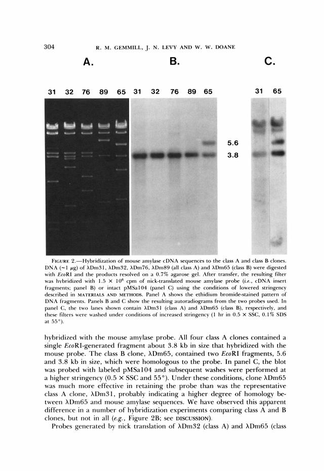
304 R. M. CEMMILL, J. N. LEVY AND W. W. DOANE
A. B. C.
31 32 76 89 65 31 32 76 89 65 31 65
F i ~ i x k : ~ . - l l ~ t ) ~ i ( l i ~ ~ i t i ~ ~ i i of inouse amylase cDNA sequences to the class A and class B clones. D N A (-1 pg) of X I ) m J 1 , X I h J 2 . XDm76, XDm89 (all class A) and XDm65 (class B) were digested with EcoRl and the products resolved on a 0.7% agarose gel. After transfer, the resulting filter was hvbridized with 1.5 X lo6 cpm of nick-translated mouse amylase probe (i.e., cDNA insert fragments; panel B) or intact pMSal04 (panel C) using the conditions of lowered stringency described in MATERIALS AND METHODS. Panel A shows the ethidium bromide-stained pattern of DNA fragments. Panels B and C show the resulting autoradiograms from the two probes used. In panel C, the two lanes shown contain XDm31 (class A) and XDm65 (class B), respectively, and these filters were washed under conditions of increased stringency (1 hr in 0.5 X SSC. 0.1% SDS at 55") .
hybridized with the mouse amylase probe. All four class A clones contained a single EcoRI-generated fragment about 3.8 kb in size that hybridized with the mouse probe. T h e class B clone, XDm65, contained two EcoRI fragments, 5.6 and 3.8 kb in size, which were homologous to the probe. In panel C, the blot was probed with labeled pMSa 104 and subsequent washes were performed at a higher stringency (0.5 X SSC and 55'). Under these conditions, clone XDm65 was much more effective in retaining the probe than was the representative class A clone, XDm31, probably indicating a higher degree of homology be- tween XDm65 and mouse amylase sequences. We have observed this apparent difference in a number of hybridization experiments comparing class A and B clones, but not in all (e.g., Figure 2B; see DISCUSSION).
Probes generated by nick translation of XDm32 (class A) and XDm65 (class
DROSOPHILA AMYLASE GENE ISOLATION 305
B) were hybridized to Southern blots of EcoRI-cut Canton-S DNA. Figure 1, lane 2, shows that XDm32 hybridized to a set of EcoRI fragments, including one fragment that coincides with the 3.8-kb fragment identified in lane 1 by the mouse amylase probe. Clone XDm65 (lane 3) hybridized to a different set of fragments on a similar blot, including two fragments, 3.8 and 5.8 kb in size, that coincide with two fragments identified with the mouse amylase probe in lane 1. Figure 2 demonstrates that the amylase-like sequences present in our isolates reside in a 3.8-kb EcoRI fragment in class A clones and in 3.8- and 5.6-kb EcoRI fragments in the class B clone. These three EcoRI fragments, subcloned into the EcoRI site of pBR325 (LEVY, GEMMILL and DOANE 1985), were used separately as probes against similar Southern blots. The 5.6-kb subclone of XDm65 hybridized to both the 5.8- and 3.8-kb genomic fragments homologous to the mouse amylase probe (Figure 1, lane 4). Likewise, the 3.8- kb subclone of XDm65 hybridized to the same two fragments (Figure 1, lane 5), although the relative hybridization intensity is now reversed. In contrast, the subclone from hDm32 (Figure 1, lane 6) hybridized only to the 3.8- kb genomic fragment.
Our results show that the clones we have isolated contain the sequences originally identified on Southern blots using the mouse amylase probe. The 5.6-kb EcoRI fragment present in XDm65 is 0.2 kb shorter than the corre- sponding genomic fragment. This difference is explained by the fact that this fragment is located at one end of the restriction endonuclease map of XDm65 (LEVY, GEMMILL and DOANE 1985) and it apparently contains an artificial EcoRI site introduced during library construction (MANIATIS et al. 1978). The class A and class B clones do not contain sequences corresponding to the 20- kb fragment identified in Figure 1, lane 1. If this sequence is present in the group of clones we isolated, it must be located in one of the clones that mapped to a chromosome arm other than 2R. These clones have not yet been char- acterized in detail.
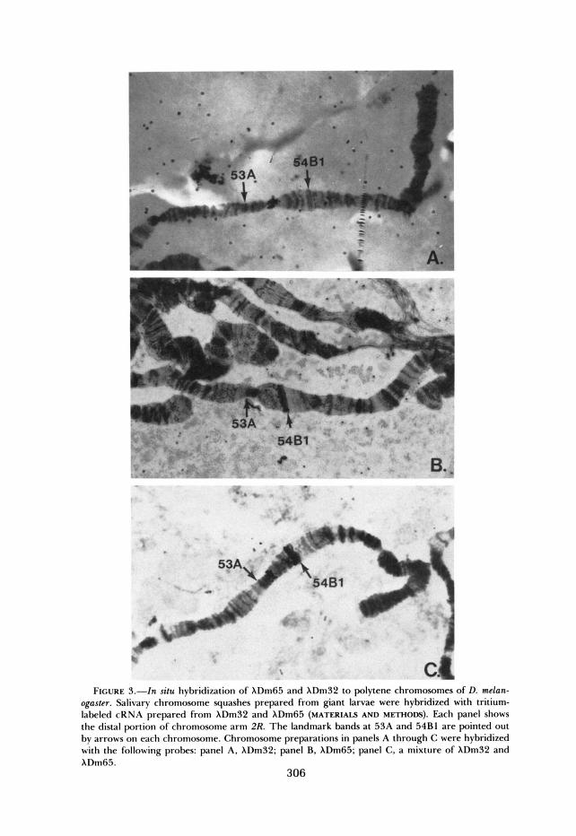
In situ hybridization of cloned sequences to polytene chromosomes: Clones XDm32 and XDm65 were hybridized in situ to polytene chromosomes from a number of strains to precisely locate their hybridization sites on the cytological map of D. melanoguster. Tritium-labeled cRNA synthesized from clone XDm32 (class A) hybridized to polytene chromosome arm 2R within section 53CD (Figure 3A). A comparable probe synthesized from XDm65 (class B) hybridized within section 54Al-B1, probably proximal to 54B1 (Figure 3B). A mixture of these two probes hybridized to both sites on chromosome 2 R , thus confirming that the sites of hybridization are distinct (Figure 3C). Our conclusions concerning the position of hybridization of these two clones have also been confirmed by hybridization to chromosomes heterozygous for In(2R)G (not shown), which has breakpoints at 50E and 54D (LINDSLEY and GRELL 1968). Both clones hybridized within the inversion loop but at separate sites consistent with each clone’s expected distance from the 54D breakpoint.
The hybridization of probes to salivary chromosome squashes was done un- der conditions of high stringency. Thus, although XDm32 and hDm65 showed homology to the mouse amylase probe and showed homology inter se under
5481 t B.
FI URE 3.-h situ hybriditation of XDm65 and XDm32 D polytene chromosomes of D. melan- ogaster. Salivary chromosome squashes prepared from giant larvae were hybridized with tritium- labeled cRNA prepared from XDm32 and XDm65 (MATERIALS AND METHODS). Fach panel shows the distal portion of chromosome arm 2R. T h e landmark bands at 53A and 54B1 are pointed out by arrows on each chromosome. Chromosome preparations in panels A through C were hybridized with the following probes panel A, XDm32: panel B, XDm65; panel C, a mixture of XDm32 and XDm65.
306
DROSOPHILA AMYLASE GENE ISOLATION 307
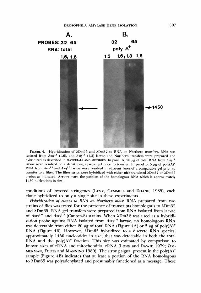
F I G U R E 4.-Hybridi7ation of XDm65 and XDm32 to R N A on Northern transfers. RNA was isolated from Amyl" (1.6). and Amyl.' (1.3) larvae and Northern transfers were prepared and hybridized as described in M A T E R I A L S A N D METHODS. In panel A, 20 pg of total RNA from Amyl4 larvae were resolved on a denaturing agarose gel prior to transfer. In panel B. 5 pg of poly(A)+ RNA from Amyl.' and Amy'.' larvae were resolved in adjacent lanes of a comparable gel prior to transfer to a filter. The filter strips were hybridized with either nick-translated XDm32 or XDm65 probes as indicated. Arrows mark the position of the homologous RNA which is approximately 1450 nucleotides in size.
conditions of lowered stringency (LEVY, GEMMILL and DOANE, 1985), each clone hybridized to only a single site in these experiments.
Hybridization of clones to RNA on Northern blots: RNA prepared from two strains of flies was tested for the presence of transcripts homologous to XDm32 and XDm65. RNA gel transfers were prepared from RNA isolated from larvae of Amy'." and Amy'.' (Canton-S) strains. When XDm32 was used as a hybridi- zation probe against RNA isolated from Amyls6 larvae, no homologous RNA was detectable from either 20 pg of total RNA (Figure 4A) or 5 pg of poly(A)+ RNA (Figure 4B). However, XDm65 hybridized to a discrete RNA species, approximately 1450 nucleotides in size, that was detectable in both the total RNA and the poly(A)+ fraction. This size was estimated by comparison to known sizes of rRNA and mitochondrial rRNA (LONG and DAWID 1979; ZIM- MERMAN, Fours and MANNING 1980). T h e strong signal present in the poly(A)+ sample (Figure 4B) indicates that a t least a portion of the RNA homologous to XDm65 was polyadenylated and presumably functioned as a message. These

308 R. M. GEMMILL, J. N. LEVY AND W. W. DOANE
results suggest that XDm65 carries a gene(s) functional in late third instar larvae, whereas XDm32 does not.
Based on visual inspection, the amount of homologous RNA present in Amy'*6 and Amy',' larvae was quite different (6 1,3 and 1,6 tracks in Figure 4B). It appears that Amy'36 larvae contained considerably more of this RNA than did Amy'.' larvae. The difference in RNA levels thus correlates well with the different amounts of amylase activity in these two strains (see fly stocks in MATERIALS AND METHODS).
DISCUSSION
Homology between a mouse amylase cDNA clone and specific DNA se- quences in D. melanogaster is weak but detectable. T o detect this homology, it was essential to use intermediate levels of stringency for both the hybridization and subsequent filter washes. In addition, it was necessary to purify the amy- lase-coding sequences away from vector sequences in order to lower the level of background hybridization (DOANE et al. 1983).
Preliminary analysis of isolated clones by restriction endonuclease digestion resulted in the grouping of clones that seemed to be related into separate classes. Clones from two of these classes, A and B, hybridized to chromosome arm 2R, known to contain the Amy region. Further analysis of these clones has shown that the two classes are quite distinct. Sequences represented by XDm32 (class A) are localized to polytene chromosome section 53CD, whereas se- quences in hDm65 (class B) are present at 54A1-B1. The only clone that detects a larval poly-adenylated RNA on Northern blots is XDm65, indicating that this clone has strong homology to a transcribed gene. The class A clones all contain a single EcoRI fragment that retains homology with mouse amylase, whereas hDm65 (class B) contains two such fragments. The degree of homol- ogy between mouse amylase and these two groups of clones appears to differ as judged by Southern blot hybridization. We have repeatedly seen XDm65 retain the mouse amylase probe at higher levels of stringency than does XDm32 (Figure 2C). Although Figure 2B appears to be an exception to this, when this filter was rewashed at a higher stringency, the probe was again retained more effectively by XDm65 than by the class A clones.
The results presented here strongly suggest that clone XDm65 contains at least one Amy gene from D. melanogaster. First, this clone retains significant homology with mouse amylase-coding sequences. Second, it hybridizes within polytene chromosome section 54Al-B1, which very likely includes the Amy region (see below). Third, it contains a gene(s) that is expressed as a polyaden- ylated RNA. Fourth, this RNA is of sufficient size (1450 nucleotides) to code for a protein the size of Drosophila amylase (molecular weight = 54,500; 1350 nucleotides required). Fifth, the relative amounts of this homologous RNA present in Amyls6 and Amy',' (Canton-S) larvae were quite different; Amy',6 larvae contained significantly more of this RNA than did Amy'*3 larvae. Since extracts of Amy'.6 larvae contain about ten-fold more total amylase activity per microgram of protein than do extracts of Amy',' larvae when reared under the same conditions as those used for RNA isolation, the strain differences in levels
DROSOPHILA AMYLASE GENE ISOLATION 309
of RNA homologous to XDM65 appear to correlate with the strain differences in total amylase activity. Sixth, the presence in XDm65 of two EcoRI fragments which hybridize with the mouse amylase probe is consistent with the presence of a duplicated amylase-coding sequence. Amy is presumed to be duplicated in strains that produce two different major amylase isozymes and yet are homo- zygous for chromosome 2R (reviewed by DOANE 1969a), including our Amy',' strain which was derived from a Canton-S stock. The genomic library that we screened (MANIATIS et al. 1978) was also derived from a Canton-S strain.
Since we have, in fact, verified that XDm65 contains the Amy region (LEVY, GEMMILL and DOANE 1985),'the in situ hybridization site of this clone raises a question about the previous cytogenetic localization of Amy. BAHN (197 la) showed that Amy is located within the translocation of strain T(1;2)OR72, which reportedly contains segment 54B-60 of chromosome 2R. The breakpoint in chromosome 2R of T(1;2)OR72 is listed by LINDSLEY and GRELL (1968) as 54B. However, this breakpoint has not been published elsewhere or corroborated, and E. BAHN (personal communication) did not verify it cytologically. Although our results may be consistent with inclusion of Amy within 54B1, it appears more likely from Figure 3B that XDm65 hybridized in 54A. Thus, the original localization of the 2R breakpoint of T(1;2)OR72 in 54B may be in error and should be reexamined. We have been unable to locate any existing strain of T(1;2)OR72 and, therefore, have not examined the cytology of the breakpoints ourselves.
Characterization of the class A clones, exemplified by hDm32, demonstrated that these clones probably do not contain functional Amy genes in spite of the homology to mouse amylase sequences. In most experiments, the degree of homology between the mouse amylase sequence and the class A clones ap- peared to be quantitatively less than between the mouse amylase sequence and the class B clone, XDm65 (Figure 2C). The polytene chromosome site at which these clones hybridized was within section 53CD, a significant distance proxi- mal to the presumptive site of Amy. We were unable to detect any RNA from third instar larvae that was homologous to XDm32. The class A clones probably represent some form of Amy pseudogene, although we cannot yet exclude the possibility that they contain some functional gene(s). DNA sequence analysis may resolve this question.
Interspecific homology has been observed for a number of essential genes, e.g., calmodulin (MUNJAAL et al. 1982), actin (SCHULER and KELLER 1981) and tubulin (CLEVELAND et al. 1980). These genes are highly conserved during evolution because their gene products form a basic and indispensable part of the cellular machinery. Amylase, however, is a dispensable activity in D. me- lanogaster (HAJ-AHMAD and HICKEY 1982) and is assumed to evolve relatively rapidly (KARN and MALACINSKY 1978). In spite of this, previous results (DOANE et al. 1983), as well as those shown in Figure 1, suggest that amylase structural gene sequences may be sufficiently conserved to retain observable homology between the amylase genes in mouse and Drosophila. In our companion paper we show conclusively that hDm65 contains the Canton-S Amy genes. Our find- ing that a mouse a-amylase probe can be used to identify an amylase gene in a library of genomic clones from D. mehogas te r raises the hope that it will be
310 R. M. GEMMILL, J. N. LEVY AND W. W. DOANE
possible to isolate many such dispensable genes from a variety of organisms by taking advantage of partial homology.
We are grateful to J. L. MARSH and to U. SCHIBLER, respectively, for the Drosophila library and the recombinant mouse clone, to SUSAN RHODES and TIMOTHY COOK for technical assistance and to LYNDA G. TREAT-CLEMONS for providing unpublished data. This work was supported by United States Public Health Service research grant GM-25255 to W.W.D. and by National Insti- tutes of Health Biomedical Research Support grant SO7 RR07 l 12 to Arizona State University.
LITERATURE CITED
ABRAHAM, I. and W. W. DOANE, 1978
AVIV, H. and P. LEDER, 1972
BAHN, E., 1967
Genetic regulation of tissue-specific expression of amylase
Purification of biologically active globin messenger RNA by chro-
Crossing over in the chromosomal region determining amylase isozymes in Dro-
Cytogenetical localization of the Amylase region in Drosophila melanogaster by
Position-effect variegation for an isoamylase in Drosophila melanogaster. Here-
Methylmercury as a reversible denaturing agent for agarose gel electrophoresis. Anal. Biochem. 7 0 75-85.
Screening Xgt recombinant clones by hybridization to single plaques in situ. Science 196 180-182.
A rapid alkaline extraction procedure for screening recom- binant plasmid DNA. Nucleic Acids Res. 7: 1513-1523.
T h e developmental regulation of amylase expression by the map locus in the midgut of Drosophila melanogaster. Ph.D. Thesis, State University of New York, Buffalo, New York.
Isolation of biologically active ribonucleic acid from sources enriched in ribonuclease. Biochemistry 18
CLEVELAND, D. W., M. A. LOPATA, R. J. MACDONALD, N. J. COWAN, W. J. RUTTER and M. W. KIRSCHNER, 1980 Number and evolutionary conservation of a- and b-tubulin and cytoplasmic @- and 7-actin genes using specific cloned cDNA probes. Cell 2 0 95-105.
DOANE, W. W., 1969a Drosophila amylases and problems in cellular differentiation. pp. 73-109. In: RNA in Development, Edited by E. W. HANLY. University of Utah Press, Salt Lake City, Utah.
DOANE, W. W., 1969b Amylase variants in Drosophila melanogaster: linkage studies and charac-
Midgut amylase activity patterns in Drosophila: nomenclature. Drosophila
Purified Drosophila cu-amylase isozymes: genetical, biochemical, and molecular characterization. pp. 585-607. In: Isozymes: Current Topics in Biological and Medical Research, Vol. 4. Academic Press, New York.
DOANE, W. W., L. G. TREAT-CLEMONS, R. M. GEMMILL, J. N. LEVY, S. A. HAWLEY, A. M. Genetic mechanism for tissue-specific control of alpha-
structural genes in Drosophila melanogaster. Proc. Natl. Acad. Sci. USA 7 5 4446-4450.
matography on oligothymidylic acid-cellulose. Proc. Natl. Acad. Sci. USA 69: 1408-141 2.
sophila melanogaster. Hereditas 5 8 1-12.
means of translocations. Hereditas 67: 75-78. BAHN, E., 1971a
BAHN, E., 197 1 b
BAILEY, J. M. and N. DAVIDSON, 1976
BENTON, W. D. and R. W. DAVIS, 1977
BIRNBOIM, H. C. and J. DOLY, 1979
BUCHBERG, A. M., 1983
ditas 67: 79-82.
CHIRGWIN, J. M., A. E. PRZYBYLA, R. J. MACDONALD and W. J. RUTTER, 1979
5294-5299.
terization of enzyme extracts. J. Exp. 2001. 171: 321-342.
Inform. Serv. 55: 36-39. DOANE, W. W., 1980
DOANE, W. W., I. ABRAHAM, M. M. KOLAR, R. E. MARTENSON and G. E. DEIBLER, 1975
BUCHBERC and K. PAIGEN, 1983

DROSOPHILA AMYLASE GENE ISOLATION 311
amylase expression in Drosophila melanogaster. pp. 63-90. In: Isozymes: Current Topics in BW- logical and Medical Research, Vol. 9. Alan R. Liss, New York.
HAGENBUCHLE, O., R. BOVEY and R. A. YOUNG, 1980 Tissue-specific expression of mouse a- amylase genes: nucleotide sequence of isozyme mRNAs from pancreas and salivary gland. Cell 21: 179-187.
HAJ-AHMAD, Y. and D. A. HICKEY, 1982 A molecular explanation of frequency-dependent selec- tion in Drosophila. Nature 299 350-352.
HAYASHI, S., I. C. GILLAM, A. D. DELANEY and G. M. TENER, 1978 Acetylation of chromosome squashes of Drosophila melanogaster decreases the background in autoradiographs from hybrid- ization with ['Z51]-labelled RNA. J. Histochem. Cytochem. 26: 677-679.
HERTZBERG, K. M., R. M. GEMMILL, J. JONES and J. M. CALVO, 1980 Cloning of an EcoRI- generated fragment of the leucine operon of Salmonella typhimurium. Gene 8 135-152.
KARN, R. C. and G. M. MALACINSKY, 1978 The comparative biochemistry, physiology, and ge- netics of animal a-amylase. Adv. Comp. Physiol. Biochem. 7: 1-103.
KIKKAWA, H., 1964 An electrophoretic study on amylase in Drosophila melanogaster. Jpn. J. Genet.
KIKKAWA, H. and K. ABE, 1960 Genetic control of amylase in Drosophila melanogaster. Annot. Zool. Jpn. 33: 14-23.
KUPERSZTOCH-PORTNOY, Y. M., M. A. LOVETT and D. R. HELINSKY, 1974 Strand and site spec- ificity of the relaxation event for the relaxation complex of the antibiotic resistance plasmid R6k. Biochemistry 13: 5484-5490.
LEVY, J. N., R. M. GEMMILL and W. W. DOANE, 1985 Molecular cloning of a-amylase genes from Drosophila melanogaster. 11. Clone verification and organization. Genetics 110 31 3-324.
LINDSLEY, D. L. and E. H. GRELL, 1968 Genetic variations of Drosophila melanogaster. Carnegie Inst. Wash. Publ. 627.
LONG, E. 0. and I. B. DAWID, 1979 Expression of ribosomal DNA insertions in Drosophila
MANIATIS, T., R. C. HARDISON, E. LACY, J. LAUER, C. O'CONNELL, D. QUON, G. K. SIMM and A. The isolation of structural genes from libraries of eukaryotic DNA. Cell
MUNJAAL, R. P., T . CHANDRA, S. L. C. Woo, J. R. DEDMAN and A. R. MEANS, 1982 A cloned calmodulin structural gene probe is complementary to DNA sequences from diverse species. Proc. Natl. Acad. Sci. USA 78: 2330-2334.
Nucleic acid hybridization to the DNA of cytological preparations. Methods Cell Biol. 1 0 1-16.
Labelling deoxyribonucleic acid to high specific activity in vitro by nick translation with DNA polymerase I. J. Mol. Biol. 113:
Two promoters of different strengths control the transcription of the mouse alpha-amylase gene Amy-I" in the parotid gland and the liver. Cell 33: 501-508.
SCHIBLER, U., A-C. PITTET, R. A. YOUNG, 0. HAGENBUCHLE, M. T ~ I , S. GELLMAN and P. K. WELLAUER, 1982 The mouse a-amylase multigene family: sequence organization of members expressed in the pancreas, salivary gland, and liver. J. Mol. Biol. 155: 247-266.
SCHULER, M. A. and E. B. KELLER, 1981 The chromosomal arrangement of two linked actin genes in the sea urchin S. purpuratus. Nucleic Acids Res. 9: 591-604.
1975 Detection of specific sequences among DNA fragments separated by gel electrophoresis. J. Mol. Biol. 9 8 503-517.
39: 401-411.
melanogaster. Cell 18 1185-1 196.
EFSTRATIADIS, 1978 1 5 687-701.
PARDUE, M. L. and J. G. GALL, 1975
RIGBY, P. W. J., M. DIECKMAN, C. RHODES and P. BERG, 1977
237-251.
SCHIBLER, U., 0. HAGENB~CHLE, P. K. WEUAUR and A. C. PITTET, 1983
SOUTHERN, E. M.,

31 2
THOMAS, P. S., 1980
R. M. GEMMILL, J. N. LEVY AND W. W. DOANE
Hybridization of denatured RNA and small DNA fragments transferred to
A system for mapping
Nucleotide sequence analysis of DNA. Methods
Rapid bacteriophage sedimentation in the presence of polyethylene glycol and its application to large- scale virus purification. Virology 40: 734-744.
Characterization and sequence analysis of a recombination site in the hybrid virus Ad2:NDl. J. Mol. Biol. 120: 13-31.
Evidence for a complex class of nonadenylated mRNA in Drosophila. Genetics 95: 673-691.
nitrocellulose. Proc. Natl. Acad. Sci. USA 77: 5201-5205.
WENSINK, P. C., D. J. FINNEGAN, J. E. DONELSON and D. S. HOGNFS, 1974 DNA sequences in the chromosomes of Drosophila melanogaster. Cell 3: 315-325.
Cancer Res. 12: 87-176. Wu, R., E. JAY and R. ROYCHOUDHURY, 1976
YAMAMOTO, K. R., B. M. ALBERTS, R. BENZINGER, L. LAWHORNE and G. TREIBER, 1970
ZAIN, B. S. and R. J. ROBERTS, 1978
ZIMMERMAN, J. L., D. L. FOUTS and J. E. MANNING, 1980
Communicating editor: W. W. ANDERSON