Embed Size (px)
Citation preview
Vol. 141, No. 1, 1986
November 26, 1986
BIOCHEMICAL AND BIOPHYSICAL RESEARCH COMMUNICATIONS
Pages 185-1 90
MOLECULAR CLONING AND NUCLEOTIDE SEflUENCE OF THE LIPASE GENE FROM PSEUDOHONAS FRAGI
IWataru Kugimiya, lYasuo Otani, lYukio Hashimoto andZYasuyuki Takagi
1Biochemical Research Laboratory, Fuji Oil Co. Ltd., Sumiyoshi cho, Izumisano shi, Osaka 598, and ZBepartment of Biochemistry, Kyushu University 60 School of Medicine, Fukuoka 812, Japan
Received October 14, 1986
The gene coding for the lipase of Pseudomonas fragi ~as cloned into Escherichia coli JM83 by inserting Sau3A-generated DNA fragments into the BamHI si te of pUC9. The plasmid isolated, pK~O, was restr ict ion mapped and the position of the lipase gene on the 2.0 kb insert was pinpointed by subcloning. DNA sequencing revealed that the open reading frame comprises 405 nucleotides and gives a preprotein of 135 amino acids with a predicted Mr of 14643. By comparing the putative lipase amino acid sequence with porcine pancreatic, rat lingual and Staphylococc~ h yicus lipases the amino acid sequence around the reactive serine was found to be common among the types of lipase which have been reported. ® 1986 Academic P r e s s , I nc .
Lipase [E.C.3.1.1.3 ) catalyzes the hydrolysis of fats to give free fat ty
acid, p a r t i a l g lycer ides and g lyce ro l , and in reverse the formation of
glycerides from glycerol and free fa t ty acid under cer tain conditions (1).
Furthermore, i t can be used as a catalyst for the in teres ter i f ica t ion of oi ls
and fats (2,3). Use of some lipases having 1"3 positional spec i f i c i ty gives
products which have valuable properties for the oil and fat industry (4,5).
Lipases have been found in many spec i e s of animal, p l an t and
microorganisms, and some have been par t ia l ly purified. However, perhaps due to
the i n s t a b i l i t y of this enzyme, there have been only a few reports of the
primary structure, and the relat ion between the structure and the function of
the enzyme is unknown.
In the present paper, the molecular cloning of the l ipase gene from
Pseudomonas fragi which is known to have 1"3 positional speci f ic i ty (6,7) and
its amino acid sequence predicted from nucleotide sequencing are described. I t
is suggested that the amino acid sequence around the act ive center of the
enzyme is conserved.
0006-291x/86 $1.50 Copyright © ]986 by Academ~ Press, Inc.
185 Aft righ~ of reproduct~n in any jorm reserve~

Vol. 141, No. 1, 1986 BIOCHEMICAL AND BIOPHYSICAL RESEARCH COMMUNICATIONS
Materials and Methods
Bacterial strains, plasmids and medium
Pseudomonas f r ag i IFO 3458 was obtained from the I n s t i t u t e for Fermentation, Osaka, and used as a cloning host organism. The vector for cloning experiments was plasmid put9 harboring ampici l l in res is tance (8). Escherichia coli JM83 was employed as the host for recombinant plasmids (9). L broth ( log peptone, 5g yeast extract and 5g NaCl per l i ter of dis t i l led water, pH 7.2 ) was the basal medium for g.col i s t rains and for the P.fragi s t ra in which were grown at 37°c and 25% respectively.
Recombinant DNA techniques
DNA was extracted from P.f ragi by the modified method of Marmur as described by Coleman et al (10). Plasmid DNA was prepared as described by glein et al (11). Restriction digests and ligation reactions were performed according to the manufacturersrecommendation. All enzymes were purchased from Takara Shuzo Co.,Kyoto.
Cloning and screening procedures
The t r i bu ty r ine d i f fus ion agar method is more convenient and more sens i t ive for the detect ion of l ipase a c t i v i t y than other exis t ing assay techniques (12,13,14). This method was used to clone the lipase gene from P. fragi . 0.5% (V/V) of t r ibutyr ine were.added to 25 ml of L-broth agar whic-h contained ampiciltin (50 fig/M) in a test tube. Mter vigorous vortexing, the solution was immediately poured into a Petri dish. Tributyrine was diffused homogeneously in the agar. E.col___j_iJM83 transformed with a DNA library of P. frag_i carried by pUC9 was grown on a tr ibutyrine agar plate for 24 hrs at 37°C, and the colony forming a clear zone was isolated.
DNA sequencing analysis
For nucleotide sequencing, fragments of DNA were subcloned into pUC9 or pUC8 and transformed into JM83 for sequence determination by the dideoxy procedure (15).
~esult and Discussion
Cloning of the P. fragi lipase gene
P.fragi produces an extracellular lipase (7 ,16). In order to isolate the
coding gene, chromosomal DNA prepared from the Pseudomonas host was par t ia l ly
digested with Sau3A and fragments of 2 to 6 kb in length were isolated by means
of sucrose gradient centrifugation. The sized DNA was ligated with alkaline
phosphatase-treated, gamHI-cut pUC9 and transformed into E.coli JH83. Then,
about 3000 recombinant colonies were repeatedly screened on tributyrine agar
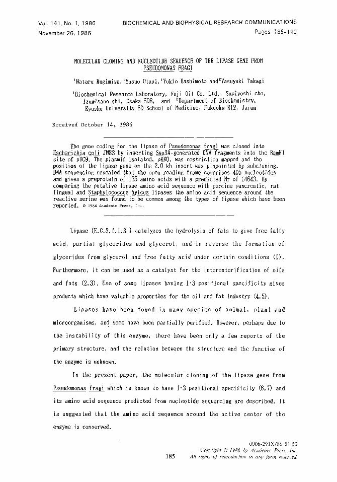
plates, and a single halo-forming colony was detected. In Fig. la the lipase
production of E.coli JM83 containing put9 (negative control) , E.coli JNg3
harboring plasmid (pKKO) and P.fragi host (lipase gene donor strain) are shown.
186

Vol. 141, No. 1, 1986 BIOCHEMICAL AND BIOPHYSICAL RESEARCH COMMUNICATIONS
b
.ind,,,l ( KKO EcoRI
i EcoRI Smal
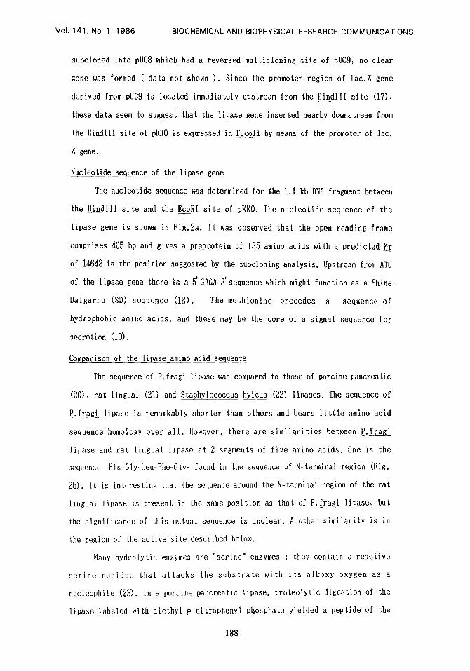
F~g.la. Lipase production by various strains on the tributyrine agar plate (see Materials and Methods). After 24 hrs incubation at 37~ and 25 ~ for E.coli and for Pseudomonas strains, respectively, visible clear zones due to lipase act ivi ty are formed around the colonies. (1)E.coli JM 83 containing pUC9 ( negative control ) . (2) E.coli JM 83 harboring pKKO. (3) P . ~ [lipase gene donor strain) . b ; Restriction map of pKKO. pKKO consists of the 2.7 kb of plasmid vector pUC9 (thin line) and the 2.0 kb of p. fragi DNfi fragment (double line ) which was cloned in the BamHI site of pUCg. The tipase gene is located within the 1.1 kb HindIIl-EcoRl DNA fragment (dashed region).
Class ica l r e s t r i c t i o n mapping of the recombinant plasmid, pKKO, revealed the
presence of a 2.0 kb inser t of Pseudomonas DNA (Fig. lb) .
Subcloning of the l ipase gene in E.coli
DNA was prepared from pKKO and cleaved with the r e s t r i c t i o n endonucleases
HindIII and EcoRI. These fragments were then subcloned in to EcoRI and EcoRI-
HindI I I s i t e s of pUC9, r e s p e c t i v e l y , and t ransformed in to E. co l i JM83.
plasmid carrying the 0.9 kb EcoRI DNA fragment did not make a clear zone on the
t r ibu ty r ine agar pla te . However, a plasmid with the 1.1 kb DN~ fragment between
H{ndIII s i t e and EcoRI s i t e made a c lear zone at the same level as pK~O ( data
not shown ) . Furthermore, when the HindIII -EcoRI 1.1 kb DNA fragment was
187
Vol. 141, No. 1, 1986 BIOCHEMICAL AND BIOPHYSICAL RESEARCH COMMUNICATIONS
subcloned into pUC8 which had a reversed multicloning s i te of pucg, no clear
zone was formed ( data not shown ). Since the promoter region of lae.Z gene
derived from pUC9 is located immediately upstream from the HindIII s i te (17),
these data seem to suggest that the lipase gene inserted nearby downstream from
the HindlII s i te of pKKO is expressed in E.coli by means of the promoter of lac.
Z gene.
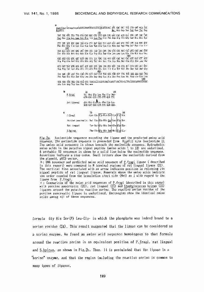
Nucleotide sequence of the lipase__gene
The nucleotide sequence was determined for the 1.1 kb DNA fragment between
the HindlII s i t e and the EcoJI s i te of pKKO. The nucleotide sequence of the
lipase gene is shown in Fig.2a. I t was observed that the open reading frame
comprises 405 bp and gives a preprotein of 135 amino acids with a predicted Mr
of 14643 in the position suggested by the subcloning analysis. Upstream from ATG
f
of the lipase gene there is a 5-GAGA-3 sequence which might function as a Shine-
Dalgarno (SD) sequence (18). The methionine precedes a sequence of
hydrophobic amino acids, and these may be the core of a signal sequence for
secretion (19).
Comparison of the lipase amino acid sequence
The sequence of P. fragi lipase was compared to those of porcine pancreatic
(20), rat lingual (21) and Staphylococcus h_yicus (22) lipases. The sequence of
P. fragi l ipase ' i s remarkably shorter than others and bears l i t t l e amino acid
sequence homology over a l l . However, there are s imi la r i t ies between P. fragi
lipase and ra t lingual lipase at 2 segments of five amino acids. One is the
sequence -His-Gly-Leu-Phe-Gly- found in the sequence of N-terminal region (Fig.
2b). It is interesting that the sequence around the N-terminal region of the rat
lingual lipase is present in the same position as that of P. frg_gi lipase, but
the significance of this mutual sequence is unclear. Another s imilar i ty is in
the region of the active s i te described below.
Many hydrolytic enzymes are serlne" enzymes ; they contain a reactive
ser ine res idue that a t t acks the subs t r a t e with i t s alkoxy oxygen as a
nucleophile (23). In a porcine pancreatic lipase, proteolytic digestion of the
lipase labeled with diethyl p-nitrophenyl phosphate yielded a peptide of the
188
Vol. 141, No. 1, 1986 BIOCHEMICAL AND BIOPHYSICAL RESEARCH COMMUNICATIONS
a
aa~cttggcigcaggtcga6gGATCAAAAACACGCTGCGAG~TTGAAC ATG GAC GAT TCG GTA Hi_~ndlll ~e__!tAsp Asp Ser Val
1QO TAT CCG ATC TTA TTG GTA T_jff. Pro Ile Leu Leu Val
TTT CAT GGT ATC hAG CAA Phe His Gly Ile Lys Gln
zoo TCA GCG GCC AAC GAG hAT Ser Ala Ala ASh Asp ASh
GGC AGG CAG GTC GGT GCA Arg Arg Gin Val Gly Ala
GCG CGT T~T GTG GCG GGC Ala Arg Tyr Val Ala Ala
CCC AAC CAC GGC TCC GAG Pro Asn Itis Gly Ser Glu
CAC GGG GTT TTT GGA His GIy Lep Phe Gly
GCA CTC hAT gAG TGC Ala Leu ASh Glu Cys
GAA GCC CGA GGC GAT Glu Ala Arg Gly Asp
CAG CGT GTC AAC CTG Gin Arg Val Asn Leu
ATC GGC CCT GAA GTG lie Ala Pro Glu Leu
4Q0 CTG GCC GAT CGT TGC
TTC GAC CGG ~TA GGC TCG Phe Asp Arg Ile G]y Ser
GGT GCC AGC GTC TTC GTT Gly Ala Set Yal Phe Val
CAA CTG CTC hAG CA~ ATC Gin Leu Leu Lys Gin Ile
300 ATC GGC CAC AGC CA~ GGC lie GIy His Ser Gin Gly
ATC GCC TCG GTG AGG TCA Ile Ala Ser Ral Thr Ser
GCC TGG CCT TTG TC~ CGG
GCG AA~ CGG TGG CTG CC~ Ala Lys Arg Trp Leu Pro
hAT ACA ASh Th__£r
CAT CAC His Ris
CCA ATC Pro Ile
CAC AAC His ASh
GCC CTT Ala Leu
GTC AGT Val Ser
GGA GGC
CGC Arg
TAC Tyr
ATT [le
CTG Leu
ACC Thr
GGC GIy
TTG Leu Ala Asp Arg Cys Ala Trp Pro Leu Ser Arg Gly GIy Leu
CCC T6ACCACCTCGTTCAGCGCATTTTTATCCGCCCTCA~GGCCACAccIO~Gc Pro ***
14 20 P. f r ~ -Val-His Gly~Leu-Phe Gly Phe-
GTA-CAC-GGC-CTT-TTT-GGA TTC-
Rat lingual -gla His-Gly~Leu-Phe-GIy-Lys- -GCA-CAT GGC~CTA-TTT-GGA AAA-
79 86 P.f ragi -Leu- 1 l e ~ G l n ~ - ~ , l a
Porcine p . . . . . . t i c - , a l lleffly-His-Ser~LeulGly~Ser-
Rat lingual Tyr-Val- IIy-His SerIGlnlGl ~ Thr-
S.h_yicus Phe- Ile-~ly-His Se~-Met G~_~Gly-
Fig.2a. Nucleotide sequence encoding the lipase and the predicted amino acid sequence. The nucleotide sequence is presented from Hi ndIII site (nucleotide 1). The amino acid sequence is shown beneath the nucleotide sequence. Hydrophobic amino acids in the putative signal peptide (amino acids 1 to 18) are underlined. A probable SD sequence is shown by a solid line below the nuCleotide sequence. Asterisks indicate a stop codon. Small letters show the nucleotide derived from the plasmid, pUC9 vector. b; DNA sequence and predicted amino acid sequence of P. frag ! lipase ( described in this report) were compared to N-terminal regione of rat lingual lipase (21). The ver t i ca l l ine associated with an arrow indicates position in releasing i ts signal peptide of ra t l ingual l ipase. Numerals above the amino acids indicate the order counted from the translation s tar t s i te (Met) as 1 with regard to the lipase from P. f ragi. c ; Comparison of the amino acid sequences of P. fragi (described in this paper) with porcine pancreatic (20), ra t l ingual (21) and S_taphylococcus hyicus (22) l ipases around the putative reactive serine. The reactive serine residue of the porcine pancreatic l ipase is underlined. Rectangles show the identical amino acids among a l l of these sequences.
formula -Gly-His-Ser(P)-Leu-Gly- in which the phosphate was indeed bound to a
ser ine residue (24). This r e s u l t suggested that the l ipase can be considered as
a s e r i n e enzyme. We found an amino acid sequence homologous to that formula
around the r e a c t i v e se r ine in an equ iva len t pos i t i on of P. f r ag i , r a t l ingual
and S.hyicus, as shown in Fig.2c. Thus, i t is postulated that the l ipase is a
'~erine" enzyme, and that the region including the r eac t ive ser ine is common to
many types of l ipases .
189
VoI. 141, No. 1, 1986 BIOCHEMICAL AND BIOPHYSICAL RESEARCH COMMUNICATIONS
Acknowledgement
We thank Dr. T. Imanaka, Osaka University for his useful discussions.
Refer enc es
1. Tsujisaka,Y.,Okumura,S. and Iwai,N.(1977) Biochim. Biophys. Acta 489,415- 422.
2. Stevenson, ti.~., Luddy,F.E. and tiothbart,H.L. (1979) J. Am. 0il Chem. Soc. 56, 676-680.
3. Macrae,N.B. (1983) 3. Am. Oil Chem. Soc. 60, 291-294. 4. Coleman,~.H. and Nacrae,A.R. (1980) U.K. Patent 1,577,933. 5. Natsuo,T.,Sawamura,N.,Hashimoto,K and Hashida,W. (1980) U.g.Patent 35,359
6. Alford,J.A., Pierce, D.A. and Suggs,F.G. (1964) J. Lipid Res. 5, 390-394. 7. Mencher, J.R. and Alford,J.A. (1967) J. Gen. Microbiology 48, 317-328. 8. Vieira,J. and ~essing,J. (1982) Gene 19,259-268. 9. Nessing, J. Recombinant DNA Technical Bulletin, NIH Publication No.79-99,
2,No.2 (1979) 43-48. 10. Coleman,K., Dougan,G. and Arbuthnott, J.P. (1983) J. Bacteriology 153, 909-
915. 11. Klein,ti.D., Selsing,E. and Wells,KD. (1980) Plasmid 3, 88-91. 12. Lawbence,R.C., Frayer,m.P. and Reiter,B. (1967) Nature 25,1264-1265. 13. Freyer,T.F.,Lawrence, R.C. and Reiter,B. (1967) 3. Dairy Sci. 50, 477-484. 14. Berner,D.L. and Hammond,E.G. (1968) Lipids 5,558-562. 15. Hattori,~. and Sakaki,Y.(1986) Analytical Biochem. 152, 232-238. 16. Lu,J.Y. and Liska,B.J. (1969) Appliedflicrobiology 18, i04-i07. 17. Yanish,-Perron, C., Vieira, J. and Nessing,J. (1985) Gene 33, 110-115. 18. Shine, J. and Dalgarno,b. (1974) Proc. Natl. Acad. Sci. USA 71, 1342-1346. 19. Watson. M.E.E. (1984) Nucleic Acids Res. 12, 5145-5153. 20. De Caro, J., Boudouard,N., Bonicel,J., Guidoni,A., Desnuelle,P. and ffovery,
M. (1981) Biochim. Biophys. Acta 671,129-138. 21. Docherty,A.J.P., Bodmer,M.W., Angal,S., Verger,K, Riviere,C., Lowe, P.A.,
Lyons,A., Emtage,J.S. and Barris,T.J.R. (1985) Nucleic Acids ties. 13, 1891 -1903.
22. Gotz,F., Popp,F., Korn,E. and Schleifer,K.B. (1985) Nucleic Acids ties. 13, 5895-5906.
23. Bender,N.L. and Kezdy,F.J. (1965) Ann. tier. Biochem. 34, 49-76. 24. Guidoni,A., Benkouka,F., Decaro,J. and tiovery,N. (1980) giochim. Biophys.
Acta 660, 148-150.
190


![University of Groningen Molecular redesign of Baeyer-Villiger ...Pseudomonas stutzeri WM88 was designed using Gene Designer.[37] The nucleotide sequence of this mutant was optimized](https://img.pdfslide.us/doc/110x75/602728d96b45c5510f6fb43e/university-of-groningen-molecular-redesign-of-baeyer-villiger-pseudomonas-stutzeri.jpg)