Embed Size (px)
Citation preview

Copyright 0 1990 by the Genetics Society of America
Molecular Cloning and Genetic Mapping of the t complex responder Candidate Gene Family
Daniel C. Bullard and John C. Schimenti Department of Genetics, Case Western Reserve University, Cleveland, Ohio 44106
Manuscript received June 16, 1989 Accepted for publication December 22, 1989
ABSTRACT Male transmission ratio distortion (TRD) is a property of mouse t haplotypes requiring the t complex
responder locus (Tcr). Tcr maps to the central region oft haplotypes, and is embedded within a series of large duplicated tracts of DNA known as “T66 elements.” In previous work, a family of genes (the “T66” genes) was identified within this region that encodes male germ cell-specific transcripts. Genetic and molecular data indicate that one of these genes represents Tcr. Here, we describe the molecular cloning of the four members of the T66 gene family, the genetic mapping of these genes to three adjacent t haplotype loci, and comparative restriction enzyme analysis of the genes. The results indicate that these genes are highly similar to one another, and were created by recent, complex duplication events. This suggests that a minor alteration(s) could have been responsible for conferring “mutant” responder activity upon Tcr, while the other homologs retained “wild-type” biochemical function. In addition, we have identified and mapped three T66 genes in wild-type t complexes. They reside in two separate loci at the opposite ends of the proximal t complex inversion, and are separated by at least 3 cM.
..
t haplotypes are variant forms of the t complex, a 15- cM stretch of DNA located in the proximal third
of mouse chromosome 17 (for a review, see SILVER 1985). This represents approximately 0.5-1% of the mouse genome. Although most t haplotypes contain at least one recessive developmental lethal mutation, and males heterozygous for two complementing t haplotypes are sterile, these variant chromosomes propagate to high frequencies in wild mouse popula- tions due to male transmission ratio distortion (TRD). This causes male mice heterozygous for a t haplotype and a wild-type form of the t complex ( + / t ) to transmit the t chromosome to nearly all of their offspring.
TRD is believed to occur through the action of at least four trans-acting t complex distorter (Tcd) loci upon a t complex responder (Tcr) locus (LYON 1984; SILVER and REMIS 1987; see Figure 1). If a male carries all the Tcd loci and is heterozygous for Tcr, the Tcr-containing chromosome 17 homolog can be transmitted to progeny at frequencies greater than 95%. However, if a male carries the Tcr locus in the absence of Tcd loci, the frequency is reversed, and the Tcr-containing homolog is transmitted to less than 20% of the offspring (LYON and MASON 1964; DUNN and BENNETT 1968). The distorters act in an additive fashion: as doses are removed, Tcr transmission de- clines (LYON 1984). Finally, if the Tcr locus is absent from both chromosomes, each is transmitted at 5076, irrespective of the presence or absence of distorters.
of page charges. This article must therefore be hereby marked “aduertwernent” T h e publication costs of this article were partly defrayed by the payment
i n accordance with 18 U.S.C. 5 1734 solely to indicate this fact.
Genetics 124: 9.57-966 (April, 1990)
The presence of at least four rearrangements of genetic material in t relative to wild-type chromo- somes suppresses recombination in + / t heterozygotes throughout the t complex region (ARTZT, SHIN and BENNETT 1982; PLA and CONDAMINE 1984; HERR- MANN et al. 1986; SARVETNICK et al. 1986; HAMMER, SCHIMENTI and SILVER 1989). This region of recom- bination suppression formally defines the t complex (SILVER 1985), and allows t haplotypes to propagate as single genetic units in mouse populations.
Tcr has been mapped to a small region in the center of t haplotypes called the Dl7Leh66b locus (abbrevi- ated throughout the text as T66B) by molecular and genetic analyses of recombinant chromosomes known as partial t haplotypes (Figure 1). Partial t haplotypes are the products of rare recombination events be- tween a t haplotype and a wild-type form of the t complex. These recombinants contain only a portion of t haplotype DNA. By using DNA probes which detect restriction fragment length polymorphisms (RFLPs) between wild-type and t haplotype loci, the recombination breakpoints of partial t haplotypes have been mapped relative to one another. Combined with the genetic testing of these partial t haplotypes for responder activity, Tcr was localized to T66B (LYON and MASON 1964; LYON 1984; Fox et al. 1985; HERR- MANN, BARLOW and LEHRACH 1987; LYON and ZEN- THON 1987).
The T66B locus is part of a family (the T66 family) of large, duplicated blocks of DNA sequences called T66 elements. Individual members of the T66 family can be up to 1 10 kb in length, and are not found

958 D. C. Bullard and J. C. Schimenti
TRD Loci: Tcd-1 Tcd-4 Tcr Tcd-3 Tcd-2
MHC tf Pgk-2
4 8 T c p - 1 1 1 9 6 6 A 6 6 B 6 6 C 8 9
T66A T66B T66C
“I
T66A-a T66B-a T66C-a T66C-g3
h2, h49 +
t 3, complete
FIGURE 1.-Breakpoint chromosomes in the Tcr region. A genetic map of the t haplotype form of the t complex is at the top of the diagram. The t complex is flanked by the centromere on the left (empty circle) and Pgk-2 (phosphoglycerate kinase-2) distally. The genetic loci 9k (quaking), major histocompatibility complex (MHC), and tftufted) are shown above the chromosome, while the following DNA microclone m;lrkers are shown below: 48(D17Leh48; Fox et a f . 1985), 119 (D17Leh119; HERRMANN et a f . 1986). 89 (D17Leh89, SILVER and REMIS 1987), and 66A/66R/66C (DI7Leh66AIBIC; FOX et a f . 1985). Also shown are loci involved in transmission ratio distortion: the t complex distorters Tcd-1-Tcd-4. and the t complex responder. Tcr (LYON 1984: SILVER and REMIS 1987). Tcd-2 is located somewhere between T66C and the distal end of the t complex. The central region of the t haplotype is expmded in the middle of the figure to emphasize the three loci T66A, T66B and T66C, first characterized by Fox et a f . 1985. Tcr is localized to the T66B region as indicated. Various partial t haplotypes having breakpoints ur4lich have been used to define these loci are indicated on the left: th5’, t””’, t”’, t“’, 1’ (FOX et a f . 1985); t’”””’ (SILVER et a f . 1987; SCHIMENTI et of. 1987). The locations of the T66 genes discussed in this report are shown at the bottom. The hatched region at the right side of each box represents T66 gene coding and about 15 kb of 5’ flanking sequences (see text: SCHIMENTI et al. 1987; L. SNYDER,J. SCHIMENTI, and L. SILVER, unpublished observations). The diagonally striped portions in T66A-a, T66B-a and T66C-a represent homologous “d ike“ sequences upstream of the T66 genes. The corresponding portion of the T66C-g3 gene is vertically striped to indicate its dissimilar ‘+like” sequences when compared to the other three genes (SCHIMENTI et a f . 1987). The order (relative to the centromere) of the two genes i n the T66C region are unknown and are shown arbitrarily. Sizes of the genes are greatly exaggerated with respect to the actual regions in which they reside.
elsewhere in the genome (SCHIMENTI et al. 1987; J. SCHIMENTI, unpublished observations). The T66B lo- cus is flanked on the centromeric and distal sides by the T66A and T66C loci, respectively, which also con- tain T66 elements (see Figures 1 and 4). The contig- uous T66A, T66B, and T66C loci contain one, two, and eight T66 elements, respectively (see Figure 4). A detailed analysis of these elements through molec- ular cloning and genomic blotting grouped them into three subclasses-a, ,d and y-based on relative similar- ity to one another (SCHIMENTI et al. 1987). These elements arose by a complex series of large scale duplications. The divergence between the a, P , and y subclasses suggest that considerable periods of time passed between early duplications, followed by more recent events that created multiple subclass members.
Some T66 elements contain sequences (genes) which hybridize to a species of RNA transcripts found
specifically in male germ cells (SCHIMENTI et al. 1988). Several cDNA clones representing these transcripts were isolated, sequenced, and compared to portions of two cloned genomic genes or pseudogenes (SCHI- MENTI et al. 1988). One of these genes, called the T66B-a gene, maps to T66B. Molecular cloning of the T66B locus has revealed that it is approximately 220 kb in length (J. SCHIMENTI, unpublished observa- tions). The T66B-a gene is a strong candidate for Tcr since it maps to the responder locus, is expressed exclusively in male germ cells, and is the only detect- able gene within the 220 kb T66B locus.
In this report, we present the genomic identifica- tion, molecular cloning, genetic mapping, and com- parative restriction enzyme analysis of the 4 structural members of the T66 gene family. These results indi- cate a strong sequence similarity among the genes, which appear to have been created as parts of larger

t complex responder Gene Family 959
TABLE 1
Restriction fragments of the T66 genes
Gene
Probe Enzyme T66A-a T66B-a T66C-a T66Gg3
Tcr l6 Ban11 4.2 + 1.5 1.8 + 0.4 1.75 + 0.4 2.1 + 1.8
PstI 6.25 6.3 7.1 6.3 + 0.6 BamHI 3.4 6.0 6.0 6.0 + 2.0
R/H , 2
Cg3-79 Ban11 1.5 1.3 1.3 1.2 Pst 1 1.6 1.0 1.6 1.5
Restriction fragment sizes are in kilobases. Boldfaced numbers denote that the fragment is t-specific, ie., it was not detected in the wild-type chromosomes used in this study.
duplication events. If indeed only one of these genes represents the responder, the molecular change(s) which have conferred this biochemical property must be quite minor, and may be revealed by further de- tailed comparative analyses. This report is a step to- ward determining the nature of such mutations.
MATERIALS AND METHODS
Southern blotting: Mouse genomic DNA (5 pg) was di- gested with the appropriate restriction enzyme, electropho- resed on 0.8% agarose gels, soaked 30-60 min in 1.5 M NaCI, 0.5 M NaOH and transferred to Genescreen mem- branes (New England Nuclear) overnight in the same solu- tion. Following a 1 min neutralization in 1.5 M NaCI, 0.5 M (Tris pH 7.6), membranes were baked at 80" for 2 hr, then UV cross-linked 15 sec at a distance of 15 cm by 2-254 nm bulbs. The blots were prehybridized at least 5 min in "Church" buffer (CHURCH and GILBERT 1984) at 65", and hybridized overnight at 65" to random primed probes (FEINBERG and VOGELSTEIN 1984). The blots were washed four times for 5 min in 2X SSC, 0.1 % SDS at room temper- ature, followed by 2-30 min washes in 0.1 X SSC, 0.1 % SDS at 65".
Cosmid cloning: Two mouse cosmid libraries containing different t haplotypes (genotypes: tW2/ tw2 and t ' " * ' / tw5) were constructed in the vector pWE15 (EVANS and WAHL 1987). Spleen DNAs were prepared as described (BLIN and STAF- FORD 1976). Partial Sau3A digests were performed on these DNAs, followed by size fractionation on 0.4% low gelling temperature agarose gels. DNA in the 30-50-kb size range was purified and ligated to BamHI-cut, phosphatased pWE15 cosmid vector DNA and packaged in vitro using Gigapack Gold extracts (Stratagene Cloning Systems). Pack- aged cosmids were infected into either Escherichia coli 490A or NM554 (a gift from Heidi Short, Stratagene cloning systems), and plated onto nonsterilized, Millipore HATF 137 mm nitrocellulose membranes placed on top of LB agar plates containing 25 pg/ml kanamycin. The filters were briefly rinsed in sterile water prior to plating. Colonies were grown for 12 hr, then two replicas were made onto nonster- ile but rinsed nylon membranes (Nytran, Schleicher & Schuell). The masters were regrown for 2-3 hr at 37", sandwiched against a second membrane soaked in glycerol, placed between two moist filters (Whatman 3"), and frozen at -70". Colonies on the replica filters were lysed according to the instructions outlined by the manufacturer (Schleicher & Schuell), but the DNA was fixed by UV cross- linking as described above. All clones refixed by "CW" in this report were isolated from the t'"E/tu'5 library. Clones
prefixed by "CD" were isolated from the tw'/tw2 library. Restriction mapping: Cosmids were restriction mapped
for the enzymes EcoRI, BamHI and Hind111 by an indirect end labeling/partial digestion method (SCHIMENTI et al. 1987). These results were compared to those obtained from complete digests of the clones on ethidium bromide stained agarose gels.
DNA Probes: Three probes were used in this study. Tcr16R/H.2 is a 426-bp Hind111 fragment extending from bases 510-936 ofa cDNA clone called Tcrl6. The sequence of this clone has been published (SCHIMENTI et al. 1988). This fragment spans 4.5 kb of genomic DNA (see Figure 3). Cg3-79 is a 400 bp subclone derived from the 5' end of the D17Leh66c-g3 gene (SCHIMENTI et al. 1987; Figure 3). Cg3-100 is a 534-bp EcoRI/BamHI fragment from the 3' end of the D17Leh66c-g3 gene (Figure 3). It corresponds to bases 1621-2155 of the cDNA sequences in Figure 4 of SCHIMENTI et al. (1 988).
Nomenclature: The five genetic subregions discussed in this report are formally designated D l 7Leh66a, D l 7Leh666, Dl7Leh66c, D17Leh66d and Dl7Leh66e. In this text, how- ever, and in earlier publications, they are abbreviated as T66A, T66B, T66C, T66D and T66E, respectively. The genes described in this text are informally named with regard to the DNA element of which they are a part. These elements, designated D l 7Leh66aa, D l 7Leh66ba, D l 7Leh66ca and Dl7Leh66cg-3 are abbreviated in this report as T66A-a, T66B-a, T66C-a and T66C-g3. Since it has been shown that at least one of the members of this gene family is translated, this family of genes has been designated t complex protean-10 (Tcp-10; SCHIMENTI et al. 1988). The different family mem- bers are named Tcp-loa, Tcp-lob, Tcp-lOc and Tcp-lOd in order from the centromere. However, since the order of the Tcp-lOc/Tcp-lOd (T66C-a/T66C-g3, or vice versa) genes is not yet known, we will at present refrain from using this terminology.
RESULTS
Isolation of T66 gene family genomic clones: Prior to this work, a cosmid clone had been isolated which contained the entire genomic coding region of a T66 gene, T66C-g3 (map location 78-101 in Figure 2 of SCHIMENTI et al. 1987). Fragments from this clone were used to identify the T66 family of RNA transcripts (SCHIMENTI et al. 1988). Comparison of cDNA sequences to those from the genomic clone allowed an approximate determination of the 5' and 3' termini of this gene (SCHIMENTI et al. 1988; J. SCHIMENTI and L. SILVER, unpublished observations).
T o isolate the remaining members of the family, a cDNA probe corresponding to the central portion of the gene (Tcr16R/H.2; Figure 3) was used to screen genomic cosmid libraries constructed from t 'ub'/tw.5 and tW2/tu2 mice. Positive clones were tested for hy- bridization to the probes Cg3-79 and Cg3-100 (see Figure 3) , which contain sequences corresponding to the 5' and 3' ends, respectively, of the presumably canonical T66C-g3 gene. Clones hybridizing to both sequences would therefore contain an entire T66 gene.
Mapping of genes and cosmids to t haplotype subregions: T o map the genetic location of the cosmid clones, we used a strategy which is routinely employed
960 D. C. Bullard and J. C. Schimenti
Ban II
A
"
Tcrl6 R/H.2 Probe
Barn HI
c;
F s s % N
o o o o
Pst I B
1 - 1.0 r !
Cg3-79 Probe
Pst I D
I
-- -2.0L / 2.1
Tcr16RlH.2 Probe
TcrlGR/H.:! probe
FIGURE 2.--Mappiq of T66 genes and cosmid clones by Southern blotting. Genolnic DNAs from various f complexes. indicated at the top of ~ h c 1a11es. have I)een ;Ibbreviated a s follows: h51 (th"/+); h53 ( fh5 ' / th5 , ' ) ; Tuw32 (I"""'?/l17"''"); h2 ( th"/fh'); h4Y (th"'/th4'); 13 (t.'); lubI/ 7 1 ~ 5 ( / " 'h l / tw , ' ) : 7112 ( t""); 129 (129/Su); +ff(+ff/+ff-;~ noninbred s;lmple of wild-type chromoson1e 17 kept in the colonv). Cosmid clone DNAs i ~ r e prcfisctl I ) \ *'CiV.'' l'he sizes of bands are show1 in kilobases. A, Ban11 digests probed with Tcrl6K/H.2. B. Psfl digests probed with C- g % i 9 . (;. h m l I I digests probed with Tcrl6R/H.2, D, fstl digests probed with Tcr16K/H.2.
for the mapping o f t complex DNA probes (Fox et al. three exist in various inbred mouse strains, and three 198.5; HERRMANN et al. 1986; SCHIMENTI et al. 1987). to four in t haplotypes-data presented in this manu- First, probes for the T66 genes were identified which script) and the high similarity between them. Third, detect RFLPs between t haplotypes and wild type these probes were hybridized to panels of DNAs from forms of the t complex. Second, an RFLP specific for various partial t haplotypes (rare recombinant chro- each t haplotype gene was identified, in part by prob- mosomes with only a portion oft haplotype chromatin) ing restriction enzyme digests of cosmid clones. The known to have breakpoints within the T66 loci T66A, identification of gene-specific RFLPs was complicated T66B and T66C. The haplotypes used and their break- bv the presence of numerous genes (at least two or points within this region are diagrammed in Figures

t complex responder Gene Family 96 1 R R B B H B B B HH B B R E R R B B B H R R
I 1 1 I I I I I I 1 I 1 I I I I I I 1 I I J T66A-a
I I 11 I 1 1 1 I I 1 I 1 I \ \ I l l I I I 1 I ' 1 T66B-a
L I I I 1 1 1 I I 1 I 1 I I l l I 1 I 1 I 1 1 T66C-a
n .lkb
R R EB B H R B B H H B E R E R R R E E 8 H R R
\ \
-.3kb
R R E B H R B B H H B B R E R R R B B B H R R
T66 GENE TRANSCRIPTION UNIT
1 1 2 1 3 1 4 I 5 1 6 1 7 1 8 1 9 1 1 0 1 111 121 1 3 1 1 4 1 1 5 1 1 6 1 171 1 8 1 191 2 0 1 2 1 1 2 2 1 231 2 4 1 251 2 6 1 271
FIGURE 3.-Restriction enzyme map of the T66 genes. The restriction maps of the four T66 genes and a few kilobases of flanking sequences are aligned. The location and orientation of the transcription unit is indicated, and the size in kilobases is shown on the bottom of the diagram. A relative insertion of 0.1 kb is present in T66A-a as indicated by a triangle below the map. A 0.3-kb deletion is indicated in the T66B-a gene. The maps of the genes were derived from the following cosmid clones: T66A-a-CW2, CW12; T66B-a-CW6, CW7, and cosu, a cosmid clone isolated from a different t'""/twJ cosmid library which has been described (SCHIMENTI et al. 1987); T66C-a-CW9, CDlO (derived from the t"'?library); T66C-g;l-CWl, CW4, CWl1. The locations of probes Cg-79 and Cg-100 are indicated by solid black rectangles below the map. The genomic sequences spanned by the cDNA probe TcrlGR/H.P are shown as a hatched rectangle.
1 and 4, and fall into three classes: those containing just T66A, those containing T66A and T66B but not T66C, and those containing all three loci (including complete t haplotypes). These haplotypes are scored for the presence or absence of gene-specific RFLPs. It is then possible to localize a RFLP. For example, if an RFLP is present in a partial t haplotype which spans both the T66A and T66B loci, but is absent in a partial t haplotype which contains only T66A, then it must map to T66B. Finally, the cosmid clones can be mapped by scoring for the corresponding RFLPs. It should be noted that the partial t haplotypes used in this study contain wild-type T66 loci, and will there- fore contain fragments representative of wild-type genes (presented below; Figure 6).
Two probes were informative for these analyses: Cg3-79, which represents the 5' region of T66 genes, and Tcr 16R/H.2, representing the central portion (see MATERIALS AND METHODS; Figure 3). Eleven re- striction enzymes were tested for the identification of RFLPs: BamHI, BanII, BgZII, DraI, EcoRI, EcoRV, HindIII, PstI, PvuII, ScaI and TaqI. Using RFLPs identified by some of these restriction enzymes, we were able to detect and map four T66 genes in some t haplotypes, and three in others. One gene maps to the T66A subregion, one to T66B, and one or two (depending on the t haplotype) within T66C.
The gene in the T66A region, T66A-a, is character- ized by t-specific 4.2-kb and 1.5-kb BanII fragments detected by the Tcr16R/H.2 probe (Figure 2A; Table 1). These bands on Southern blots are present in all partial t chromosomes that extend into T66A but not T66B, such as t h5.7 and t T u w : j p (Figures 1 and 2A). These
RFLPs are displayed by the cosmid clones CW2, CW3 and CWI 2 (Figure 2A). This probe similarly hybrid- izes to a T66A-specific 3.4-kb BamHI band in the same cosmid clones (Figure 2C; Table 1). In addition, T66A- a can be mapped by a 1.5-kb BanII RFLP detected by the probe Cg3-79 (Table 1). This RFLP also correlates with the CW2, CW3 and CW 12 cosmid clones (not shown).
The T66B-a gene was mapped by a 1.0-kb PstI RFLP detected by the Cg3-79 probe. This t-specific band is not present in tTuW"', a partial t haplotype which has a breakpoint between the T66A and T66B regions, but is contained in those with breakpoints between T66B and T66C, such as t h2 and t h4.9 (Figure 2B; Figure 1 ; Table 1 ; t h2 is not shown). This fragment is contained in cosmids CW6 and CW7 (Figure 2B).
The cumulative data indicate the presence of two genes in the T66C interval of the t lub' and t w 5 haplo- types: T66C-g3 and T66C-a. The T66C-g3 gene is characterized by four t-specific RFLPs indicated in Table 1: 0.6-kb PstI, 2.1-kb BanII and 2.0-kb BamHI fragments with the Tcr16R/H.2 probe (see Figure 2, A and C for the Ban11 and BamHI RFLP data, re- spectively, and Table 1 for PstI) and a 1.2-kb BanII fragment with the Cg3-79 probe (Table 1). These RFLPs are present in DNA from the compound het- erozygote t lub'/tw5, but not in partial t haplotypes which have a breakpoint between T66B and T66C (Figures 1 and 2, A and C). They are contained in the cosmid clones CWI , CW4, CWIO, CW11 and other previously isolated cosmids corresponding to the T66C-g3 element (Figures 2, A and C; SCHIMENTI et al. 1987, 1988).

962 D. C. Bullard and J. C. Schimenti
ThP 1 I
T T48 RP17
qk Tcpl f"-T66D - n 1119 I T66E 766Ell T11911 " ' gI l l b l g2 " b 2 g 3 ' + o I I
I 1
orf
t "U 1 I 1 a I I b 4 I I b a fb, gI-g4, XI I
T48 Tcpl RP17 TI19 T66A T66B T66C TGr
FIGURE 4.-Recombination breakpoints of partial t haplotypes. Maps of the proximal portion of wild-type and t haplotype forms of the t con~plex are aligned. The relative locations of genetic intervals which have been defined by recombination data are indicated by brackets. Relevant molecular markers for each of these intervals are indicated. The sizes o f genetic intervals are not to scale. The classical distance between Brachyury ( T ) and quaking ( q k ) is about 3 cM. The physical size of the TZ 19-T66E-T66EII-TIZYII segment is approximately 1300 kb (HERRMANN, BARLOW and LEHRACH 1987). Individual "T66 elements" in the T66A, E , C and D loci are listed within brackets. The relative order of T66 elements in the T66C locus is not known, but the order in the T66B locus is as indicated (J. SCHIMENTI, unpublished observations). The relative order of neither the T66D-g2/T66D-bl nor T66D-g3/T66D-b2 pairs are known. a, b and g stand for a, 6, and 7, respectively, as originally designated by SCHIMENTI et al. (1987). Formal nomenclature designations and references on the mapping of the loci T4K and TZZY are given in the Figure 1 legend. Other markers are: RP17 (DI7RpZ7; MANN, SILVER and ELLIOTT 1986); and Tcp-I (SILVER 1981). An example of formal nomenclature for the various T 6 6 element loci is: D17Leh66Aa for the "a" element in T66A. The vertical lines connecting the chromosomes represent the breakpoints of various partial t haplotypes. The abbreviated names (they should be preceded by a " t " ) of these haplotypes are shown. t h P , th4,', th49, tks", Tt"" and tu',' contain t haplotype-derived centromeric ends, while the Centromeric end of tw'"b2 is wild type. The region of DNA deleted by TIP is bracketed. Published reports which have characterized the breakpoints of these recombinant chromosomes are as follows: th"', tns.', th2 , th4', t X (FOX et al. 1985); t'""' (SILVER et al. 1987; SCHIMENTI et al. 1987); Tt"" and t"""h' (SARVETNICK et al. 1986); th'"J and (HERRMANN, BARLOW and LEHRACH 1987).
In contrast, we have not obtained evidence for the presence of the T66C-g3 gene in the t"' haplotype. This is in agreement with earlier work which reported a deletion of the T66C-g3 DNA element in this hap- lotype (SCHIMENTI et al. 1987). This apparent deletion was identified by analysis with the probes Cg3-38, Bb- 40 and Tu66; these probes are situated approximately 40, 38 and 25 kb upstream, respectively, of the T66C- g3 gene (SCHIMENTI et al. 1987; this report). t"" lacks the 1.5-kb PstI fragment detected by the Cg3-79 probe, which is contained in tiub'/tm5 mice and the T66C-g3 gene cosmids CW 1, CW4, CW 10 and CW 11 (Figure 2B). Similar results have been obtained for t " , a partial t chromosome which has a breakpoint distal to the T66 family of DNA sequences (FOX et al. 1985). We have probed Southern blots of t W 2 homozygous DNA cut with 14 restriction enzymes using the Tcr16R/H.2 probe. Seven of these enzymes (BglII, EcoRI, HindIII, NcoI, PstI, TaqI and XbaI) generated three bands of apparently equal intensities, while none yielded four bands (data not shown). The transmission frequency of tu' in heterozygous males is 95% (DUNN and SUCKLING 1956), indicating that this gene (which is believed to be a pseudogene-see DISCUSSION) does not participate in TRD.
A fourth gene, T66C-a, was identified and mapped indirectly. Because no partial t haplotypes have been
described with breakpoints in T66C (to a resolution determined by the current availability of DNA probes), demonstration of multiple genes in this locus depended on a compilation of the following pieces of data: (1) identification of nonallelic RFLPs mapping to this region, (2) restriction patterns of cosmid clones, and (3) physical linkage to diagnostic RFLPs outside the T66 gene sequences.
The presence of the T66C-a gene was predicted from previous studies of the T66 family of DNA elements and T66 gene sequences. The T66C-a ele- ment contains sequences homologous to the 5' end of T66 gene transcripts (SCHIMENTI et al. 1988). Cloned DNA upstream of these sequences were clearly dis- tinct from those flanking the T66C-g3 gene (see DIS-
CUSSION pertaining to alpha vs gamma T66 elements). One cosmid clone isolated from the t'"*'/t"' library, CW9, has a set of restriction fragments which differ from the T66A-a, T66B-a and T66C-g3 genes (Table 1). CW9 has a unique 7.1-kb PstI fragment which hybridizes to the Tcr 16R/H. 2 probe (Table 1 ; Figure 2D). While this fragment is present in DNA from both wild type and t fub'/tw,5 mice, it is not contained in the partial t haplotypes tTuw.", t h 4 9 or th' (Figure 2D; th' is not shown). This indicates that the fragment does not map to the T66A or T66B loci (Figure 1). We conclude that this fragment (and therefore the CW-9 cosmid)

t complex responder Gene Family 963
maps to T66C, and is representative of the T66C-a gene.
Because the cosmid library and genomic blots were made with compound heterozygote t’ub’/tw.i DNA, it remains possible that the T66C-a and T66C-g3 genes are alleles. However, several pieces of data indicate that these two genes are nonallelic. First, for all five combinations of probes and restriction enzymes listed in Table 1, the two cloned genes have different pat- terns. Second, these two genes are linked to vastly different upstream sequences-T66C-a to “a”-like and T66C-g3 to “y”-like DNA (SCHIMENTI et al. 1987; see DISCUSSION). Thirdly, it has been shown that the t l u b l
and t u’5 haplotypes contain sequences representative of both the T66C-a and T66C-g3 elements (SCHIMENTI et al. 1987).
Comparative restriction enzyme mapping and gene boundaries: Our cosmid clones were mapped with three enzymes, EcoRI, BamHI and HindIII. The analysis is shown in Figure 3. It is evident that all four genes are highly similar in DNA sequence. I f the genes are superimposed, a total of 26 different restriction enzyme sites are present in the region examined. Twenty of these are shared by each of the four genes, while five represent a presence or absence of a site specific to only one of the genes. In only one case (a BamHI site at position 1.5) is a site shared by two genes and absent in the others. We detected one relative insertion in the T66A-a gene and one relative deletion in the T66B-a gene after comparison to the others. The nature of these size differences is un- known.
T66 genes in wild-type t complexes: Although we have not cloned wild-type T66 gene homologs, they can be identified and genetically mapped by Southern blot analysis of recombinant chromosomes. An in- formative probe for these analyses is Cg-100, which was isolated from the 3‘ end of the T66C-g3 gene (Figure 3). When hybridized to Southern blots of TaqI digested t’uh‘/tu’.i DNA, a single fragment of 9.6 kb is detected (see Figure 5). This demonstrates that all T66 genes in these t haplotypes possess a fragment of this size. 129/Sv mice display three bands of 9.4, 7.2 and 5.8 kb, whereas C3H/He mice have 9.6- and 7.2- kb bands (Figure 5).
The wild-type-specific 7.2-kb Tu91 band, which is shared by 129/Sv and C3H/He, is present only in those recombinant chromosomes which contain wild- type DNA proximal to the RP17 locus. These includes
absent from partial t haplotypes which contain all
not T66E (Figures 4 and 5). These data allow the localization of this fragment between the wild-type breakpoints of T“p and tor‘ (Figure 4), a segment of DNA approximately 500 kb in length (HERRMANN, BARLOW and LEHRACH 1987). This genomic region
t h 4 5 , tu”uhP, and T“p (see Figures 4 and 5). I t is
( ~ ~ o r l ) or part ( th5 .Y, tTtrusJP , th4’) of the T66D locus, but
Taq I
I
FIGURE 5.--Mapping of wild-type T(i(i g e n a by Southern blot- ting. All of the DNAs shown have been digested wi th Tug1 and probed with Cg-100. The molecular weight of the four bands are shown in kilobases. The abbreviated names of various f haplotypes (they should be preceded by a “I”) are shown. 129 stands for 129/ Sv, and C3H for C3H/He. The tu”’ chromosome contains only the f forms of the T66 loci (BUCAN et al. 1987).
contains a large inverted duplication of DNA, part of which contains T66 element sequences (HERRMANN, BARLOW and LEHRACH 1987; SCHIMENTI et al. 1987). The order of loci in this region was determined to be: centromere-TI 19I-T66E-T66EII-T11911 (Figure 4; HERRMANN, BARLOW and LEHRACH 1987). The T66 gene represented by the 7.2-kb Tu91 fragment must map to either T66E or T66EII.
The 5.8- and 9.4-kb bands present in 129/Sv must be interpreted with caution since they are not present in C3H/He. This discrepancy is indicative of poly- morphism in wild-type t complexes. One of these two bands may be allelic to the 9.6 kb band in C3H/He (Figure 5), and the other may represent a gene absent in this strain. Support for this interpretation comes from the observation that C3H/He mice are deleted for the T66D-b1 and T66D-g2 loci (SCHIMENTI et al. 1987). However, it must be considered that one of the C3H/He bands actually represents a doublet.
The interpretation most consistent with our data maps the 5.8 kb fragment to the T66D-gl locus. All of the proximal partial t haplotypes tested which con- tain wild-type chromatin distal, but not proximal, to T66D-gl do not contain this band (Figures 4 and 5). I t is present in the to‘’ and th45 haplotypes, which possess the entire T66D locus (Figures 4 and 5). It is absent in tw‘uhP, which contains wild-type chromatin proximal to the Tcp-1 locus. These data localize the 5.8 kb band distal to Tcp-1 and proximal to T66D-b1/ T66D-g2. T66D-gl represents the only characterized locus containing a T66 element in this region, and is the likely location of the T66 gene homolog contain- ing the 5.8-kb fragment. Interestingly, this band is absent from the deletion chromosome pp (Figure 5), which is known to be deleted for Tcp-1 (SILVER,

964 D. C. Bullard and J. C. Schimenti
WHITE and ARTZT 1980), but not sequences in T66D detected by a probe called p66M-RT (HERRMANN, BARLOW and LEHRACH 1987). This probe hybridizes to a 1 O-kb wild-type specific BamHI fragment in th2 but not th49 DNA (HERRMANN et al. 1986). This would appear to localize this 10-kb BamHI fragment to the T66D-bIIT66D-g2 locus (Figure 4). We conclude that the distal Th* deletion breakpoint lies between T66D- g l and T66D-blIT66D-g2, thereby deleting the T66D- g l locus and a resident T66 gene homolog repre- sented by the 5.8-kb Taq1 restriction fragment.
The partial t haplotype Tt Or’ seems to contradict the mapping of a T66 gene to T66D-gl. This chromosome does not have the T66E loci, but does contain the entire T66D locus (Figure 4). It does not exhibit the 5.8-kb TaqI fragment (see the Ttor’/twP and Tt OT1/tw5 samples in Figure 5). In isolation, this might suggest that the 5.8-kb band maps to T66E or T66EII. How- ever, neither tw’ub2 nor th45 , both of which contain T66E and T66EII, have this band. It is likely that polymorphism in the wild-type T66 gene loci, such as the variation seen between 129/Sv and C3H/He, is responsible for this discrepancy. Specifically, allelism between the 9.6 kb C3H/He and 5.8 kb 129/Sv TaqI fragments could explain the disconcordance (Figure
The remaining 129/Sv 9.4-kb TaqI band appears to map to the T66D-g2/T66D-b1 subregion. It is pres- ent in the recombinant chromosomes which contain the entire T66D locus ( tae5, th45 and Tt’“), and tTuw3’, which has a breakpoint between T66D-gl and the distal T66D subregions (Figures 4 and 5). It is absent in the partial t haplotypes th4g and th53, which recom- bined between T66D-bl/T66D-g2 and T66-bZ/T66-g3, as well as twiub2, which has none of the T66D loci. Finally, it has been reported that the C3H/He chro- mosome is deleted for the T66D-bl and T66D-gZ elements (SCHIMENTI et al. 1987), which could account for the absence of this bandlgene.
In summary, we have found evidence for the exist- ence of three T66 genes in wild-type forms of the t complex. One maps to T66EIT66EII (the T66E gene), a second to T66D-gl (the T66D-gl gene), and a third to T66D-bIIT66D-g2 (the T66D-blg2 gene). Contrary to a report which cited preliminary evidence for the presence of a T66 gene associated with the T66D-g3 element (SCHIMENTI et al. 1988), we have not found any evidence for such a gene in this study.
5).
DISCUSSION
Evolution of the T66 genes: The genomic analyses and cloning data presented here demonstrate the ex- istence of three to four T66 gene homologs in those t haplotypes examined. The comparative restriction mapping shows that these genes are highly related, and suggest that this gene family was either recently created, or has been subject to concerted evolution.
Rat and Mus spretus appear to have only one T66 gene copy as assayed by Southern blot analysis (J. SCHIMENTI, unpublished observations). While this sug- gests a recent expansion in t haplotype gene copy number, the possibility that these species have deleted genes subsequent to diverging cannot be ruled out. As we have found in this study, different inbred strains appear to have different numbers of T66 genes. The variety in copy number suggests that unequal recom- bination has been a mechanism for expansion and/or contraction of this gene family (SCHIMENTI et al. 1987).
The restriction site analyses of the genes presented here are insufficient to allow a determination of their evolutionary relatedness. Closer molecular examina- tion of flanking and intron regions, coupled with a complete linkage map of the various elements, may elucidate the events leading to the creation of this multigene family. Similar work on the wild-type loci must be performed in order to reconstruct events which have occurred in the T66 family since the divergence of + and t chromosomes. These questions are also significant in regard to the T66 family’s ostensible involvement in the “proximal” t complex inversion event (SCHIMENTI and SILVER 1986; SCHI- MENTI et al. 1987). Recent work with interspecific crosses has supported the hypothesis that the proximal inversion actually occurred in the wild-type lineage, with a breakpoint in the T66 family that resulted in the transposition of two T66 DNA elements several centiMorgans towards the centromere (HAMMER, SCHIMENTI and SILVER 1989). It appears that the T66E gene was contained in this inversion, resulting in its separation from the other T66 genes by at least 3 cM.
Relationship of T66 genes to T66 elements: The initial studies of the T66 region of t haplotypes re- sulted in the identification of a family of large, dupli- cated tracts of DNA called “T66 elements” (SCHI- MENTI et al. 1987). These elements were classified into three subfamilies (a , p and y) based on differential hybridization to various probes. The size of individual elements range up to 110 kb (J. SCHIMENTI, unpub- lished observations), and in some cases include T66 genes. It is likely that the T66 genes expanded in copy number by inclusion in larger scale T66 element duplications. Three of the T66 genes described here, T66A-a, T66B-a and T66C-a, comprise portions of the a-class elements. In contrast, the T66C-g3 gene adjoins distinct y-type sequences; these sequences map about 20 kb 5’ (at map position 60 in SCHIMENTI et al. 1987) to the transcription initiation site (L. SNYDER, J. SCHI- MENTI and L. M. SILVER, unpublished results). The a and y sequences have diverged to a degree where they do not cross-hybridize over large stretches (SCHI- MENTI et al. 1987). The T66C-g3 gene and its adjacent sequences may have arisen by either of two means: (1)
t complex responder Gene Family 965
a partial duplication event not involving an entire a element, or (2) a deletion which removed the up- stream 5’ a sequences, resulting in juxtaposition to y- type DNA.
Two of the wild-type T66 gene homologs, T66D-gl and T66D-bIlg2, colocalize with y elements. The T66E gene resides within a region which contains both an a and /3 element (SCHIMENTI et al. 1987). Although the wild-type genes and their flanking sequences have not been cloned, we can speculate that the T66D-gl and T66D-bllg2 genes are parts of y elements (as the t haplotype T66C-g3 gene), and the T66E gene is part of an a element (as the T66A-a, T66B-a and T66C-a genes). That t haplotypes have three a-associated (and sometimes one y-associated) T66 genes while wild type appears to have two ys and one a indicates that a dramatic series of evolutionary changes occurred in this gene family since the divergence oft and + forms of the t complex. A clear understanding of the exact structural relationships between the two t complex forms will require a higher resolution analysis of the wild-type T66 loci.
Function of the T66 gene family members: The apparent absence of the T66C-g3 gene from some t haplotypes suggests that its function is either redun- dant, unnecessary, or nonexistent. Since the t”’ hap- lotype (which is deleted for T66C-g3) has a 96% distortion ratio in males, this gene does not seem to play a role in TRD. The predicted amino acid coding sequence of a portion of this gene is divergent com- pared to sequenced cDNAs (SCHIMENTI et al. 1988), and the sequences of several PCR-amplified T66 tran- scripts do not match that of T66C-g3 (L. SILVER per- sonal communication). These early indications raise the possibility that T66C-g3 is a pseudogene.
I f indeed the T66B-a gene represents the responder as postulated, what is the function of the other T66 genes? There are three likely possibilities: (1) They are pseudogenes; (2) They also possess “mutant” re- sponder-like activity; and (3) They play a “normal” or wild-type functional role in spermatogenesis. Al- though the T66C-g3 gene may be a pseudogene, both the T66A-a and T66C-a genes are transcribed (SCHI- MENTI et al. 1988; J. THOMAS and L. SILVER, personal communication). However, it is unknown whether the messages they encode are translated.
The cumulative genetic evidence argues against the possibility that the T66A-a and T66C-a genes have responder activity. The T66A locus, in which the T66A-a gene maps, does not appear to play a role in TRD (SILVER and REMIS 1987). Although the dis- torter gene Tcd-3 maps to T66C, it has been impossible to assay potential responder activity associated with this locus due to the non-existence of recombinant haplotypes which contain T66C but not T66B. Never- theless, several studies have indicated that the T66B locus possesses full ability to mediate TRD (Fox et al.
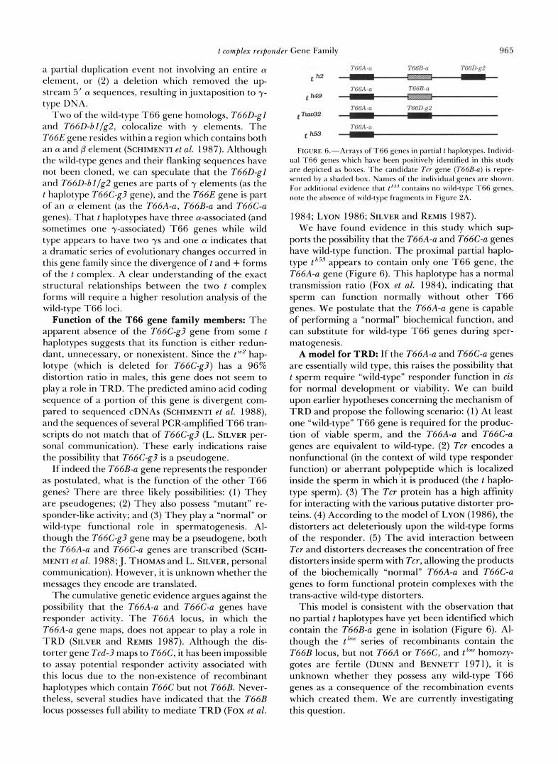
T66.4-a T66B-a T66D-g2
t w 2
th49 1 .. I 7’6‘6/\ -a T66B-a
t-2
t m
7’b‘(j,,l CI T(XD-fi2
7’tj(b\.(1
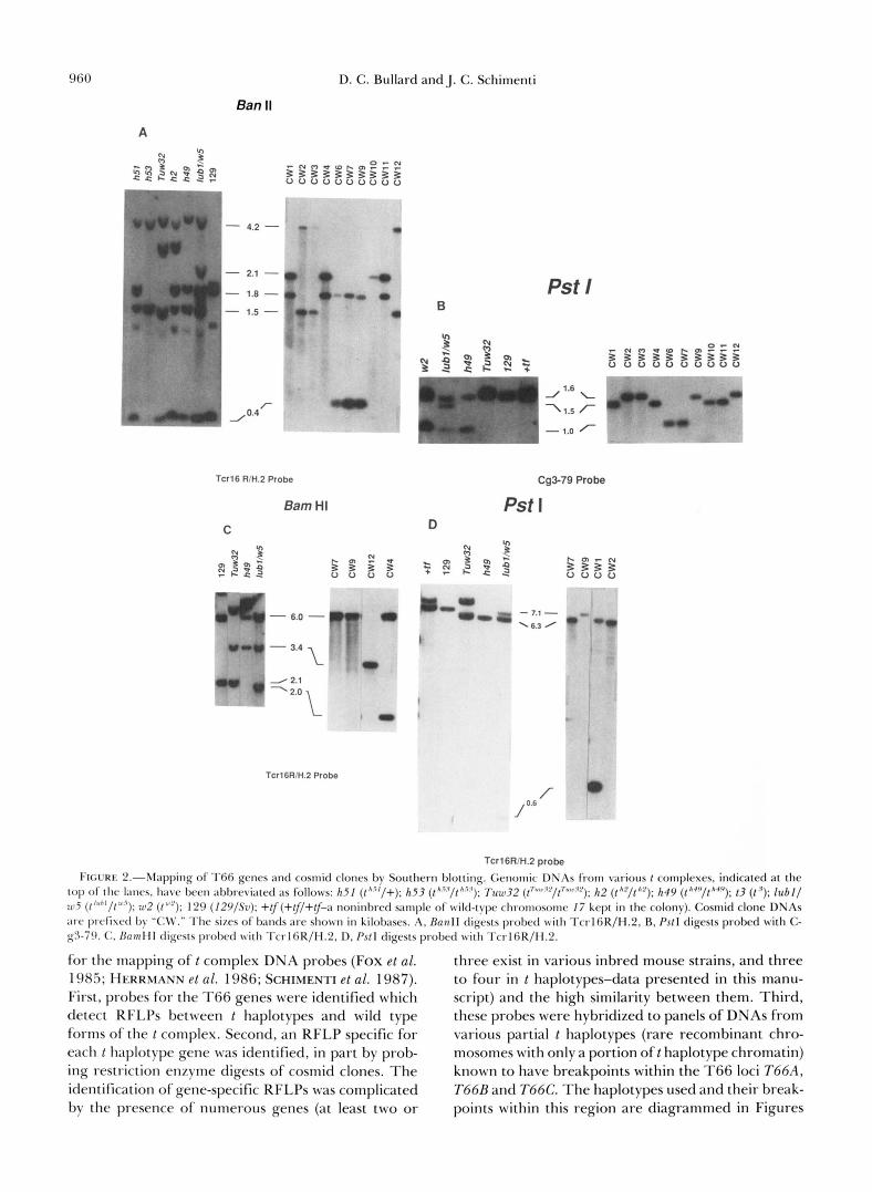
FIGURE 6.-Arravs of T66 genes in partial 1 haplotypes. Individ- ual T66 genes which have been positively identified in this study are depicted as boxes. The candidate Tcr gene (T66R-a) is repre- sented by a shaded box. Names of the individual genes are shown. For additional evidence that t””j contains no wild-type T66 genes, note the absence of wrild-type fragments in Figure 2A.
1984; LYON 1986; SILVER and REMIS 1987). We have found evidence in this study which sup-
ports the possibility that the T66A-a and T66C-a genes have wild-type function. The proximal partial haplo- type th“.? appears to contain only one T66 gene, the T66A-a gene (Figure 6). This haplotype has a normal transmission ratio (Fox et al. 1984), indicating that sperm can function normally without other T66 genes. We postulate that the T66A-a gene is capable of performing a “normal” biochemical function, and can substitute for wild-type T66 genes during sper- matogenesis.
A model for TRD: I f the T66A-a and T66C-a genes are essentially wild type, this raises the possibility that t sperm require “wild-type” responder function in cis for normal development or viability. We can build upon earlier hypotheses concerning the mechanism of TRD and propose the following scenario: (1) At least one “wild-type” T66 gene is required for the produc- tion of viable sperm, and the T66A-a and T66C-a genes are equivalent to wild-type. (2) Tcr encodes a nonfunctional (in the context of wild type responder function) or aberrant polypeptide which is localized inside the sperm in which it is produced (the t haplo- type sperm). (3) The Tcr protein has a high affinity for interacting with the various putative distorter pro- teins. (4) According to the model of LYON (1 986), the distorters act deleteriously upon the wild-type forms of the responder. (5 ) The avid interaction between Tcr and distorters decreases the concentration of free distorters inside sperm with Tcr, allowing the products of the biochemically “normal” T66A-a and T66C-a genes to form functional protein complexes with the trans-active wild-type distorters.
This model is consistent with the observation that no partial t haplotypes have yet been identified which contain the T66B-a gene in isolation (Figure 6). Al- though the t’“” series of recombinants contain the T66B locus, but not T66A or T66C, and t Iw homozy- gotes are fertile (DUNN and BENNETT 1971), it is unknown whether they possess any wild-type T66 genes as a consequence of the recombination events which created them. We are currently investigating this question.

966 D. C. Bullard and J. C. Schimenti
Due to the unique structure and properties of t haplotypes, it has been impossible to generate ideal recombinant chromosomes which would allow us to address the functional roles of the individual T66 genes. It is clear that more modern approaches, such as transgenesis and gene disruption by homologous recombination, will be required to thoroughly under- stand the T66 gene family. Such experiments are under way.
We thank LEE SILVER and colleagues for sharing their prelimi- nary data, and for providing several of the mice used in this study. The Southern blot filter in Figure 3 was prepared by J.S. in the lab of LEE SILVER at Princeton University. We also thank JIM FOREJT for suggesting the presence of a T66 gene in the T66E locus indicated by his R l line mapping. This work was supported by a grant from the U.S. Public Health Service National Institute of Child Health and Human Development (R01 HD24374-02), the March of Dimes Basil O’Connor Starter Scholar Research Award (# 5-699), and the Searle Scholars Program/The Chicago Commu- nity Trust to J.S.
LITERATURE CITED
ARTZT, K . , H . 4 . SHIN and D. BENNETT, 1982 Gene mapping within the T/t complex in the mouse. 11. Anomalous position of the H-2 complex in t haplotypes. Cell 28: 471-476.
BLIN, N., and D. W. STAFFORD, 1976 Isolation of high molecular- weight DNA. Nucleic Acids Res. 3: 2303-2308.
BUCAN, M., B. HERRMANN, A,”. FRISCHAUF, V. BAUTCH, V. BODE, L. M. SILVER, G. MARTIN and H. LEHRACH, 1987 Deletion and duplication of DNA sequences associated with the embry- onic lethal phenotype of the t ‘ complementation group of the mouse t complex. Genes Dev. 1: 376-385.
CHURCH, G. M . , and W. GILBERT, 1984 Genomic sequencing. Proc. Natl. Acad. Sci. USA 81: 1991-1995.
DUNN, I,. C., and D. BENNETT, 1968 A new case of transmission ratio distortion in house mice. Proc. Natl. Acad. Sci. USA 61: 570-573.
DUNN, L. C., and D. BENNETT, 1971 Further studies o fa mutation (Low) which distorts transmission ratios in the house mouse. Genetics 67: 543-558.
DUNN, L. C., and J. SUCKLING, 1956 Studies of the genetic varia- bility in populations of wild house mice. I. Analysis of seven alleles at locus T. Genetics 41: 344-352.
EVANS, G., and G. WAHL, 1987 Cosmid vectors for genomic walking and rapid restriction mapping. Methods Enzymol. 152:
FEINRERG, .4. P., and B. VOGELSTEIN, 1984 A technique for radiolabeling DNA restriction endonuclease restriction frag- ments to high specific activity. Anal. Biochem. 137: 266-267.
Fox, H., G. MARTIN, M. F. LYON, B. HERRMANN, A,”. FRISCHAUF, H. LEHRACH and L. M. SILVER, 1984 Molecular probes define different regions of the mouse t complex. Cell 40: 63-69.
HAMMER, M. H.,J. C. SCHIMENTIand L. M. SILVER, 1989 On the origins o f t complex inversions. Proc. Natl. Acad. Sci. USA 86: 3261-3265.
604-6 10.
HERRMANN, B. G., D. P. BARLOW and H. LEHRACH, 1987 A large inverted duplication allows homologous recombination be- tween chromosomes heterozygous for the proximal t complex inversion. Cell 48: 813-825.
HERRMANN, B., M. BUCAN, P. E. MAINS, A,”. FRISCHAUF, L. M. SILVER and H. LEHRACH, 1986 Genetic analysis of the prox- imal portion of the mouse t complex: evidence for a second inversion within t haplotypes. Cell 44: 469-476.
LYON, M. F., 1984 Transmission ratio distortion in mouse t hap- lotypes is due to multiple distorter genes acting on a responder locus. Cell 37: 621-628.
LYON, M . F., 1986 Male sterility of the mouse t complex is due to homozygosity of the distorter genes. Cell 44: 357-363.
LYON, M . F., and I . MASON, 1964 Information on the nature o f t haplotypes from the interaction of mutant haplotypes in male fertility and segregation ratio. Genet. Res. 50: 255-266.
LYON, M. F., and J. ZENTHON, 1987 Differences in or near the responder region of complete and mouse partial t haplotypes. Genet. Res. 50: 29-34.
MANN, E. A, , L. M. SILVER and R. W. ELLIOT, 1986 Genetic analysis of a t complex locus that is homologous to a kidney cDNA clone. Genetics 114: 993-998.
PLA, M., and H. CONDAMINE, 1984 Recombination between two mouse t haplotypes (t”’”tfand t l U b ’ ) : mapping of the H-2 com- plex relative to the centromere and tufted (tf) locus. Immuno- genetics 17: 445-455.
SARVETNICK, N., H. Fox, E. MANN, P. MAINS, R. ELLIOTT and L. M. SILVER, 1986 Nonhomologous pairing in mice heterozy- gous for a t haplotype can produce recombinant chromosomes with duplications and deletions. Genetics 113: 723-724.
SCHIMENTI, J., and L. SILVER, 1986 Amplification and re- arrangement of DNA sequences during the evolutionary diver- gence of t haplotypes and wild-type forms of mouse chromo- some 17. Curr. Top. Microbiol. Immunol. 127: 247-252.
SCHIMENTI, J., L. VOLD, D. SOCOLOW and L. M. SILVER, 1987 An unstable family of large DNA elements in the center of nlouse t haplotypes. J . Mol. Biol. 194: 583-594.
SCHIMENTI, J., J. CERRA-THOMAS, C. L. DECKER, S. D. ISLAM, S. H. PILDER and L. M. SILVER, 1988 A candidate gene family for the mouse t complex responder (Tcr) locus responsible for haploid effects on sperm function. Cell 55: 71-78.
SILVER, L. M., 1981 A structural gene (Tcp-1) within the mouse t complex is separable from effects on tail length and lethality but may be associated with effects on spermatogenesis. Genet. Res. 38: 1 15-1 23.
SILVER, L. M., 1985 Mouse t haplotypes. Annu. Rev. Genet. 1 9 179-208.
SILVER, L. M., and D. REMIS, 1987 Five of the nine genetically defined regions of mouse t haplotypes are involved in trans- mission ratio distortion. Genet. Res. 49: 51-56.
SILVER, L. M., M. WHITE and K. ARTZT. 1980 Evidence for unequal crossing over with the mouse T/ t complex. Proc. Nat. Acad. Sci. USA. 77: 6077-6080.
SILVER, L. M., M. HAMMER, H. Fox, J. GARRELS, M. BUCAN, B. HERRMANN, A,”. FRISCHAUF, H. LEHRACH, H. WINKING, F. FIGUEROA, and J. KLEIN 1984. Molecular evidence for the rapid propagation of mouse t haplotypes from a single, recent, ancestral chromosome. Mol. Biol. Evol. 4: 473-482.
Communicating editor: R. E. GANSCHOW