Embed Size (px)
Citation preview

HAL Id: hal-00531921https://hal.archives-ouvertes.fr/hal-00531921
Submitted on 4 Nov 2010
HAL is a multi-disciplinary open accessarchive for the deposit and dissemination of sci-entific research documents, whether they are pub-lished or not. The documents may come fromteaching and research institutions in France orabroad, or from public or private research centers.
L’archive ouverte pluridisciplinaire HAL, estdestinée au dépôt et à la diffusion de documentsscientifiques de niveau recherche, publiés ou non,émanant des établissements d’enseignement et derecherche français ou étrangers, des laboratoirespublics ou privés.
Molecular characterization of two sea bass gonadotropinreceptors: cDNA cloning, expression analysis, and
functional activityAna Rocha, Ana Gómez, Silvia Zanuy, José Miguel Cerdá-Reverter, Manuel
Carrillo
To cite this version:Ana Rocha, Ana Gómez, Silvia Zanuy, José Miguel Cerdá-Reverter, Manuel Carrillo. Molecu-lar characterization of two sea bass gonadotropin receptors: cDNA cloning, expression analysis,and functional activity. Molecular and Cellular Endocrinology, Elsevier, 2007, 272 (1-2), pp.63.�10.1016/j.mce.2007.04.007�. �hal-00531921�

Accepted Manuscript
Title: Molecular characterization of two sea bass gonadotropinreceptors: cDNA cloning, expression analysis, and functionalactivity
Authors: Ana Rocha, Ana Gomez, Silvia Zanuy, Jose MiguelCerda-Reverter, Manuel Carrillo
PII: S0303-7207(07)00160-8DOI: doi:10.1016/j.mce.2007.04.007Reference: MCE 6645
To appear in: Molecular and Cellular Endocrinology
Received date: 1-8-2006Revised date: 19-4-2007Accepted date: 21-4-2007
Please cite this article as: Rocha, A., Gomez, A., Zanuy, S., Cerda-Reverter, J.M.,Carrillo, M., Molecular characterization of two sea bass gonadotropin receptors:cDNA cloning, expression analysis, and functional activity, Molecular and CellularEndocrinology (2007), doi:10.1016/j.mce.2007.04.007
This is a PDF file of an unedited manuscript that has been accepted for publication.As a service to our customers we are providing this early version of the manuscript.The manuscript will undergo copyediting, typesetting, and review of the resulting proofbefore it is published in its final form. Please note that during the production processerrors may be discovered which could affect the content, and all legal disclaimers thatapply to the journal pertain.
Acce
pted
Man
uscr
ipt
1
Molecular characterization of two sea bass gonadotropin
receptors: cDNA cloning, expression analysis, and functional
activity.
Ana Rocha, Ana Gómez*, Silvia Zanuy, José Miguel Cerdá-Reverter and Manuel Carrillo Department of Fish Physiology and Biotechnology, Instituto de Acuicultura de Torre la Sal, Consejo Superior de Investigaciones Cientificas (CSIC), 12595 Torre la Sal, Ribera de Cabanes, Castellón, Spain. *Address for correspondence: Ana Gómez
Instituto de Acuicultura de Torre la Sal. Ribera de Cabanes s/n. 12595 Torre la Sal. Castellón. Spain. Phone: +34 964 319500. Fax: +34 964 319509 e-mail: [email protected]
* Manuscript
Page 1 of 43

Acce
pted
Man
uscr
ipt
2
Key words: Follicle-stimulating hormone; luteinizing hormone; G-protein coupled receptor; gonadal development; reproduction; teleost fish
Abstract
The follicle-stimulating hormone (FSH) and the luteinizing hormone (LH) play central
roles in vertebrate reproduction. They act through their cognate receptors to stimulate
testicular and ovarian functions. The present study reports the cloning and characterization of
two sea bass (Dicentrarchus labrax) cDNAs encoding a FSH receptor (sbsFSHR) and a LH
receptor (sbsLHR). The mature proteins display typical features of the glycoprotein hormone
receptor family members, but the sbsFSHR also contains some remarkable differences when
compared with other fish or mammalian FSHRs. Among them, a distinct extracellular N-
terminal cysteine domain as regards to its length and cysteine number, and the presence of an
extra leucine-rich repeat. Expression analysis revealed that the sbsFSHR is exclusively
expressed in gonadal tissues, specifically in the follicular wall of previtelogenic and early-
vitelogenic follicles. On the contrary, sbsLHR mRNA was found to be widely distributed in
sea bass somatic tissues. When stably expressed in mammalian cell lines, sbsFSHR was
specifically stimulated by bovine FSH, while sbsLHR was activated by both bovine LH and
FSH. Nevertheless, specific stimulation of the sbsLHR was observed when recombinant sea
bass gonadotropins were used. The isolation of a FSHR and a LHR in sea bass opens new
ways to study gonadotropin action in this species.
Page 2 of 43

Acce
pted
Man
uscr
ipt
3
Introduction
It is well established that the follicle-stimulating hormone (FSH) and the luteinizing
hormone (LH), secreted by the pituitary gland, play central roles in vertebrate reproduction
(Chappel and Howles, 1991). In ovarian follicles, FSH regulates granulosa cell proliferation,
the synthesis of cell cycle-regulatory proteins and induces the expression of differentiation-
specific genes (Richards, 1994). On the other hand LH promotes follicular maturation,
ovulation and the synthesis of ovarian steroid hormones. In the testes, FSH determines Sertoli
cell proliferation and attends germ cell maturation (Allan and Handelsman, 2005), while LH
supports Leydig cell functions and stimulates the synthesis of androgens, regulating the final
stages of spermatogenesis. FSH and LH are members of the glycoprotein hormone family,
which also includes the thyroid stimulating hormone (TSH) and the chorionic gonadotropin
(CG) (Pierce and Parsons, 1981).
The glycoprotein hormones exert their biological actions by interacting with specific
receptors present on target cell surfaces. Accordingly, the FSH receptor (FSHR) gene is
expressed only in granulosa cells in the ovary and in Sertoli cells in the testis. The LH
receptor (LHR) is expressed primarily in theca and granulosa cells of ovarian preovulatory
follicles and in the Leydig cells in the testes (reviewed in refs. Themmen and Huhtaniemi,
2000; Ascoli et al., 2002; Vassart et al., 2004). In primates and equines, it also binds the CG,
a placental hormone essential for the maintenance of pregnancy, structurally similar to LH
(Bousfield et al., 1994). These receptors are encoded by paralogous genes belonging to the
large family of G protein-coupled receptors (GPCRs) (Simoni et al., 1997; Ascoli et al., 2002;
Dias et al., 2002; Szkudlinski et al., 2002), which includes in addition the TSH receptor that
binds TSH. They constitute the subfamily of glycoprotein hormone receptors (GpHRs),
themselves members of the wider leucine-rich repeat containing GPCR (LGR) family. The
members of this family are characterized by a large extracellular (EC) domain with multiple
Page 3 of 43

Acce
pted
Man
uscr
ipt
4
imperfect leucine-rich repeats (LRRs), flanked by N- and C-terminal cysteine-rich
subdomains. This is followed by a rhodopsin-like domain of seven transmembrane (7TM)
helices and a C-terminal intracellular tail (Hsu et al., 2000).
Evidence for the presence of gonadotropin receptors in fish was first demonstrated
more than ten years ago, by binding studies of coho salmon FSH and LH to membranes of
isolated granulosa cells and theca-interstitial layers of coho salmon ovary (Yan et al., 1992;
Miwa et al., 1994). The isolation of the cDNAs from these receptors remained elusive for
years. Finally, the cloning of two types of gonadotropin receptors from the ovaries of amago
salmon was reported (Oba et al., 1999a; Oba et al., 1999b), thereby providing new
experimental tools to understand fish GpHRs biology.
Ligand binding and receptor activation appear to be distinct activities of the GpHRs.
Ligand binding specificity and affinity are determined by residues present in the β-strands of
the LRRs (Braun et al., 1991; Smits et al., 2003), whereas the TM domain is responsible for
receptor activation and signal transduction through G proteins. Hormone binding to the EC
domain of a GpHR causes changes in the 7TM domain promoting cAMP accumulation via
activation of heterotrimeric Gs, thereby initiating a signalling cascade that leads to steroid
synthesis (Ascoli et al., 2002; Dias et al., 2002; Szkudlinski et al., 2002).
Ligand specificity of the mammalian GpHRs is well defined, with no cross-stimulation
occurring under physiological conditions. Conversely, promiscuous activation of fish GpHRs
has been described. In vitro experiments with African catfish, channel catfish and zebrafish
recombinant FSHRs suggest that these are not selective for FSH, as LH can also activate
them, whereas, LHRs are highly selective for homologous LH (Bogerd et al., 2001; Kumar et
al., 2001a; Kumar et al., 2001b; Vischer and Bogerd, 2003; Kwok et al., 2005). These data are
consistent with results obtained in coho salmon, whereby type I receptor (i.e., the putative
FSHR), localized in both the theca and granulosa cells, did not discriminate between FSH and
Page 4 of 43

Acce
pted
Man
uscr
ipt
5
LH, while a type II receptor i.e., the putative LHR, located only in granulosa cells, was highly
selective for LH (Yan et al., 1992; Miwa et al., 1994). However, the ligand selectivity of the
amago salmon receptors appeared to be different from this (Oba et al., 1999a; Oba et al.,
1999b). Heterologous cells transiently transfected with the amago salmon FSHR were
specifically activated by chum salmon FSH but not by LH, whereas LHR transfected cells
were highly responsive to LH and in a lesser extend to FSH.
This different ligand selectivity could imply that GpHR actions in teleosts are not
totally overlapping those of their mammalian counterparts, and reinforces the need of learning
more from fish, the most diverse group of vertebrates with dissimilar modes of gonadal
development and reproductive strategies. Although the cloning of a FSHR and a LHR has
been reported in two species of Percirforms, Nile tilapia and seabream (Oba et al., 2001;
Wong et al., 2004), there is no information regarding the functional characterization of these
receptors in this linage of fish.
To bring more knowledge on fish gonadotropin receptors, in the present study we
report the isolation and functional characterization of the cDNAs coding for a sea bass FSHR
(sbsFSHR) and a LHR (sbsLHR).
Materials and Methods
Isolation of Nucleic Acids
Sexually mature female and male sea bass (Dicentrarchus labrax) were obtained from
the stock raised in the Instituto de Acuicultura de Torre la Sal facilities. Animals were treated
in accordance with the European Union Animal Care Regulations.
Total RNA was isolated from sea bass tissues (n = 2 animals/ tissue) following the
TRI Reagent protocol (Molecular Research Center, Inc. Cincinnati, OH). Poly(A)+ RNA was
isolated using PolyATtract mRNA Isolation Systems (Promega Corp., Southampton, UK).
Page 5 of 43

Acce
pted
Man
uscr
ipt
6
Genomic DNA (gDNA) was isolated from sea bass whole blood, using a protein salting-out
method (Martínez et al., 1998).
Reverse transcription-Polymerase Chain Reaction (RT-PCR) and Polymerase Chain
Reaction
To perform PCR amplification on sea bass gDNA 150 ng were used as template.
Superscript II reverse transcriptase (Invitrogen Corp., Carlsbad, CA) was used for
cDNA synthesis by priming 4 µg of RQ1-DNase (Promega Corp.) treated total RNA with 100
ng of random hexamers in a total volume of 20 µl. Briefly, RNA was mixed with 100 ng of
random hexamers, 1 µl of dNTPs (10 mM each dNTP) and denatured at 65°C for 5 min. After
a quick chilling on ice, 40 units of RNAsin (Promega Corp.), and 200 units of SuperScript II
reverse transcriptase were added. Incubation took place at 42ºC for 50 min. The reaction was
stopped by heating to 70ºC for 15 min.
One to 2.5 µl of the cDNA were used for PCR reactions. The sequences of the primers
used in this study are shown in Table 1. Touchdown PCR programs comprising a 10 to 15ºC
span of annealing temperatures were used following these conditions: an initial denaturation
at 94ºC for 2 min followed by 30 (15ºC span) or 20 (10ºC span) cycles of 94ºC for 30 s, the
highest annealing temperature for 30 s, and 72ºC for 45 s. The annealing temperature was
decreased 0.5ºC per cycle.
Primer pairs fshr25-fshr26 and lhr35-lhr23 (Table 1) were designed to amplify an
intron-containing region in order to exclude false positive bands arising from potential
contaminating gDNA. Also, they share limited sequence identity. As internal control a 495 bp
fragment of the sea bass 18S ribosomal RNA (rRNA) gene was amplified, using primers
5’18S and 3’18S . Cycling conditions consisted of 25 cycles at 94ºC for 30 s, 65ºC for 45 s,
and 72ºC for 45 s.
Page 6 of 43

Acce
pted
Man
uscr
ipt
7
The resulting PCR products were cloned into the pGEM-T Easy Vector (Promega
Corp.) and sequenced.
Screening of a sea bass cDNA testicular library
A 1175 bp cDNA fragment from the sbsFSHR and a 678 bp cDNA fragment of the
sbsLHR were labelled with [α-32P]-dCTP by random octamer priming (Rad Prime DNA
Labelling System Kit, Invitrogen Corp.) and used as probes to screen, following standard
methods (Sambrook & Russel 2001), a sea bass testicular directional cDNA library
constructed in the UNI-ZAP XR vector.
Prehybridizations and hybridizations were done at high stringency conditions (50%
formamide, 42ºC). Final washes were carried out at 55ºC with 1xSSPE and 0.5% SDS.
Several positive clones were obtained after first round screening of approximately 1x10⁶
phages. Secondary and tertiary screenings were performed and the resulting positive phages
were in vivo excised from the Lambda ZAP XR vector as pBluescript SK (-) phagemids,
following the protocol described by the manufacturer (Stratagene Corp.).
DNA Sequence and Phylogenetic analysis
DNA sequences were determined for both strands on an automated ABI PRISM 3730
DNA Analyser (Applied Byosistems) using the Rhodamine Terminator Cycle Sequencing Kit
(Perkin-Elmer Inc., Wellesley, MS). The generated sequences were assembled using
Sequencher version 4.0.5 software (Gene Codes Corpor., Ann Arbor, MI). BLASTN and
BLASTP (version 2.2.9, National Center for Biotechnology Information) were used for
database searching. The presence and location of the putative signal peptide cleavage site, the
7TM helices, Ser, Thr, and Tyr phosphorylation sites and potential N-glycolylation sites in the
amino acid sequences, were predicted using the prediction servers of the Center for Biological
Page 7 of 43

Acce
pted
Man
uscr
ipt
8
Sequence Analysis (http://www.cbs.dtu.dk/services/). The Protein Families (Pfam) Database
of the Sanger Institute (UK) was used to search for common protein domains. The UniProt
name for the sbsFSHR and the sbsLHR are Q4L192_DICLA and Q4L191_DICLA
respectively. Multiple sequence alignments were carried out using ClustalX version 1.81
(Thompson et al., 1997). Phylogenetic analyses, of full length amino acid sequences, were
conducted using MEGA version 2.0 (Kumar et al., 2001). A rooted phylogenetic tree was
constructed by means of the Neighbor-joining algorithm, using the LGR sequence of
Caenorhabditis elegans as an outgroup. Gaps or missing data were pairwise deleted. One
thousand bootstrapping pseudo replicates were used to assess the robustness of the inferred
nodes of the tree.
Northern blot analysis
Poly (A)+ RNA (10 µg) from sea bass ovaries and total RNA (30 µg) from testes and
ovaries were denatured and electrophoresed in the presence of formaldehyde using standard
conditions (Sambrook and Russel, 2001). RNAs were blotted on a Hybond-NX nylon
membrane (Amersham Biosciences UK Ltd.) and stained with methylene blue to control for
equal loading. Prehybridization and hybridization were carried out overnight at 42ºC with
50% formamide. The full length sbsFSHR and sbsLHR cDNAs were labelled with [α-32P]-
dCTP and used as probes. Final washes were performed in 2xSSC /0.5% SDS at 50 ºC. The
membranes were exposed to Hyperfilm TM MP autoradiography films (Amersham
Biosciences UK Ltd.).
In situ hybridization
In situ hybridization experiments were carried out as described previously (Cerda-
Reverter et al., 2003). Briefly, sea bass gonads were fixed with 4% paraformaldehyde (PAF)
Page 8 of 43

Acce
pted
Man
uscr
ipt
9
overnight at 4ºC, dehydrated, and embedded in Paraplast (Sherwood, St. Louis, MO). Before
hybridization, 6µm sections were deparaffinized, rehydrated, and postfixed. Slides were later
washed in phosphate buffer (PB) and treated with Proteinase-K. Next, they were washed in
PB and post fixed again in PAF, subsequently rinsed in sterile water, and acetylated in a
triethanolamine (0.1 M, pH 8)/acetic anhydride solution. Sections were then dehydrated and
dried at room temperature.
A pBluescript SK (-) phagemid containing an 800 bp fragment from the sbsFSHR 3’
UTR was used to prepare riboprobes. Antisense and sense RNA probes were synthesized in
vitro by linearizing the phagemid with XbaI or XhoI, and in vitro transcription was carried out
with T7 or T3 RNA polymerase, respectively. The probes were labelled with 35S-UTP using
the Riboprobe In Vitro Transcription System Kit (Promega Corp.). After in vitro RNA
synthesis, samples were treated with 1U of RQ1-DNase RNAse-free (Promega Corp.) and
purified on Sephadex G50 columns. The two fractions containing the highest radioactivity
were pooled, precipitated, stored at -20 ºC and used within 1 week.
The 35S-UTP riboprobes were pelleted and dissolved in an appropriate volume of 100
mM DTT to obtain 2x10⁵ cpm/µ l. After 5 minutes incubation at 80ºC, 35S-UTP riboprobes
were diluted 1/10 (final concentration of probes, 10 mM DTT and 2x10⁴ cpm/µl) in
hybridization buffer containing 50% formamide, 300 mM NaCl, 20 mM Tris-HCl (pH 8), 5
mM EDTA (pH 8), 10% Dextan sulphate, 1X Denhardt s solution and 0.5 µg/ml of yeast
RNA type III. Subsequently, 60 µl of hybridization solution were added to each pre-treated
slides (see above) that were cover-slipped and incubated in a humidified chamber overnight at
55ºC.
After washing and ribonuclease treatment, slides were finally dehydrated and dried at
room temperature. Finished the hybridization process, slides were dipped in Hypercoat
Photographic Emulsion RPN40 (Amersham Biosciences UK Ltd) and exposed under dry
Page 9 of 43

Acce
pted
Man
uscr
ipt
10
conditions at 4ºC for 15–20 days, developed in Kodak D-19 Film Developer and
counterstained with Cleveland & Wolfe staining (Herlaut 1960). Anatomical locations were
confirmed by reference to Mayer et al. (1990) and Alvariño et al. (1992).
Expression plasmids, cell culture and transfections
The pcDNA-sbsFSHR plasmid was generated by inserting a 2.8 kb fragment
containing the complete open reading frame (ORF) of the sbsFSHR into the pcDNA3
expression vector (Invitrogen Corp.). The expression vector pcDNA-sbsLHR, containing the
complete ORF of the sbsLHR was generated in several steps. First, two fragments
corresponding to the EC domain of sbsLHR were PCR amplified with primer pairs lhr30-
lhr24 and lhr35-lhr21 respectively (Table 1). Equal quantities of each fragment were used as
template in an overlapping PCR reaction with primers lhr30-lhr21 (Table 1). The resulting
PCR product was inserted into the pPCR-Script Amp SK (+) cloning vector (PCR-Script®
Amp Cloning Kit, Stratagene, La Jolla, CA). A HindIII-VspI fragment from this plasmid
containing most of the EC domain (nucleotides 1 to 1048) was in-frame ligated to a VspI-
EcoRV fragment (nucleotides 1048 to 3172) obtained from a cDNA library phagemid, which
corresponds to the rest of the sbsLHR cDNA. Finally, they were introduced into the HindIII-
EcoRV-digested pcDNA3 vector (Invitrogen Corp.). All the above PCR reactions were
performed with the proofreading PfuTurbo® DNA polymerase (Stratagene, La Jolla, CA) and
further checked by sequencing. pCRE-luc (BD Clontech, Palo Alto, CA) contains the firefly
luciferase gene under the control of a promoter with cAMP Responsive Elements (CRE)
binding sites. The tgCMV/HyTK plasmid harbours a hygromycin resistance gene (Wellbrock
et al., 1998).
Human embryonic kidney (HEK) 293 cells were grown in DMEM (Invitrogen Corp)
containing 10% foetal bovine serum, penicillin (100 units/ml) and streptomycin (100 µg/ml)
Page 10 of 43

Acce
pted
Man
uscr
ipt
11
in a humidified atmosphere of 5% CO2 at 37ºC. Cells were transfected using a modified
calcium phosphate transfection method (Chen and Okayama, 1987) or Lipofectamine 2000
reagent according to manufacturer’s guidelines. pcDNA-sbsFSHR and tgCMV/HyTK were
co-transfected (50:1) in cells growing in 24-well plates. pcDNA-sbsLHR, pCRE-luc and
tgCMV/HyTK were co-transfected (50:50:1) in cells growing in 6 well culture plates. To
generate stable clones, 48 hours after transfection cells were replated in 96-well plates and
selected in medium containing 400 µg/ml of hygromycin B (Invitrogen). Hygromycin
resistant colonies were isolated 2 weeks after transfection and subsequently expanded. In the
sbsFSHR stable clones, the expression level of this gene was analyzed by Northern blot.
Transient transfections of the pCRE-luc plasmid were performed in 9 cm dishes. Forty eight
hours after transfection, cells were spread in 24-well plates and stimulated by adding the
specified amounts of hormones in the growth medium for the indicated times.
The sbsLHR single clones were screened for luciferase activity by treating the cells
with 100 µM forskolin (Sigma-Aldrich Co.) for five hours. Untreated cells were used as
controls. The clone presenting the highest difference in luciferase activity between treated and
untreated cells (LHR-LUC10) was further expanded. For functional characterization of the
sbsLHR, LHR-LUC10 cells were spread in 48 well plates and stimulated by adding the
specified amounts of hormones on the growth medium for five hours.
Bovine FSH (bFSH, AFP-5332B, biopotency 68 x NIH-FSH-S1) and bovine LH
(bLH, AFP-11743B, biopotency 2,3 x NIH-LH-S1) were obtained from the National
Hormone & Peptide Program, Harbor-UCLA Medical Center (California, USA). Cells were
also challenged with conditioned medium of cultured Chinese hamster ovary (CHO-K1)
stable clones producing recombinant sea bass LH and FSH (Gómez et al., unpublished).
Page 11 of 43

Acce
pted
Man
uscr
ipt
12
Luciferase assay
To measure luciferase activity, cells were lysed in Reporter Lysis Buffer (Promega
Corp.) as indicated by the manufacturer. Cell debris were separated by centrifugation for 30 s
at 15000xg, and 20 µl of the supernatant were mixed with 200 µl of luciferin reagent (20 mM
TricineKOH, pH 7.8, 0.1 mM EDTA, 8 mM MgCl2, 33.3 mM DTT, 270 µM CoA, 530 µM
ATP, 400 µM luciferin). The light emitted was measured in a luminometer (Junior, EG&G,
Berthold) and expressed as relative light units (RLU).
Results
Cloning and sequence analysis of a sbsFSHR and a sbsLHR cDNAs
Two pairs of degenerate oligonucleotides (fshr6-fshr2 and fshr1-fshr2) were designed
based on published fish and mammalian FSHR and LHR sequences respectively, and
corresponding to a highly conserved and intronless region of the TM domain. They were used
as primers in a PCR reaction using sea bass gDNA as template. A 330 bp and a 680 bp
products were amplified, and subsequently subcloned and sequenced. The obtained sequences
displayed the highest identity to other FSHRs and LHRs respectively. A sbsFSHR specific
primer (fshr9), based on the sequence of the amplified product, was used in a PCR reaction on
testis cDNA in combination with a degenerated primer designed on the EC domain (fshr8).
The amplified fragment was about 1.1 Kb. Screenings of a cDNA testicular library using the
sbsFHSR 1.1 Kb and the sbsLHR 680 bp fragments as probes rendered a high number of
positive clones for the sbsFSHR and a single specific clone for the sbsLHR.
The sbsFSHR sequence. PCR and sequencing analysis of the 5’ region of the sbsFSHR
clones showed that eight of them were full length and contained the ATG initiation codon.
One of them was chosen for in vivo excision and sequencing. This phagemid contained a
3134 bp fragment (Fig. 1), consisting of an ORF of 2109 nucleotides that was flanked by
Page 12 of 43

Acce
pted
Man
uscr
ipt
13
leader and trailer sequences of 206 and 819 nucleotides respectively. This ORF encodes a
protein of 702 amino acids of which the first 20 amino acids were predicted to constitute the
putative signal peptide (Fig. 1). The mature protein displayed typical features of members of
the GpHR family. The predicted EC domain contains 377 amino acids, including the signal
peptide, followed by a putative 7TM domain and an intracellular C-terminal domain.
The sbsFSHR protein showed the highest identity with FSHRs of other fish species
(81%-53%), followed by mammalian FSHRs (46%-45%), chicken FSHR (45%), mammalian
LHRs (44%-43%), chicken LHR (41%), fish LHRs (41%-38%), reptile FSHRs (47%-46%)
and fish and mammalian TSHRs (39%-35%). Amino acid sequence alignment of the
sbsFSHR with other glycoprotein receptors, made clear that the most conserved regions of the
receptor are the TM domain and the intracellular loop between TM I and TM II, whereas the
least conserved areas are the EC domain (Fig. 2) and the intracellular loop between TM V and
TM VI. The ClustalX alignment also revealed the presence of specific signature sequences
(e.g. 316CCAF, 364FNPCED, 477ERW, 590FTD, 634NPFLY), highly conserved in GpHRs
(Vassart et al., 2004) (Fig. 2).
When aligning the amino acid sequence of the sbsFSHR with other fish and human
FSHR sequences, two insertions and one deletion were identified in the EC domain of the
sbsFSHR (Fig. 2). The first insertion, of 9 amino acids, is placed just after the signal peptide.
The second one, considerable larger (25 amino acids), begins in amino acid position 80.
Finally, a 30 amino acid deletion, with respect to the human FSHR (hFSHR), is easily
identified in the C-terminal region of the EC domain, in the middle of the cysteine-rich
cluster. This deletion is present in other fish FSHRs.
Predictions for N-glycosylation sites identified five motifs in the sbsFSHR at positions
62NTT, 232NGT, 309NLT, 325NRS and 351NST (Fig. 1). In other fish species, these potential
Page 13 of 43
Acce
pted
Man
uscr
ipt
14
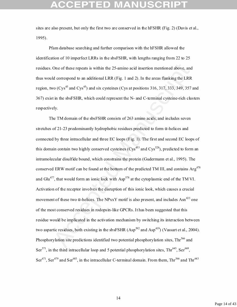
sites are also present, but only the first two are conserved in the hFSHR (Fig. 2) (Davis et al.,
1995).
Pfam database searching and further comparison with the hFSHR allowed the
identification of 10 imperfect LRRs in the sbsFSHR, with lengths ranging from 22 to 25
residues. One of these repeats is within the 25-amino acid insertion mentioned above, and
thus would correspond to an additional LRR (Fig. 1 and 2). In the areas flanking the LRR
region, two (Cys45 and Cys65) and six cysteines (Cys at positions 316, 317, 333, 349, 357 and
367) exist in the sbsFSHR, which could represent the N- and C-terminal cysteine-rich clusters
respectively.
The TM domain of the sbsFSHR consists of 263 amino acids, and includes seven
stretches of 21-23 predominantly hydrophobic residues predicted to form α-helices and
connected by three intracellular and three EC loops (Fig. 1). The first and second EC loops of
this domain contain two highly conserved cysteines (Cys453 and Cys528), predicted to form an
intramolecular disulfide bound, which constrains the protein (Gudermann et al., 1995). The
conserved ERW motif can be found at the bottom of the predicted TM III, and contains Arg478
and Glu477, that would form an ionic lock with Asp578 at the cytoplasmic end of the TM VI.
Activation of the receptor involves the disruption of this ionic look, which causes a crucial
movement of these two α-helices. The NPxxY motif is also present, and includes Asn633 one
of the most conserved residues in rodopsin-like GPCRs. It has been suggested that this
residue would be implicated in the activation mechanism by switching its interaction between
two aspartic residues, both existing in the sbsFSHR (Asp592 and Asp419) (Vassart et al., 2004).
Phosphorylation site predictions identified two potential phosphorylation sites, Thr566 and
Ser571, in the third intracellular loop and 5 potential phosphorylation sites, Thr643, Ser669,
Ser671, Ser679 and Ser683, in the intracellular C-terminal domain. From them, Thr566 and Thr643
Page 14 of 43

Acce
pted
Man
uscr
ipt
15
are potencial phosphorylation sites for protein kinase C, while Ser571 and Ser683 are potential
sites for protein kinase A phosphorylation.
The sbsLHR sequence. The single isolated clone for the sbsLHR contained a cDNA
insert of 2228 bp. Sequence analysis revealed that this clone did not contain the full length
sbsLHR cDNA, as it was lacking coding sequence at its 5’ end. Based on information
obtained from the genomic sequence of the sbsLHR (unpublished data), a specific primer
(lhr30) annealing to the 5’ UTR of sbsLHR gene was designed. It was used for PCR
amplification of testis cDNA in order to obtain the remaining sbsLHR cDNA sequence. The
combination of this PCR product plus the cDNA sequence obtained from the library yield a
sbsLHR cDNA sequence of 3172 bp. It consists of an ORF of 2166 bp that codes for a 721
amino acid polypeptide, flanked by 5’ UTR and 3’ UTR of 158 bp and 848 bp, respectively.
The first 21 amino acids were predicted to constitute the putative signal peptide. The complete
nucleotide and deduced amino acid sequence are shown in Fig. 3. sbsLHR contains all the
features of a GpHR. The large EC amino-terminal domain consists of 388 amino acids,
including the signal peptide. Searches in the Pfam database and alignments with other GpHRs
allowed us to identify, within this domain, nine imperfect LRRs, with sizes raging from 21 to
25 amino acids each. These LRRs are flanked by ten conserved cysteines, four of them in an
N-terminal cluster (Cys at positions 27, 31, 33 and 40) and six in a C-terminal group (Cys at
positions 283, 284, 308, 360, 368 and 378). The predicted TM domain, of 261 amino acids,
includes 7 stretches of hydrophobic residues. The intracellular C-terminal domain consists of
72 amino acids and has two highly conserved contiguous cysteines (Cys668 and Cys669). Three
N-glycosylation motifs were identified in the sbsLHR EC domain at positions 42NVT, 103NLS,
and 199NGT (Fig. 3). Only two of them, the second and third, are conserved between
mammalian and sea bass LHRs. (Vu-Hai et al., 2000). NetPhosK 1.0 prediction server found
one site, Thr590, in the third intracellular loop of the TM, and six sites, Thr652, Ser671, Ser674,
Page 15 of 43

Acce
pted
Man
uscr
ipt
16
Ser683, Ser693 and Ser711 in the intracellular C-terminal domain of the sbsLHR, predicted to be
phosphorylated by protein kinase C.
The sbsLHR protein has the highest identity to LHRs of other fish species (86%-
45.5%), followed by chicken LHR (51%), mammalian LHRs (49%-48%), mammalian FSHRs
(46%-45%), reptile FSHRs (44.5%-38%), fish FSHRs (43.5%-37%) and mammalian and fish
TSHRs (41%-37%). Sea bass FSHR and LHR EC domains are 28% identical, while their TM
and intracellular domains are 64% identical. The overall amino acid identity between these
two sea bass proteins is 41%. Alignment of the sbsLHR amino acid sequence with other
LHRs, showed that the most divergent areas are the C-terminus of the EC domain and the
intracellular domain (data not shown). Similarly to the sbsFSHR, highly conserved GpHR
signature sequences (e.g. 375FNPCEDIMSA, 488ERW, 601FTD, 644NPFLY) (Vassart et al.,
2004) were also found in the sbsLHR.
Phylogenetic analysis
The evolutionary relationship of the sbsFSHR and sbsLHR to other members of the
GpHR family was inferred by performing a phylogenetic analysis by the Neighbour-joining
method. The topology of the resulting rooted tree (Fig. 4) shows three main groups: LHR,
FSHR and TSHR lineages. The LHRs are separated from the other GpHRs in 73% of the
replicates while the FSHR and TSHR clades presented a bootstrap value of 78. As predicted
by fish evolutionary relationships (Nelson, 1994), sbsFSHR and sbsLHR sequences exhibit
the closest relation with the seabream gonadotropin receptors.
Expression analysis
The tissue expression pattern of the sbsFSHR and sbsLHR was analyzed by RT-PCR
(Fig. 5A, B). sbsFSHR mRNA was only detected in gonads, and higher levels were seen in
Page 16 of 43

Acce
pted
Man
uscr
ipt
17
testis compared to the ones observed in ovary. No transcripts were amplified in the somatic
tissues analyzed (Fig. 5A). On the other hand, the sbsLHR showed the highest expression in
gonadal tissues, but lower expression levels were detected in different somatic tissues, from
which the head and ventral kidneys as well as the spleen presented the strongest amplification
signal (Fig. 5B). Sequencing results from two randomly selected RT-PCR products (liver and
gills) confirmed the authenticity of these extragonadal amplicons (Fig. 5B).
Northern blot analysis of poly (A)+ RNA and total RNA from sea bass gonads, using the full
length sbsFSHR cDNA as a probe, revealed two faint hybridization signals corresponding to
two different transcripts. One of approximately 3 kb, which corresponds to the full length
mRNA and another of 1.8 kb that would be consistent with the existence of an alternatively
spliced sbsFSHR transcript (Fig. 5C). When the sbsLHR cDNA was used as a probe, a faint
and diffuse hybridization signal, of approximately 3 kb, was detected; this could contain
sbsLHR transcripts slightly different in size (Fig. 5C). No signal could be detected in Northern
blot when total RNA was used (data not shown). The level of expression obtain for both
receptors in the Northern blot analysis, indicates that the sbsLHR is expressed to a much
lower level than the sbsFSHR.
To establish the cellular localisation and maturation stage dependent expression of the
sbsFSHR, we carried out an in situ hybridization on ovary sections of mature sea bass female.
When an antisense probe was used, a strong expression was observed in previtelogenetic
oocytes (Fig. 6A). Positive signals were also detected in the follicular cells of oocytes in early
and late stages of the vitelogenesis process (Fig. 6B, C, D). Little or no expression was found
in mature oocytes. No specific signal was generated with a sbsFSHR-cRNA sense probe (data
not shown).
Page 17 of 43

Acce
pted
Man
uscr
ipt
18
Activation of the sbsFSHR and sbsLHR by gonadotropins
To test the functionality of the isolated sbsFSHR and sbsLHR we expressed their
cDNAs in HEK293 cells. It has been described (Tao et al., 2000) that the expression of a high
number of gonadotropin receptors per cell can result in an increase in the basal level of
activation of the receptor. With the aim of obtaining low amounts of receptor expressed per
cell we developed stable HEK293 clones containing the sbsFSHR or the sbsLHR cDNAs. In
the case of the sbsFSHR we analyzed those clones by Northern blot (data not shown) and
selected the one that rendered the lowest expression of the receptor. On this clone we
performed transient transfections with the pCRE-luc plasmid. To assess the functionality of
the sbsLHR, we developed stable transfected HEK293 cells expressing the sbsLHR and the
luciferase based reporter construct pCRE-luc (LHR-LUC10 cells). The activation of
gonadotropin receptors results in the activation of the cAMP pathway (reviewed in Means et
al., 1980) what finally leads to the expression of genes containing CRE binding sites in their
promoters. Thus, the increase in luciferase activity as a result of its expression from the
pCRE-luc plasmid constitutes an indirect measurement of the increase in intracellular cAMP.
The sbsFSHR/pCRE-luc double transfectants were stimulated with different amounts
(0-5 µg/ml) of bFSH and bLH. When stimulated with bFSH we observed that the luciferase
activity increased following a dose-response curve, while those cells treated with bLH did not
show an increase in luciferase activity different from the untreated cells, even at high
concentration (5 µg/ml) (Fig. 7B). In a time course experiment we show that maximum
luciferase activity is obtained 6 hours after bFSH stimulation and no increase in luciferase
activity is obtained in those cells treated with bLH (Fig. 7A). All together these data show that
the sbsFSHR is specifically stimulated by FSH but not by LH.
LHR-LUC10 cells were stimulated with different amounts (0-5 µg/ml) of bLH and
bFSH. Treatment with bLH increased luciferase activity in a clear dose-dependent manner.
Page 18 of 43

Acce
pted
Man
uscr
ipt
19
Interestingly, bFSH was also effective in stimulating the sbsLHR in a similar way as the one
observed for the bLH, with luciferase activity being only slightly lower (Fig. 8A). On the
contrary, when LHR-LUC10 cells were stimulated with conditioned medium from CHO-K1
stable clones producing recombinant sbsFSH, no increase in intracellular cAMP was observed
compared to the cells treated with CHO-K1 wild type medium. (Fig. 8B). On the other hand,
conditioned medium of CHO-K1 stable clones producing recombinant sbsLH was effective in
stimulating the sbsLHR (Fig. 8B).
Discussion
This study describes the cloning of two sea bass cDNAs that code for a FSHR and a
LHR. They contain all the general structural features of a GpHR, but important differences
were observed in the sbsFSHR. Unlike hFSHR, sbsLHR and many other LRR-containing
proteins that have four cysteines in their N-terminal cysteine-rich region, the sbsFSHR has
only two cysteines (Cys45 and Cys65). This allows the formation of a single disulphide bridge
in the sbsFSHR with a different location than the two bridges found in the hFSHR (Fan and
Hendrickson, 2005), what suggests a different folding for each receptor in this region. In other
fish FSHRs the number of cysteines in this region is variable (Fig 2). Besides, the sbsFSHR,
as well as Nile tilapia and seabream FSHRs, have an insertion of nine amino acids in this
domain, precisely where one of the two cysteines is found (Fig. 2).
The N-terminal region is followed by the LRR domain. LRRs are usually involved in
protein-protein interactions, and are constituted of β-strand/α-helix units connected by a turn
and arranged in a horseshoe-like configuration (Kobe & Kajava 2001). We identified nine
LRR motifs in the sbsLHR, which match with the nine LRRs described for the hLHR. In the
sbsFSHR ten LRRs were identified, nine of them correspond with repeats of the hFSHR, and
the additional one is encoded by the 25-amino acid insertion described previously (Fig. 2).
Page 19 of 43

Acce
pted
Man
uscr
ipt
20
The presence of this extra LRR indicates that the binding domain of the sbsFSHR could have
a distinct curvature and size, influencing hormone recognition.
The crystal structure of the human FSH (hFSH) in complex with a fragment of the
hFSHR EC domain (hFSHR1–250) has revealed how the hFSH-α and -β subunits bind the
receptor making contacts with all the β-strands of the LRRs (Fan and Hendrickson, 2005).
From the residues buried at the receptor/ligand interface either by one subunit alone or by
both hFSH-α and -β, few are conserved in the sbsFSHR. A higher preservation is found in
sbsFSHR LRR5 (amino acids 163 to 171) that is predicted to interact mostly with FSH-α
residues. A key feature in this area is an aromatic ring interaction between Tyr88 of hFSH-α
and Tyr124 of hFSHR, which correspond to Tyr90 and Tyr164 in their sea bass counterparts. In
addition, Asn129 and Gly131 make important contacts with different residues in the α-subunit,
and both are conserved in sbsFSHR (Asn169 and Gly171).
Although collective interfaces may contribute to the specificity of binding, three
potential determinants of specificity have been identified in the hFSHR (Fan and
Hendrickson, 2005), namely residues Leu55, Lys179 and the combination of Glu76 and Arg101.
In the hFSHR, Leu55 makes hydrophobic contacts with Arg42 of hFSH-α , and Leu99 and
Tyr103 of hFSH-β. In the sbsFSHR, Gln70 is equivalent to hFSHR Leu55, which still allows
hydrophobic interactions with Lys45 of sbsFSH-α and Gly94 and Ser98 of sbsFSH-β (Mateos et
al., 2003). In the second specificity pocket of the hFSHR the side chain of Lys179 hydrogen
bonds with Ser89 and Asp90 of hFSH-β. In sbsFSH-β, Thr84 corresponds to human Ser89; both
hydroxyl side chain amino acids, and human Asp90 is substituted by Glu85, both negatively
charged. Thus, these sbsFSH residues could host the side chain of sbsFSHR Arg220, a polar
hydrophilic positively charged residue equivalent to hFSHR Lys179. The sides of this channel
are formed by the basic residues Lys54 and Lys93 of sbsFSH-α (Lys51 and Lys91 in hFSH-α),
which would make salt bridges with two conserved acidic residues in the sbsFSHR, Asp190
Page 20 of 43

Acce
pted
Man
uscr
ipt
21
and Asp193. The third specificity determinant is based on polar interactions of the contiguous
residues Arg97 and Val96 of hFSH-β with the hFSHR residues Glu76 and Arg101 respectively.
These two residues are spatially adjacent in the human receptor, however, in the sbsFSHR the
additional LRR would originate an extra turn there, spatially separating the equivalent sea
bass residues, and therefore preventing the formation of equivalent bonds. Consistently, those
human residues are not conserved in sea bass.
For the LHR there is no crystal structure available, but six ionizable amino acids of the
rat LHR (rLHR) EC domain involved in ligand binding (Bhowmick et al., 1999) are
conserved in the sbsLHR, or substituted by residues with similar size, shape and chemical
composition. These are rLHR Glu132, Asp135, Lys158, Lys183, Glu184 and Asp206, which
correspond to Asp158, Asp161, Asn184, Lys209, Asn210 and Asp232 in the sbsLHR. Besides, two
residues located in the LRR3 of the hLHR, namely Asn104 that is an LH-selective determinant
(Vischer et al., 2003a), and Ile114 that is essential for ligand binding (Leung et al., 2006), are
conserved in sbsLHR (Asn 111, Ile118). On the other hand, the rat and human LHRs have in
their LRRs two cysteines important for hormone binding (Cys109 and Cys134 in the rLHR),
which are probably bonded (Zhang et al., 1996). But, sbsLHR, and all known fish LHRs,
contain a serine (sbsLHR Ser133) instead of rLHR Cys109, what prevents disulfide bonding.
Even so, the sbsLHR, like other fish LHRs, can be activated by mammalian LH, indicating
that its presence is not essential for fish LHR functionality.
Following the LRRs is the C-terminal cysteine-rich domain, which has six conserved
cysteines in all known FSHRs and LHRs, including the ones of sea bass. A crucial ligand
binding site has been found in hFSHR between the third and fourth cysteine of this domain
(Kene et al., 2005), but, interestingly, this site does not exist in fish FSHRs, due to a 30 amino
acid deletion (Fig. 2). Unfortunately, direct evidence of specific contacts is not available, as
the crystal structure of the hFSH-FSHR1-250 complex does not contain this domain.
Page 21 of 43

Acce
pted
Man
uscr
ipt
22
Despite the distinct features found in the EC regions of fish and mammalian FSHRs,
fish receptors respond to mammalian FSH, suggesting that those differences can be important
but not fundamental for ligand binding. Besides, this is consistent with the existence of
multiple FSH selective determinants in the hFSHR (Vischer et al., 2006).
sbsFSHR expression was exclusively detected in gonads, as found in other fish species
(Oba et al., 1999b; Bogerd et al., 2001). On the contrary, the sbsLHR is mainly expressed in
gonads, but also in other sea bass tissues, which is in line with reports in mammals (Frazier et
al., 1990; Meduri et al., 1997; Rao, 2001) and different fish species (Oba et al., 1999a; Kumar
et al., 2001b; Vischer and Bogerd, 2003; Kwok et al., 2005). In the sea bass, extragonadal
expression of LHR was remarkable in head kidney. Similarly, LHR expression in African
catfish was found to be higher in head kidney than in gonads (Vischer and Bogerd, 2003).
Moreover, in coho salmon two gonadotropin preparations effectively stimulated cortisol and
androstenedione secretion by the interrenal tissue, which it is located in the head kidney
(Schreck et al., 1989). Despite many reports on the presence of LHR in diverse extragonadal
tissues, their significance remains poorly understood due to the absence of in vivo data on
their functionality (Pakarainen et al., 2005).
In situ hybridization showed a strong expression of the sbsFSHR in previtelogenic and
early vitelogenic oocytes, as observed in Nile tilapia (Oba et al., 2001) and zebrafish ovaries
(Kwok et al., 2005). This follicle-stage dependent expression strongly suggests a role for the
sbsFSHR in oocyte growth. However, in adult sea bass (Mateos et al., 2003) and other fish
species (Yoshiura et al., 1997; Kajimura et al., 2001; So et al., 2005), maximum levels of
FSH-β expression are detected at the final stages of gamete maturation. This mismatch
between the expression of FSH-β and sbsFSHR could be explained by the spawning strategy
of sea bass. The sea bass has a group-synchronous ovarian development and spawns several
times per reproductive season. Steady high levels of FSH would ensure follicle growth of
Page 22 of 43

Acce
pted
Man
uscr
ipt
23
different clutches of oocytes during a rather long period, but only those ones producing FSHR
would be responsive.
In mammals, glycoprotein hormone/receptor interactions are very specific.
Conversely, functional studies in salmonids, catfish and cyprinids question the binding
specificity between teleost gonadotropins and their respective receptors (Yan et al., 1992,
Miwa et al., 1994; Oba et al., 1999a,b; Bogerd et al., 2001; Vischer et al., 2003b; Kumar et
al., 2001a,b; Basu and Bhattacharya, 2002; Kwok et al., 2005). In this study, sbsFSHR
displayed ligand selectivity as it was only activated by bFSH, but not by bLH. Besides, the
same results were obtained with conditioned medium of CHO-K1 stable clones producing
recombinant sea bass FSH and LH; only recombinant sbsFSH could stimulate cAMP
production (unpublished data). On the contrary, the sbsLHR was activated by both bLH and
bFSH. The same behaviour was observed in zebrafish and amago salmon, bLH and bFSH
could activate their LHRs, while the FSHRs were only responsive to bFSH (Kwok et al.,
2005, Oba et al., 1999a,b). Nevertheless, other mammalian hormones can act in a different
way in other fish receptors (Kumar et al., 2001a,b; Bogerd et al., 2001; Vischer and Bogerd,
2003). This complex cross-activation warns about the use of mammalian hormones in
aquaculture, where they have been applied pharmacologically to overcome reproductive
dysfunctions caused by confinement.
Despite the promiscuous activation of the sbsLHR by bovine gonadotropins, this
receptor was only activated by recombinant sbsLH, but not sbsFSH. This divergent response
of the sbsLHR to bFSH and sbsFSH is probably due to sequence differences in their β-
subunits. In the mammalian LH β-subunits, the presence of a net positive charge in the
stretch between conserved Cys10-11 (small seat-belt loop) is important for LHR binding.
However, bFSH, sbsFSH and sbsLH, like other mammalian FSHs, have a clear negative net
charge in this area; thus, charge differences are not related to the promiscuous activation of
Page 23 of 43

Acce
pted
Man
uscr
ipt
24
the sbsLHR. Indeed, fish β-subunits also contain a specificity determinant in this loop, but it
is not related to charge differences (Vischer et al., 2004). Considering the whole sequence of
the bovine and sea bass β-subunits, bFSH is more similar to sbsLH than to sbsFSH,
particularly between Cys1-2 and Cys6-7. Furthermore, sbsFSH has five amino acids less than
the other three gonadotropins between Cys6-8, resulting in a shorter β3 loop. This fact could
influence hormone binding, as it has been suggested that the tips of β1 and β3 loops in the
hCG contact the receptor (Moyle et al., 2004). Another different feature of the sbsFSH is the
spatial position of the C-terminal seat-belt loop, between Cys11-12. In the bovine subunits and
sbsLH-β these residues are near β1 and β3 loops, due to a disulfide bond between Cys12 and
Cys3. Nevertheless, in sbsFSH, as in other perciform and salmonid species, Cys3 is missing
(Mateos et al., 2003; Swanson et al., 2003). Instead, a cysteine is found in the very N-
terminus, which bonds with Cys12, placing the tail of the seat-belt far from β1 and β3 loops.
This difference could denote a different binding mode for the gonadotropin receptors of sea
bass and other fish species.
In summary, we have characterized, for the first time, both gonadotropin receptors in a
Perciform species. sbsLHR sequence is more conserved than sbsFSHR when compared to
other fish and mammalian receptors; particularly in the EC domain, where the sbsFSHR
contains an additional LRR. While sbsFSHR is exclusively expressed in gonads, sbsLHR
shows a lower and more ubiquitous expression. Finally, the sbsLHR was activated by both
bovine gonadotropins, but our results suggest that the interactions between the sea bass
gonadotropins and their receptors are specific. However, additional experiments with different
hormone doses are needed to further assess this specificity.
Page 24 of 43

Acce
pted
Man
uscr
ipt
25
Acknowledgements
We acknowledge Prof. Adelino Canario from the University of Algarve, Portugal for
providing us the sea bass cDNA testicular library and the NIDDK’s National Hormone &
Peptide Program and A.F. Parlow for providing bovine FSH and LH.
This work was financially supported by a Spanish Ministry of Science and Technology
grant (AGL 2001-1257) and a Generalitat Valenciana grant (Grupos 04/80) to MC. The
European Social Fund and Portuguese National funds under Portuguese National Science
Foundation (FCT) POCI-2010 SFRH/BD/6901/2001 covered a fellowship received by AR.
References
Allan, C.M., Handelsman, D.J., 2005. In vivo FSH actions. In: Skinner, M.K. and Griswold, M.D. (eds.), Sertoli Cell Biology. Elsevier Academic Press, San Diego, CA, USA, pp. 171-197.
Alvarino, J.M.R., Carrillo, M., Zanuy, S., Prat, F., Mañanós, E., 1992. Pattern of sea bass oocyte development after ovarian stimulation by LHRHa. J. Fish Biol. 41, 965-970.
Ascoli, M., Fanelli, F., Segaloff, D.L., 2002. The Lutropin/Choriogonadotropin receptor, a 2002 perspective. Endocr. Rev. 23, 141-174.
Basu, D., Bhattacharya, S., 2002. Purification of two types of gonadotropin receptors from carp ovarian follicles: overlapping recognition by two different ligands. Gen. Comp. Endocrinol. 129, 152-162.
Bhowmick, N., Narayan, P., Puett, D., 1999. Identification of Ionizable Amino Acid Residues on the Extracellular Domain of the Lutropin Receptor Involved in Ligand Binding. Endocrinology 140, 4558-4563.
Bogerd, J., Blomenrohr, M., Andersson, E., van der Putten, H.H.A.G., Tensen, C.P., Vischer, H.F., Granneman, J.C.M., Janssen-Dommerholt, C., Goos, H.J.T., Schulz, R.W., 2001. Discrepancy between molecular structure and ligand selectivity of a testicular follicle-stimulating hormone receptor of the African Catfish (Clarias gariepinus). Biol. Reprod. 64, 1633-1643.
Bousfield, G.R., Perry, W.M., Ward, D.N., 1994. Gonadotropins: chemistry and biosynthesis. In: Knobil, E., Neill, J.D. (eds.), The Physiology of Reproduction, Raven Press, New York, pp. 1749–1792.
Page 25 of 43

Acce
pted
Man
uscr
ipt
26
Braun, T., Schofield, P.R., Sprengel, R., 1991. Amino-terminal leucine-rich repeats in gonadotropin receptors determine hormone selectivity. EMBO J. 10, 1885-1890.
Cerdá-Reverter, J.M., Ling, M.K., Schioth, H.B., Peter, R.E., 2003. Molecular cloning, characterization and brain mapping of the melanocortin 5 receptor in the goldfish. J. Neurochem. 87, 1354-1367.
Chappel, S.C., Howles, C., 1991. Reevaluation of the roles of luteinizing hormone and follicle- stimulating hormone in the ovulatory process. Hum. Reprod. 6, 1206-1212.
Chen, C., Okayama, H., 1987. High-efficiency transformation of mammalian cells by plasmid DNA. Mol. Cell. Biol. 7, 2745-2752.
Davis, D., Liu, X., Segaloff, D.L., 1995. Identification of the sites of N-linked glycosylation on the follicle-stimulating hormone (FSH) receptor and assessment of their role in FSH receptor function. Mol. Endocrinol. 9, 159-170.
Dias, J.A., Cohen, B.D., Lindau-Shepard, B., Nechamen, C.A., Peterson, A.J., Schmidt, A., 2002. Molecular, structural, and cellular biology of follitropin and follitropin receptor (Vitamins and Hormones, vol 64), pp. 249-322, Academic Press.
Fan, Q.R., Hendrickson, W.A., 2005. Structure of human follicle-stimulating hormone in complex with its receptor. Nature 433, 269-277.
Frazier, A.L., Robbins, L.S., Stork, P.J., Sprengel, R., Segaloff, D.L., Cone, R.D., 1990. Isolation of TSH and LH/CG receptor cDNAs from human thyroid: regulation by tissue specific splicing. Mol. Endocrinol. 4, 1264-1276.
Gudermann, T., Nurnberg, B., Schultz, G., 1995. Receptors and G proteins as primary components of transmembrane signal transduction. Part 1. G-protein-coupled receptors: structure and function. J. Mol. Med. 73, 51-63.
Herlaut, M., 1960. Étude critique de deux techniques nouvelles destine à metre en evidence les différents categories cellulaires presénte dans le glande pituitaire. Bulletin de Microscopies Appliquée. 10, 37-44.
Hsu, S.Y., Kudo, M., Chen, T., Nakabayashi, K., Bhalla, A., van der Spek, P.J., van Duin, M., Hsueh, A.J.W., 2000. The Three Subfamilies of Leucine-Rich Repeat-Containing G Protein-Coupled Receptors (LGR): Identification of LGR6 and LGR7 and the Signaling Mechanism for LGR7. Mol. Endocrinol. 14, 1257-1271.
Kajimura, S., Yoshiura, Y., Suzuki, M., Aida, K., 2001. cDNA cloning of two gonadotropin beta subunits (GTH-Ibeta and -IIbeta) and their expression profiles during gametogenesis in the Japanese flounder (Paralichthys olivaceus). Gen. Comp. Endocrinol. 122, 117-129.
Kene, P.S., Dighe, R.R., Mahale, S.D., 2005. Delineation of regions in the extracellular domain of follicle-stimulating hormone receptor involved in hormone binding and signal transduction. Am. J. Reprod. Immunol. 54, 38-48.
Kobe, B. and Kajava, A.V., 2001. The leucine-rich repeat as a protein recognition motif. Curr. Opin. Struct. Biol. 11, 725-732.
Page 26 of 43

Acce
pted
Man
uscr
ipt
27
Kumar, R.S., Shigeho, I., Trant, J.M., 2001a. Molecular Biology of Channel Catfish Gonadotropin Receptors: 1. Cloning of a Functional Luteinizing Hormone Receptor and Preovulatory Induction of Gene Expression. Biol. Reprod. 64, 1010-1018.
Kumar, R.S., Ijiri, S., Trant, J.M., 2001b. Molecular Biology of the Channel Catfish Gonadotropin Receptors: 2. Complementary DNA Cloning, Functional Expression, and Seasonal Gene Expression of the Follicle-Stimulating Hormone Receptor. Biol. Reprod. 65, 710-717.
Kumar, S., Tamura, K., Jakobsen, I.B., Nei, M., 2001. MEGA2: molecular evolutionary genetics analysis software. Bioinformatics 17, 1244-1245.
Kwok, H.F., So, W.K., Wang, Y., Ge, W., 2005. Zebrafish Gonadotropins and Their Receptors: I. Cloning and Characterization of Zebrafish Follicle-Stimulating Hormone and Luteinizing Hormone Receptors-- Evidence for Their Distinct Functions in Follicle Development. Biol. Reprod. 72, 1370-1381.
Leung, M.Y.-K., Steinbach, P.J., Bear, D., Baxendale, V., Fechner, P.Y., Rennert, O.M., Chan, W.Y., 2006. Biological Effect of a Novel Mutation In The Third Leucine-Rich Repeat of Human Luteinizing Hormone Receptor. Mol. Endocrinol. 20, 2493-2503.
Martinez, G., Shaw, E.M., Carrillo, M., Zanuy, S., 1998. Protein salting-out method applied to genomic DNA isolation from fish whole blood. Biotechniques 24, 238-239.
Mateos, J., Mañanos, E., Martinez-Rodriguez, G., Carrillo, M., Querat, B., Zanuy, S., 2003. Molecular characterization of sea bass gonadotropin subunits (alpha, FSHbeta, and LHbeta) and their expression during the reproductive cycle. Gen. Comp. Endocrinol. 133, 216-232.
Mayer, I., Shackley, S.E., Witthames, P.R., 1990. Aspects of the reproductive biology of the bass, Dicentrarchus labrax L. II. Fecundity and pattern of oocyte development. J. Fish Biol. 36, 141-148.
Means, A.R., Dedman, J.R., Tash, J.S., Tindall, D.J., van Sickle, M., Welsh, M.J., 1980. Regulation of the testis sertoli cell by follicle stimulating hormone. Annu. Rev. Physiol. 42, 59-70.
Meduri, G., Charnaux, N., Loosfelt, H., Jolivet, A., Spyratos, F., Brailly, S., Milgrom, E., 1997. Luteinizing hormone/human chorionic gonadotropin receptors in breast cancer. Cancer Res. 57, 857-864.
Miwa, S., Yan, L., Swanson, P., 1994. Localization of two gonadotropin receptors in the salmon gonad by in vitro ligand autoradiography. Biol. Reprod. 50, 629-642.
Moyle, W.R., Xing, Y., Lin, W., Cao, D., Myers, R.V., Kerrigan, J.E., Bernard, M.P., 2004 Model of glycoprotein hormone receptor ligand binding and signaling. J. Biol. Chem. 279, 44442-44459.
Nelson, J.S.,1994. Fishes of the World, Wiley, New York.
Page 27 of 43

Acce
pted
Man
uscr
ipt
28
Oba, Y., Hirai, T., Yoshiura, Y., Kobayashi, T., Nagahama, Y., 2001. Fish gonadotropin and thyrotropin receptors: the evolution of glycoprotein hormone receptors in vertebrates. Comp. Biochem. Physiol. B Biochem. Mol. Biol. 129, 441-448.
Oba, Y., Hirai, T., Yoshiura, Y., Yoshikuni, M., Kawauchi, H., Nagahama, Y., 1999a. Cloning, Functional Characterization, and Expression of a Gonadotropin Receptor cDNA in the Ovary and Testis of Amago Salmon (Oncorhynchus rhodurus). Biochem. Biophys. Res. Commun. 263, 584-590.
Oba, Y., Hirai, T., Yoshiura, Y., Yoshikuni, M., Kawauchi, H., Nagahama, Y., 1999b. The Duality of Fish Gonadotropin Receptors: Cloning and Functional Characterization of a Second Gonadotropin Receptor cDNA Expressed in the Ovary and Testis of Amago Salmon (Oncorhynchus rhodurus). Biochem. Biophys. Res. Commun. 265, 366-371.
Pakarainen, T., Zhang, F.P., Poutanen, M., Huhtaniemi, I., 2005. Fertility in luteinizing hormone receptor-knockout mice after wild-type ovary transplantation demonstrates redundancy of extragonadal luteinizing hormone action. J. Clin. Invest. 115, 1862-1868.
Pierce, J.G. and Parsons, T.F., 1981. Glycoprotein Hormones: Structure and Function. Annu. Rev. Biochem. 50, 465-495.
Rao, C.V., 2001. An overview of the past, present, and future of nongonadal LH/hCG actions in reproductive biology and medicine. Semin. Reprod. Med. 19, 7-17.
Richards, J.S., 1994. Hormonal control of gene expression in the ovary. Endocr. Rev. 15, 725-751.
Sambrook, J., Russell, D.W., 2001. Molecular Cloning: A Laboratory Manual, Cold Spring Harbor Laboratory Press, New York.
Schreck, C.B., Bradford, C.S., Fitzpatrick, M.S., Patiño, R., 1989. Regulation of the interrenal of fishes: non-classical control mechanisms. Fish Physiol. Biochem. 7, 259-265.
Simoni, M., Gromoll, J., Nieschlag, E., 1997. The Follicle-Stimulating Hormone Receptor: Biochemistry, Molecular Biology, Physiology, and Pathophysiology. Endocr. Rev. 18, 739-773.
Smits, G., Campillo, M., Govaerts, C., Janssens, V., Richter, C., Vassart, G., Pardo, L., Costagliola, S., 2003. Glycoprotein hormone receptors: determinants in leucine-rich repeats responsible for ligand specificity. EMBO J. 22, 2692-2703.
So, W.K., Kwok, H.F., Ge, W., 2005. Zebrafish gonadotropins and their receptors: II. Cloning and characterization of zebrafish follicle-stimulating hormone and luteinizing hormone subunits--their spatial-temporal expression patterns and receptor specificity. Biol. Reprod. 72, 1382-96.
Swanson, P., Dickey, J.T., Campbell, B., 2003. Biochemistry and physiology of fish gonadotropins. Fish Physiol Biochem. 28, 53-59.
Page 28 of 43

Acce
pted
Man
uscr
ipt
29
Szkudlinski, M.W., Fremont, V., Ronin, C., Weintraub, B.D., 2002. Thyroid-Stimulating Hormone and Thyroid-Stimulating Hormone Receptor Structure-Function Relationships. Physiol. Rev. 82, 473-502.
Tao, Y.X., Abell, A.N., Liu, X., Nakamura, K., Segaloff, D.L., 2000. Constitutive Activation of G Protein-Coupled Receptors as a Result of Selective Substitution of a Conserved Leucine Residue in Transmembrane Helix III. Mol. Endocrinol. 14, 1272-1282.
Themmen, A.P.N. and Huhtaniemi, I.T., 2000. Mutations of gonadotropins and gonadotropin receptors: elucidating the physiology and pathophysiology of pituitary-gonadal function. Endocr. Rev. 21, 551-583.
Thompson, J.D., Gibson, T.J., Plewniak, F., Jeanmougin, F., Higgins, D.G., 1997. The CLUSTAL X windows interface: flexible strategies for multiple sequence alignment aided by quality analysis tools. Nucleic Acids Res. 25, 4876-4882.
Vassart, G., Pardo, L., Costagliola, S., 2004. A molecular dissection of the glycoprotein hormone receptors. Trends Biochem. Sci. 29, 119-126.
Vischer, H.F., Bogerd, J., 2003. Cloning and Functional Characterization of a Gonadal Luteinizing Hormone Receptor Complementary DNA from the African Catfish (Clarias gariepinus). Biol. Reprod. 68, 262-271.
Vischer, H.F., Granneman, J.C.M., Bogerd, J., 2003a. Opposite Contribution of Two Ligand-Selective Determinants in the N-Terminal Hormone-Binding Exodomain of Human Gonadotropin Receptors. Mol Endocrinol 17, 1972-1981.
Vischer, H.F., Granneman, J.C., Linskens, M.H., Schulz, R.W., Bogerd, J., 2003b. Both recombinant African catfish LH and FSH are able to activate the African catfish FSH receptor. J. Mol. Endocrinol. 31, 133-140.
Vischer, H.F., Marques, R.B., Granneman, J.C.M., Linskens, M.H.K., Schulz, R.W., Bogerd,
J., 2004. Receptor-selective determinants in catfish gonadotropin seat-belt loops. Mol. Cell. Endocrinol. 224, 55-63.
Vischer, H.F., Granneman, J.C.M., Bogerd, J., 2006. Identification of FSH-selective {beta}-strands in the N-terminal hormone-binding exodomain of human gonadotropin receptors. Mol. Endocrinol. 20, 1880-1893.
Vu-Hai, M.T., Huet, J.C., Echasserieau, K., Bidart, J.M., Floiras, C., Pernollet, J.C., Milgrom, E., 2000. Posttranslational modifications of the lutropin receptor: mass spectrometric analysis. Biochemistry 39, 5509-5517.
Wellbrock, C., Geissinger, E., Gomez, A., Fischer, P., Friedrich, K., Schartl, M., 1998. Signalling by the oncogenic receptor tyrosine kinase Xmrk leads to activation of STAT5 in Xiphophorus melanoma. Oncogene 16, 3047-3056.
Wong, T.T., Gothilf, Y., Zmora, N., Kight, K.E., Meiri, I., Elizur, A., Zohar, Y., 2004. Developmental Expression of Three Forms of Gonadotropin-Releasing Hormone and Ontogeny of the Hypothalamic-Pituitary-Gonadal Axis in Gilthead Seabream (Sparus aurata). Biol. Reprod. 71, 1026-1035.
Page 29 of 43

Acce
pted
Man
uscr
ipt
30
Yan, L., Swanson, P., Dickhoff, W.W., 1992. A two-receptor model for salmon gonadotropins (GTH I and GTH II). Biol. Reprod. 47, 418-427.
Yoshiura, Y., Kobayashi, M., Kato, Y., Aida, K., 1997. Molecular cloning of the cDNAs encoding two gonadotropin beta subunits (GTH-I beta and -II beta) from the goldfish, Carassius auratus. Gen. Comp. Endocrinol. 105, 379-389.
Zhang, R., Buczko, E., Dufau, M.L., 1996. Requirement of Cysteine Residues in Exons 1-6 of the Extracellular Domain of the Luteinizing Hormone Receptor for Gonadotropin Binding. J. Biol. Chem. 271, 5755-5760.
Page 30 of 43

Acce
pted
Man
uscr
ipt
31
Figure Legends
Figure 1 Nucleotide and deduced amino acid sequence of the sbsFSHR cDNA. Numbers on
the left refer to position of the amino acid (top) and the nucleotide residues (bottom). Amino
acid numbering begins with the proposed initial methionine. The predicted signal peptide is
indicated in bold italics. Cysteine residues of the N- and C-terminal cysteine-rich regions of
the extracellular domain are indicated by grey boxes. The ten β-strand motifs (X-L-X-L-X) of
the LRRs, identified by Pfam Blast and sequence alignment with the hFSHR, are shown as
arrows. Two potential N-linked glycosylation sites, conserved in the hFSHR, are indicated by
open boxes. The position of the seven predicted transmembrane helixes is shown as black
boxes. The nucleotide sequence has been submitted to the GenBank and is available under the
accession number AY642113.
Figure 2 Amino acid sequence alignment of the extracellular domain and the region that links
with the transmembrane domain of the FSHRs. Accession numbers: human (Hum)
AY429104, sea bass (Sbs) AY642113, Nile tilapia (Til) AB041762, seabream (Sb)
AY587262, amago salmon (As) AB030012, Atlantic salmon (Ats) AJ567667, rainbow trout
(Rtr) AF439405, zebrafish (Zbf) AY278107 African catfish (Ac) AJ012647, channel catfish
(Cc) AF285182. The numbers on the right refer to amino acid position. sbsFSHR (Sbs)
sequence is highlighted in bold letters. Amino acid residues conserved in all sequences are
boxed in grey. Asterisks indicate the cysteines conserved in the sbsFSHR. Black bars over the
sequence indicate the β-strand motif (X-L-X-L-X) of the LRRs.
Figure 3 Nucleotide and deduced amino acid sequence of the sbsLHR cDNA. Numbers on
the left refer to position of the amino acid (top) and the nucleotide residues (bottom). Amino
acid numbering begins with the proposed initial methionine. The predicted signal peptide is
Page 31 of 43

Acce
pted
Man
uscr
ipt
32
indicated in bold italics. Cysteines of the N- and C-terminal cysteine-rich regions of the
extracellular domain are indicated by grey boxes. Two conserved adjacent cysteines, present
in the intracellular domain and predicted to be palmitoylated are also indicated by grey boxes.
The nine β-strand motifs (consensus sequence: X-L-X-L-X) of the LRRs, found by Pfam
Blast and sequence alignments, are shown as arrows. Two potential N-linked glycosylation
sites, conserved in the hLHR, are indicated by open boxes. The position of the seven
predicted transmembrane domains is shown as black boxes. The nucleotide sequence has been
submitted to the GenBank and is available under the accession number AY642114 .
Figure 4 Phylogenetic tree of fish GpHR amino acid sequences inferred from the Neighbor-
Joining method. Accession numbers: sbsLHR (AY642114), amago salmon LHR (AB030005),
channel catfish LHR (AF285181), Nile tilapia LHR (AB041763), gilthead seabream LHR
(AY587261), African catfish LHR (AF324540), rainbow trout LHR (AF439404), Atlantic
salmon LHR (AJ579790), zebrafish LHR (AY424302), amago salmon TSHRa (AB030954),
amago salmon TSHRb (AB030955), striped sea bass TSHR (AF239761), Nile tilapia TSHR
(AB047390), Atlantic salmon TSHR (AF406603), African catfish TSHR (AY129556),
sbsFSHR (AY642113), African catfish FSHR (AJ012647), channel catfish FSHR
(AF285182), amago salmon FSHR (AB030012), Atlantic salmon FSHR (AJ567667), rainbow
trout FSHR (AF439405), Nile tilapia FSHR (AB041762), gilthead seabream FSHR
(AY587262), zebrafish FSHR (AY278107). The LGR sequence from the Caenorhabditis
elegans (AF224743) was used as the outgroup. Bootstrap values (in %) from 1000 replicates
are indicated for each tree node.
Figure 5 RT-PCR expression analysis of sbsFSHR (A) and sbsLHR (B) in adult fish tissues.
Specific primers encoding a segment of the transmembrane or extracellular domain were used
Page 32 of 43

Acce
pted
Man
uscr
ipt
33
for cDNA amplification of sbsFSHR and sbsLHR respectively. The presence of introns in
these areas of the genes guarantees that amplification comes exclusively from the mRNA. The
integrity of the RNAs was verified by uniform amplification of the sea bass 18S rRNA
transcript. (C) Northern blot analysis. Poly (A)+ RNA from ovary (lane 1) and total RNA
from testis (lanes 2 and 3) and ovary (lane 4) from 2 adult animals were probed with the full
length sbsFSHR cDNA (left panel). Poly (A)+ RNA from ovary was probed with the
complete sbsLHR cDNA (right panel), the RNA in this membrane is identical to lane 1 in the
left panel. This blot was much longer exposed than the sbsFSHR blot. The numbers on the left
correspond to the localization of size marker RNAs (in Kb). Arrows indicate the position of
two sbsFSHR mRNAs of about 1.8 and 3 Kb, and one sbsLHR transcript of about 3 Kb.
Figure 6 In situ hybridization of sbsFSHR antisense riboprobe in ovary sections of sexually
mature females. Expression of sbsFSHR (dark grains) was observed on the follicular wall of
oocytes in previtelogenesis (A), in cortical alveoli stage (B) and in later stages of
vitelogenesis (C, D). Higher magnification in photomicrograph D clearly shows expression in
the follicular wall (fw) while the surrounding areas are free of signal (zp, zona pellucida; cyv,
cortical alveoli; yv, yolk vesicle; oo , ooplasma). Scale bar: 50 µm.
Figure 7 Functional analysis of sbsFSHR expressed in HEK293 cells. A stable clone
containing the sbsFSHR was transiently transfected with the reporter plasmid pCRE-Luc. (A)
Temporal luciferase activity in response to 1 µg/ml of bFSH, bLH or untreated (Control). (B)
Luciferase activity after 6-h treatment with different dosis of bFSH and bLH.
Figure 8 Functional analysis of sbsLHR expressed in HEK293 cells. A double stable clone
expressing the sbsLHR and the reporter construct pCRE-Luc was developed. (A) Luciferase
Page 33 of 43

Acce
pted
Man
uscr
ipt
34
activity after 5-h treatment with different doses of bFSH and bLH. (B) Luciferase activity
after 5-h treatment with a single concentration of conditioned medium of cultured CHO-K1
wild type cells (Control) or stable clones producing recombinant sea bass LH or FSH.
Page 34 of 43
Acce
pted
Man
uscr
ipt
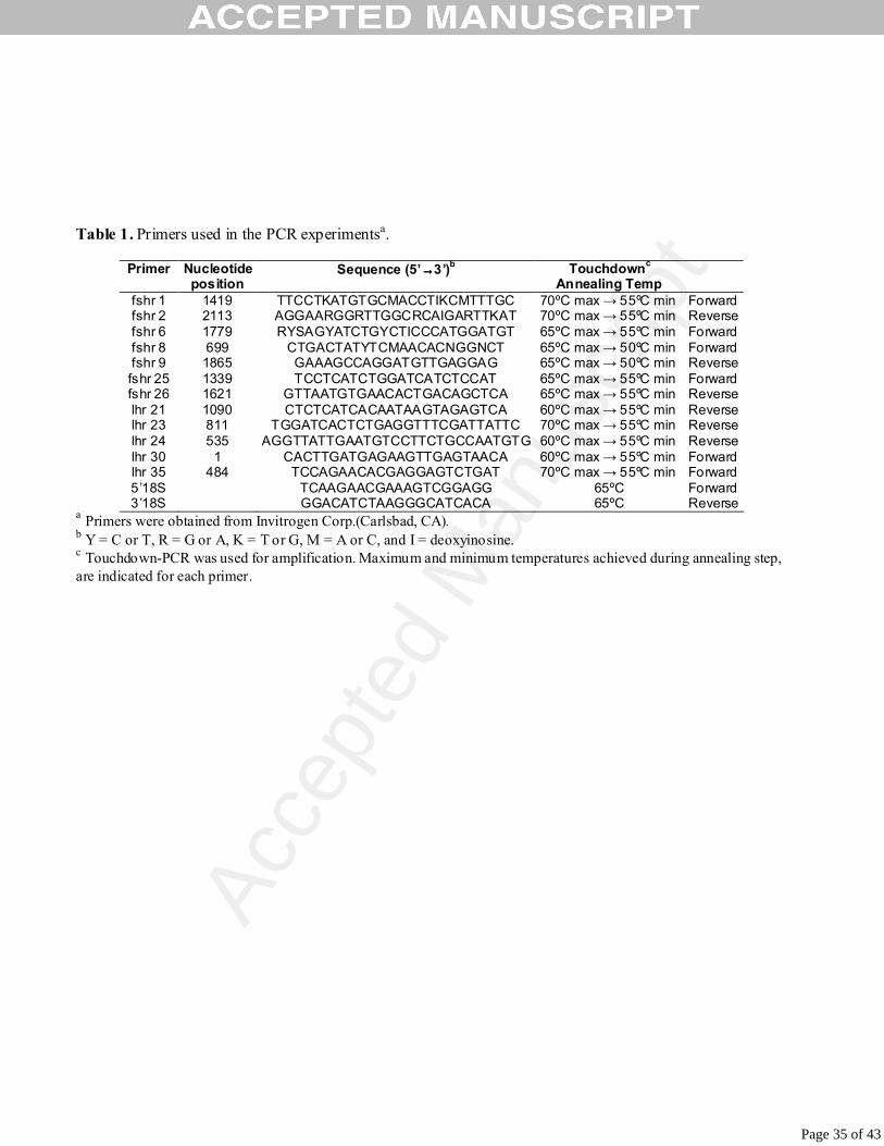
Table 1. Primers used in the PCR experimentsa.
Primer Nucleotide position
Sequence (5’→→→→3’)b Touchdownc Annealing Temp
fshr 1 1419 TTCCTKATGTGCMACCTIKCMTTTGC 70ºC max → 55ºC min Forward fshr 2 2113 AGGAARGGRTTGGCRCAIGARTTKAT 70ºC max → 55ºC min Reverse fshr 6 1779 RYSAGYATCTGYCTICCCATGGATGT 65ºC max → 55ºC min Forward fshr 8 699 CTGACTATYTCMAACACNGGNCT 65ºC max → 50ºC min Forward fshr 9 1865 GAAAGCCAGGATGTTGAGGAG 65ºC max → 50ºC min Reverse fshr 25 1339 TCCTCATCTGGATCATCTCCAT 65ºC max → 55ºC min Forward fshr 26 1621 GTTAATGTGAACACTGACAGCTCA 65ºC max → 55ºC min Reverse lhr 21 1090 CTCTCATCACAATAAGTAGAGTCA 60ºC max → 55ºC min Reverse lhr 23 811 TGGATCACTCTGAGGTTTCGATTATTC 70ºC max → 55ºC min Reverse lhr 24 535 AGGTTATTGAATGTCCTTCTGCCAATGTG 60ºC max → 55ºC min Reverse lhr 30 1 CACTTGATGAGAAGTTGAGTAACA 60ºC max → 55ºC min Forward lhr 35 484 TCCAGAACACGAGGAGTCTGAT 70ºC max → 55ºC min Forward 5’18S TCAAGAACGAAAGTCGGAGG 65ºC Forward 3’18S GGACATCTAAGGGCATCACA 65ºC Reverse
a Primers were obtained from Invitrogen Corp.(Carlsbad, CA). b Y = C or T, R = G or A, K = T or G, M = A or C, and I = deoxyinosine. c Touchdown-PCR was used for amplification. Maximum and minimum temperatures achieved during annealing step, are indicated for each primer.
Table 1
Page 35 of 43

Acce
pted
Man
uscr
ipt
1 CACACTTCTTCCCTCTTCAACCTGGCGGTGAACCCAGAGTTGTGAGACGAAAAAAAAAAACAACCCATTCATGATATCAAATGTGGAGTGTGGGCTCTGT 101 GAGAAGGCCTGAACACACGTGAACACAGAGGGAAACAACAGTGAGAGAGTGTGAGGAGAAGCAGAAGAAGAAGAAGAGAAACAACCTGGACTGTGGACAG 1 M M M V M I L I M L M I L M I K T A T A S V P G P E M D V K P 201 ACTGCGATGATGATGGTGATGATTCTCATAATGTTGATGATTCTGATGATAAAGACGGCAACAGCCTCGGTGCCTGGCCCAGAGATGGACGTTAAACCAG 32 G V E T S L A K R T L S F .C. Y Q L K F G V T E I P S S I S S N T T .C 301 GAGTGGAGACCAGCTTGGCCAAACGAACCCTGAGCTTCTGCTACCAGTTGAAGTTTGGGGTCACAGAGATTCCCTCCAGCATCTCCAGCAACACCACATG 66 L E V K Q T E I V V I P Q G A L N S L Q H L R K L T I W E N D K L 401 CCTGGAAGTTAAGCAGACGGAGATCGTAGTGATTCCCCAGGGCGCCCTCAACAGCCTGCAGCACCTCAGGAAACTCACCATATGGGAGAACGACAAGCTG 99 E S I N E F A F A S L S Q L T D I F I S G N V A L K N I G A F A F 501 GAGAGTATCAATGAGTTTGCCTTCGCCAGCCTCTCTCAGCTCACCGATATCTTCATCTCTGGGAATGTTGCGTTAAAAAATATAGGAGCTTTTGCTTTCT 132 S D L P E L T E I T I T K S K H L T H I N P D A F K D I V K L K Y L 601 CTGATCTGCCTGAACTCACTGAGATAACCATAACAAAGTCAAAACACCTGACACACATCAATCCAGATGCATTCAAGGACATTGTGAAACTGAAGTATCT 166 T I A N T G L R L F P D F T K I H S T G L L L F D L H D N S H I E 701 GACCATCGCCAACACTGGACTGAGGCTTTTTCCAGACTTCACCAAGATCCACTCCACAGGCCTGCTTCTGTTTGACCTGCACGACAACAGCCACATAGAG 199 R V P A N A F K G L C T Q T I P E I R L T R N G I K E V A S D A F 801 AGAGTCCCCGCCAATGCTTTCAAAGGCCTCTGCACTCAAACTATCCCTGAGATACGGCTCACCAGAAATGGCATCAAGGAGGTGGCAAGTGACGCCTTCA 232 N G T K M H R L F L R G N K Q L T H I N P N A F V G S S E L V V L D 901 ACGGCACAAAGATGCACAGATTGTTCCTAAGAGGCAACAAACAGCTTACTCACATCAATCCCAACGCCTTTGTGGGTTCCAGTGAGTTGGTGGTACTGGA 266 I S Q T A L S S L P D Y I L G G L Q K L I A E S A P N L K E L P P 1001 CATTTCCCAGACAGCCCTCAGCTCCCTGCCGGACTACATCCTCGGTGGACTCCAGAAGCTGATCGCAGAGTCCGCCCCCAATCTGAAGGAGCTTCCTCCT 299 L E L F T K L H Q A N L T Y S S H .C..C. A F H N I H R N R S K W N S 1101 CTGGAGCTCTTCACCAAACTGCACCAGGCCAACCTGACGTACTCGTCACACTGCTGCGCCTTCCATAACATACACAGGAACAGATCAAAGTGGAACTCCC 332 L .C. S H P D A Q G N L H F Y R D Y .C. S N S T S I I .C. T P T Q D D F N 1201 TGTGCTCCCACCCCGATGCTCAGGGCAACCTTCACTTCTACAGGGACTACTGCTCCAACTCCACCTCCATCATCTGCACCCCAACCCAAGATGACTTTAA TM I 366 P .C. E D I M S A V P L R V L I W I I S I L A L L G N T V V L L V L 1301 CCCCTGTGAGGATATCATGTCCGCCGTCCCCCTGCGGGTCCTCATCTGGATCATCTCCATCCTCGCACTGCTGGGGAACACAGTGGTACTTCTGGTCTTA TM II 399 L G S R T K L T V P R F L M C H L A F S D L C M G I Y L V V I A T:: 1401 TTAGGCAGCCGCACCAAACTGACTGTTCCCCGTTTCCTCATGTGCCACTTGGCCTTTTCTGACCTCTGCATGGGCATCTACCTGGTAGTCATAGCAACTG TM III 432 V D M L T Q G Q Y Y N H A I D W Q T G L G C S V A G F F T V F A S E 1501 TAGACATGCTCACTCAAGGCCAGTACTACAACCACGCCATAGACTGGCAGACGGGCCTCGGCTGCAGTGTTGCGGGCTTCTTCACGGTGTTTGCCAGTGA 466 ..L S V F T L T A I T L E R W H T I T H A L R L D R K L R L R H A C. 1601 GCTGTCAGTGTTCACATTAACAGCGATCACCCTGGAGCGTTGGCACACCATCACACATGCTCTGCGGCTCGACCGTAAACTTCGTCTGAGACATGCCTGT TM IV 499 .I V M T A G W I F S A V A A L L P T V G V S S Y G K V S I C L P M 1701 ATTGTCATGACAGCAGGGTGGATCTTCTCCGCCGTAGCTGCTTTGCTTCCCACAGTCGGAGTCAGCAGCTACGGCAAGGTGAGTATCTGTCTGCCCATGG TM V 532 D V E F L G S Q V Y V V S L L L L N I L A F F C V C G C Y L S I Y L 1801 ACGTGGAGTTTCTGGGGTCTCAGGTCTATGTGGTGTCACTTCTCCTCCTCAACATCCTGGCTTTCTTCTGCGTGTGCGGCTGTTACCTCAGCATCTACCT TM VI 566 T V R N P S S A P A H A D T R V A Q R M A V L I F T D F V C M A P. 1901 GACCGTCCGCAACCCCTCGTCAGCGCCGGCCCACGCCGACACACGTGTGGCCCAACGCATGGCAGTCCTCATCTTCACCGACTTTGTCTGCATGGCTCCC TM VII 599 I S F F A I S A A L K L P L I T V S D A K L L L V L F Y P I N S C.. 2001 ATCTCCTTCTTTGCCATCTCAGCAGCCCTCAAGCTCCCTCTCATCACAGTGTCAGACGCCAAACTCCTGCTGGTCCTCTTCTACCCGATCAACTCCTGCT 632 S N P F L Y A F F T R T F R R D F F L L A A R F G L F K T R A Q I Y 2101 CCAACCCCTTCCTGTACGCCTTCTTCACACGCACCTTCAGGCGGGATTTCTTTCTCCTCGCAGCTCGCTTCGGCCTGTTTAAGACCCGAGCACAGATTTA 666 R T E S S S C Q Q P A W T S P K S S H V M L Y S L A N A L S L E G 2201 CCGGACAGAGAGTTCCTCCTGTCAGCAGCCGGCGTGGACCTCTCCAAAGAGCAGCCACGTGATGCTGTACTCCTTGGCCAATGCATTAAGTCTAGAAGGG 699 K P E F * 2301 AAACCAGAGTTCTGACTGTTATTCAAAAGCAGATACAAACTTTTTGCTTAATTTTGTTGGTTTTCATGTATCTCTGATATGTTGCCAGTATTTTTGGCCA 2401 GCACATCAGCTGCTGATTCTTCAGACCTGACCTCAGACCTGCAACAACACAAGTATCCAGTGTCCTTCTTAAACCATGTGTGACAACTCTACTGTATGTA 2501 CAGTGTGTATAGAAAACCAAGTAATAGCTACAGAACAACTGCAGATACTTTGACCAAATCTAGGTCAAATGGCTGTCCCAGATAATTCCTTGTGTTTGTT 2601 TTAATTTGATTGGAACGCCAGATGGGTGGAGTTTACTGCATAATAACAATTGAGTTCACAAGTAAATGTAGGAGGAGTATCAGAGGAGGTCAACTGACTT 2701 TTACTTTCATGCATTGTCTTATATAACATCCATCTGGTGTCCCAAGATAATGAAACAAGCTTAAATACTTTGAATCTGTTTAATAAAGGTTTGACACTCG 2801 GGATATCAGCATATGAACGTCAAGAAATCAACATTCATTTAACATTCAACGTCATGATTGAATGATATTCATTAAGAAGCACACCGCAGGGACTTACTGT 2901 TTAATAATCTCCTCATACACCGTCTGCTGTAGCTCTACATCCCTCCACATCCCTCTTACCCTTCAGTGTCCATATTTTTTTAATGAAGGACGTGTAATTA 3001 TCTTTGTAATGTAATTAGGTTGTGTAAATGAAAGTACCACTGTTTTGTATATGTGTGTTTTGTACAGAATATATCTTTGAGATGTAATTTCATGTCATTT 3101 ATAAATTTGTTTATTCAAAAAAAAAAAAAAAAAA
Figure 1
Page 36 of 43

Acce
pted
Man
uscr
ipt
▄▄▄▄▄ ▄▄ Hum -------MALLLVSLLAFLSLGSGCHHRICHCSNRVFLCQESK---------VTEIPSDLPRNAIELRFVLTKLRVIQKGAFSGFGDLEKI 75 Sbs MMMVMILIMLMILMIKTATASVPGPEMDVKPGVETS-LAKRTLSFCYQLKFGVTEIPSSISSNTTCLEVKQTEIVVIPQGALNSLQHLRKL 90 Sb -MMAMILIMVTIVMIKMAGASAPDAETDVKPGADESVLAKRTLSICNQLPPEVTEIPSNISSDTECLEVKQTQIGVIPRGAVHRLQHLRIL 90 Til MMLVMTLMMLLIVTIKMAAASAHGSEMDIRPGFHPS-LAKQTSCLSYQVMFGVTAFPSNIS-NAQCLEVKQTQIREIQQGTLSSLQHLMEL 89 As -MMKMKKIMKMLLCVLGCVSMSQAEVAMVNSGTTFTYLCMGNT---------ITHMPTHIPKNTTDLEFKQTHIRVFPQEAFTNLQQLTAI 81 Ats -MMKMKKIMKMLLCVLGCVCVSQAEVAMVNSGTTFTYLCMGNT---------ITHMPTHIPKNTTNLEFKQTHIRVFPREAFTNLLQLTAI 81 Rtr -MMKMKKIMKMLLCMLGCVCVSQAEVAMVNSGTTFTYLCMGNT---------ITHMPTHIPKNTTDLEFKQTHIRVFPREAFTNLQQLTAI 81 Zbf MVLSMMLCFILGCSIANTEDTLAASQFCAFNGSTRSFICLGNK---------VHEIPRRIPTNTTFVEIKLTQISVFRRAALSELHELKRI 82 Ac -----MLRYILSWLVMHTGNMFLGSYACLASGTTRSFLCLGSK---------VHQMPHHIPINTTYVEIKLTQIIIFPYRAMSSLHDLKRI 77 Cc -----MMCFILSWLMMHAGNMCLGSYACLANGTTRSFLCLGSK---------VHQMPYHIPKNTTYVEIKLTRIIMLPSRAMSSLHDLKRI 77 ▄▄▄ ▄▄▄▄▄ ▄▄▄▄▄ ▄▄▄▄▄ Hum EISQNDVLEVI-------------------------EADVFSNLPKLHEIRIEKANNLLYINPEAFQNLPNLQYLLISNTGIKHLPDVHKI 141 Sbs TIWENDKLESINEFAFASLSQLTDIFISGNVALKNIGAFAFSDLPELTEITITKSKHLTHINPDAFKDIVKLKYLTIANTGLRLFPDFTKI 181 Sb IISKNEVLESIGAFAVAGLPQITNIFISENAELASIGAFAFSDLPELTEMTITKSKHLRHIHPDAFRNIVKLRVLIISNTGLRMFPDFTKI 181 Til TISENDLLESIGAFAFSGLPHLTKILISKNAALRNIGAFVFSNLPELSEIIITKSKHLSFIHPDAFRNMARLRFLTISNTGLRIFPDFSKI 180 As VLTENGMLESI-------------------------GAFAFANLPRLTEITITKSKHLVIIHHQAFIGLPKLSHLTICNTGLRVLPNFSRI 147 Ats VLTENGMLESI-------------------------GAFAFANLPRLTEITITKSKHLVVIHHQAFMGLPKLSHLTICNTGLRVLPNFSRI 147 Rtr VLTENGMLESI-------------------------GAFAFANLPRLTEITITKSKHLVIIHHQAFIGLPKLSHLTICNTGLRVLPNFSRI 147 Zbf VVSENGALERI-------------------------EALAFFNLTELEEITITKSKNLV-MHKDAFWRLPKLRYLTISNTGLKILPDFSQI 147 Ac MVSENGALQRI-------------------------EAYAFANLTILEEITITKSKNLVSMDRDTFWGLPKLRYLTISNTGLTVLPDFSKV 143 Cc LVSENGVLQRI-------------------------EAYAFANLTKLEEITITKSKNLVCMDRHTFWGLPKLRYLTISNTGLTVLPDFSKV 143 ▄▄▄▄▄ ▄▄▄▄▄ ▄▄▄▄▄ ▄▄▄▄▄ Hum HSLQ-KVLLDIQDNINIHTIERNSFVGLSFESV-ILWLNKNGIQEIHNCAFNGTQLDELNLSDNNNLEELPNDVFHGASGPVILDISRTRI 230 Sbs HSTG-LLLFDLHDNSHIERVPANAFKGLCTQTIPEIRLTRNGIKEVASDAFNGTKMHRLFLRGNKQLTHINPNAFVGSSELVVLDISQTAL 271 Sb HSTAPDFLFGLQENSHIERVPVNAFRGLCTQTISEIRLTRNGIKEVASDAFNGTKMHRLRLSGNQQLTHINPNAFVGSSELVVLDISHTAL 272 Til HSTA-CFLLDLQDNSHIKRVPANAFRGLCTQTFAEIRLTRNGIKEVASDAFNGTKMHRLFLGGNRQLTHISPNAFVGSSELVVLDVSETAL 270 As HSAAMTFLLDLQDNVHIVIIPSNAFLGLTTNTIDELRLTKNGISEVESHAFNGTKIHKLYLMGNLQLSHMHNNSFKGAEGPGFLDISRTAL 238 Ats HSTALTFLLDLQDNVHIVIIPSNAFLGLTTNTIDELRLTKNGISEVESHAFNGTKIHKLFLMGNLQLSHMHNNSFKGAEGPGFLDISRTAL 238 Rtr HSAALTFLLDLQDNVHIVIIPSNAFLGLTTNTIDELRLTKNGISEVESHAFNGTKIHKLFLMGNLQLSHMHNNSFKGAEGPGFLDISRTAL 238 Zbf NSAALEFLFDLQDNMHIERIPSNAFLGLTNATITELRLTKNGIREIDSHAFNGTKIGKLFLMGNQQLNHIHSYAFKGAEGPVVLDISRTAV 238 Ac QSAAFEFLFDLEDNMHIEVIHSNAFAGLTSGTITELRLTKNGITEVERNAFNGTKMEKLFLMGNQQLKRIDNHAFLGAEGPLVLDISRTAI 234 Cc QSAAFEFLFDLEDNMHIEVIPSNAFAGLTSGTITELRLTKNGITEVDRNAFNGTKIEKLFLMGNQQLKRIHNYAFLGAEGPLVLDISRTAI 234 ▄▄▄▄▄
**
*
Hum HSLPSYGLENLKKLRARSTYNLKKLPTLEKLVALMEASLTYPSHCCAFANWRRQISELHP-ICNKSILRQEVDYMTQTRGQRSSLAEDNES 320 Sbs SSLPDYILGGLQKLIAESAPNLKELPPLELFTKLHQANLTYSSHCCAFHNIHRNRSKWNS-LCSHPDAQGNLHFY---------------- 345 Sb SSLPENILGGLQKLMAESVFRLKELPPLQLFNKLQEVHLTYPSHCCAFHNVHRNRSSWNALLCSHPDAPGMPNFY---------------- 347 Til TSLPDSILDGLKRLIAESAFNLKELPPIQLFTKLHQAKLTYPSHCCAFLNMHRNRSRWHS-LCDNPEAKNNLHFF---------------- 344 As SSLPESVLGEVEHLSAVSVFSLRTLPPLSLFTKLRQANLTYPSHCCAFHKHQRNRTFRMTSACFKPGAQNNLHFF---------------- 313 Ats SSLPESVLGEVEHLSAVSVFSLRALPPLSLFTKLRQANLTYPSHCCAFHKHQRNRTFRMNSACFKPGAQDNLHFF---------------- 313 Rtr SSLPESVLGEVEHLSAVSVFSLRALPPLSLFTKLRQANLTYPSHCCAFHKHQRNRTFRMTSACFKPGAQNNLHFF---------------- 313 Zbf HTLPESMLKTLKLLMAVSVYSLRKLPSLELFTELTQANLTYPSHCCAFKNFKKHKSVKNQ-MCNVTGAHEEPDFFNFF------------- 315 Ac SSLPENMLRRLKLLIATSVYSLRWLPNLEIFAELTQANLTYPSHCCAFKNFKKNKLEKNH-LCNVSTIRNQEPYF--F------------- 309 Cc SSLPENMLRRLKLLIATSVYSLRWLPNLEIFTELTQANLTYPSHCCAFKNFKKNKSEKNR-LCNDSTIRNQEPYF--F------------- 309
* * * Hum SYSRGFDMTYTEFDYDLCNEVVDVTCSPKPDAFNPCEDIMGYNILRVLIWFISILAITGNIIVLVILTTSQYKLTVPRFLMCNLAFADLCI 411 Sbs --------------RDYCSNSTSIICTPTQDDFNPCEDIMSAVPLRVLIWIISILALLGNTVVLLVLLGSRTKLTVPRFLMCHLAFSDLCM 422 Sb --------------KDHCSNSTAIICTPTQDEFNPCEDIMSAVPLRILIWIISILALLGNAAVLLVLLGSRCKLTVPRFLMCHLAFSDLCM 424 Til --------------REYCSNSTNITCSPAPDDFNPCEDIMSATPLRILIWIISVLALLGNAVVLLVLLGSRYKLTVPRFLMCHLAFADLCM 421 As --------------MDFCLNWTSVACSPAPDAFNPCEDIMGSAPLRVLIWIISVLALLGNTIVLLVLLGSRAKMTVPRFLMCHLSFADLCM 390 Ats --------------MDFCLNWTSVACSPAPDAFNPCEDIMGSAPLRVLIWIISVLALLGNTIVLLVLLGSRAKMTVPRFLMCHLSFADLCM 390 Rtr --------------MDFCLNWTSVACSPAPDAFNPCEDIMGSAPLRVLIWITSVLALLGNTIVLLVLLGSRAKMTVPRFLMCHLSFADLCM 390 Zbf --------------NDHCKDVIEVTCYPTPDAFNPCEDIMGFTFLRVLIWFISVLAIVGNTVVLLVLLTSRYKLTVPRFLMCHLAFADLCM 392 Ac --------------EEHCKDVIEVSCYPEPDAFNPCEDIMGFTFLRVLIWFISVLAVLGNFTVLLVLLSSHTKLTVPKFLMCHLAFADFCM 386 Cc --------------EEHCKDVIEVRCYPEPDAFNPCEDIMGFTFLRVLIWFISVLAVLGNFTVLLVLLSSRTKLTVPKFLMCHLAFADFCM 386
Figure 2
Page 37 of 43
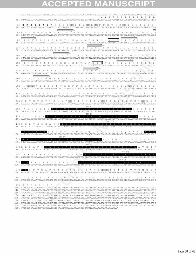
Acce
pted
Man
uscr
ipt
1 CACTTGATGAGAAGTTGAGTAACAGGTGGCTGACATTGTCCTCCGCACGGCTTCGATGACGATCTGCTGCGGTTGCAGCAAAAGAGACTTTAAAGAAGGG M W T S L P A L L F L S V L 101 CCAGAGATCTTAATGCGCAGCGCTCAGCGGGCATCCCGGCTTCTGTGCGTTTTACAGAATGTGGACCTCGCTGCCGGCGCTCTTGTTCCTGTCTGTTTTG 15 G F Y G C K C A P G F G .C. P R I .C. R .C. F S N T I R .C. N N V T Q G S 201 GGCTTCTACGGATGCAAGTGCGCTCCGGGTTTCGGCTGTCCGAGGATCTGCCGCTGTTTCTCCAACACAATCAGATGCAACAATGTAACTCAAGGGTCGG 48 A L M M D H R D K R L F F Y H L S L N T I S S H S F D G L K G V Q R 301 CGCTGATGATGGATCACAGAGATAAAAGACTGTTTTTCTATCATTTGTCTTTGAACACCATTTCCAGCCATTCTTTTGATGGTTTAAAAGGAGTCCAGAG 82 I E I A Q S V T L E T I E A L A F N N L L N L S E I S V Q N T R S 401 GATTGAGATTGCTCAGAGTGTAACCCTGGAAACCATTGAGGCATTAGCCTTTAATAACCTTCTCAACCTCTCTGAAATCTCAGTCCAGAACACGAGGAGT 115 L M H I G R R T F N N L P K L H Y L S I S N T G I T L F P D I T Y 501 CTGATGCACATTGGCAGAAGGACATTCAATAACCTCCCCAAACTACATTACTTGAGCATCTCAAATACTGGGATAACCCTTTTCCCTGACATTACATATA 148 I N S L E S E F I L D I C D N L Y L L E I P P N A F I G L T K E Y V 601 TCAATTCTCTTGAATCCGAGTTTATCTTGGATATATGTGATAACCTGTATCTACTGGAAATACCTCCTAACGCTTTCATCGGACTGACAAAGGAGTATGT 182 T M N L Y N N G I R E I H D Y A F N G T K I D K L V L K N N R N L 701 TACAATGAACCTGTATAACAACGGCATCAGAGAAATACACGACTACGCCTTCAACGGGACAAAGATAGATAAGCTGGTATTAAAGAATAATCGAAACCTC 215 R V I H R Y A F E G A T G P G V L D V S A T A L T K L P P Q G L E 801 AGAGTGATCCACAGATATGCTTTCGAAGGAGCCACAGGACCTGGAGTCTTGGACGTTTCTGCGACAGCTCTCACAAAGTTGCCACCGCAGGGACTGGAGT 248 S V L V L F A Q S A Y A L K S L P P L Q G L W S L R E A H L T Y N S 901 CAGTTCTGGTGTTGTTTGCTCAGTCAGCATATGCCTTGAAGAGTCTGCCTCCTCTGCAGGGACTGTGGAGCCTGCGAGAGGCCCATCTCACCTACAACAG 282 H .C..C. A L L S W N T H R D F S I N P A Y N N D S T Y .C. D E S D Q L 1001 TCACTGTTGTGCGCTGCTGAGCTGGAACACCCACAGGGACTTTTCCATTAATCCTGCATATAATAATGACTCTACTTATTGTGATGAGAGTGACCAATTA 315 A R V Q R V I G G S A D T T L V M D M P F F S D V D L S E D E G F 1101 GCAAGGGTTCAGCGTGTGATTGGAGGTTCAGCAGACACGACTCTAGTTATGGATATGCCATTTTTTTCAGACGTTGATTTATCTGAGGACGAGGGCTTTG 348 G D V N F H Y P E L D F .C. Q T R P T L V .C. T P E A D A F N P .C. E D I 1201 GAGACGTGAATTTCCACTATCCAGAGCTGGACTTCTGTCAGACCCGACCAACTTTGGTTTGCACACCTGAAGCAGATGCTTTCAATCCCTGTGAGGACAT TM I 382 A G F S F L R .V A I W F I N I L A I T G N L T V L L V. S F T S R N 1301 TGCAGGTTTCAGTTTTCTCAGAGTAGCCATTTGGTTTATCAACATACTGGCCATCACAGGCAACCTGACAGTCCTCCTAGTCTCCTTCACCAGCCGCAAC TM II 415 K L T V P R .F L M C H L A F A D L C I G I Y L L M I A T V. D F R T 1401 AAGCTGACGGTGCCTCGCTTCCTCATGTGCCACCTGGCTTTTGCAGACCTCTGCATTGGGATCTACCTCCTAATGATAGCCACAGTAGACTTCCGCACAC TM III 448 Q G H Y S Q H A I E .W Q T G S G C S A A G F L S V F G G E L S V Y T 1501 AGGGCCACTACAGTCAGCATGCTATTGAATGGCAAACAGGGTCTGGCTGTAGTGCTGCAGGCTTCCTGTCTGTGTTTGGTGGGGAGCTGTCAGTCTACAC 482 L S T I T L. E R W H T I T N A L Q I E R H L V L T Q A A S I .M A A. 1601 GCTTTCCACCATCACCCTGGAGCGCTGGCACACCATCACCAACGCTCTGCAGATAGAGCGCCATCTGGTACTCACACAGGCAGCGAGCATAATGGCGGCT TM IV 515 .G W I I C L G M G M L P L F G V S S Y A. K V S M C L P M D I E T P 1701 GGGTGGATCATCTGTCTGGGGATGGGGATGCTGCCTCTGTTTGGCGTGAGCAGTTACGCCAAAGTCAGCATGTGCTTACCAATGGACATAGAGACCCCCC TM V 548 L A Q A F I .I L I L L F N V G A F V V V C V C Y V L I Y L. A V K N P 1801 TGGCTCAGGCCTTCATCATACTCATCCTGCTCTTCAACGTGGGCGCCTTTGTTGTTGTGTGTGTTTGTTACGTGCTGATCTATCTGGCTGTGAAAAACCC TM VI 582 E F P R R S A D T K I A K R M .A V L I F T D F L C M A P I S F F A. 1901 AGAGTTCCCCAGAAGAAGCGCTGACACCAAGATTGCAAAGCGCATGGCTGTGCTCATCTTCACTGATTTCCTCTGCATGGCTCCCATCTCCTTCTTTGCC TM VII 615 .I S. A A F K V P L I T V T N .S K I L L V L F F P I N S C A N P F L.. 2001 ATCTCTGCTGCATTCAAGGTCCCCCTCATCACAGTCACTAACTCCAAGATCCTGCTGGTCCTCTTCTTCCCCATCAATTCTTGTGCCAATCCCTTCCTTT 648 Y A. I F T K A F R K D A Y Q L M S A M G .C C. K S K A S V Y R M K A Y 2101 ACGCTATCTTCACCAAGGCTTTTAGAAAGGACGCATATCAGCTCATGAGTGCCATGGGATGCTGTAAAAGCAAGGCCAGTGTTTATCGCATGAAGGCCTA 682 C S E N A I K S N L G S N N K E S G T G G R R A A V A G Q S H H L 2201 CTGCTCCGAAAATGCAATCAAGAGCAACCTGGGATCCAATAATAAAGAATCAGGCACAGGAGGGCGGCGGGCTGCGGTGGCAGGGCAGAGCCATCACCTG 715 K E E R E L T * 2301 AAAGAGGAGAGGGAGCTCACCTGAAATAAAAAGATTCAAAAGTCTTTTAATTGGACAATTGGTCAAGAGAAACTAAGGAAAAAAACAACTCATGCTGGAT 2401 TTGGAGGAAATTACTCTAACATGTTGACATTGATGGCATATTTTAATCTGCACTGTTGGAGATATTTGTATTAGAAAGTATAAGAAAATCTTATTGCCTT 2501 TTCCAAATCTGATATCAGTTCAACCCAATAAAAAAATAGTTTTTTTGTTGACTGGTTATGACAGGAAAAGTAAAACGAATAACACTGATGATCGCTCTGT 2601 TGTTTCAGGTCCACCTGTAGTTTCATATGTCAAAATGTCTTGTAAATATACAGAAGAAAGCTCTGGAAACAGCTACTAAATGGACCTTTATGGACCTTTG 2701 TTGTATTGTCAGCTATGAACTGTCATATCTCAGTATTTAATGGGATGACCTAAACGAACAAGAAGACTTTAAAAAGCTGCCAAAAAACTGAAATTTGAAA 2801 CATCACCTGTTCAGATCTACTGATTGTATGACACAGTGTTAAACTCCTTGTGGTGAGACTGAGTGATCTGCTTCATCCTAGCTTCATCTCCAAAGTTCAT 2901 CTGAATGAGAATCAAACCAGAGCTGTCAGTTTATCTCAATGTTATTGATGTATGTGAAATAAATTGTCCGCTTCATTCTGGGTGTGAAATTAAGAACATC 3001 TATAGCCATGGTTAGTATTTAAAGGTTATAGCCTATAGTTGACCACGTGGTGGAGTAGTTGGGAGGAAAACTTCTACACCACTTGAGTTTTTGTTGTTCT 3101 TTTGGCCTAAGGGTTTTTGGGATTATGTAATTCAAAATAAAAGACTTCACCTACAAAAAAAAAAAAAAAAAA
Figure 3
Page 38 of 43

Acce
pted
Man
uscr
ipt
Figure 4
Page 39 of 43

Acce
pted
Man
uscr
ipt
Figure 5
Page 40 of 43

Acce
pted
Man
uscr
ipt
Figure 6
Page 41 of 43
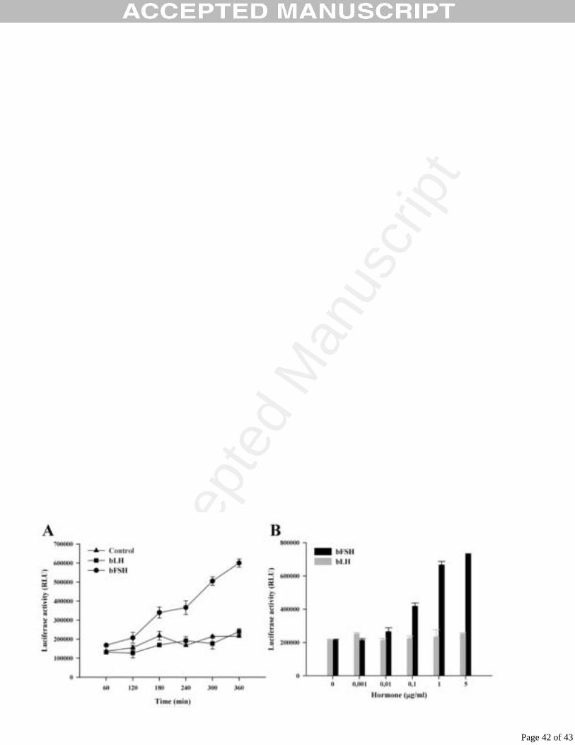
Acce
pted
Man
uscr
ipt
Figure 7
Page 42 of 43
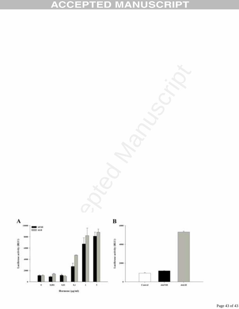
Acce
pted
Man
uscr
ipt
Figure 8
Page 43 of 43