Embed Size (px)
Citation preview
INFECTION AND IMMUNITY, Oct. 2003, p. 5803–5813 Vol. 71, No. 100019-9567/03/$08.00�0 DOI: 10.1128/IAI.71.10.5803–5813.2003Copyright © 2003, American Society for Microbiology. All Rights Reserved.
Molecular Characterization of the Acute Inflammatory Response toInfections with Gram-Negative versus Gram-Positive Bacteria
Robert J. Feezor,1 Caroline Oberholzer,1,2 Henry V. Baker,3 Daniela Novick,4Menachem Rubinstein,4 Lyle L. Moldawer,1* John Pribble,5 Sonia Souza,5
Charles A. Dinarello,6 Wolfgang Ertel,2 and Andreas Oberholzer1,2
Department of Surgery1 and Department of Molecular Genetics and Microbiology,3 University of Florida College ofMedicine, Gainesville, Florida 32610; Department of Trauma and Reconstructive Surgery, Benjamin Franklin
Medical Center, Berlin, Germany2; Department of Molecular Genetics, Weizmann Institute of Science,Rehovot, Israel4; ICOS Inc., Bothell, Washington 980215; and Department of Medicine,
University of Colorado Health Sciences Center, Denver, Colorado 802626
Received 10 February 2003/Returned for modification 14 April 2003/Accepted 27 June 2003
Sepsis caused by gram-negative bacteria and that caused by gram-positive bacteria often manifest similarclinical features. We investigated plasma proinflammatory cytokine profiles in patients with sepsis due togram-positive and gram-negative bacteria and studied the cytokine production and differential gene regulationof leukocytes stimulated ex vivo with Escherichia coli lipopolysaccharide or heat-killed Staphylococcus aureus.Concentrations of tumor necrosis factor alpha, interleukin 1 receptor antagonist (IL-1Ra), IL-8, IL-10, IL-18binding protein, procalcitonin, and protein C in plasma did not differ between patients with sepsis due togram-negative and gram-positive bacteria. However, plasma IL-1�, IL-6, and IL-18 concentrations weresignificantly higher in patients with sepsis due to gram-positive bacteria. Ex vivo stimulation of whole bloodwith heat-killed S. aureus markedly increased IL-1� and IL-18 levels more than E. coli lipopolysaccharidestimulation. Microarray analysis revealed at least 359 cross-validated probe sets (genes) significant at the P< 0.001 level whose expression discriminated among gram-negative-organism-stimulated, gram-positive-or-ganism-stimulated, and unstimulated whole-blood leukocytes. The host inflammatory responses to gram-negative and gram-positive stimuli share some common response elements but also exhibit distinct patterns ofcytokine appearance and leukocyte gene expression.
Despite improvements in hemodynamic monitoring, antibi-otics, and other supportive therapies, sepsis remains the mostcommon cause of death within intensive care units (27) and the13th leading cause of death overall in the United States (10).More than 500,000 people are diagnosed with sepsis each yearin the United States, and the observed incidence is still rising(3, 26, 35), with financial costs exceeding $16 billion each year(3).
Although there have been modest successes in treating sep-sis in animal models, little advancement has been made inhuman studies (1, 4, 5, 36). Possible reasons for this failureinclude heterogeneity of the patient population, improper tim-ing of attempted therapies, and, more recently appreciated, thecomplex redundancy of molecular pathways of inflammationinduced by different microbial pathogens (18, 32). Indeed,there is growing recognition that gram-positive organisms areresponsible for an ever-increasing number of septic events (9,11, 31).
The American College of Physicians and the Society of Crit-ical Care Medicine have adopted consensus definitions of sep-sis and the systemic inflammatory response syndrome (SIRS)in an attempt to standardize patient characterization (2). Se-vere sepsis is a known or suspected microbial infection associ-
ated with fever, leukocytosis or leukopenia, hypotension, in-creased cardiac output, and decreased peripheral vascularresistance (2, 16). Injury severity scoring systems measure clin-ical parameters to stratify the severity of patient illness, deter-mine level of intervention, and potentially predict patient out-come. However, these scoring systems primarily measure thephysiological effect of the infection on the host, not the micro-bial, biochemical, or genetic mechanism(s) of the organ injuryresponse (23). Similar clinical pictures result from the actionsof any number of inflammatory stimuli, both noninfectious andinfectious, including different classes of organisms. Specifically,SIRS can be caused by infection with gram-positive or gram-negative bacteria, multiple trauma, ischemia-reperfusion in-jury, pancreatitis, severe thermal injury, and other noxiousstimuli.
The successful use of anticytokine or anti-inflammatorytherapies to interrupt the development of the sepsis responserequires a more thorough understanding of the initiating me-diators involved in its induction. Recent studies on the role ofthe Toll-like receptor (TLR) family have shown that these cellsurface proteins differentially recognize and signal the pres-ence of products of gram-positive and gram-negative bacteriaand have highlighted the complexity of the host response todifferent microbial pathogens (28, 29). Although sepsis andSIRS are mediated in some part by the early production of avariety of proinflammatory cytokines, including tumor necrosisfactor alpha (TNF-�) and interleukin 1 (IL-1), IL-12, andIL-18, among others, there is growing evidence to suggest that
* Corresponding author. Mailing address: Department of Surgery,University of Florida College of Medicine, P.O. Box 100286, Room6116, Shands Hospital, 1600 SW Archer Rd., Gainesville, FL 32610-0286. Phone: (352) 265-0494. Fax: (352) 265-0676. E-mail: [email protected].
5803
the patterns of early cytokine and mediator production may bedependent upon the specificity of the microbial pathogens andthe host recognition pathways invoked (19, 24, 25).
In the present study, we sought to examine the diversity inhost response to gram-negative and gram-positive pathogensby focusing on the differential proinflammatory cytokine re-sponses in the plasma of patients with sepsis due to gram-negative or gram-positive bacteria, as well as assessing ex vivostimulation of whole blood with gram-negative and gram-pos-itive microbial products. Moreover, an initial genome-widesurvey of gene expression patterns was performed on wholeblood from three subjects stimulated ex vivo with a represen-tative product from an individual gram-negative or gram-pos-itive organism.
These studies suggest that distinct patterns of cytokine pro-duction may be induced by products of different gram-negativeand gram-positive microbes. In particular, sepsis due to gram-positive bacteria and exposure of whole blood to heat-killedStaphylococcus aureus appeared to preferentially stimulate theproduction of members of the IL-1 superfamily, includingIL-1� and IL-18. Examination of the early (2-h) genome-widegene expression patterns of whole-blood leukocytes stimulatedwith either bacterial lipopolysaccharide (LPS) (Escherichiacoli) or heat-killed S. aureus revealed 359 genes whose differ-ential expression discriminated among unstimulated and stim-ulated whole blood. Several of these differentially expressedgenes encoded proinflammatory as well as anti-inflammatorycytokines. Although the physiological responses of the host tosepsis due to gram-negative and gram-positive bacteria mayappear similar, these studies suggest that the early inflamma-tory and immunological response to these pathogens may differdramatically depending upon the inciting organism.
MATERIALS AND METHODS
Patient enrollment. Fifty-two patients were enrolled prospectively from 33different institutions as part of the PAFase (ICOS Inc., Bothell, Wash.) ARDSPrevention Study. The criteria for patient inclusion included the following: age of�18 years, clinical evidence of infection within 3 days of enrollment in the study,fulfillment of SIRS criteria, and persistent hypoperfusion attributable to theinfection. The SIRS criteria, as established by the consensus statement from theAmerican College of Chest Physicians and the Society of Critical Care Medicine,require that the patient have two of the following: (i) core body temperature of�38°C or �36°C, (ii) tachycardia (�90 beats per min), (iii) tachypnea (�20breaths per min or an arterial CO2 pressure of �32 mm Hg), (iv) leukocytosis orleukopenia (white blood cell count of �12,000 cells/mm3 or �4,000 cells/mm3) or�10% immature forms (1). Informed consent was provided by the patient or anappropriate surrogate, and the conduct of the study was approved by eachindividual institutional review board. Permission to receive and analyze blindedsamples from these institutions was granted by the Institutional Review Board atthe University of Florida. Patients who met any of the following criteria wereexcluded from the study: (i) radiographic bilateral infiltrates with a ratio ofarterial O2 pressure to the fraction of inspired O2 of �200 within 72 h ofenrollment, (ii) known or suspected infection with human immunodeficiencyvirus, (iii) Child-Pugh grade C liver dysfunction or known esophageal varices, (iv)Glasgow coma scale of �8 arising from a closed head injury, (v) cardiopulmonaryarrest within 3 days of enrollment, (vi) administration of pentoxifylline, granu-locyte-colony stimulating factor, immunoglobulins, or inhaled nitric oxide within3 days of enrollment, (vii) administration of corticosteroid doses of �1.0 mg ofprednisone (or its dose equivalent) per kg of body weight per day or any immu-nosuppressive therapy within 3 days of enrollment, (viii) participation in a clin-ical trial within 28 days of enrollment, (ix) positive blood or urine pregnancy test,suspicion of pregnancy, or lactation at the time of enrollment, (x) prior fulfill-ment of the protocol requirements for inclusion during the present hospitaliza-tion, (xi) presence of a “do not resuscitate” order or lack of a commitment of
aggressive intensive care, and (xii) prognosis of an underlying disease that limitslife expectancy to less than 3 months.
Blood was collected from these patients at the time of enrollment, sampleswere centrifuged, and the resulting plasma was frozen at �70°C until analysis.
Measurements of inflammatory mediators in plasma. Levels of TNF-�, IL-1�,IL-1, IL-1 receptor antagonist (IL-1Ra), IL-6, IL-8, IL-10, IL-18, IL-18 bindingprotein (IL-18BP) (21), procalcitonin, and total protein C in plasma were de-termined for all patients at baseline (at the time of enrollment prior to drugadministration). Workers who performed cytokine measurements were blindedto patient identity and etiology of sepsis. TNF-�, IL-6, IL-8, and IL-10 weremeasured by enzyme-linked immunosorbent assay (ELISA) using PharmPaksobtained from R&D Systems (Minneapolis, Minn.) according to the manufac-turer’s instructions. Procalcitonin was determined using a luminescence-basedimmunoassay from Brahms Diagnostica GmbH (Atlanta, Ga.). The IL-18ELISA kits were purchased from Medical and Biological Laboratories Co., Ltd.(Watertown, Mass.). IL-18BP levels were determined with a technique devel-oped in the laboratory of Daniela Novick at the Weizmann Institute in Rehovot,Israel, which uses a double-antibody ELISA to calculate free IL-18 concentra-tions on the basis of the law of mass action (22). This ELISA does not recognizeeither IL-18 or pro-IL-18, and there is no interference with IL-18BP measure-ments with increasing concentrations of IL-18 (22). In 34 of the 52 patients whereremaining sample was available, total protein C levels were determined byELISA by using kits provided by Diagnostica Stago, Inc. (Parsippany, N.J.).
Whole blood ex vivo bacterial stimulation. After informed consent was ob-tained, venous whole blood was obtained from three healthy young adults byvenipuncture into heparinized tubes. For gene expression analyses, venous bloodwas stimulated with either 1 �g of LPS per ml or 0.1% heat-killed S. aureusCowan for 2 h at 37°C. Control samples were processed in parallel withoutstimulation, and each sample was evaluated without pooling of the samples. Inone of the three subjects, the blood draw was repeated a total of three times toevaluate the reproducibility of gene expression patterns from the same individ-ual. Because of the volume of blood required, the three separate blood drawswere performed 1 week apart. By analyzing replicates from three independentsubjects and from a single subject three times, the variation in gene expressiondue to individual genetic polymorphisms or preexisting conditions (comorbidi-ties) could be distinguished from the variation due to analytical replication.Correlation coefficients for gene expression (for the 12,558 probe sets in theU95Av2 GeneChip) with replicates obtained from the same individual rangedbetween 0.97 to 0.99 (R2), whereas the same correlation coefficients betweendifferent individuals ranged between 0.91 to 0.97.
The blood from each individual collection was divided into three aliquots andeither left unstimulated or stimulated for 2 h with LPS or heat-killed S. aureusCowan. After the 2-h incubation, the whole blood was centrifuged at 400 � g for10 min at 4°C, and the buffy coat layer was removed. Contaminating erythrocyteswere removed by lysis with hypotonic saline and the buffy coat pellet was recov-ered by centrifugation. Total cellular RNA was isolated from the buffy coat witha commercial kit (RNeasy; Qiagen, Inc.); purity was confirmed by spectropho-tometry (A260/A280 ratio), examination of an ethidium bromide stained-RNAagarose gel, and reverse transcription-PCR amplification of a housekeeping gene(Cu/Zn superoxide dismutase) (30).
cRNA synthesis and chip hybridization. cRNA synthesis was performed by asingle investigator starting with 10 �g of total cellular RNA, based on theprotocol outlined by Affymetrix, with few modifications. After the mRNA hadbeen selected by hybridization with the primer T7-(dT)24 (Genset Oligos,Genset Corp., La Jolla, Calif.), double-stranded cDNA was synthesized accord-ing to a standardized protocol (SuperScript double-stranded cDNA synthesis kit;Invitrogen Corp., Carlsbad, Calif.). cRNA was transcribed in vitro, incorporatingbiotinylated nucleotides by using a BioArray high-yield RNA transcript labelingkit (T7) (Enzo Life Sciences, Inc., Farmingdale, N.Y.), and hybridized ontoHu95aVer2 oligonucleotide arrays (Affymetrix). Each sample was studied inparallel, and the samples were not pooled. The chips were hybridized for 16 h at45°C, stained and washed according to the Affymetrix protocol (EukGE-WS2v4)using an Affymetrix fluidics station, and scanned with an Affymetrix scanner.
Microarray data analysis. Scanned images (.dat files) were analyzed withAffymetrix Microarray Suite version 5. Raw Q scores ranged from 2.2 to 5;average background ranged from 50 to 144. Chip-to-chip normalization wasaccomplished by using global normalization with an average target gene intensityof 500. Scaling factors ranged from 4 to 18.
Expression filter. Probe sets that were flagged as absent on all arrays analyzedin this study by the Affymetrix Microarray Suite version 5 software with defaultsettings were removed from the datasets and not included in the analyses re-ported here. (The signal intensity measurements as detected reflect the degree ofhybridization of synthesized cRNA to the probe sets on the microarray chip.
5804 FEEZOR ET AL. INFECT. IMMUN.
These probe sets represent genes or DNA sequences within genes. Some genesare represented by more than one probe set on a given microarray, and henceprobe sets are not uniquely correlated to genes. However, for ease of discussion,we use the terms “probe sets” and “genes” interchangeably.)
Variation filter, normalization, and cluster analysis. The signal intensities ofthe probe sets remaining after applying the expression filter were ranked accord-ing to the coefficient of variation, and the 50% of the data set with the greatestcoefficient of variation were then normalized to a mean of 0 and a standarddeviation of 1. k-mean determinations and hierarchical cluster analyses wereperformed with the variance-normalized data set and viewed with the algorithmsin the software packages Cluster and TreeView developed by Eisen et al. (14).
Supervised learning, discrimination analysis, and cross validation. The hy-bridization signal intensities of the genes passing the initial expression filter wereanalyzed (in part) with BRB Array Tools 3.01 (developed by Richard Simon andAmy Peng) to identify genes that differentiated among the three treatmentclasses: unstimulated, stimulated with heat-killed S. aureus Cowan, and stimu-lated with LPS (P � 0.001). The ability of gene identification to predict treatmentclass was assessed by “leave-one-out” cross-validation using each of four methodsof class prediction: nearest-neighbor prediction, three-nearest-neighbors predic-tion, linear discriminant analysis, and nearest-centroid analysis.
Blood cytokine analyses from ex vivo stimulation. Whole blood from threehealthy subjects was either unstimulated or stimulated with 1 ng, 1 �g, or 10 �gof E. coli O111:B4 LPS endotoxin (Sigma Fine Chemicals, St. Louis, Mo.) per mlor 0.001, 0.01, or 0.1% heat-killed S. aureus Cowan overnight in a closed systemon a wig-wag in a 37°C incubator. At the end of the incubation, the blood wascentrifuged at 400 � g for 8 min at 4°C, and the resulting plasma was stored at�70°C for subsequent cytokine analyses.
Statistical analyses. Microarray data were analyzed as described above. Re-maining data were analyzed by either the Mann-Whitney rank sum test, Student’st test, or analysis of variance (ANOVA), as stated in the figure legends andtables. For ANOVA, post hoc multiple-range tests were performed according tothe Student-Newman-Keuls method, and significance was designated at the 95%confidence level.
RESULTS
Patient studies. Of the 52 septic patients studied at baseline,25 (48.1%) had documented infections with gram-negative or-ganisms and 27 (51.9%) had infections with gram-positive or-
ganisms. Table 1 summarizes the microbial source for the twoexperimental groups. The mean ages were similar, and bothgroups had a male predominance (Table 2). The meanAPACHE II scores and multiple organ dysfunction scores(MODS) were also not different between patients with sepsisdue to gram-negative and gram-positive bacteria (Table 2).
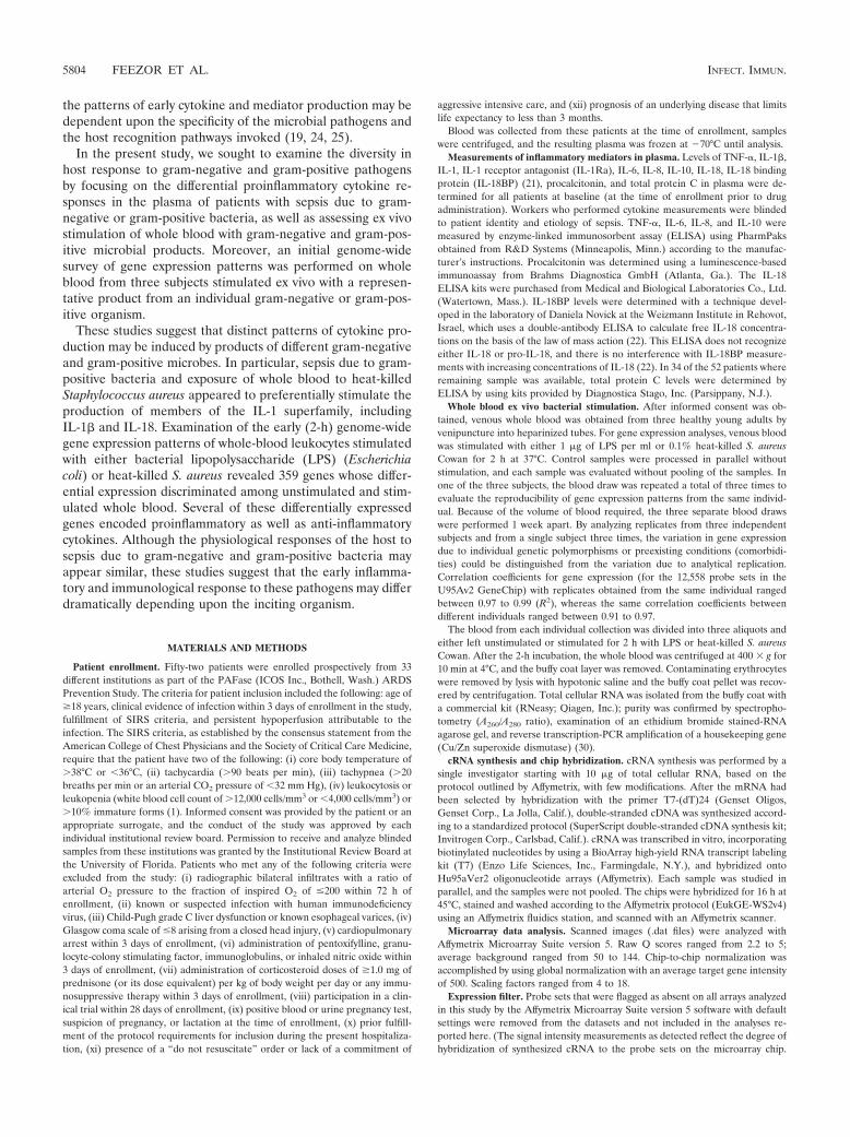
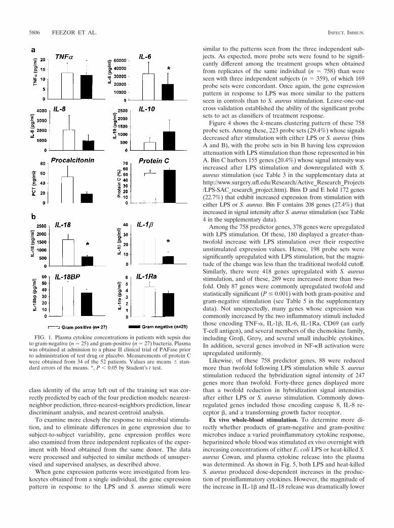
Baseline levels of TNF-�, IL-1Ra, IL-8, IL-10, IL-18BP,procalcitonin, and protein C in plasma were not significantlydifferent between septic patients with gram-positive and gram-negative infections. In contrast, plasma IL-1�, IL-6, and IL-18concentrations were significantly higher among patients withsepsis due to gram-positive bacteria than patients with sepsisdue to gram-negative bacteria (Fig. 1) despite no significantdifferences in the magnitude of the physiologic response(APACHE II score), the degree of organ injury (MODSscore), or other proinflammatory cytokines. Similarly, the con-centration of free IL-18 in plasma was significantly higher inpatients with sepsis due to gram-positive bacteria than thosewith sepsis due to gram-negative bacteria (median, 232 pg/mlfor gram positive versus 81 pg/ml for gram negative by theMann-Whitney rank sum test, P � 0.05). These findings, there-fore, suggest that the patterns of plasma cytokine appearancemay differ between patients with sepsis due to gram-negativeand gram-positive bacteria.
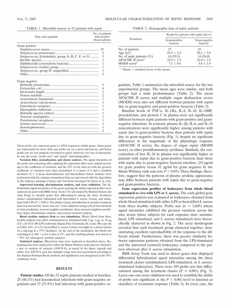
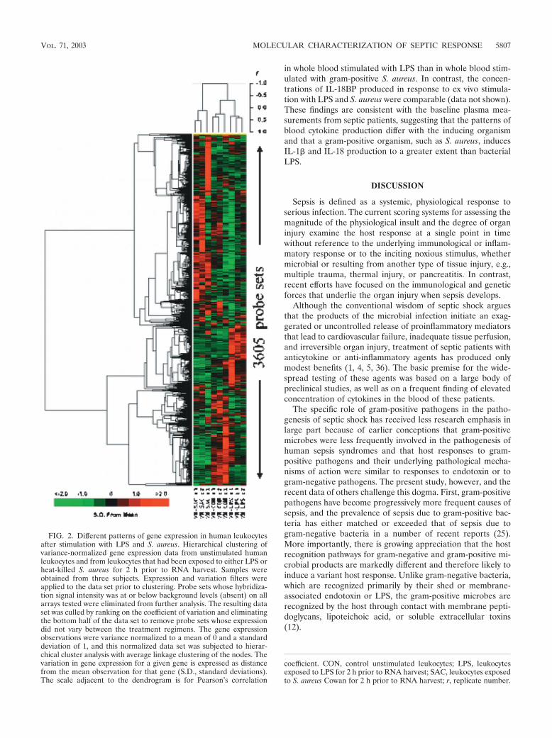
Gene expression profiles of leukocytes from whole bloodstimulated ex vivo with LPS or S. aureus. The early global geneexpression pattern was evaluated in leukocytes obtained fromwhole blood stimulated with either LPS or heat-killed S. aureusfrom three healthy subjects. Probe sets (n 3,605) whosesignal intensities exhibited the greatest variation across thenine arrays (three subjects for each response class: unstimu-lated, LPS stimulated, and S. aureus stimulated) were hierar-chically clustered as shown in Fig. 2. The array dendrogramrevealed that each treatment group clustered together, dem-onstrating excellent reproducibility of the response to the dif-ferent stimuli. Furthermore, there was greater similarity be-tween expression patterns obtained from the LPS-stimulatedand the untreated (control) leukocytes, compared to the pat-tern observed after S. aureus stimulation.
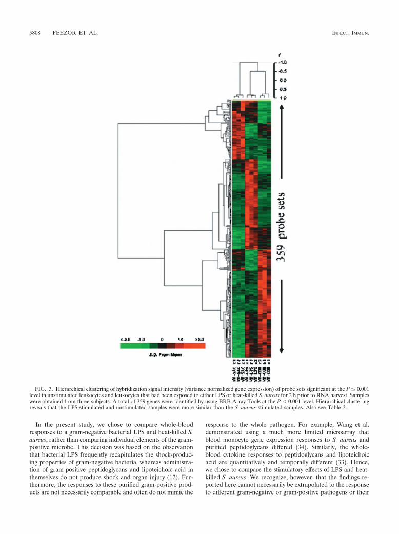
BRB Array Tools was used to detect genes that displayeddifferential hybridization signal intensities among the threetreatment classes (unstimulated, LPS-stimulated, or S. aureus-stimulated leukocytes). There were 359 probe sets that differ-entiated among the treatment classes (P � 0.001) (Fig. 3).Leave-one-out cross validation was used to establish the abilityof probe sets significant at the P � 0.001 level to function asclassifiers of treatment response (Table 3). In every case, the
TABLE 1. Microbial sources in 52 patients with sepsis
Type and organismNo. of patients
with positiveblood cultures
Gram positiveStaphylococcus aureus.............................................................. 15Streptococcus pneumoniae ....................................................... 3Streptococcus, �-hemolytic, group A, B, C, F, or G ........... 3Bacillus species......................................................................... 1Diphtheroids (coryneform bacteria) ..................................... 1Streptococcus viridans group................................................... 1Streptococcus, group D, unspecified ...................................... 1Other ......................................................................................... 2
Gram negativeKlebsiella pneumoniae.............................................................. 5Escherichia coli......................................................................... 4Bacteroides fragilis .................................................................... 2Proteus mirabilis ....................................................................... 2Acinetobacter baumannii ......................................................... 1Acinetobacter calcoaceticus ..................................................... 1Enterobacter aerogenes ............................................................. 1Haemophilus influenzae ........................................................... 1Klebsiella species (other)......................................................... 1Neisseria meningitides............................................................... 1Pseudomonas aeruginosa ......................................................... 1Serratia marcescens .................................................................. 1Stenotrophomonas .................................................................... 1Other ......................................................................................... 3
TABLE 2. Demographic data of septic patients
Parameter
Result for patients with sepsis due to:
Gram-positivebacteria
Gram-negativebacteria
No. of patients 27 25Age (yr)a 54.3 3.4 58.1 3.8No. of male patients (%) 16 (59.3) 14 (56.0)APACHE II scorea 24.9 1.4 21.0 1.9MODS scorea 7.1 0.6 5.6 1.9
a Means standard errors of the means.
VOL. 71, 2003 MOLECULAR CHARACTERIZATION OF SEPTIC RESPONSE 5805
class identity of the array left out of the training set was cor-rectly predicted by each of the four prediction models: nearest-neighbor prediction, three-nearest-neighbors prediction, lineardiscriminant analysis, and nearest-centroid analysis.
To examine more closely the response to microbial stimula-tion, and to eliminate differences in gene expression due tosubject-to-subject variability, gene expression profiles werealso examined from three independent replicates of the exper-iment with blood obtained from the same donor. The datawere processed and subjected to similar methods of unsuper-vised and supervised analyses, as described above.
When gene expression patterns were investigated from leu-kocytes obtained from a single individual, the gene expressionpattern in response to the LPS and S. aureus stimuli were
similar to the patterns seen from the three independent sub-jects. As expected, more probe sets were found to be signifi-cantly different among the treatment groups when obtainedfrom replicates of the same individual (n 758) than wereseen with three independent subjects (n 359), of which 169probe sets were concordant. Once again, the gene expressionpattern in response to LPS was more similar to the patternseen in controls than to S. aureus stimulation. Leave-one-outcross validation established the ability of the significant probesets to act as classifiers of treatment response.
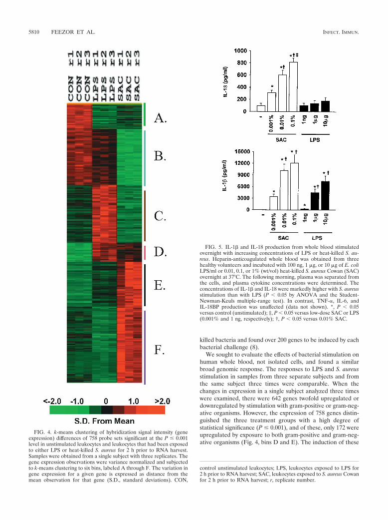
Figure 4 shows the k-means clustering pattern of these 758probe sets. Among these, 223 probe sets (29.4%) whose signalsdecreased after stimulation with either LPS or S. aureus (binsA and B), with the probe sets in bin B having less expressionattenuation with LPS stimulation than those represented in binA. Bin C harbors 155 genes (20.4%) whose signal intensity wasincreased after LPS stimulation and downregulated with S.aureus stimulation (see Table 3 in the supplementary data athttp://www.surgery.ufl.edu/Research/Active_Research_Projects/LPS-SAC_research_project.htm). Bins D and E hold 172 genes(22.7%) that exhibit increased expression from stimulation witheither LPS or S. aureus. Bin F contains 208 genes (27.4%) thatincreased in signal intensity after S. aureus stimulation (see Table4 in the supplementary data).
Among the 758 predictor genes, 378 genes were upregulatedwith LPS stimulation. Of these, 180 displayed a greater-than-twofold increase with LPS stimulation over their respectiveunstimulated expression values. Hence, 198 probe sets weresignificantly upregulated with LPS stimulation, but the magni-tude of the change was less than the traditional twofold cutoff.Similarly, there were 418 genes upregulated with S. aureusstimulation, and of these, 289 were increased more than two-fold. Only 87 genes were commonly upregulated twofold andstatistically significant (P � 0.001) with both gram-positive andgram-negative stimulation (see Table 5 in the supplementarydata). Not unexpectedly, many genes whose expression wascommonly increased by the two inflammatory stimuli includedthose encoding TNF-�, IL-1�, IL-6, IL-1Ra, CD69 (an earlyT-cell antigen), and several members of the chemokine family,including Gro�, Gro�, and several small inducible cytokines.In addition, several genes involved in NF-�B activation wereupregulated uniformly.
Likewise, of these 758 predictor genes, 88 were reducedmore than twofold following LPS stimulation while S. aureusstimulation reduced the hybridization signal intensity of 247genes more than twofold. Forty-three genes displayed morethan a twofold reduction in hybridization signal intensitiesafter either LPS or S. aureus stimulation. Commonly down-regulated genes included those encoding caspase 8, IL-8 re-ceptor �, and a transforming growth factor receptor.
Ex vivo whole-blood stimulation. To determine more di-rectly whether products of gram-negative and gram-positivemicrobes induce a varied proinflammatory cytokine response,heparinized whole blood was stimulated ex vivo overnight withincreasing concentrations of either E. coli LPS or heat-killed S.aureus Cowan, and plasma cytokine release into the plasmawas determined. As shown in Fig. 5, both LPS and heat-killedS. aureus produced dose-dependent increases in the produc-tion of proinflammatory cytokines. However, the magnitude ofthe increase in IL-1� and IL-18 release was dramatically lower
FIG. 1. Plasma cytokine concentrations in patients with sepsis dueto gram-negative (n 25) and gram-positive (n 27) bacteria. Plasmawas obtained at admission to a phase II clinical trial of PAFase priorto administration of test drug or placebo. Measurements of protein Cwere obtained from 34 of the 52 patients. Values are means stan-dard errors of the means. *, P � 0.05 by Student’s t test.
5806 FEEZOR ET AL. INFECT. IMMUN.
in whole blood stimulated with LPS than in whole blood stim-ulated with gram-positive S. aureus. In contrast, the concen-trations of IL-18BP produced in response to ex vivo stimula-tion with LPS and S. aureus were comparable (data not shown).These findings are consistent with the baseline plasma mea-surements from septic patients, suggesting that the patterns ofblood cytokine production differ with the inducing organismand that a gram-positive organism, such as S. aureus, inducesIL-1� and IL-18 production to a greater extent than bacterialLPS.
DISCUSSION
Sepsis is defined as a systemic, physiological response toserious infection. The current scoring systems for assessing themagnitude of the physiological insult and the degree of organinjury examine the host response at a single point in timewithout reference to the underlying immunological or inflam-matory response or to the inciting noxious stimulus, whethermicrobial or resulting from another type of tissue injury, e.g.,multiple trauma, thermal injury, or pancreatitis. In contrast,recent efforts have focused on the immunological and geneticforces that underlie the organ injury when sepsis develops.
Although the conventional wisdom of septic shock arguesthat the products of the microbial infection initiate an exag-gerated or uncontrolled release of proinflammatory mediatorsthat lead to cardiovascular failure, inadequate tissue perfusion,and irreversible organ injury, treatment of septic patients withanticytokine or anti-inflammatory agents has produced onlymodest benefits (1, 4, 5, 36). The basic premise for the wide-spread testing of these agents was based on a large body ofpreclinical studies, as well as on a frequent finding of elevatedconcentration of cytokines in the blood of these patients.
The specific role of gram-positive pathogens in the patho-genesis of septic shock has received less research emphasis inlarge part because of earlier conceptions that gram-positivemicrobes were less frequently involved in the pathogenesis ofhuman sepsis syndromes and that host responses to gram-positive pathogens and their underlying pathological mecha-nisms of action were similar to responses to endotoxin or togram-negative pathogens. The present study, however, and therecent data of others challenge this dogma. First, gram-positivepathogens have become progressively more frequent causes ofsepsis, and the prevalence of sepsis due to gram-positive bac-teria has either matched or exceeded that of sepsis due togram-negative bacteria in a number of recent reports (25).More importantly, there is growing appreciation that the hostrecognition pathways for gram-negative and gram-positive mi-crobial products are markedly different and therefore likely toinduce a variant host response. Unlike gram-negative bacteria,which are recognized primarily by their shed or membrane-associated endotoxin or LPS, the gram-positive microbes arerecognized by the host through contact with membrane pepti-doglycans, lipoteichoic acid, or soluble extracellular toxins(12).
FIG. 2. Different patterns of gene expression in human leukocytesafter stimulation with LPS and S. aureus. Hierarchical clustering ofvariance-normalized gene expression data from unstimulated humanleukocytes and from leukocytes that had been exposed to either LPS orheat-killed S. aureus for 2 h prior to RNA harvest. Samples wereobtained from three subjects. Expression and variation filters wereapplied to the data set prior to clustering. Probe sets whose hybridiza-tion signal intensity was at or below background levels (absent) on allarrays tested were eliminated from further analysis. The resulting dataset was culled by ranking on the coefficient of variation and eliminatingthe bottom half of the data set to remove probe sets whose expressiondid not vary between the treatment regimens. The gene expressionobservations were variance normalized to a mean of 0 and a standarddeviation of 1, and this normalized data set was subjected to hierar-chical cluster analysis with average linkage clustering of the nodes. Thevariation in gene expression for a given gene is expressed as distancefrom the mean observation for that gene (S.D., standard deviations).The scale adjacent to the dendrogram is for Pearson’s correlation
coefficient. CON, control unstimulated leukocytes; LPS, leukocytesexposed to LPS for 2 h prior to RNA harvest; SAC, leukocytes exposedto S. aureus Cowan for 2 h prior to RNA harvest; r, replicate number.
VOL. 71, 2003 MOLECULAR CHARACTERIZATION OF SEPTIC RESPONSE 5807
In the present study, we chose to compare whole-bloodresponses to a gram-negative bacterial LPS and heat-killed S.aureus, rather than comparing individual elements of the gram-positive microbe. This decision was based on the observationthat bacterial LPS frequently recapitulates the shock-produc-ing properties of gram-negative bacteria, whereas administra-tion of gram-positive peptidoglycans and lipoteichoic acid inthemselves do not produce shock and organ injury (12). Fur-thermore, the responses to these purified gram-positive prod-ucts are not necessarily comparable and often do not mimic the
response to the whole pathogen. For example, Wang et al.demonstrated using a much more limited microarray thatblood monocyte gene expression responses to S. aureus andpurified peptidoglycans differed (34). Similarly, the whole-blood cytokine responses to peptidoglycans and lipoteichoicacid are quantitatively and temporally different (33). Hence,we chose to compare the stimulatory effects of LPS and heat-killed S. aureus. We recognize, however, that the findings re-ported here cannot necessarily be extrapolated to the responseto different gram-negative or gram-positive pathogens or their
FIG. 3. Hierarchical clustering of hybridization signal intensity (variance normalized gene expression) of probe sets significant at the P � 0.001level in unstimulated leukocytes and leukocytes that had been exposed to either LPS or heat-killed S. aureus for 2 h prior to RNA harvest. Sampleswere obtained from three subjects. A total of 359 genes were identified by using BRB Array Tools at the P � 0.001 level. Hierarchical clusteringreveals that the LPS-stimulated and unstimulated samples were more similar than the S. aureus-stimulated samples. Also see Table 3.
5808 FEEZOR ET AL. INFECT. IMMUN.
constituents. Clearly, the response to microbial pathogens var-ies depending upon the specific pathogen or microbial product,its concentration, and the duration of the exposure. The find-ings here, however, illustrate the potential differences in theexpression patterns of whole-blood leukocytes in response totwo disparate, but common, stimulants.
The discovery of the TLR family has provided unequivocalevidence that the recognition pathways for gram-negative andgram-positive pathogens are different. Although not fully re-solved, there is accumulating consensus that endotoxin ofgram-negative bacteria is primarily recognized through theCD14-TLR4 cell surface complex, whereas peptidoglycans andlipoteichoic acid are recognized by TLR2 and, to a more lim-ited extent, TLR4. The intracellular signaling pathways forTLR4 and TLR2 are generally similar, as both receptors aremembers of the IL-1 receptor superfamily and employ IL-1receptor-associated kinases. However, TLR4 is apparentlyunique in that intracellular signaling can occur through bothMyD88 (a TLR-associated adapter protein)-dependent and-independent pathways (17).
There has been some recognition that the host responses togram-negative and gram-positive pathogens can vary, althoughthe majority of the studies were performed on isolated cellpopulations with purified products from gram-positive andgram-negative pathogens. Almost 10 years ago, Bjork and col-leagues examined cytokine production in individual peripheralblood mononuclear cells stimulated with endotoxin or Staph-ylococcus enterotoxin A and showed divergent cytokine re-sponses (6). Although endotoxin produced a potent earlyproinflammatory cytokine response characterized by produc-tion of TNF-�, IL-1�, IL-1�, and IL-8, T-cell cytokines such asTNF�, IL-2, IL-4, and gamma interferon were not detected. Incontrast, it was shown that enterotoxin A induced a differentcytokine pattern, with TNF-�, IL-1�, TNF�, IL-4, and gammainterferon production predominating (6). The production ofIL-8, IL-6, and TNF did not discriminate between infectionswith gram-positive and gram-negative bacteria (6).
In the present study, using ex vivo whole blood stimulation,we reached comparable conclusions with the observation thatincreased IL-1� and IL-18 production, not TNF-�, IL-8, orIL-10, discriminated between gram-positive (heat-killed S. au-
reus) and gram-negative (E. coli LPS) stimulants. The ability ofheat-killed S. aureus to induce IL-18 production is consistentwith a recent study by Bocker et al. (7). The present studysuggests that concentrations of these two cytokines (as well asIL-6, to a lesser extent) in plasma may discriminate amongpatients presenting with sepsis due to gram-negative and gram-positive bacteria. Although the total number of patients stud-ied was relatively small (n 52), the concentrations of IL-1�and IL-18 in plasma were nearly two- to threefold higher inpatients with sepsis due to gram-positive bacteria than in thosewith sepsis due to gram-negative bacteria. In this regard, thestudy extends the earlier work of Oberholzer et al. (24) dem-onstrating significantly elevated plasma IL-18 concentrationsin patients with sepsis compared to severely injured traumapatients and healthy humans. Furthermore, those authorsfound that septic patients who died and patients with septicshock exhibited higher levels of IL-18 than survivors and septicpatients without shock. In addition, septic patients with gram-positive organisms had significantly higher plasma IL-18 levelsthan patients with gram-negative organisms (24). In unpub-lished findings, Lungstras-Butler et al. observed in patientswith necrotizing fasciitis of predominantly gram-positive bac-terial origin that circulating levels of IL-18 and IL-1� weremarkedly higher in patients who died than in those who sur-vived (C. A. Dinarello, personal communication).
We can extend these findings to a larger, independent pa-tient population and confirm that despite these different IL-18and IL-1� responses, the plasma IL-18BP concentrations werenot different in patients with sepsis due to gram-positive andgram-negative bacteria. IL-18BP is a constitutively expressedand secreted protein that binds specifically to mature IL-18and neutralizes its activity. We observed no difference in IL-18BP levels in the plasma of patients with sepsis due to gram-negative and gram-positive bacteria, nor did either LPS or heatkilled S. aureus induce an ex vivo IL-18BP release from wholeblood (data not shown). Furthermore, free IL-18 was higheramong patients with gram-positive sepsis than those withgram-negative sepsis. Taken together, these findings suggestthat the host response to a gram-positive pathogen like S.aureus is a preferential induction of IL-18 in the absence of anyincreased production of one of its natural inhibitors, such asIL-18BP.
To further elucidate the differential host response to gram-negative and gram-positive stimulation, we employed Af-fymetrix microarrays to globally explore gene expression pat-terns in whole blood in response to ex vivo LPS and S. aureusstimulation. Samples were obtained from three healthy sub-jects, and for one of the subjects, the analyses were repeatedthree times. In this manner, the variation in gene expressionbetween and within healthy donors could be evaluated. Al-though microarray technology is relatively new, it has beenused to compare responses by inflammatory cells to variousstimuli. Nau et al. evaluated gene expression responses tomacrophages cultured with gram-positive, gram-negative, andmycobacterial stimuli (20). Of the genes they studied, 977exhibited altered expression due to the action of at least one ofthese stimulants but only 132 were commonly upregulatedamong the eight stimuli tested (20). Similarly, Boldrick et al.stimulated blood mononuclear cells with LPS and diverse heat-
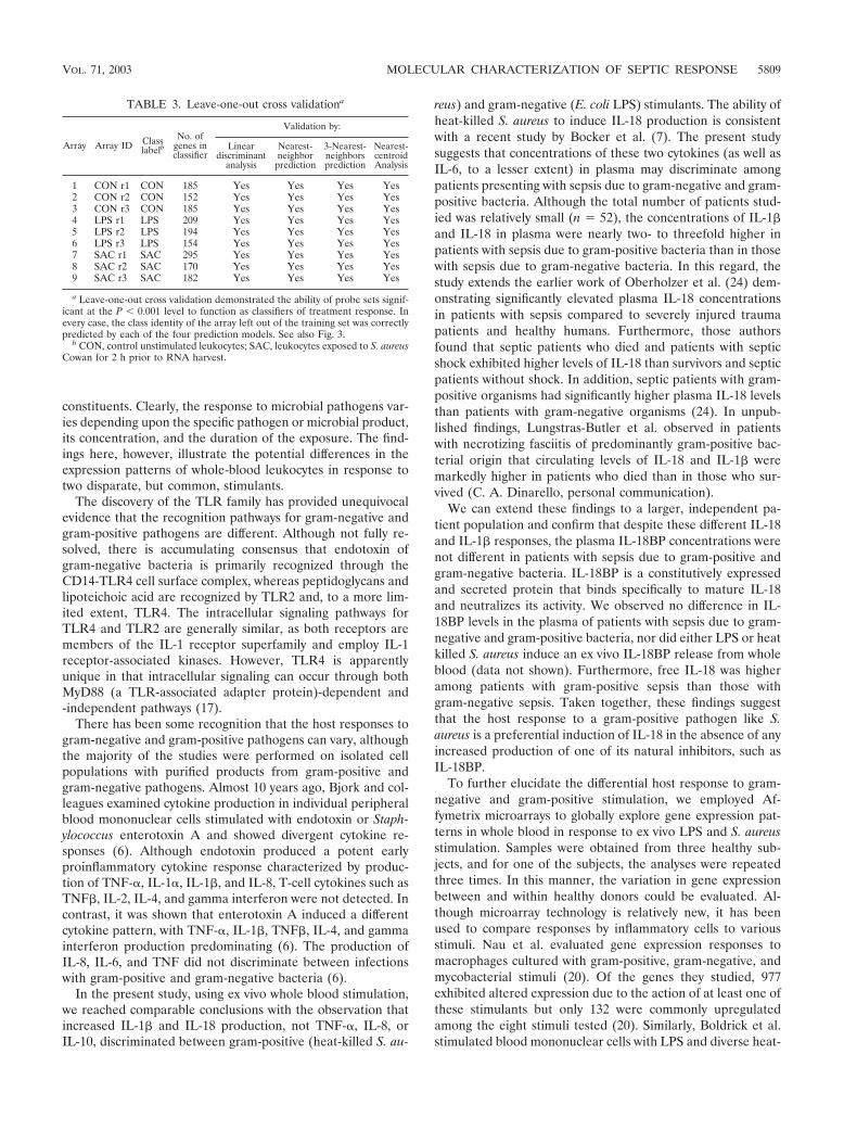
TABLE 3. Leave-one-out cross validationa
Array Array ID Classlabelb
No. ofgenes inclassifier
Validation by:
Lineardiscriminant
analysis
Nearest-neighbor
prediction
3-Nearest-neighborsprediction
Nearest-centroidAnalysis
1 CON r1 CON 185 Yes Yes Yes Yes2 CON r2 CON 152 Yes Yes Yes Yes3 CON r3 CON 185 Yes Yes Yes Yes4 LPS r1 LPS 209 Yes Yes Yes Yes5 LPS r2 LPS 194 Yes Yes Yes Yes6 LPS r3 LPS 154 Yes Yes Yes Yes7 SAC r1 SAC 295 Yes Yes Yes Yes8 SAC r2 SAC 170 Yes Yes Yes Yes9 SAC r3 SAC 182 Yes Yes Yes Yes
a Leave-one-out cross validation demonstrated the ability of probe sets signif-icant at the P � 0.001 level to function as classifiers of treatment response. Inevery case, the class identity of the array left out of the training set was correctlypredicted by each of the four prediction models. See also Fig. 3.
b CON, control unstimulated leukocytes; SAC, leukocytes exposed to S. aureusCowan for 2 h prior to RNA harvest.
VOL. 71, 2003 MOLECULAR CHARACTERIZATION OF SEPTIC RESPONSE 5809
killed bacteria and found over 200 genes to be induced by eachbacterial challenge (8).
We sought to evaluate the effects of bacterial stimulation onhuman whole blood, not isolated cells, and found a similarbroad genomic response. The responses to LPS and S. aureusstimulation in samples from three separate subjects and fromthe same subject three times were comparable. When thechanges in expression in a single subject analyzed three timeswere examined, there were 642 genes twofold upregulated ordownregulated by stimulation with gram-positive or gram-neg-ative organisms. However, the expression of 758 genes distin-guished the three treatment groups with a high degree ofstatistical significance (P � 0.001), and of these, only 172 wereupregulated by exposure to both gram-positive and gram-neg-ative organisms (Fig. 4, bins D and E). The induction of these
FIG. 4. k-means clustering of hybridization signal intensity (geneexpression) differences of 758 probe sets significant at the P � 0.001level in unstimulated leukocytes and leukocytes that had been exposedto either LPS or heat-killed S. aureus for 2 h prior to RNA harvest.Samples were obtained from a single subject with three replicates. Thegene expression observations were variance normalized and subjectedto k-means clustering to six bins, labeled A through F. The variation ingene expression for a given gene is expressed as distance from themean observation for that gene (S.D., standard deviations). CON,
FIG. 5. IL-1� and IL-18 production from whole blood stimulatedovernight with increasing concentrations of LPS or heat-killed S. au-reus. Heparin-anticoagulated whole blood was obtained from threehealthy volunteers and incubated with 100 ng, 1 �g, or 10 �g of E. coliLPS/ml or 0.01, 0.1, or 1% (wt/vol) heat-killed S. aureus Cowan (SAC)overnight at 37°C. The following morning, plasma was separated fromthe cells, and plasma cytokine concentrations were determined. Theconcentrations of IL-1� and IL-18 were markedly higher with S. aureusstimulation than with LPS (P � 0.05 by ANOVA and the Student-Newman-Keuls multiple-range test). In contrast, TNF-�, IL-6, andIL-18BP production was unaffected (data not shown). *, P � 0.05versus control (unstimulated); ‡, P � 0.05 versus low-dose SAC or LPS(0.001% and 1 ng, respectively); †, P � 0.05 versus 0.01% SAC.
control unstimulated leukocytes; LPS, leukocytes exposed to LPS for2 h prior to RNA harvest; SAC, leukocytes exposed to S. aureus Cowanfor 2 h prior to RNA harvest; r, replicate number.
5810 FEEZOR ET AL. INFECT. IMMUN.
genes is consistent with what Nau and colleagues have definedas a “shared activation program” primarily responsible fortransforming the leukocyte into “a cell primed to interact withits environment and mount an immune response” (20). Ourdata perhaps showed fewer common response elements in partdue to the method of statistical analysis chosen, which identi-fied genes that were capable of discrimination among the threeexperimental treatment classes.
More surprising, however, was the divergence of the re-sponse to bacterial product stimulation. k-means clustering ofthe same data set revealed 155 probe sets to be upregulatedwith LPS stimulation and downregulated with S. aureus Cowanstimulation (Fig. 4, bin C; see also Table 3 in the supplemen-tary data) and 208 that were upregulated with Staphylococcusstimulation and downregulated with LPS (Fig. 4, bin F; see alsoTable 4 in the supplementary data). As expected, the genesupregulated by LPS included genes involved in proinflamma-tory signaling, such as those encoding interferon-regulatoryfactors, interferon-stimulated proteins, mitogen-activated pro-tein kinase-activating proteins, platelet-activating factor recep-
tor, protein C kinase, and signal transduction genes such asNF-�B and I-�B. This pattern of gene expression may havereflected the blood monocyte being the primary target of LPSin whole blood rather than the T lymphocyte. In contradistinc-tion to plasma cytokine measurements, the expression of IL-18was upregulated with stimulation by gram-negative organisms.
In contrast, there were several T-cell genes specifically up-regulated to a greater extent with S. aureus than with LPS,including those for granzyme A, an activated T-cell nuclearfactor, and a T-cell receptor. These responses are not generallyunexpected, given that T-cell activation occurs rapidly in re-sponse to S. aureus. However, there were marked increases inthe expression of several of the heat-shock proteins and 52different ribosomal proteins, which were not seen with LPSstimulation.
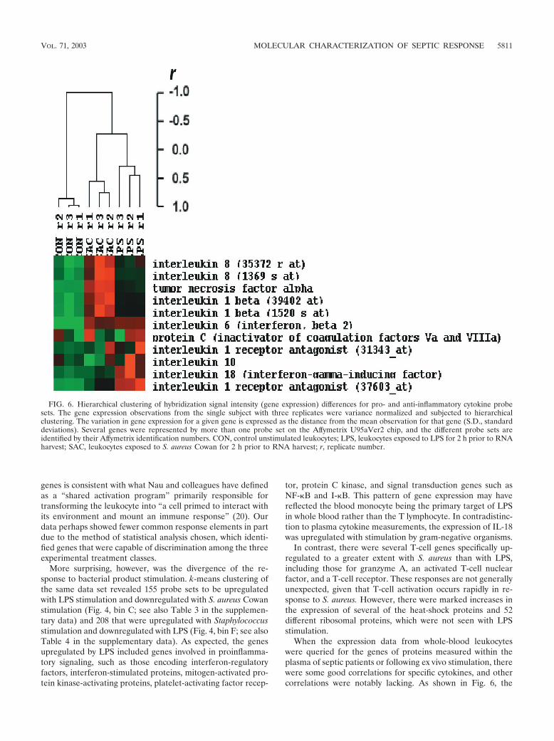
When the expression data from whole-blood leukocyteswere queried for the genes of proteins measured within theplasma of septic patients or following ex vivo stimulation, therewere some good correlations for specific cytokines, and othercorrelations were notably lacking. As shown in Fig. 6, the
FIG. 6. Hierarchical clustering of hybridization signal intensity (gene expression) differences for pro- and anti-inflammatory cytokine probesets. The gene expression observations from the single subject with three replicates were variance normalized and subjected to hierarchicalclustering. The variation in gene expression for a given gene is expressed as the distance from the mean observation for that gene (S.D., standarddeviations). Several genes were represented by more than one probe set on the Affymetrix U95aVer2 chip, and the different probe sets areidentified by their Affymetrix identification numbers. CON, control unstimulated leukocytes; LPS, leukocytes exposed to LPS for 2 h prior to RNAharvest; SAC, leukocytes exposed to S. aureus Cowan for 2 h prior to RNA harvest; r, replicate number.
VOL. 71, 2003 MOLECULAR CHARACTERIZATION OF SEPTIC RESPONSE 5811
expression of the genes encoding the pro- and anti-inflamma-tory cytokines that were measured in the plasma were in-creased by both gram-negative and gram-positive stimuli com-pared to unstimulated controls. However, in many cases, themagnitude of the increase did not necessarily correlate withthe increase in protein concentration seen in the plasma or inex vivo-stimulated whole blood. For example, IL-8, IL-1�, andTNF-� gene expression was increased further by S. aureusCowan than by E. coli LPS, but only IL-1� levels were in-creased with the gram-positive organism. Similarly, LPS ap-peared to induce whole-blood leukocyte expression of IL-18 toa greater extent than S. aureus, whereas the protein responseswere markedly greater with the gram-positive organism.
These disparate results between gene expression and proteinproduction reflect a general limitation of experimental ap-proaches based on mRNA expression, since they do not con-sider posttranscriptional regulation. It is possible that this find-ing of diminished IL-18 gene expression upon stimulation withgram-positive bacteria relative to that with gram-negative bac-teria reflects the importance of posttranscriptional modifica-tion of IL-18 protein. IL-18 and IL-1�, for that matter, requireprocessing from a higher-molecular-weight intracellular spe-cies to the biologically active secreted protein (13). Regulationat this level of IL-18 processing may be rate limiting for itssecretion and plasma appearance.
In addition, an overall discrepancy between leukocyte geneexpression and plasma cytokine concentrations may occur be-cause whole-blood leukocytes are not necessarily the primarysource for many of the cytokines in systemic circulation. Al-though TNF-� expression in blood leukocytes was higher fol-lowing S. aureus stimulation than LPS stimulation, TNF-� lev-els in the blood of patients with sepsis due to gram-positivebacteria were not elevated compared to levels in patients withsepsis due to gram-negative bacteria. It may well be that bloodleukocytes are not the primary source of TNF-� in the circu-lation of septic patients. We showed, for example, that morethan 60% of the TNF-� in the peripheral blood of patientsfollowing an in vivo endotoxin challenge originated from thevisceral organs and the hepatic venous circulation, rather thanthe systemic circulation (15).
What these studies do underscore, however, is the incom-pleteness of our fundamental understanding of the host re-sponse to gram-negative and gram-positive pathogens. Al-though the results are limited by the study of a single gram-positive and gram-negative stimulant, they do reveal that thereare common elements in the responses to these pathogens,including the release of several proinflammatory cytokines,including TNF-�, IL-6, and some chemokines in the blood ofseptic patients. In response to both in vivo and ex vivo stimu-lation, however, the host responses to the two different classesof pathogens are probably as disparate as they are similar. Wemay have been inappropriately reassured that the responseswere similar based on the comparable clinical presentations, aswell as a limited number of cytokines in the blood, includingIL-6 and TNF-�. Although IL-1� and IL-18 are two clearexamples of where the host responses to LPS and heat killed S.aureus differ significantly, the gene expression data suggest thatthis may be only the beginning. Examining the global geneexpression pattern suggests that the responses are considerablymore diverse than originally predicted and that the genes
which make up the shared activation program are relativelylimited in number and are characterized by many of the pre-viously known mediators, TNF-�, IL-6, chemokines, etc. Ifanything, these studies emphasize that future research shouldfocus on furthering our understanding of the regulation ofpathogen-specific genes and how their products contribute tothe host response. These complexities in the host response todifferent pathogens will make it more difficult to formulatebiological response modifiers for patients with sepsis withbroad action against various classes of pathogens and theunique host responses they elicit.
ACKNOWLEDGMENTS
This study was supported in part by grants GM-40586 (L.L.M.),AI-15614 (C.A.D.), and HL-68743 (C.A.D.) awarded by the NationalInstitutes of Health (UPSHS). A.O. was supported in part by a grantfrom the Swiss National Science Foundation. R.J.F. was supported inpart by a training grant (T32 GM-80721) from the National Institute ofGeneral Medical Sciences in burn and trauma research.
REFERENCES
1. Abraham, E. 1999. Why immunomodulatory therapies have not worked insepsis. Intensive Care Med. 25:556–566.
2. American College of Chest Physicians/Society of Critical Care Medicine.1992. American College of Chest Physicians/Society of Critical Care Medi-cine Consensus Conference: definitions for sepsis and organ failure andguidelines for the use of innovative therapies in sepsis. Crit. Care Med.20:864–874.
3. Angus, D. C., W. T. Linde-Zwirble, J. Lidicker, G. Clermont, J. Carcillo, andM. R. Pinsky. 2001. Epidemiology of severe sepsis in the United States:analysis of incidence, outcome, and associated costs of care. Crit. Care Med.29:1303–1310.
4. Baue, A. E. 1997. Multiple organ failure, multiple organ dysfunction syn-drome, and systemic inflammatory response syndrome. Why no magic bul-lets? Arch. Surg. 132:703–707.
5. Bernard, G. R., J. L. Vincent, P. F. Laterre, S. P. LaRosa, J. F. Dhainaut, A.Lopez-Rodriguez, J. S. Steingrub, G. E. Garber, J. D. Helterbrand, E. W.Ely, and C. J. Fisher, Jr. 2001. Efficacy and safety of recombinant humanactivated protein C for severe sepsis. N. Engl. J. Med. 344:699–709.
6. Bjork, L., J. Andersson, M. Ceska, and U. Andersson. 1992. Endotoxin andStaphylococcus aureus enterotoxin A induce different patterns of cytokines.Cytokine 4:513–519.
7. Bocker, U., T. Manigold, J. M. Watson, M. V. Singer, and S. Rossol. 2001.Regulation of Staphylococcus aureus-mediated activation of interleukin-18in peripheral blood mononuclear cells. Eur. Cytokine Netw. 12:631–638.
8. Boldrick, J. C., A. A. Alizadeh, M. Diehn, S. Dudoit, C. L. Liu, C. E. Belcher,D. Botstein, L. M. Staudt, P. O. Brown, and D. A. Relman. 2002. Stereotypedand specific gene expression programs in human innate immune responses tobacteria. Proc. Natl. Acad. Sci. USA 99:972–977.
9. Bone, R. C. 1994. Gram-positive organisms and sepsis. Arch. Intern. Med.154:26–34.
10. Centers for Disease Control and Prevention. 1990. Increase in NationalHospital Discharge Survey rates for septicemia—United States. 1979–1987.Morb. Mortal. Wkly. Rep. 39:31–34.
11. Cockerill, F. R., III, J. G. Hughes, E. A. Vetter, R. A. Mueller, A. L. Weaver,D. M. Ilstrup, J. E. Rosenblatt, and W. R. Wilson. 1997. Analysis of 281,797consecutive blood cultures performed over an eight-year period: trends inmicroorganisms isolated and the value of anaerobic culture of blood. Clin.Infect. Dis. 24:403–418.
12. De Kimpe, S. J., M. Kengatharan, C. Thiemermann, and J. R. Vane. 1995.The cell wall components peptidoglycan and lipoteichoic acid from Staphy-lococcus aureus act in synergy to cause shock and multiple organ failure.Proc. Natl. Acad. Sci. USA 92:10359–10363.
13. Dinarello, C. A. 2001. Novel targets for interleukin 18 binding protein. Ann.Rheum. Dis. 60(Suppl. 3):iii18–iii24.
14. Eisen, M. B., P. T. Spellman, P. O. Brown, and D. Botstein. 1998. Clusteranalysis and display of genome-wide expression patterns. Proc. Natl. Acad.Sci. USA 95:14863–14868.
15. Fong, Y. M., M. A. Marano, L. L. Moldawer, H. Wei, S. E. Calvano, J. S.Kenney, A. C. Allison, A. Cerami, G. T. Shires, and S. F. Lowry. 1990. Theacute splanchnic and peripheral tissue metabolic response to endotoxin inhumans. J. Clin. Invest. 85:1896–1904.
16. Fry, D. E. 2000. Sepsis syndrome. Am. Surg. 66:126–132.17. Kaisho, T., K. Hoshino, T. Iwabe, O. Takeuchi, T. Yasui, and S. Akira. 2002.
Endotoxin can induce MyD88-deficient dendritic cells to support T(h)2 celldifferentiation. Int. Immunol. 14:695–700.
5812 FEEZOR ET AL. INFECT. IMMUN.
18. Krishnagopalan, S., and R. P. Dellinger. 2001. Innovative therapies forsepsis. BioDrugs 15:645–654.
19. Muller-Alouf, H., J. E. Alouf, D. Gerlach, J. H. Ozegowski, C. Fitting, andJ. M. Cavaillon. 1994. Comparative study of cytokine release by humanperipheral blood mononuclear cells stimulated with Streptococcus pyogenessuperantigenic erythrogenic toxins, heat-killed streptococci, and lipopolysac-charide. Infect. Immun. 62:4915–4921.
20. Nau, G. J., J. F. Richmond, A. Schlesinger, E. G. Jennings, E. S. Lander, andR. A. Young. 2002. Human macrophage activation programs induced bybacterial pathogens. Proc. Natl. Acad. Sci. USA 99:1503–1508.
21. Novick, D., S. H. Kim, G. Fantuzzi, L. L. Reznikov, C. A. Dinarello, and M.Rubinstein. 1999. Interleukin-18 binding protein: a novel modulator of theTh1 cytokine response. Immunity 10:127–136.
22. Novick, D., B. Schwartsburd, R. Pinkus, D. Suissa, I. Belzer, Z. Sthoeger,W. F. Keane, Y. Chvatchko, S. H. Kim, G. Fantuzzi, C. A. Dinarello, and M.Rubinstein. 2001. A novel IL-18BP ELISA shows elevated serum IL-18BP insepsis and extensive decrease of free IL-18. Cytokine 14:334–342.
23. Oberholzer, A., C. Oberholzer, R. M. Minter, and L. L. Moldawer. 2001.Considering immunomodulatory therapies in the septic patient: should ap-optosis be a potential therapeutic target? Immunol. Lett. 75:221–224.
24. Oberholzer, A., U. Steckholzer, M. Kurimoto, O. Trentz, and W. Ertel. 2001.Interleukin-18 plasma levels are increased in patients with sepsis comparedto severely injured patients. Shock 16:411–414.
25. Opal, S. M., and J. Cohen. 1999. Clinical gram-positive sepsis: does itfundamentally differ from gram-negative bacterial sepsis? Crit. Care Med.27:1608–1616.
26. Parrillo, J. E. 1993. Pathogenetic mechanisms of septic shock. N. Engl.J. Med. 328:1471–1477.
27. Parrillo, J. E., M. M. Parker, C. Natanson, A. F. Suffredini, R. L. Danner,R. E. Cunnion, and F. P. Ognibene. 1990. Septic shock in humans. Advances
in the understanding of pathogenesis, cardiovascular dysfunction, and ther-apy. Ann. Intern. Med. 113:227–242.
28. Takeuchi, O., and S. Akira. 2001. Toll-like receptors; their physiological roleand signal transduction system. Int. Immunopharmacol. 1:625–635.
29. Takeuchi, O., K. Hoshino, T. Kawai, H. Sanjo, H. Takada, T. Ogawa, K.Takeda, and S. Akira. 1999. Differential roles of TLR2 and TLR4 in recog-nition of gram-negative and gram-positive bacterial cell wall components.Immunity 11:443–451.
30. Tannahill, C. L., K. Fukuzuka, T. Marum, Z. Abouhamze, S. L. MacKay,E. M. Copeland III, and L. L. Moldawer. 1999. Discordant tumor necrosisfactor-alpha superfamily gene expression in bacterial peritonitis and endo-toxemic shock. Surgery 126:349–357.
31. Valles, J., C. Leon, F. Alvarez-Lerma, et al. 1997. Nosocomial bacteremia incritically ill patients: a multicenter study evaluating epidemiology and prog-nosis. Clin. Infect. Dis. 24:387–395.
32. Vincent, J. L. 1998. Search for effective immunomodulating strategiesagainst sepsis. Lancet 351:922–923.
33. Wang, J. E., P. F. Jorgensen, M. Almlof, C. Thiemermann, S. J. Foster, A. O.Aasen, and R. Solberg. 2000. Peptidoglycan and lipoteichoic acid from Staph-ylococcus aureus induce tumor necrosis factor alpha, interleukin 6 (IL-6), andIL-10 production in both T cells and monocytes in a human whole bloodmodel. Infect. Immun. 68:3965–3970.
34. Wang, Z. M., C. Liu, and R. Dziarski. 2000. Chemokines are the mainproinflammatory mediators in human monocytes activated by Staphylococ-cus aureus, peptidoglycan, and endotoxin. J. Biol. Chem. 275:20260–20267.
35. Wheeler, A. P., and G. R. Bernard. 1999. Treating patients with severe sepsis.N. Engl. J. Med. 340:207–214.
36. Zeni, F., B. Freeman, and C. Natanson. 1997. Anti-inflammatory therapies totreat sepsis and septic shock: a reassessment. Crit. Care Med. 25:1095–1100.
Editor: F. C. Fang
VOL. 71, 2003 MOLECULAR CHARACTERIZATION OF SEPTIC RESPONSE 5813