Embed Size (px)
Citation preview

VIROLOGY 167, 361-369 (1988)
Molecular Characterization of a Prominent Antigen of the Vaccinia Virus Envelope
JAMES GORDON, THOMAS KOVALA, AND SAMUEL DALES 1
Cytobiology Group, The University of Western Ontario, London, Ontario, Canada N6A 5C1
Received Apri129, 1988;accepted July26, 1988
During vaccinia virus (VV) assembly a major polypeptide migrating with an apparent MW of 35K, designated Ag35, is expressed as an early function and becomes an integral component of the lipoprotein envelope surrounding the mature virion. In a previous study evaluating humoral immunity to VV, a prominent response against Ag35 was invariably detected in immunized mice. In the context of our continuing investigations of the structure and function of the vaccinia envelope, with a view to alteration in antigenicity of this agent when used as a vaccine vector for foreign antigens, we carried out detailed mapping of the Ag35 gene, as well as determination of the nucleotide sequence. Use of hybridiza- tion-arrested translation, coupled with immunoprecipitation, located this gene within a 2.7-kbp EcoRI fragment of the larger 8.7-kbp Hindlll H fragment. By means of $1 endonuclease resistance analysis a viral transcript was identified at the site of the Ag35 gene, where the occurrence of an open reading frame (ORF), corresponding to the transcript, was deduced from DNA sequence determination. However, the ORF encodes a polypeptide of only 22,300 Da predicted MW, which is much lower than the apparent MW estimated from SDS-polyacrylamide gel electrophoresis. The size discrepancy is not due to glycosylation or phosphorylation of Ag35 but may result from a proline-rich sequence which occurs in this polypeptide. To confirm that the ORF recognized in this study does, indeed, encode Ag35, the gene was expressed as a fl-galactosidase fusion protein in pUC19; Escherichia coil transformed with the relevant clones ex- pressed a polypeptide of the appropriate molecular weight and antigenicity, when tested by Western blots. Regarding secondary structure and hydropathicity it can be predicted from the DNA sequence that Ag35 is highly hydrophilic but contains a hydrophobic region at the carboxy terminus, perhaps providing the stretch involved in membrane insertion. Computer search of a bank of protein sequences revealed an unusually strong similarity of 68% between the Ag35 at amino acid positions 44-121 and the G glycoprotein of respiratory syncytial virus at positions 189-264. © 1988 Academic Press, Inc.
INTRODUCTION
The orthopoxviruses are a group of large, structurally complex DNA-containing viruses which replicate in the cytoplasm of infected cells. Vaccinia virus has been the most extensively studied of these viruses. The vaccinia virion has been shown to contain at least 100 polypep- tides, including a large number of enzymes required for the immediate early phase of infection (Reviewed by Dales and Pogo, 1981). Replication and maturation of the virus occur in viral factories or virosomes located in the cytoplasm. The assembly of virions occurs by a unique mechanism involving the virus-induced de novo synthesis and assembly of bilayer membranes which are formed into the viral envelope. Although some viri- ons disseminated from the host cell may acquire an external wrapping membrane derived from the Golgi, the cytoplasmic form of the mature virus particle, en- closed within the virus-specified envelope, is fully in- fectious.
The vaccinia virus genome for which a restriction map is available (DeFillipes, 1982) is a 187-kbp mole- cule of linear double-stranded DNA with inverted termi-
nal repeats and covalently closed ends. A review of the molecular biology and genetics of this agent can be found in Dales and Pogo (1981) and Moss (1985). Vac- cinia genes appear to be closely spaced, contiguous stretches of DNA with no evidence of any significant grouping of viral genes along temporal or functional grounds. Nor has mRNA splicing been shown to occur, although recent studies have suggested that a novel mechanism of discontinuous transcription may be in- volved in the synthesis of late vaccinia m RNA (Bertholet et aL, 1987; Schwer et al., 1987).
Much interest has been generated in the use of vac- cinia recombinants as foreign gene vectors for vaccine purposes (for review see Mackett and Smith, 1986). Widespread, safe application of such vaccines neces- sitates a better understanding of host cell-virus inter- actions, especially those pertaining to the viral anti- gens recognized during the immune response of the host. In this study, we report the physical mapping and DNA sequencing of the gene encoding a major vac- cinia antigen which is a component of the viral enve- lope. 2
1 To whom requests for reprints should be addressed. 2 A preliminary report on Ag35 mapping was given at the Sixth Pox-
virus/Iridovirus Workshop, Cold Spring Harbor, 1986.
361 0042-6822/88 $3.00 Copyright © 1988 byAoademic Press, Inc. All rights of reproduction in any form reserved.

362 GORDON, KOVALA, AND DALES
MATERIALS AND METHODS
Cells and virus
Mouse L2 fibroblasts were maintained in nutrient media (NM) consisting of Eagle's modified MEM sup- plemented with 10% fetal bovine serum. IHD-W, the hemagglutinin-deficient (HA-) variant of vaccinia virus (VV) was propagated and titrated by plaque assay on L cells, as previously described (Weintraub et aL, 1974). Purification of virus from infected cells was carried out using potassium tartrate gradients, as previously de- scribed (Stern and Dales, 1974).
Isolation of DNA and RNA
Vaccinia DNA was isolated from purified virions, or viral cores, by digestion with 50 #g/ml proteinase K in 10 mMTris-HOl, pH 7.5, 1 mM EDTA, 0.1% SDS for 1 hr at 50 °, then by repeated extraction with buffer-satu- rated phenol (10 mMTris, pH 8.0), which was followed by dialysis against TE (10 mM Tris, 1 mM EDTA), pH 7.5.
Plasmid DNA was prepared by the alkaline lysis pro- cedure of Thompson et aL (1983) and concentrated in CsCI gradients, according to Maniatis eta/. (1982).
Total RNA was extracted from VV-infected cells, as described by Cheley eta/. (1981). Poly(A) + RNA was separated using oligo(dT) column chromatography (Collaborative Research), as described by Maniatis et al. (1982).
Preparation of antisera to viral envelope
Viral envelope components were solubilized using Nonidet-P40, as previously described (Stern and Dales, 1976).
Antiserum to these components was obtained from male albino rabbits immunized by conventional proce- dures, as in Wilton eta/. (1986).
Protein gels and electroblotting
SDS-polyacrylamide gel electrophoresis (SDS- PAGE) was carried out as described by Laemmli (1970). Transfer of polypeptides from polyacrylamide gels to nitrocellulose filters was carried out according to Towbin eta/. (1979). Antigen was detected on West- ern blots, as in Batteiger et aL (1982), as modified by Wilton eta/ . (1986), using ~251-conjugated protein A (New England Nuclear), to detect specific antibody binding.
Restriction endonuclease digestion and agarose gel electrophoresis
Restriction endonuclease digestions and DNA gel electrophoresis followed the methods adopted by Gor-
don and Carstens (1984). The individual DNA frag- ments produced by restriction endonucleases were isolated and purified from agarose gels by the method of Vogelstein and Gillespie (1979) and ligated into re- stricted pUC19 DNA or M13 mp18 or mp19 DNA (Ya- nisch-Perron etaL, 1985) as previously described (Gor- don and Carstens, 1984), then were employed to trans- form competent Escherichia coil JM109 cells, as described by Hanahan (1983).
Hybridization-arrested translation
Physical mapping of Ag35 was carried out by hybrid- ization-arrested translation, according to Patterson et aL (1977). Plasmid clones representing most of the ge- nome of VV strain WR (Jones and Moss, 1984) were kindly supplied by B. Moss (National Institutes of Health). Poly(A) + RNA from infected cells was hybrid- ized to denatured restriction fragments of VV DNA in 80% formamide, 50 mM Pipes, pH 6.4,400 mM NaCI, 1 mM EDTA for 3 hr at 37 °. The RNA:DNA hybrids were precipitated with ethanol, resuspended in H20, and translated in vitro in the presence of [36S]methionine, employing rabbit reticulocyte lysates (New England Nuclear).
The translation mixture was diluted in immunopre- cipitation buffer (IP) (Shida and Dales, 1982). The su- pernatant was treated with the antiserum to the viral envelope for 16-18 hr, then with goat anti-rabbit anti- serum for 2 hr. This and all subsequent steps were car- ried out at 4 °. The mixture was centrifuged at 15,000 g for 30 min, and the pellets formed were washed three times with IP buffer, resuspended in dissociation buffer, and used in SDS-PAGE analysis. The gels were processed for fluorography using Enhance (New En- gland Nuclear).
Analysis of DNA-RNA hybrids by resistance to S1 nuclease
The 5' end of viral mRNA was identified using $1 nuclease to digest molecular hybrids formed by an as- sociation between 5' end-labeled DNA and poly(A) + RNA. Plasmid clones were isolated possessing inserts with nonidentical restriction endonuctease flanking sites within the DNA region of interest. These recombi- nant plasmids were linearized by digestion at one of the flanking restriction endonuclease sites, labeled at the 5' end by incubation, first with calf intestinal alkaline phosphatase, then with T4 polynucleotide kinase, in the presence of [~.-32P]ATP, as described by Maniatis etaL (1982). These series of reactions produced linear DNA molecules with label at both termini but with VV sequences at only one end. The preparation of 5' end- labeled fragments in this manner eliminated the need
VACCINIA ANTIGEN CHARACTERIZATION 363
to digest the end-labeled DNA with a second restriction endonuclease and purify labeled fragments from aga- rose gels. After denaturation by boiling and rapid cool- ing the linearized DNA was allowed to form hybrids in the presence of 80% formamide, 50 mM Pipes, pH 6.4, 400 mM NaCI, 1 mM EDTA for 3 hr at 42 ° with poly(A) + RNA, obtained from virus-infected cells. Then the reac- tion mixture was diluted with 10 vol of cold (4 ° ) Sl buffer (280 mM NaCI, 50 mM NaOAc, pH 4.6, 4.5 mM ZnSO4, 20 #g/ml BSA) containing 50-150 u of $1 nuclease and incubated at 37 ° for 30 min. After addi- tion of 7.5 M NH4OAc to stop $1 nuclease activity the nucleic acid was precipitated with ethanol, and the pre- cipitates were sedimented into pellets, resuspended, and analyzed by electrophoresis using 5% PAGE and autoradiography.
DNA sequence analysis
M 13 clones for sequence analysis were created by means of a progressive digestion with nuclease BAL31 as described by Poncz et al. (1982). Deletions were prepared from the EcoRV site upstream of the putative Ag35 coding region and from the Xbal site within the coding region. The preparation of single-stranded M 13 phage DNA, sequencing procedures, and gel analysis were carried out according to standard protocols (Big- gin etaL, 1983; Carlson and Messing, 1984). Oligonu- cleotide primers for DNA sequence analysis were ob- tained from the Regional DNA Synthesis Laboratory (University of Calgary). Plasmid sequencing was car- ried out as described by Chen and Seeburg (1985). Computer analysis of DNA sequence used the pack- age described by Pustell and Kafatos (1984).
RESULTS
Identification by antiserum of Ag35 as a component of the viral envelope
A polyvalent antiserum, obtained by immunizing rab- bits with material extracted by NP-40 from purified virus (Stern and Dales, 1976), when used for electroblotting of cell lysates or purified VV, recognized a spectrum of viral antigens similar to that identified by hyperimmune sera from mice (Wilton et aL, 1986). The most promi- nent among these antigens was the Ag35 polypeptide (data not shown), demonstrating that this prominent envelope component elicited a vigorous humoral re- sponse regardless of the species immunized.
Temporal regulation of Ag35 synthesis
The de novo cytoplasmic assembly of VV envelopes from proteins expressed as early functions becomes evident prior to the initiation of viral DNA synthesis
1 2 3
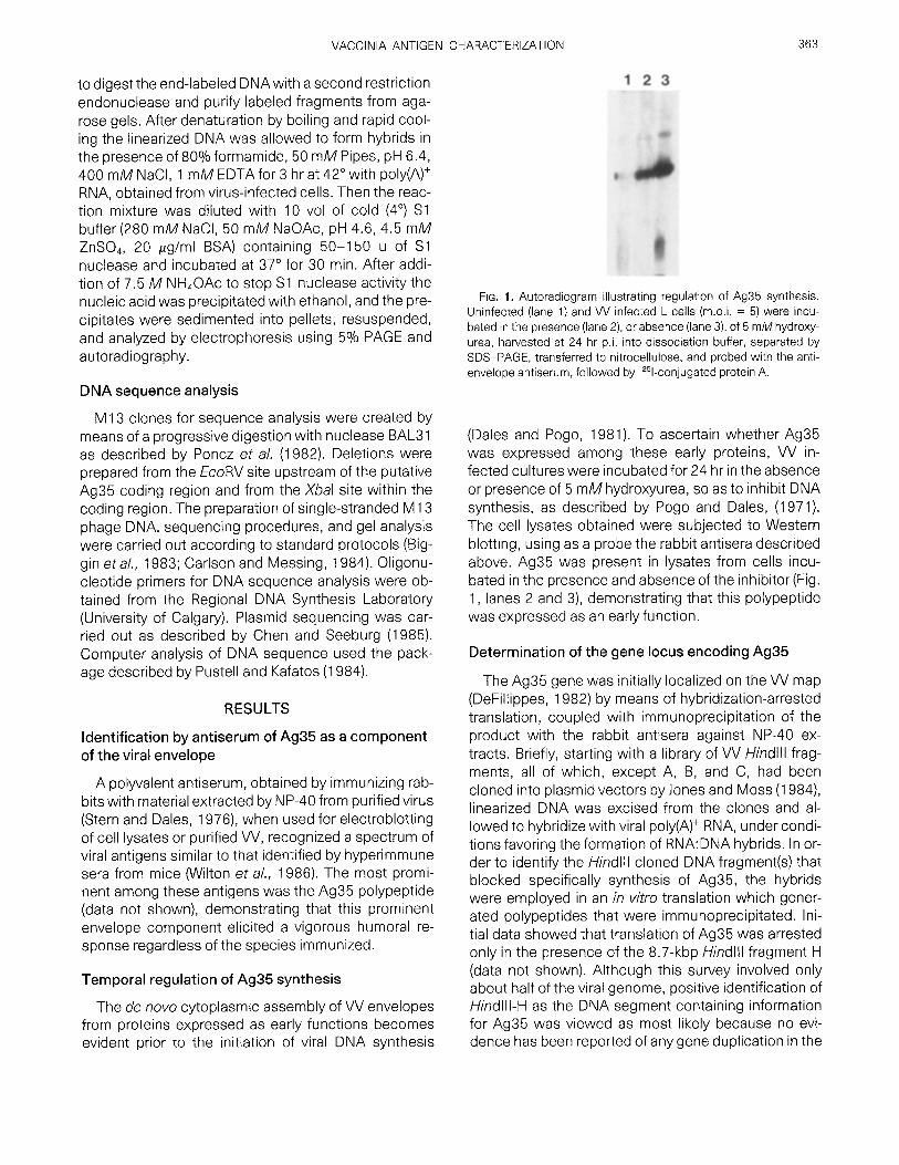
FiG. 1. Autoradiogram illustrating regulation of Ag35 synthesis. Uninfected (lane 1) and VV infected L cells (m.o.i. = 5) were incu- bated in the presence (lane 2), or absence (lane 3), of 5 mM hydroxy- urea, harvested at 24 hr p.i. into dissociation buffer, separated by SDS-PAGE, transferred to nitrocellulose, and probed with the anti- envelope antiserum, followed by 12%conjugated protein A.
(Dales and Pogo, 1981). To ascertain whether Ag35 was expressed among these early proteins, VV in- fected cultures were incubated for 24 hr in the absence or presence of 5 mM hydroxyurea, so as to inhibit DNA synthesis, as described by Pogo and Dales, (1971). The cell lysates obtained were subjected to Western blotting, using as a probe the rabbit antisera described above. Ag35 was present in lysates from cells incu- bated in the presence and absence of the inhibitor (Fig. 1, lanes 2 and 3), demonstrating that this polypeptide was expressed as an early function.
Determination of the gene locus encoding Ag35
The Ag35 gene was initially localized on the VV map (DeFillippes, 1982) by means of hybridization-arrested translation, coupled with immunoprecipitation of the product with the rabbit antisera against NP-40 ex- tracts. Briefly, starting with a library of VV Hindlll frag- ments, all of which, except A, B, and C, had been cloned into plasmid vectors by Jones and Moss (1984), linearized DNA was excised from the clones and al- lowed to hybridize with viral poly(A) + RNA, under condi- tions favoring the formation of RNA:DNA hybrids. In or- der to identify the Hindlll cloned DNA fragment(s) that blocked specifically synthesis of Ag35, the hybrids were employed in an in vitro translation which gener- ated polypeptides that were immunoprecipitated. Ini- tial data showed that translation of Ag35 was arrested only in the presence of the 8.7-kbp Hindlll fragment H (data not shown). Although this survey involved only about half of the viral genome, positive identification of HindllI-H as the DNA segment containing information for Ag35 was viewed as most likely because no evi- dence has been reported of any gene duplication in the

364 GORDON, KOVALA, AND DALES
1 2 3 4 5 6 7 8
H E E EH H i n d l l i . H I i i i t
5 6 7
Fic. 2. Physical mapping of Ag35. Poly(A) + RNA from vaccinia in- fected L cells was hybridized to the cloned restriction endonuclease fragments, identified by capital letters. The hybrids were added to an in vitro rabbit reticulocyte lysate and translation carried out in the presence of [35S]methionine. The products were immunoprecipi- tated with antiserum against the viral envelope and separated by SDS-polyacrylamide gel electrophoresis. Lane 1, Lysate of vaccinia virus infected L cells pulse labeled with [35S]methionine from 12 to 16 hr p.i. to label vaccinia polypeptides; lane 2, total in vitro transla- tion products from vaccinia mRNA; lane 3, immunoprecipitated in vitro translation products; lane 4, HindllI-H; lane 5, 2.8-kbp EcoRI Hindlll fragment; lane 6, 2.7-kbp EcoRI fragment; lane 7, 0.5-kbp EcoRI-Hindlll fragment; lane 8, pUC 18. The arrow marks the posi- tion of Ag35. A restriction map of HindllI-H is shown underneath the blot indicating the origin of the DNA fragments used in lanes 5 7.
central regions of the VV genome, although duplication of information does exist within the inverted terminal repeats (Venkatsesan et aL, 1982).
To localize Ag35 more precisely a physical map of HindllI-H was established for several additional restric- tion enzymes (Fig. 2). The HindllI-H fragment was cleaved at three sites by EcoRI, yielding two internal fragments of 2.7 kbp and terminal EcoRI-Hindlll frag- ments of 2.8 kbp and 0.5 kbp. The internal 2.7-kbp frag- ments could be distinguished from each other by the patterns of subfragments produced after simultaneous hydrolysis with EcoRI and Xbal. To obtain further preci- sion in these mapping studies, subfragments derived from the cloned EcoRI fragments were integrated into plasmid vectors as follows: the termini were cloned into pUC18; the right-hand 2.7-kbp segment was cloned into pBR 325. Despite repeated attempts no clones of the left-hand 2.7-kbp EcoRI fragment were identified. When used for hybridization-arrest transla- tion, as described above, of these cloned subfrag-
ments of HindllI-H DNA only the clone containing the right-hand 2.7-kbp EcoRI fragment prevented Ag35 synthesis, whereas clones containing the terminal fragments did not interfere with the expression of this polypeptide (Fig. 2), indicating that the gene encoding Ag35 must occupy a position either entirely or partly within the right-hand 2.7-kbp EcoRI fragment.
For detailed mapping of the Ag35 gene, Sl nuclease-protection assays were conducted. Two clones spanning the EcoRI to Xbal sites, comprising the original 2.7-kbp EcoRI clone identified by hybridiza- tion-arrested translation, as well as clones flanking this region (Fig. 4), were used. The linearized clones were labeled at the 5' position, denatured, and hybridized to isolated VV poly(A) + RNA. The position of the labeled 5' ends in cloned DNAs involved in $1 protection analysis data, shown in Fig. 3, is illustrated on the map dia- grammed below the figure. The DNA:RNA hybrids which had formed were digested with nuclease $1, then were separated on neutral polyacrylamide gels. As indicated in Fig. 3, a 520-bp $1-protected fragment was evident as a consequence of poly(A) + RNA hybrid- ization to DNA which was 5' end-labeled at the Xbal site within the 2.7-kbp EcoRI fragment. This result indi- cated that the transcript within the RNA:DNA hybrid
1 2 3 4 5 6 7 8
1632-
517- 506-
298- 220-
154-
(1 E X E H 3 5 7
1 2 4 6 8
kbp 0.4 1.6 1.1 0,5
FiG. 3. Mapping the 5' end of the Ag35 transcript. Poly(A) + RNA, isolated from infected cells was hybridized to restriction fragments labeled at the 5' end with 32p. After digestion of hybrids with $1 nuclease and separation of the products on a 5% polyacrylamide gel an autoradiogram was prepared. A restriction map of the region un- der analysis and flanking regions is shown underneath. Arrows indi- cate location of the 5' end-label within the DNA fragments, while the numbers indicate the lane, in the gel above, in which the correspond- ing end-labeled and Sl-digested fragment was analyzed, pBR 322 DNA digested with Hinfl and end labeled with 32p provides the size marker. E, EcoRI; H, Hindlll; X, Xbal.

VACCINIA ANTIGEN CHARACTERIZATION 365
C NMK F F 0 I G L J H D A B A I l l I I IL I I I I I L I
J L
R~BS X E X E X E ~4 ' I i I i I o.s ! B I I I 2.8 2.7 j 2.7
I /
/
/ m R N A ~, I
)~ E V X E H
C [o,4 I 1.6 I I i.i I 0.5[
FIG. 4. Detailed mapping of Ag35 ORF in the genome of strain WR VV. (A) Hindlll map; (B) position of Ag35 ORF in the HindllI-H frag- ment at higher resolution B, BamHI; E, EcoRI; H, Hindlll; S, Sa/I; V, EcoRV; X, Xbal; (C) detailed map of the 2.7 kbp and adjacent frag- ments used in our $1 mapping study. Arrow indicates the location of Ag35 mRNA.
must have been oriented with its 5' end 520 bp to the left of the Xbal site, whereby the 3' end of this transcript was presumably positioned to the right of the Xbal site. The 3' end of the transcript was not actually identified by this procedure, but deduced from DNA sequencing data (see below). The map location of the gene encod- ing Ag35 is summarized in Fig. 4.
Sequencing of the Ag35 gene
The region of the VV genome, shown by the pre- ceeding mapping studies to encode Ag35, was sub- jected to DNA sequence analysis. For this purpose ad- ditional clones were prepared containing subfrag- ments of the Hindlll H fragment of strain IHD-W VV, extensively employed in this laboratory. It was deter- mined, however, that Ag35 of WR virus, the source of the plasmids, was indistinguishable with respect to molecular weight or serological reactivity from the component in IHD-W VV examined by us (unpublished data). Clones were prepared in M 13 which contained I H D-W DNA with "nested" BAL31 deletions to the right and left of the EcoRV and Xbal sites. Results of DNA sequencing are summarized in Fig. 5, from which it can be deduced that a single ORF is present, correspond- ing to the region which was identified as the $1 endo- nuclease protected region. The sequence TTTTTAT, identified as a transcriptional stop site by Yuen and Moss (1987), is located 11 bp downstream of the trans- lational stop site. This ORF contains information suffi- cient to encode a polypeptide of 203 amino acid resi- dues, of 22,300 Da predicted molecular weight. The predicted molecular weight is, therefore, much lower than the MW of 35 kDa estimated from SDS-PAGE. This discrepancy cannot be due to the commonly em countered polypeptide modifications, among them gly- cosylations and phosphorylations, which are absent from Ag35 (Essani and Dales, 1979). A hydropathicity
plot made from the predicted composition of this poly- peptide, as described by Pustell and Kafatos (1984), revealed this protein to be predominantly hydrophilic, possessing a hydrophobic domain near the carboxy terminus (Fig. 7).
Expression of Ag35 in E. coil
The discrepancy in molecular weight between that estimated by SDS-PAGE and that predicted from the DNA sequence makes it necessary to demonstrate clearly that the open reading frame identified and se- quenced does, indeed, encode the polypeptide recog-
-180 -170 -160 -150 -140 -130 * * * * * *
GTTACAAACAACTAGGAAATTGGTTTATGATGTATAATTTTTTTAGTT~TATAGATTCT
-120 -110 -100 -90 -80 -70 * * * * * *
TTATTCTATACTTAAAAAATGAAAATAAATACAAAGGTTCTTGAGGGTTGTGTTAAATTG
-60 -50 -40 -30 -20 -10 , * * * * *
AAAGCGAGAAATAATCATAAATTATTTCATTATCGCGATATCCGTTAAGTTTGTATCGTA
10 20 30 40 50 68 * * * * * *
ATGGCGTGGTCAATTACAAATAAAGCGGATACTAGTAGCTTCACAAAGATGGCTGAAATC M A W S I T N K A D T S S F T K M A E I
70 80 90 100 110 120 * * * * * *
AGAGCTCATCTAAAAAATAGCGCTGAAAATAAAGATAAAAACGAGGATATTTTCCCGGAA R A H L K N S A E N K D K N E D I F P E
130 140 150 160 170 180 * * * * * *
GATGTAATAATTCCATCTACTAAGCCCAAAACCAAACGAGCCACTACTCCTCGTAAACCA D V I I P S T K P K T K R A T T P R K P
190 280 210 220 230 240 * * * * * *
GCGGCTACTAAAAGATCAACCAAAAAGGAGGAAGTGGAAGAAGAAGTAGTTATAGAGGAA A A T K R S T K K E E V E E E V V I E E
250 260 270 280 290 300 * * * * * *
TATCATCAAACAACTGAAAAAAATTCTCCATCTCCTGGAGTCAGCGACATTGTAGAAAGC y H Q T T E K N S P S P G V S D I V E 8
310 320 330 340 350 360 * * * * * *
GTGGCCGCTGTAGAGCTCGATGATAGCGACGGGGATGATGAACCTATGGTACAAGTTGAA V A A V E L D D S D G D D E P M V Q V E
370 380 390 400 410 420 * * * * * *
GCTGGTAAAGTAAATCATAGTGCTAGAAGCGATCTTTCTGACCTAAAGGTGGCTACCGAC A C K V N H S A R S D L S D L K V A T D
430 440 450 460 470 480 * * * * * *
AATATCGTTAAAGATCTTAAGAAAATTATTACTAGAATCTCTGCAGTATCGACGGTTCTA N I V K D L K K I I T R I S A V S T V L
490 500 510 520 530 540 * * * * * *
GAGGATGTTCAAGCAGCTGGTATCTCTAGACAATTTACTTCTATGACTAAAGCTATTACA E D V Q A A G I S R Q F T S M T K A I T
550 560 570 500 590 600 * * * * * *
ACACTATCTGATCTAGTCACCGAGGGAAAATCTAAAGTTGTTCGTAAAAAAGTTAAAACT T L S D L V T E G K S K V V R K K V K T
610 620 630 640 650 660 * * * * * *
TGTAAGAAGTAAATGCGTGCACTTTTTTATAAAGATGGTAAACTCTTTACCGATAATAAT C K K -
670 680 690 700 710 720 * * * * * *
TTTTTAAATCCTGTATCAGACGATAATCCAGCGTATGAGGTTTTGCAACATGTTAAAATT
730 740 750 760
CCTACTCATTTAACAGATGTAGTAGTATATGAACAAACGTGGGA
FIG. 5. DNA sequence and predicted amino acid sequence of the gene encoding Ag35.

366 GORDON, KOVALA, AND DALES
nized by the antiserum directed against the viral enve- lope. In order to confirm that the Ag35 gene identified on the map encodes the 35K envelope antigen, "ex- pression clones" were prepared and tested in E. coll. The plasmid, pHEV 16, encompassing a stretch of DNA from the EcoRV site, upstream of the Ag35 coding region, to the EcoRI site downstream, was digested with EcoRV. The linearized clone was reacted for vary- ing times with exonuclease BAL31, to remove varying numbers of bases within the coding region. Use of BAL31 in this manner allows the removal of an in-frame termination codon which is located a short distance upstream from the translational start site and would re- sult in premature termination of translation. Treatment with BAL31 also allows the preparation of translational fusions representing all three reading frames from the Ag35 locus. Following exposure to BAL31, the partially deleted insert was excised with EcoRI, then separated by electrophoresis. DNA fragments in which deletions up to 200 bp occurred were purified from the gels, then ligated into pUC 19 DNA, predigested with EcoRI and Smal in order to yield one cohesive and one blunt end. This allowed the orientation of the insert such that the BAL31-derived ends were ligated adjacent to the lac promoter of the vector. It was anticipated that transla- tion from the lac promoter of the vector would generate a fusion protein consisting of 17 amino acids from the amino terminus of the pUC 19-derived fl-galactosidase protein (Yanisch-Perron et aL, 1985) linked to the re- maining coding region of Ag35. Competent E. coil JM 109 cells were transformed with the mixture of clones containing the various ligated VV-fl-galactosidase DNAs. Ampicillin resistant, /3-galactosidase- clones were screened for presence of the appropriate size in- serts. After identification Of the relevant transformed E. co~i, the cells were grown overnight into small cultures, from which lysates were prepared for separation by SDS-PAGE, and Western blotting with antisera to VV envelopes.
Among clones expressing polypeptides reactive with the serum against the NP-40-extracted envelope one clone, pAgB3, expressing a polypeptide migrating only slightly faster than authentic Ag35 was selected for fur- ther analysis, pAgB3 expressed a polypeptide of 34K as observed by Western blot analysis only when grown in the presence ofthe lac inducer IPTG (Fig. 6A)indicat- ing that the transcription and translation of the vaccina virus-derived gene had been placed under the control of this E. co~i-derived promoter. Direct plasmid DNA se- quence analysis of pAgB3 using the M13 reverse se- quencing primer (Guo et al., 1983) located the site of fusion between the pUC19-derived /~-galactosidase gene and the Ag35-derived sequence. This analysis re- vealed that 17 amino acids derived from fl-galactosi-
dase were fused in-frame to the Ag35 sequence at leu- cine 24 (Fig. 6B). The predicted molecular weight of the fusion protein derived from the DNA sequence was 21,600. As in the case of the authentic Ag35, the pre- dicted molecular weight of the fusion protein was sig- nificantly less than that estimated from the polypep- tide's migration by SDS-PAGE. This provides further evidence that the disparity between the migration of Ag35 and its molecular weight does not result from post-translational modification in the infected cell but must result from some inherent characteristic of the polypeptide itself.
DISCUSSION
Hyperimmune sera from rabbits and mice (Wilton et al., 1986) detected a prominent antigen with an appar- ent MW of 35K (Ag35) which can be released from VV envelopes by NP-40. This result revealed the occur- rence of a vigorous host humoral response against components on the viral surface. With this evidence at hand we examined the physical characteristics of Ag35 on the assumption that any new information about the polypeptide could be relevant to an under- standing about immune surveillance responses during early stages of VV infection.
Expression of Ag35 in the presence of hydroxyurea shows that this component is an early viral protein, consistent with it being utilized for assembly of VV en- velopes during early stages in formation of immature virions (Pogo and Dales, 1971).
Initial mapping by hybridization-arrested translation, coupled with immunoprecipitation, localized the Ag35 gene near the right end of the 8.7-kbp Hindlll fragment H. By means of $1 nuclease protection analysis of RNA:DNA hybrids and DNA sequencing, this gene was shown to be transcribed with its 5' end corresponding closely in position to an ORF.
From the sequence data the ORF should encode as the gene product a polypeptide of 22,300 Da MW, which is inconsistent with the 35K MW estimated from SDS-PAGE and Western blotting. A similar wide dis- parity between deduced and observed molecular weights was also pointed out recently by Rosel et al. (1986) who made a comparison between the coding capacity of ORFs in the entire HindllI-H DNA of WR VV and the uncharacterized polypeptides from in vitro translation. At present the basis for the molecular weight discrepancy remains obscure but is not likely to be the consequence of post-translational glycosyl- ation, phosphorylation, or fatty acid esterification, be- cause these modifications usually do not occur during in vitro translation by the rabbit reticulocyte lysate sys- tem, unless provisions are made for them. Further-

VACCINIA ANTIGEN CHARACTERIZATION 367
A 2 3
B
pOC 19 ATG ACCATGATTACGCCAAGCTTGCATGCCTG CAGG TCG ACTCTAGAGG ATCCCCGGG TACCGAGCTCG AATTCA \ 11 \11 \11 \11 \11 \11 \11 \11 \11 \11 \11 \11 \11 \11 \1 / \11 \11 \ T M I T P S L H A C R S T L E D P L K N S A E N
I V I V I V I X I I \ / I V I X Ag35 AAGATGG C TG AAATCAGAGCTCATCTAAAAAATAGCGCTGAAAAT
FIG. 6. Expression of the Ag35 gene as a/~-galactosidase fusion protein in Escherichia coll. (A) Western blot analysis of authentic Ag35 or the fusion polypeptide. Lysates of VV infected L cells (lane 1 ) or E. cofiJM 109 cells transformed with the clone pAgB3 grown in the absence (lane 2) or continuous presence (lane 3) of the lac operon inducer IPTG (1 mM) were separated by SDS-PAGE, transferred to nitrocellulose, and probed with the anti-envelope antiserum which had been preadsorbed with a lysate of untransformed E. coliJM109 cells to prevent nonspecific signals followed by 12%conjugated protein A. (B) Predicted amino acid sequence of the/3-galactosidase-Ag35 fusion protein expressed by pAgB3. The plasmid was sequenced using the M13 reverse sequencing primer which anneals to the left of the multiple cloning site of the plasmid and allows DNA sequencing to be carried out in the same direction as translation from the lac promotor. The amino acid sequence of the amino terminus of the fusion protein predicted from the DNA sequence is shown in the center with the DNA sequence of the vector (top) and the Ag35 gene (bottom) indicating the contribution of each DNA sequence to the fusion protein.
more, the/3-ga lactos idase-35K fusion protein, synthe- sized by means of the pUC1 9 expression system, dem- onstrated a similar molecular weight discrepancy. Apparently, the change in mobility from 22.3K to 35K is inherent in some property of the polypept ide itself. One possible cause for anomalous migration in SDS- PAGE could be the presence of five proline residues within a 22 amino acid stretch of the polypept ide be- cause a segment rich in prolines might create an "arm- l ike" extension or kinkiness of the polypept ide back- bone. Ferguson e t a / . (1984) have suggested that proline-rich polypept ides may exhibit migrational anomalies.
The structure of the Ag35, as predicted from the DNA sequence (Fig. 7), is rather unusual for a viral en- velope polypeptide: it is highly hydrophil ic, indeed very basic, for two-thirds of its length on the amino terminal end; and an amino terminal hydrophobic sequence, of the type generally involved in t ransmembrane inser- tion, is absent. The latter feature might be connected with the mechanism of synthesis of Ag35, which does not involve the endoplasmic reticulum or membrane- bound polysomes. Instead, the polypept ide produced
in the cytosol is most probably integrated with host lip- ids into de novo assembled envelopes in a process characterist ic for these unique poxvirus membranes (Dales and Pogo, 1981). Ag35 is hydrophobic along one-third of the earboxy terminal end, except at the ex- treme terminus, where the sequence is hydrophil ic.
5,0 17o 15o 2?0 A 4 - Hydrophobic
2-
O-
- 2 -
-4-
B
Hydrophilic
, !
a a a a 13 a
FIG. 7. Hydropathicity and secondary structure of the Ag35 as pre- dicted from the DNA sequence. (A) Hydrophebic and hydrophilic do- mains of the polypeptide. (B) Secondary structure: The solid line cor- responds to regions where there is strong tendency toward folding into an o~-helix or/3 sheets. Regions where this tendency is weaker are indicated by dots.

368 GORDON, KOVALA, AND DALES
30 4~ 50 60 70 80 9Z 100 110 120 13Z
RSV-G I P~_E_~K~(3EKjT T~TK~q~K P[E~L~- ~ T K K Dp K p~K S ~ Kip[f EiB~ TII N TLN T T_~-~_NJI T~TTL~L IS N T TG N pN L T S ML~.M E ~
190 200 210 220 23~ 240 250 260
FIG. 8. Amino acid sequence homology between Ag35 at position 44-121 and glycoprotein G of respiratory synoytial virus, residues 189- 264. Within the framed areas single dots indicate a conservative change and double dots complete identity.
The hydrophilic, predominantly basic characteristic of the polypeptide is reflected in a very high predicted pl of 8.68 as determined from the sequence. Although we lack direct evidence to indicate that the hydrophobic region is integrated within a lipid bilayer of the viral en- velope, easy and efficient extraction with NP-40 of this polypeptide suggests that this is a possibility. At pres- ent we do not have any direct information about any higher order conformation of this polypeptide. The de- duced secondary structure of Ag35, based on the cri- teria of Chou and Fassman (1974), predicts that it should possess almost exclusively ~ helix and random coil configurations (Fig. 7). Since the predicted amino acid sequence contains only a single cysteine residue it is not possible that an intrachain disulfide bond could be formed. Interchain disulfide bridges also do not ap- pear to exist, as Western blot analysis of purified virus shows that after dissociation and electrophoresis mi- gration of Ag35 to an apparent 35K MW position is not altered, whether reducing or nonreducing conditions prevail (data not shown).
Rosel et al. (1986) carried out a thorough survey of ORFs and transcripts within Hindlll fragment H. Their results for the region we have identified as the Ag35 gene were similar to ours with the exception that their in vitro translation product was estimated from SDS- PAGE to be 40K. Comparison of the DNA sequence analyses revealed a single base-pair change, from G to T at position 474 within the ORF, that did not affect the predicted amino acid sequence.
Computer search through the bank of protein se- quences by means of the Protein Information Re- sources data bank (Brookhaven Laboratories) for any homology to the predicted amino acid sequence of Ag35 uncovered as the most significant match a stretch of 78 residues, corresponding to 75 residues in the envelope glycoprotein G of respiratory syncytial virus (RSV) (Satake etaL, 1985; Wertz etaL, 1985). The homologous stretch of the two viral materials contains conservative changes in 68% of the amino acids in- cluding 24% amino acid identity. Within this homolo- gous region an even greater match involves 26 amino acid residues, containing 73% conserved sequences which are 46% identical (Fig. 8). Although Ag35 and RSV G glycoprotein are both membrane components,
the significance of their chemical relatedness is un- clear at this time because the envelope structure in which they are present and the general architecture of the two viruses involved are quite dissimilar.
In considering the information about Ag35 under present investigation, care should be taken to distin- guish this polypeptide from otherVV products of similar molecular weight. One such component, identified and mapped by Hirt et aL (1986) is a 37 kDa late antigen, encoded within Hindlll fragment F which occurs as part of the wrapping membranes originating from the Golgi apparatus (Dales and Pogo, 1981). Another compo- nent is the recently characterized, immunodominant 39K viral core polypeptide of Maa and Esteban (1987) the gene of which is present near the junction of Hindlll fragments A and D. It is quite likely that this core anti- gen corresponds to that estimated by us as 40K-42K, recognized by hyperimmune mouse antisera against IHD VV (Wilton eta/., 1986).
ACKNOWLEDGMENTS
We thank Dr. G. Mackie for his valuable advice and Dr. M. Clarke for his help in the analysis of the DNA sequence data. This work was supported by the Medical Research Council of Canada. J.G. was the recipient of an Ontario Graduate Scholarship.
REFERENCES
BATTEIGER, B., NEWHALL, W. J., and JONES, R. B. (1982). The use of Tween-20 as a blocking agent in the immunological detection of proteins transferred to nitrocellulose membranes. J. Immunol. Methods 55,297-307.
BERTHOLET, C., VAN MEIR, E., TEN HEGGELER-BORDIER, B., and WITTEK, R. (1987). Vaccinia virus produces late mRNAs by discontinuous synthesis. Cell 50, 153-162.
BIGGIN, M. D., GIBSON, T. J., and HONG, G. F. (1983). Buffer gradient gels and 35S label as an aid to rapid DNA sequence determina- tions. Proc. Nat/. Acad. ScL USA 80, 3963-3965.
CARLSON, J., and MESSING, J. (1984). Efficiency in cloning and se- quencing using the single-stranded bacteriophage M13. J. Bio- tech. 1,253-254.
CHELEY, S., ANDERSON, R., CUPPLES, M. J., LEE CHAN, E. C. M., and MORRIS, V. L. (1981). Intracellular murine hepatitis virus-specific RNAs contain common sequences. Virology 112,596-604.
CHEN, E. Y., and SEEBURG, P. H. (1985). Supercoil sequencing: Afast and simple method for sequencing plasmid DNA. DNA 4, 165- 170.
CNOU, P. Y., and FASMAN, G. D. (1974). Prediction of protein confor- mation. Biochemistry 13, 222-245.

VACCINIA ANTIGEN CHARACTERIZATION 369
DALES, S., and POGO, B. G. T. (1981). Biology of poxviruses. In "Virol- ogy Monographs" (D. W. Kingsbury and H. zur Hansen, Eds.), Vol. 18, Springer-Verlag, New York.
DEFILLIPES, F. M. (1982). Restriction enzyme mapping of vaccinia vi- rus DNA. J. Virol. 43, 136-149.
ESSANI, K., and DALES, S. (1979). Biogenesis ofvaccinia: Evidence for more than 100 polypeptides in the virion. Virology 95, 385-394.
FERGUSON, B., JONES, N., RICHTER, J., and ROSENBURG, M. (1984). Ade- novirus Ela gene product expressed at high levels in Escherichia coil is functional. Science 224, 1343-1346.
GORDON, J. D., and CARSTENS, E. B. (1984). Phenotypic characteriza- tion and physical mapping of a temperature-sensitive mutant of Autographa californica nuclear polyhedrosis virus defective in DNA synthesis. Virology 138, 69-81.
Guo, L. H., YANG, R. C. A., and Wu, R. (1983). An improved strategy for rapid direct sequencing of both strands of long DNA molecules cloned into a plasmid. Nucleic Acids Res. 11,5521-5540.
HANAHAN, D. (1983). Studies on transformation of Escherichia coil with plasmids. J. MoL Biol. 166, 557-580.
HIRT, P., HILLER, G., and Wlqq-EK, R. (1986). Localization and fine structure of a vaccinia virus gene encoding an envelope antigen. J. Virol. 58, 757-764.
JONES, E. V., and Moss, B. (1984). Mapping of the vaccinia virus DNA polymerase gene by marker rescue and cell-free translation of se- lected RNA. J. ViroL 49, 72-77.
LAEMMLI, U. K. (1970). Cleavage of structural proteins during the as- sembly of the head of bacteriophage T4. Nature (London) 227, 680-684.
MAA, J. S., and ESTEBAN, M. (1987). Structural and functional studies of a 39000-Mr immunodominant protein of vaccinia virus. J. Virol. 61,3910-3919.
MACKEqq-, M., and SMITH, G. L. (1986). Vaccinia virus expression vec- tors.J. Gen. Virol. 67, 2067-2082.
MANIATIS, T., FRITSCH, E. F., and SAMBROOK, J. (1982). "Molecular Cloning, a Laboratory Manual." Cold Spring Harbor Laboratory, Cold Spring Harbor, NY.
Moss, B. (1985). Replication of poxiviruses. /n "Virology" (B. N. Fields, D. M. Knipe, R. M. Chanock, J. L. Melnick, B. Roizman, and R. E. Shope, Eds.), pp. 658-703. Raven Press, New York.
O'FARRELL, P. H., KU]q-ER, E., and NAKANISHI, M. (1980). A restriction map of the bacteriophage T4 genome. MoL Gen. Genet. 179, 421 - 435.
PAqq-ERSON, B. M., ROBERTS, B. E., and KUFF, E. L. (1977). Structural gene identification and mapping by DNA mRNA hybrid-arrested cell-free translation. Proc. Natl. Acad. Sci. USA 74, 4370-4374.
POGO, B. G. T., and DALES, S. (1971). Biogenesis of vaccinia: Separa- tion of early stages from maturation by means of hydroxyurea. Vi- rology 43, 144-151.
PONOZ, M., SOLOWlEJCZYK, D., BALLANTINE, M., SCHWARTZ, E., and SURREY, S. (1982). "Nonrandom" DNA sequence analysis in bac- teriophage M13 by the dideoxy chain-termination method. Proc. Natl. Acad. ScL USA 79, 4298-4302.
PUSTELL, J., and KAEATOS, F. C. (1984). A convenient and adaptable package of computer programs for DNA and protein sequence
management analysis and homology determination. Nucleic Acids Res. 12, 643-655.
ROSEL, J. L., EARL, P. L., WEIR, J. P., and Moss, B. (1986). Conserved TAATG sequence at the transcriptional and translational initiation sites of vaccinia virus late genes deduced by structural and func- tional analysis of the Hindlll H genome fragment. Z Virol. 60, 436- 449.
SATAKE, M., COLIGAN, J. E., ELANGO, N., NORRBY, E., and VENKATESAN, S. (1985). Respiratory syncytial virus envelope glycoprotein (G) has a novel structure. Nucleic Acids Res. 13, 7795-7812.
SCHWER, B., VISCA, P., VOS, J. C., and STUNNENBERG, H. G. (1987). Discontinuous transcription or RNA processing of vaccinia virus late messengers results in a 5' poly(A) leader. Ceil 50, 163-169.
SHIDA, H., and DALES, S. (1982). Biogenesis of vaccinia: Molecular basis for the hemagglutination-negative phenotype of the IHD-W strain. Virology 117, 219-237.
STERN, W., and DALES, S. (1974). Biogenesis of vaccinia: Concerning the origin of envelope phospholipids. Virology 62, 293-306.
STERN, W., and DALES, S. (1976). Biogenesis of vaccinia: Isolation and characterization of a surface component that elicits antibody suppressing infectivity and cell-cell fusion. Virology 75, 323-341.
THOMPSON, J. A., BLAKESLEY, R. W., DORAN, K., HOUGH, C. J., and WELLS, R. D. (1983). Purification of nucleic acids by RPC-5 analog chromatography: Peristaltic and gravity flow applications. In "Methods in Enzymology" (R. Wu, L. Grossman, and K. Moldave, Eds.), Vol. 100, pp. 368-399. Academic Press, New York.
TOWBIN, H., STAEHELIN, T., and GORDON, J. (1979). Electrophoretic transfer of proteins from polyacrylamide gels to nitrocellulose sheets: Procedure and some applications. Proc. Nat/. Acad. Sci. USA 76, 4350-4354.
VENKATESAN, S., GERSHOWITZ, A., and MOSS, B. (1982). Complete nu- cleotide sequences of two adjacent early vaccinia virus genes lo- cated within the inverted terminal repetition. J. Virol. 44, 637-646.
VOGELSTEIN, B., and GILLESPIE, D. (1979). Preparative and analytical purification of DNA from agarose. Proc. Nat/. Acad. Sci. USA 76, 615-619.
WEINTRAUB, S., and DALES, S. (1974). Biogenesis of poxviruses: Ge- netically controlled modifications of structural and functional com- ponents of the plasma membrane. Virology 60, 96-127.
WERTZ, G. W., COLLINS, P. L., HUANG, Y., GRUBER, C., LEVINE, S., and BALL, h. A. (1985). Nucleotide sequence of the G protein gene of human respiratory syncytial virus reveals an unusual type of viral membrane protein. Proc. Natl. Acad. ScL USA 82, 4075-4079.
WILTON, S., GORDON, J., and DALES, S. (1986). Identification of anti- genic determinants by polyclonal and hybridoma antibodies in- duced during the course of infection by vaccinia virus. Virology 148, 84-96.
YANISCH-PERRON, C., VIEIRA, J., and MESSING, J. (1985). Improved M 13 phage cloning vectors and host strains: Nucleotide sequences of the M 13 mpl 8 and pUC 19 vectors. Gene 33, 103-119.
YUEN, L., and Moss, B. (1987). Oligonucleotide sequence signalling transcriptional termination of vaccinia virus early genes. Proc. Natl. Acad. ScL USA 84, 6417-6421.