Embed Size (px)
Citation preview
Molecular Cell, Volume 53
Supplemental Information
Optimal Translational Termination Requires C4 Lysyl Hydroxylation of eRF1
Tianshu Feng, Atsushi Yamamoto, Sarah E. Wilkins, Elizaveta Sokolova, Luke A. Yates, Martin Münzel,
Pooja Singh, Richard J. Hopkinson, Roman Fischer, Matthew E. Cockman, Jake Shelley, David C.
Trudgian, Johannes Schödel, James S. O. McCullagh, Wei Ge, Benedikt M. Kessler, Robert J. Gilbert,
Ludmila Y. Frolova, Elena Alkalaeva, Peter J. Ratcliffe, Christopher J. Schofield, and Mathew L.
Coleman

Inventory of Supplemental Information:
Figure S1, related to Figure 1
Phylogenetic tree of the catalytic domains of all known JmjC domain proteins.
MS/MS spectrum demonstrating K63 hydroxylation of overexpressed eRF1.
Figure S2, related to Figure 2
Amino acid analyses of an eRF1 K63 hydroxylated cyclic peptide demonstrates Jmjd4 is a C4
lysyl hydroxylase.
Figure S3, related to Figure 3
MS/MS spectrum of Arg-C digested endogenous eRF1 confirms K63 hydroxylation.
Endogenous eRF1 K63 hydroxylation is inhibited by 2OG competitive inhibitors and
hypoxia.
Figure S4, related to Figure 4
Jmjd4 catalysis regulates translational termination in multiple cell types and sequence
contexts.
Supplemental Experimental Procedures
Supplemental References

C
Figure S1
A B
D
Oryctolagus cuniculus (Rabbit)Equus caballus (horse)
Bos taurus (cattle)Ovis aries (sheep)
Sus scrofa (pig) Pan troglodytes (chimpanzee) Homo sapiens (human)
Rattus norvegicus (Norway rat) Mus musculus (house mouse)
Monodelphis domestica (opossum)Taeniopygia guttata (zebra finch)Gallus gallus (chicken)
Danio rerio (zebrafish) Xenopus tropicalis (western clawed frog)
Strongylocentrotus purpuratus (sea urchin) Apis mellifera (honey bee)
Tribolium castaneum (red flour beetle)Drosophila melanogaster (fruit fly)
Caenorhabditis elegans (nematode)Arabidopsis thaliana (thale cress)Oryza sativa (rice)
Phytophthora sojae (water mold)Monosiga brevicollis (slime mold)
Mixia osmundae (fungus)Schizosaccharomyces japonicus (yeast)
Schizosaccharomyces pombe (yeast)
100
100
6885
75
40
42
95
84
49
59
60
23
29
12
39
81
100
93
15
6236
83

Figure S1, related to Figure 1. (A) BLAST analysis of the NCBI non-redundant protein
database was used to identify Jmjd4 proteins from the indicated 26 eukaryotic species
(orthologues of Jmjd4 were not found in bacteria or archaea). A multiple sequence alignment
of the JmjC domains was carried out using Clustal Omega, and a phylogenetic tree
constructed with PHYLIP using bootstrap (seqboot) and maximum likelihood (protml)
methods. Bootstrap values are listed above the internodes, and represent the percentage of
sampled trees used in the analysis that contained the consensus partition. NCBI reference
IDs: H. sapiens (NP_075383), P. troglodytes (XP_514250), M. musculus (NP_848774), R.
norvegicus (NP_001099254), B. Taurus (XP_582558), O. aries (XP_004009182), S. scrofa
(XP_003123657), M. domestica (XP_001376273), O. cuniculus (XP_002723947), E.
caballus (XP_005599806), G. gallus (NP_001026130), T. guttata (XP_002194504), D. rerio
(NP_001070096), X. tropicalis (XP_002939408), T. castaneum (XP_974646), D.
melanogaster (NP_609870), A. mellifera (XP_395655), S. purpuratus (XP_001181165), C.
elegans (NP_001255071), A. thaliana (NP_201113), O. sativa (NP_001043285), S. pombe
(NP_593806), S. japonicas (EEB07354), M. osmundae (GAA97942), M. brevicollis
(XP_001750327), P. sojae (EGZ28846). It was not possible to identify clear orthologues of
Jmjd4 (or Jmjd6) in S. cerevisiae using this approach. (B) A phylogenetic tree was
constructed using PHYLIP (Phylogeny Inference Package) from a Clustal Omega protein
sequence alignment of the JmjC domains of all known human JmjC-domain containing
proteins. The branches corresponding to ‘JmjC-only’ proteins are highlighted in blue.
Enzymes of this sub-family with an identified hydroxylase function are indicated by green
stars. (C) Summary of proteins interacting with Jmjd4 in an activity-dependent manner.
Proteomic pulldown MS data from empty vector, FLAG-Jmjd4, and FLAG-Jmjd4 H189A
(inactive) were cross-referenced and then filtered to highlight those proteins only identified in
active Jmjd4 complexes. These candidate substrates were ranked according to MS parameters

that are proportional to abundance in the sample, including peptide spectral matches (PSMs),
the number of peptides detected, and the percentage sequence coverage of the protein. eRF1
and eRF3a were the only two proteins that demonstrated significant activity-dependent
abundance. The cellular localization of each candidate is indicated (where known) for
comparison to Jmjd4 (Figure 1). (D) MS/MS spectrum of a peptide containing residues 48-65
derived from Arg-C digested overexpressed eRF1 indicating hydroxylation of K63. Upper
panel: MS/MS spectra showing the y- (blue) and b-ion (red) series for singly, doubly (2+)
and triply (3+) charged fragments. Lower panel: table of the corresponding matched y- and b-
ion fragments indicating the masses detected.

B
Figure S2
A D
E
Ci)
ii)
iii)
iv)
Jmjd4-treatedUntreated
Jmjd4-treatedJmjd4-treated spiked
with C3-OH-Lys
Jmjd4-treatedJmjd4-treated spiked
with C5-OH-Lys
Jmjd4-treatedJmjd4-treated spiked
with C4-OH-Lys
m/z m/z
Inte
nsity
Inte
nsity
Inte
nsity
Inte
nsity
No
enzy
me
+ Jm
jd4
+ Jm
jd4
H18
9A+
Jmjd
6
LADEFGTASNIKSRVNRLSVLGAI
Linear Peptide Cyclic PeptideF
b
a e
dd c
c
ppm5 4 3 2
* * c a
*
*
*e
bd
b
ppm5 4 3 2
2532 3288
2532
2548
2532
2532
3288
3288
3288
3304
3304
3304
3320+16Da+16Da

Figure S2, related to Figure 2. A cyclic eRF1 peptide supports Jmjd4-dependent C4
lysyl hydroxylation. (A) Crystal structure of eRF1 highlighting the position of K63 (yellow)
at the apex (red) of an α-helical extension within domain-1. (B) Overlay of the cyclic dimer
sequence (red ball and stick) with its predicted secondary structure (grey). Note the similarity
in predicted conformation to the native structure above. (C) Jmjd4 efficiently hydroxylates a
cyclic eRF1 peptide in vitro. Linear (left column) or cyclic (right column) eRF1 peptides
were incubated with cofactors alone (no enzyme), wildtype or inactive (H189A) recombinant
Jmjd4, or Jmjd6 prior to MALDI-TOF MS analyses to quantify the extent of hydroxylation
(indicated by an enzyme-dependent +16Da mass shift). Note that the cyclic peptide shows
two +16Da shifts, consistent with hydroxylation of both lysyl residues within the cyclic
peptide. In contrast, incubation of the linear peptide leads to a minor +16Da peak (2548Da).
Although dependent on Jmjd4 activity we were unable to definitively assign this peak as
arising from K63 hydroxylation by MS/MS or amino acid analysis due to insufficient
conversion. (D) NMR assignment of a mixture of stereoisomers of 3-hydroxylysine. 1H NMR
spectrum of the reaction mixture after treatment with H2SO4. Resonances corresponding to 3-
hydroxylysine (a-e) are highlighted. (E) NMR assignment of a mixture of stereoisomers of 4-
hydroxylysine. 1H NMR spectrum of the reaction mixture after treatment with H2SO4.
Resonances corresponding to 4-hydroxylysine (a-e) and the starting material (4,5-
dehydrolysine, asterisks) are highlighted. (F) Amino acid analysis provides evidence for
Jmjd4-dependent C4-lysyl hydroxylation. (i) LC-MS analysis of a Jmjd4-reacted cyclic eRF1
peptide following enzymatic digestion indicates a single major product (red). Spiking the
sample with known hydroxylysine standards including C3-hydroxylysine (ii), C5-
hydroxylysine (iii) or a mixture of C4- and C5-hydroxylysine (iv) assigns the peak as C4-
hydroxylysine. Two peaks are observed per standard because each is a mixture of
stereoisomers.

B
Figure S3A
HIF-1α
Jmjd4
eRF1
β-actin
Con
trol
DM
OG
HIF-1α
Jmjd4
eRF1
β-actin
21 1 0.1 0
% O2
43 44 45 46 47 48 43 44 45 46 47 48
rela
tive
abun
danc
e
1
0
>95% hydroxylation
Control
elution time (min)
1
0
~45% hydroxylation
DMOG
elution time (min)
eRF1 K63-HeRF1 K63-OH
43 44 45 46 47 48 43 44 45 46 47
43 44 45 46 47 48 43 44 45 46 47 48
rela
tive
abun
danc
e
1
0
>95% hydroxylation
21% O2
elution time (min)
1
0
~90% hydroxylation
1% O2
rela
tive
abun
danc
e
1
0
~50% hydroxylation
0.1% O21
0
<2% hydroxylation
0% O2
elution time (min)
eRF1 K63-HeRF1 K63-OH
C
100 200 300 400 500 600 700 800 900 1000 1100 1200 1300 1400 1500 1600 1700 18000
10
20
30
40
50
60
70
80
90
100
519.26y4
299.36b3
406.32y3
430.30b4
543.24b5
614.41b6
633.38y5
720.39y6
729.49b7
791.43y7
858.50b8
892.52y8
949.63y9
1005.57b9
1062.64b10
1096.53y10
1163.50b11
1225.59y11
1340.59y12
1411.63y13
1524.73y14
1655.80y15
1783.73y16
1692.80b16
1548.78b15
1435.69b14
1321.62b13
1234.54b12
1779.85b17
Rel
ativ
e A
bund
ance
vs.
m/z
V A K M L A D E F G T A S N I K S R2+
[M+2H] m/z = 977.508
b
y
OH+15.99
b3 b4 b5 b6 b7 b8 b9 b10 b11 b12 b13 b14 b15
y4y5y6y7y8y9y10y11y12y13y14y15y16y17
b17b16
y3
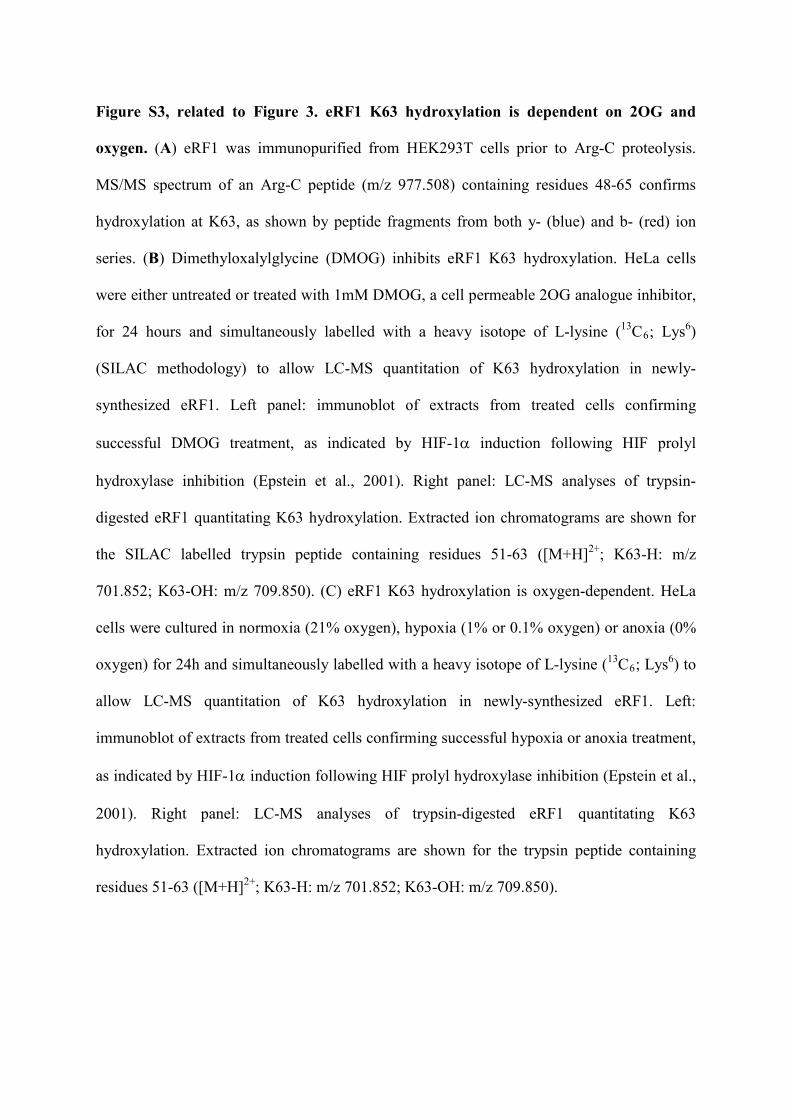
Figure S3, related to Figure 3. eRF1 K63 hydroxylation is dependent on 2OG and
oxygen. (A) eRF1 was immunopurified from HEK293T cells prior to Arg-C proteolysis.
MS/MS spectrum of an Arg-C peptide (m/z 977.508) containing residues 48-65 confirms
hydroxylation at K63, as shown by peptide fragments from both y- (blue) and b- (red) ion
series. (B) Dimethyloxalylglycine (DMOG) inhibits eRF1 K63 hydroxylation. HeLa cells
were either untreated or treated with 1mM DMOG, a cell permeable 2OG analogue inhibitor,
for 24 hours and simultaneously labelled with a heavy isotope of L-lysine (13C6; Lys6)
(SILAC methodology) to allow LC-MS quantitation of K63 hydroxylation in newly-
synthesized eRF1. Left panel: immunoblot of extracts from treated cells confirming
successful DMOG treatment, as indicated by HIF-1α induction following HIF prolyl
hydroxylase inhibition (Epstein et al., 2001). Right panel: LC-MS analyses of trypsin-
digested eRF1 quantitating K63 hydroxylation. Extracted ion chromatograms are shown for
the SILAC labelled trypsin peptide containing residues 51-63 ([M+H]2+; K63-H: m/z
701.852; K63-OH: m/z 709.850). (C) eRF1 K63 hydroxylation is oxygen-dependent. HeLa
cells were cultured in normoxia (21% oxygen), hypoxia (1% or 0.1% oxygen) or anoxia (0%
oxygen) for 24h and simultaneously labelled with a heavy isotope of L-lysine (13C6; Lys6) to
allow LC-MS quantitation of K63 hydroxylation in newly-synthesized eRF1. Left:
immunoblot of extracts from treated cells confirming successful hypoxia or anoxia treatment,
as indicated by HIF-1α induction following HIF prolyl hydroxylase inhibition (Epstein et al.,
2001). Right panel: LC-MS analyses of trypsin-digested eRF1 quantitating K63
hydroxylation. Extracted ion chromatograms are shown for the trypsin peptide containing
residues 51-63 ([M+H]2+; K63-H: m/z 701.852; K63-OH: m/z 709.850).

CAG UAG UGA UAA
Figure S4A
CAG UAG UGA UAA
Fold
incr
ease
in re
adth
roug
h
Jmjd4
eRF1
β-actin
Con
trol
Jmjd
4eR
F1
siRNA
eRF1
β-actin
Con
trol
eRF1
#27
eRF1
#29
siRNA
Jmjd4
eRF1
β-actin
Con
trol
Jmjd
4eR
F1
siRNA
Jmjd4
β-actin
Con
trol
Jmjd
4
siRNA
B
C0
1
2
3
4
5
6 si Control si Jmjd4 si eRF1
*
**
**
**
*
**
0
1
2
3
4 si Control si Jmjd4 si eRF1
**
**
**
**
**
**
CAG UGA UAG UAA
Fold
incr
ease
in re
adth
roug
h si Control si eRF1 #29 si eRF1 #27
0
2
4
6
8
10
12
14
16
18
**
**
**
**
**
**
CAG UGA UAG UAA
Fold
incr
ease
in re
adth
roug
h
si Control
si Jmjd4
0
2
4
6
8
10
12
14
16
18
20
**
**
**
0
1
2
3
4
5
6
7
8
9
Fold
ince
rase
in re
adth
roug
h
si Control si eRF1 #29 si eRF1 #27
**
**
Control + siResHA-Jmjd4
+ siResHA-Jmjd4
H189A
Fold
incr
ease
in U
GA
read
thro
ugh
si Control
si Jmjd4
0
1
2
3
4
5
****
E F
GH
*
Fold
incr
ease
in re
adth
roug
h
si Control
si Jmjd4
0
1
2
3
I
J K
Fold
incr
ease
in
read
thro
ugh
Control + siResHA-Jmjd4
+ siResHA-Jmjd4
H189A
0
1
2
3
4 **
*
L si Control
si Jmjd4
N
Fold
incr
ease
in re
adth
roug
h
D
M
CAG UGA
Fold
ince
rase
in re
adth
roug
h
0
0.5
1
1.5
2
2.5si Controlsi Jmjd4si Jmjd6si FIHsi Mina53
**
β-actin
eRF1
Mina53
FIH
Jmjd6
Jmjd4
Con
trol
Jmjd
4
siRNA
Jmjd
6
FIH
Min
a53
0
0.5
1
1.5
2
UAG UGA UAA
si Controlsi Jmjd4
Fold
cha
nge
infir
efly
/Ren
illa
luci
fera
se m
RN
A
0
0.5
1
1.5
2
UAG UGA UAA
Fold
cha
nge
inJm
jd4/
Cyc
loph
ilin
A m
RN
A
si Controlsi Jmjd4
11
422
SV40 early enhancer/promoter
490
626
chimeric intron
670
688
698
1630
T7 Renilla luciferase
1690
3339
Firefly luciferase
464 1755
recoding window
1291bp (1)
1155bp (2)
~120bp (4)~660bp (3)
no te
mpl
ate
plas
mid
si C
ontro
l
si J
mjd
4
plas
mid
si C
ontro
l
si J
mjd
4
plas
mid
si C
ontro
l
si J
mjd
4
UAG UGA UAA
12
*100200300500
1000
1517
*3
4

Figure S4, related to Figure 4. Jmjd4 knockdown induces stop codon readthrough in
multiple cell types and contexts. (A) Jmjd4 knockdown in U2OS cells induces readthrough of a
Renilla:stop:firefly reporter in which the stop codon is embedded within a leaky termination
sequence from tobacco mosaic virus. Left Panel: U2OS cells were transfected with the reporters
in Figure 4B prior to siRNA (si) transfection and dual luciferase assay. Right Panel: Immunoblot
of reporter extracts indicating Jmjd4 and eRF1 knockdown. (B) Similar analyses in (A)
performed in Hep3B cells. (C) Schematic of the Renilla-Firefly luciferase bicistronic vector used
in this study (Grentzmann et al., 1998), indicating normal splicing to remove the chimeric intron
from the full-length mRNA (1) to produce product (2). Sequences inserted within the recoding
window have potential to generate cryptic splice sites and aberrant splice variants of product (2)
(see [Holcik et al., 2005; Lemp et al., 2012]) indicated as products (3) and (4). Primers
(indicated by arrow heads) were designed to span from the chimeric intron to firefly luciferase in
order to detect the indicated transcripts and potential splice products. (D) RT-PCR analyses
demonstrate the integrity of the Renilla:stop:firefly mRNA. Total RNA was purified from HeLa
cells transfected with control or Jmjd4 siRNA and the bicistronic luciferase reporters depicted in
(C), prior to DNase digest to remove plasmid DNA followed by cDNA synthesis and end point
RT-PCR. Purified plasmid DNA was used as a positive control (‘plasmid’). Note the absence of
lower molecular weight splice variants indicating that the integrity of the reporter has not been
compromised by insertion of the TMV termination sequence. (E) Left Panel: qPCR
demonstrating that the abundance of firefly relative to Renilla luciferase mRNA is not altered by
Jmjd4 siRNA. Right Panel: siRNA knockdown of Jmjd4 was confirmed by qPCR in parallel to
analysis of the bicistronic reporter RNA (F) Closely-related JmjC hydroxylases do not regulate
translational termination. Left Panel: HeLa cells were transfected with the Renilla:UGA:firefly

reporter prior to the indicated siRNA (si) transfection and dual luciferase assay. Right Panel:
Immunoblot of reporter extracts indicating knockdown of the indicated JmjC hydroxylases. (G)
Schematic of an alternative stop codon readthrough reporter: β-galactosidase:stop:firefly
luciferase. (H) Transfection of A549 cells with two independent eRF1 siRNA sequences induces
stop codon readthrough. Left Panel: A549 cells were transfected with the reporters in (G) prior to
siRNA (si) transfection followed by β-galactosidase and firefly luciferase assays. Right Panel:
Immunoblot of reporter extracts indicating eRF1 knockdown. (I) Transfection of A549 cells with
Jmjd4 siRNA induces stop codon readthrough. Left Panel: A549 cells were transfected with the
reporters in (G) prior to siRNA (si) transfection followed by β-galactosidase and firefly
luciferase assays. Right Panel: Immunoblot of reporter extracts indicating Jmjd4 knockdown.
Note that a single siRNA sequence is presented since the specificity is confirmed in (K) and (L).
(J) Transfection of A549 cells with eRF1 (left) or Jmjd4 (right) siRNA also induces readthrough
of the β-galactosidase:stop:firefly luciferase reporter containing a stop codon embedded within a
strong termination sequence. See (H) and (I) for corresponding immunoblots. (K) Jmjd4
hydroxylase activity is required for efficient translational termination. Stop codon readthrough
assays were performed as in (H) using A549 cell lines stably expressing empty vector, siRes-
HA-Jmjd4, or siRes-Jmjd4 H189A mRNAs, and the β-galactosidase:stop:firefly luciferase
reporter with an UGA stop codon embedded within tobacco mosaic virus termination sequence.
(L) Jmjd4 hydroxylase activity is also required for efficient translational termination at a strong
termination sequence. Stop codon readthrough assays were performed as in (H) using A549 cell
lines stably expressing empty vector, siRes-HA-Jmjd4, or siRes-Jmjd4 H189A mRNAs, and the
β-galactosidase:stop:firefly luciferase reporter with a strong termination sequence. (M)
Immunoblot analyses of extracts from cells treated in (K) and (L) confirming knockdown of

endogenous Jmjd4 and re-expression of siRNA-resistant Jmjd4 (n.s. = non-specific
immunoreactivity). (N) Coomassie-stained gel of recombinant proteins demonstrate that Jmjd4-
reacted eRF1 samples were successfully depleted of contaminating Jmjd4 before measuring their
release factor activity in Figures 4E and 4F. The lanes are as follows: (1) Recombinant wildtype
Jmjd4 (to highlight the position of potential contaminant in lanes 2 to 5). The amount of Jmjd4
loaded is equivalent to the relative amount in the bulk hydroxylation reaction prior to eRF1
purification. (2) Wildtype eRF1 purified after bulk hydroxylation reaction with wildtype Jmjd4.
(3) Wildtype eRF1 purified after bulk hydroxylation reaction with Jmjd4-H189A. (4) eRF1
K63R purified after bulk hydroxylation reaction with wildtype Jmjd4. (5) eRF1 K63R purified
after bulk hydroxylation reaction with Jmjd4-H189A. In panels showing bicistronic reporter
results, data are presented as mean ± S.E.M. Statistical significance was determined by paired
two-tail Student’s t-test (I, J) for single comparisons, and analysis of variance (ANOVA)
followed by Dunett’s (A, B, F, H, J) or Bonferroni (K, L) post-hoc tests for multiple comparisons
(*P<0.05; **P<0.01).

Supplemental Experimental Procedures
Cell culture and treatments
Stable HEK293T cells overexpressing FLAG-Jmjd4 or FLAG-FIH were cultured as described in
the main text, in the presence of 1µg/ml puromycin. HeLa cell lines stably expressing pTIPZ
doxycycline-inducible siRNA-resistant HA-Jmjd4 (wildtype or H189A mutant; see below) were
cultured under the same conditions as parental HeLa cells, except that Tet system-approved fetal
calf serum (Clontech) was used in addition to 1µg/ml puromycin. RNA interference (Mission
siRNA, Sigma) was performed using two to three consecutive transfections with Oligofectamine
(for A549 and HeLa; Invitrogen) or N-TER (for U2OS; Sigma) reagent at 25nM siRNA per
transfection. Transient transfections were performed using Fugene6 (HeLa and U2OS; Roche) or
Turbofect (A549; Fermentas) according to the manufacturer’s instructions. Endogenous eRF1
hydroxylation was measured in SILAC-labelled HeLa cells (see below) treated as follows.
Hypoxia was performed at 1, 0.1 or 0% oxygen for 24 hours using a Ruskinn hypoxia station,
followed by cell lysis (see below) in the presence of 1mM N-oxalylglycine (NOG; Santa Cruz
Biotechnology). Cellular inhibition of 2OG oxygenases was achieved by treating cells with 1mM
dimethyl-N-oxalylglycine (DMOG; Frontier Scientific) for 24 hours.
Expression constructs and reporters
Site-directed mutagenesis was performed by PCR using standard procedures. Human Jmjd4 and
FIH cDNAs were cloned into the Not-I/Cla-I restriction sites of the pGIPZ vector (Open
Biosystems) using PCR primers incorporating N-terminal 3XFLAG epitope tags, thereby
replacing the GFP cDNA with the required enzyme cDNA. The human Jmjd4 cDNA was cloned

into the pTriEx2 recombinant expression vector platform (Merck) by PCR. β-
galactosidase:stop:firefly luciferase reporters were designed to encode the β-galactosidase cDNA
upstream of a recoding window and in-frame with the firefly luciferase cDNA. Plasmids were
constructed using a two-step PCR cloning strategy: the β-galactosidase coding sequence was
inserted into pEF1/V5-HisA (Invitrogen) via KpnI and BamHI sites, and the firefly luciferase
gene subsequently inserted via BamHI and NotI restriction sites with a 5’ extension on the
forward primer containing the recoding sequences. To induce basal readthrough stop codons
were positioned in the context of a minimal leaky termination signal derived from the replicase
cistron of the plant tobacco mosaic virus (TMV) (Pelham, 1978): CAA [STOP:
UAG/UGA/UAA] CAA UUA. Controls included either replacing the UAG stop codon with a
sense CAG codon for continuous readthrough, or substituting the leaky termination signal for a
strong stop codon: UGU [UAA] AGG AAA. All constructs were sequence verified prior to use.
Statistical analyses
Data were analyzed using IBM SPSS Statistics software version 21.0 and MATLAB
(Mathworks) version 7.13.0.564. Paired two-tailed Student’s t-test was performed for single
comparisons. For multiple comparisons, analysis of variance (ANOVA) was performed followed
by Dunnett’s or Bonferroni post-hoc analysis. All results are presented as mean ± SEM. To
assess whether the bicistronic reporter data were normally distributed, the probability plot
correlation coefficient (PPCC) test for normality was used (Filliben, 1975; Jacobs and Dinman,
2004). An example probability plot is shown below which visualizes how closely the data
matches a standard normal distribution (data corresponds to Figure S4B). The PPCC quantifies

the level of this correlation. All calculated PPCCs were >0.869 indicating that there is no
significant deviation from normality.
Immunoblotting
Following SDS-PAGE samples were electroblotted to polyvinylidene fluoride membrane
(Millipore). Membranes were blocked in 5% (w/v) milk powder in PBS/0.1% Tween-20 prior to
probing with the following primary antibodies; anti-HA-HRP (Roche), anti-FLAG-HRP
(Sigma), anti-Jmjd4 (Sigma), anti-eRF1 (Santa Cruz Biotechnology), anti-FIH (Stolze et al.,
2004), anti-Jmjd6 (Santa Cruz Biotechnology), anti-MINA53 (Invitrogen) and anti-β-actin-HRP
(Abcam). HRP-conjugated secondary antibodies were purchased from Dako. Signals were
developed using SuperSignal West Pico, Dura, or Femto chemiluminescent substrates (Thermo
Fisher Scientific).
Mapping and quantitation of hydroxylation sites by proteomics
Jmjd4 pulldowns to identify candidate substrates were performed by immunoprecipitating
extracts derived from control, FLAG-Jmjd4, or FLAG-Jmjd4 H189A overexpressing cells with
anti-FLAG affinity resin (Sigma) overnight at 4°C. Identification of hydroxylation sites in the
eRF1/eRF3a complex was performed by first transiently transfecting HEK293T cells with V5-
eRF1 and HA-eRF3a in the presence or absence of HA-Jmjd4 for 48 hours, prior to
immunoprecipitation of extracts with anti-V5 affinity resin (Sigma) overnight at 4°C.
Immunocomplexes from both of the above experiments were thoroughly washed in lysis buffer
prior to elution in ammonium hydroxide and precipitation with methanol/chloroform.
Endogenous eRF1 was purified from various sources by extracting samples in lysis buffer prior
to immunoprecipitation with mouse anti-eRF1 monoclonal antibody (Santa Cruz Biotechnology)
overnight at 4°C. These immunocomplexes were thoroughly washed in lysis buffer prior to
elution in Laemmli buffer, SDS-PAGE separation, and coomassie blue staining. All samples
were then digested in bicarbonate buffer using sequencing-grade trypsin (Sigma) or Arg-C
(Roche) (Cockman et al., 2009). SILAC was performed by pulse-labelling cells with lysine-
deficient DMEM (Thermo Fisher Scientific) containing 10% (v/v) dialyzed fetal bovine serum
(Appleton Woods) supplemented with ‘heavy’ L-lysine (U-13C6; Lys6; Cambridge Isotope
Laboratories) for the duration of the treatment.
Mass spectrometry analysis
Samples were resuspended in 20 µl of Buffer A (2% acetonitrile, 0.1% formic acid) and
subjected to LC-MS/MS (Waters, Acquity, 75 µm x 250 mm, 1.7 µm particle size) analysis
using a Thermo LTQ Orbitrap Velos (Thermo Scientific) at a Resolution of 30000. MS/MS

spectra were acquired in CID mode, selecting up to 20 precursors. Peptides were separated by a
linear gradient of 1-40% Acetonitrile in 120 min at a flow rate of 250nl/min (Fischer et al.,
2012). MS/MS spectra were extracted from raw files by ProteoWizard MSConvert software
(http://proteowizard.sourceforge.net/) using the 200 most intense peaks in each and converted
into MGF files. The peaklists were searched against the IPI human database (v3.87, 91464
entries) using Mascot (http://www.matrixscience.com/) v2.3.01, allowing one missed cleavage
and 20ppm/0.5 Da mass deviations in MS MS/MS. Carbamidomethylation of cysteine was a
fixed modification. Oxidation of methionine and lysine as well as deamidation of asparagine and
glutamine were variable modifications. Annotation of oxidized methionines and lysines was
performed manually and assisted with ModLS (Trudgian et al., 2012) as part of data analysis
using the Central Proteomics Facilities Pipeline (Trudgian et al., 2010).
RNA analyses
Total RNA was extracted from HeLa cells using TRIzol (Sigma) following the manufacturer’s
instructions. To remove contaminating plasmid DNA, extracted RNA was treated with DNAse
turbo (Ambion) prior to cDNA synthesis by reverse transcriptase (Applied Biosystems). End
point RT-PCR assays to monitor integrity of the Renilla-stop-firefly mRNA were performed
using Q5 high-fidelity DNA polymerase (New England Biolabs) and the following primers:
forward 5’-CAGAAGTTGGTCGTGAGGCA-3’ and reverse 5’-
TCCAGCGGTTCCATCCTCTA -3’. PCR products were separated by electrophoresis on 1.5%
w/v agarose/TBE gels and visualised using ethidium bromide and UV. To measure the relative
abundance of Renilla and firefly luciferase mRNAs, qRT-PCR were performed using SYBR
green dye (Applied Biosystems), with the following primers for Renilla luciferase: forward 5’-
AAGAGCGAAGAGGGCGAGAA -3’ and reverse 5’-TGCGGACAATCTGGACGAC-3’, for
firefly luciferase: forward 5’-CGTGCCAGAGTCTTTCGACA-3’ and reverse 5’-
ACAGGCGGTGCGATGAG-3’. Jmjd4 mRNA abundance was quantified by qRT-PCR using a
TaqMan probe (Applied Biosystems) and analysed using the StepOnePlus Real Time PCR
System (Applied Biosystems) corrected against Taqman control Cyclophilin A.
In vitro hydroxylation assays with recombinant eRF1 and Jmjd4
Small scale reactions were undertaken as follows. Recombinant eRF1 (2ug) and Jmjd4 (200ng)
were incubated at 37°C in 200µl of assay buffer (50mM Tris-HCl pH 7.4, 16µM Fe[II], 160µM
2OG, and 1mM DTT) in the presence or absence of 1mM NOG. After the indicated incubation
time, the reaction was quenched with 0.1% formic acid. Proteins were extracted using methanol-
chloroform, digested with trypsin and eRF1 K63 hydroxylation quantitated by LC-MS.
Batch scale reactions for in vitro translation assays were performed as follows. Bacterial
pellets expressing recombinant protein were lysed in 50mM Tris-HCl pH8, 200mM KCl, 0.5%
Triton-X-100, 10% glycerol, 0.2mg/ml lysozyme, protease inhibitors and 6mM β-
mercaptoethanol, before sonication and centrifugation to remove insoluble debris. Supernatants
were mixed in a 1:10 volume ratio of Jmjd4 to eRF1 prior to four separate additions of 10mM
2OG and 80µM Fe(II) over a 16 hour period at room temperature. eRF1 was subsequently
purified by a two-step chromatography method using Ni-NTA and MonoQ columns as described
in the main text.

Components of the fully reconstituted in vitro translation system
The 40S and 60S ribosomal subunits, as well as eukaryotic translation factors eIF2, eIF3, eEF1
and eEF2, were purified from a rabbit reticulocyte lysate as described (Alkalaeva et al., 2006).
The eukaryotic translation factors eIF1, eIF1A, eIF4A, eIF4B, eIF4G, eIF5B, eIF5, eRF1 and its
K63R mutant were produced as recombinant proteins in E. coli strain BL21 with subsequent
protein purification on Ni-NTA agarose and ion-exchange chromatography (Alkalaeva et al.,
2006). MVHL-stop transcription vectors have been described (Alkalaeva et al., 2006).
Pre-termination complex assembly and purification
Pre-termination complexes (pre-TC) were assembled as described (Alkalaeva et al., 2006).
Briefly, 37 pmol of MVHL-stop mRNAs were incubated for 30 min in buffer A (20 mM Tris
acetate, pH 7.5, 100 mM KOAc, 2.5 mM MgCl2, 2 mM DTT) supplemented with 400 U RNase
inhibitor (RiboLock, Fermentas), 1 mM ATP, 0.25 mM spermidine, 0.2 mM GTP, 75 µg total
tRNA (acylated with Val, His, Leu and [35S]Met), 75 pmol 40S and 60S purified ribosomal
subunits, 125 pmol eIF2, eIF3, eIF4G, eIF4A, eIF4B, eIF1, eIF1A, eIF5, eIF5B each, 200 pmol
eEF1 and 50 pmol eEF2 and then centrifuged in a Beckman SW55 rotor for 95 min at 4°C and
50 000 rpm in a 10%–30% linear sucrose density gradient prepared in buffer A with 5 mM
MgCl2. Fractions corresponding to pre-TC complexes according to optical density and the
presence of [35S]Met were combined, diluted 3-fold with buffer A containing 1.25 mM MgCl2
(to a final concentration of 2.5 mM Mg2+) and used in termination efficiency assays.
Termination efficiency assay
Termination efficiency (kcat/KM) was determined from kobs as described (Eliseev et al., 2011).
Briefly, aliquots containing 0.0125 pmol of pre-TC assembled in the presence of [35S]Met-tRNA
were incubated at 37°C with 2.5 pmol of eRF1 for 0-15 min. Ribosomes and tRNA were pelleted
with ice-cold 5% TCA supplemented with 0.75% casamino acids and centrifuged at 4°C and 14
000 g. The amount of released [35S]Met-containing tetrapeptide was used for determination of
the efficiency of peptidyl-tRNA hydrolysis.
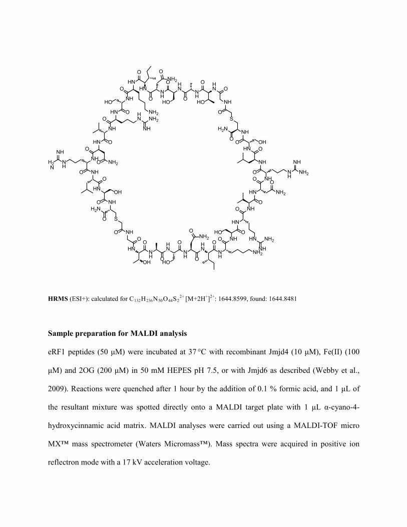
Peptide synthesis
The eRF1 24mer linear peptide (LADEFGTASNIKSRVNRLSVLGAI) was commercially
synthesized by G.L. Biochem (Shanghai, China). For synthesis of the cyclic eRF1 peptide, a
linear precursor peptide (Gly-Thr-Ala-Ser-Asn-Ile-Lys-Ser-Arg-Val-Asn-Arg-Leu-Ser-[D-Cys])
was prepared by standard solid phase synthesis on a CS Bio CS336X peptide synthesizer (100
μmol scale) using DIC as coupling reagent. After cleavage of the N-terminal Fmoc-protecting
group, a solution of 150 mg of chloromethylcarbonyloxysuccinimide (ClAc-OSu) in 4 mL DMF
was added to the resin and the mixture was shaken for 3 h. The resin was filtered off and
subsequently treated with 4 mL of deprotection solution (95 % TFA, 2.5 % Triisopropylsilane,
2.5 % water). After 3 h the volume was reduced to 1 mL under a nitrogen stream and the
peptides were crashed out with cold Et2O. The mixture was centrifuged and the supernatant
discarded. The solid was taken up in 1.5 mL of triethylammoniumacetate buffer (1 M, pH 8.5)
and the pH readjusted to >8. The mixture was heated in a microwave (Biotage Initiator) to 80°C
for 10 min, and the cyclic peptide was subsequently purified by HPLC (0-45 % MeCN in 45
min, 0.1 % TFA, Dionex Ultimate 3000 series, Grace Vydac 218TP101522 column):
NH
HN NH
HN N
H
NH2
HN N
HNH2
NH
HN
HN
NH
NH2
NH
HN NH2
NHNH
NH
NH2
NH
HN
NH
O O
OH O
O
HO O
O
O OOHO
OO
O O
O
OO
H2N
OH
NH
HNN
H
HNN
H
NH2
HNHN
NH2
NH
HN HN
NH
NH2
NH
HN
NH2NHN
H
NH
H2N
NH
HN
NH
OO
HOO
O
HOO
OO
O
O
HO
O
OO
O
OO
O
OH
OS
O
H2N
O S
O
HRMS (ESI+): calculated for C132H236N50O44S22+ [M+2H+]2+: 1644.8599, found: 1644.8481
Sample preparation for MALDI analysis
eRF1 peptides (50 μM) were incubated at 37 °C with recombinant Jmjd4 (10 μM), Fe(II) (100
μM) and 2OG (200 μM) in 50 mM HEPES pH 7.5, or with Jmjd6 as described (Webby et al.,
2009). Reactions were quenched after 1 hour by the addition of 0.1 % formic acid, and 1 μL of
the resultant mixture was spotted directly onto a MALDI target plate with 1 μL α-cyano-4-
hydroxycinnamic acid matrix. MALDI analyses were carried out using a MALDI-TOF micro
MX™ mass spectrometer (Waters Micromass™). Mass spectra were acquired in positive ion
reflectron mode with a 17 kV acceleration voltage.

Sample preparation for amino acid analysis
For amino acid analyses, the eRF1 cyclic peptide (50 μM) was incubated with recombinant
Jmjd4 (10 μM), Fe(II) (100 μM) and 2OG (200 μM) for 1 hour at 37 °C in HEPES buffer (50
mM, pH 7.5). The reaction was quenched by the addition of methanol (50 % final concentration),
incubated on ice for 2 hours and then centrifuged to pellet any precipitated Jmjd4. The
supernatant containing the cyclic peptide (as confirmed by MALDI-TOF MS analysis) was
transferred to a fresh tube and dried in a vacuum centrifuge. The dried reaction mixture was then
resuspended in double distilled water and the peptide subjected to enzymatic hydrolysis at 37 °C
with trypsin (1 μL, 100 μg mL−1; Promega) for 1 h, followed by Carboxypeptidase Y (1 μL, 5
mg mL−1, Roche) overnight.
The hydrolyzed peptides were dried by vacuum centrifugation, reconstituted in borate
buffer pH 9.0 and derivatized by 6-aminoquinolyl-N-hydroxysuccinimidyl carbamate (AQC) in
acetonitrile according to the AccQ-Tag Ultra Derivatization Protocol (Waters). LC-MS analyses
were performed as described (Mantri et al., 2011) using a Waters Acquity ultra performance
liquid chromatography system coupled to an LCT Premier XE orthogonal acceleration time-of-
flight mass spectrometer equipped with an electrospray ionization source (Waters, USA). The
gradient conditions used for separation were as follows:
t [min] flow rate % Solvent A
(AccQ Tag Ultra Phase A)
% Solvent B
(98.7% acetonitrile, 1.3% formic acid)
0.00 0.7 99.9 0.1
25.00 0.6 90.0 10.0
25.50 0.6 40.0 60.0
27.50 0.6 40.0 60.0
28.00 0.6 100.0 0.0

30.00 0.7 100.0 0.0
The conditions for ESI-MS detection were as follows: positive-ion mode, V-mode
analyser, desolvation temperature at 250 °C, source temperature at 120 °C, capillary voltage at
3000 V, sample cone voltage at 100 V, cone and desolvation gas flow at 30 and 550 L/min,
respectively. The MS data were acquired and an extracted ion chromatogram produced for the
m/z 503.1, corresponding to bis-derivatised hydroxylysine. The trace was smoothed (Savitzky-
Golley, 2 iterations, 3 points window).
Synthesis of 3-hydroxylysine
3-Hydroxylysine was synthesized as summarized in Scheme 1. Initially, 4-aminobutanal
diethylacetal was protected as the phtalimide. The aldehyde was subsequently liberated using
mild acid hydrolysis. In the key step ethylisocyanoacetate was reacted with the aldehyde using
transition metal catalysis. The resulting dihydrooxazole was cleaved with HCl; we did not
acquire evidence for elimination within limits of detection. In the final step the phtalimide was
cleaved using hydrazine to give 3-hydroxylysine as a mixture of stereoisomers:

N+O
O-C
O
ON
O
N
OH
O
N
OH
O
NH2
O O
O O
H2N
HO H2N
HO
NO O
O
NO O
O
O AcOH
(99 %) CuBr, PPh3,DIPEA(45 %)
HCl N2H4
(33 % over 2 steps)
Scheme 1: Synthesis of 3-hydroxylysine.
2-(4,4-Diethoxybutyl)isoindoline-1,3-dione
N
O
O
OO
N-Ethoxycarbonylphtalimide (8.10 g, 36.6 mmol, 1.05 mmol) was dissolved in 40 mL CH2Cl2,
4-aminobutanal dimethylacetal (6.00 mL, 35.0 mmol, 1.0 eq) and NEt3 (5.40 mL, 36.6 mmol,
1.05 eq) were added and the reaction mixture stirred at room temperature for 4 h. After
completion of the reaction the solvent was evaporated in vacuo to give 7.92 g (78 %) of 2-(4,4-
diethoxybutyl)isoindoline-1,3-dione (Teshima et al., 1991).
1H NMR (CDCl3, 400MHz): δ = 7.82 (dd, J=5.5, 3.0 Hz, 1 H), 7.69 (dd, J=5.5, 3.0 Hz, 1 H),
4.70 (broad singlet, 1 H), 4.50 (t, J=5.5 Hz, 1 H), 4.10 (q, J=7.0 Hz, 1 H), 3.70 (t, J=7.0 Hz, 1

H), 3.62 (dq, J=9.5, 7.1 Hz, 2 H), 3.47 (dq, J=9.5, 7.1 Hz, 2 H), 3.14 (q, J=7.5 Hz, 1 H), 1.70 -
1.80 (m, 1 H), 1.60 - 1.69 (m, 2 H), 1.33 (t, J=7.5 Hz, 1 H), 1.24 (t, J=7.0 Hz, 2 H), 1.17 ppm (t,
J=7.0 Hz, 4 H)
13C NMR (CDCl3,101MHz): δ = 168.4, 133.8, 132.1, 123.1, 102.4, 61.2, 37.7, 30.9, 23.9, 15.3
ppm
HRMS (ESI+): calculated for C16H21NNaO4+ [M+H+]+: 314.1363, found:314.1353.
4-(1,3-Dioxoisoindolin-2-yl)butanal
N
O
O
O
2-(4,4-Diethoxybutyl)isoindoline-1,3-dione (500 mg, 1.72 mmol, 1.0 eq) was dissolved in 10 mL
H2O and CH2CO2H (20.0 mL, 343 mmol, 200 eq) was added. The reaction mixture was stirred
for 2.5 h at room temperature, then concentrated in vacuo to afford 427 mg (yield apparently
quantitative, but residual CH2CO2H present) of 4-(1,3-dioxoisoindolin-2-yl)butanal (Iradier et
al., 2001).
1H NMR (CDCl3, 400MHz): δ = 9.76 (s, 1 H), 7.79 - 7.86 (m, 2 H), 7.67 - 7.74 (m, 2 H), 3.73 (t,
J=7.0 Hz, 2 H), 2.53 (t, J=7.5 Hz, 2 H), 2.00 ppm (quin, J=7.0 Hz, 2 H)
13C NMR (CDCl3, 101MHz): δ = 200.8, 168.3, 134.0, 131.9, 123.2, 41.0, 37.1, 21.1 ppm
HRMS (ESI+): calculated for C12H11NNaO3+ [M+Na+]+: 240.0631, found: 240.0629
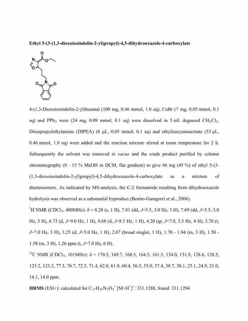
Ethyl 5-(3-(1,3-dioxoisoindolin-2-yl)propyl)-4,5-dihydrooxazole-4-carboxylate
NO O
O
NO
O
4-(1,3-Dioxoisoindolin-2-yl)butanal (100 mg, 0.46 mmol, 1.0 eq), CuBr (7 mg, 0.05 mmol, 0.1
eq) and PPh3 were (24 mg, 0.09 mmol, 0.1 eq) were dissolved in 5 mL degassed CH2Cl2.
Diisopropylethylamine (DIPEA) (8 μL, 0.05 mmol, 0.1 eq) and ethylisocyanoacetate (53 μL,
0.46 mmol, 1.0 eq) were added and the reaction mixture stirred at room temperature for 2 h.
Subsequently the solvent was removed in vacuo and the crude product purified by column
chromatography (0 – 15 % MeOH in DCM, flat gradient) to give 46 mg (45 %) of ethyl 5-(3-
(1,3-dioxoisoindolin-2-yl)propyl)-4,5-dihydrooxazole-4-carboxylate as a mixture of
diastereomers. As indicated by MS-analysis, the C-2 formamide resulting from dihydrooxazole
hydrolysis was observed as a substantial byproduct (Benito-Garagorri et al., 2006).
1H NMR (CDCl3, 400MHz): δ = 8.28 (s, 1 H), 7.81 (dd, J=5.5, 3.0 Hz, 3 H), 7.69 (dd, J=5.5, 3.0
Hz, 3 H), 6.73 (d, J=9.0 Hz, 1 H), 4.68 (d, J=8.5 Hz, 1 H), 4.20 (qt, J=7.0, 3.5 Hz, 4 H), 3.70 (t,
J=7.0 Hz, 3 H), 3.25 (d, J=5.0 Hz, 1 H), 2.07 (broad singlet, 1 H), 1.70 - 1.94 (m, 3 H), 1.50 -
1.58 (m, 3 H), 1.26 ppm (t, J=7.0 Hz, 6 H).
13C NMR (CDCl3, 101MHz): δ = 170.5, 169.7, 168.5, 164.5, 161.5, 134.0, 131.9, 128.6, 128.5,
123.2, 123.2, 77.3, 76.7, 72.3, 71.4, 62.0, 61.9, 60.4, 56.5, 55.0, 37.4, 30.7, 30.1, 25.1, 24.9, 21.0,
14.1, 14.0 ppm.
HRMS (ESI+): calculated for C17H19N2O5+ [M+H+]+: 331.1288, found: 331.1294

2-Amino-6-(1,3-dioxoisoindolin-2-yl)-3-hydroxyhexanoic acid
HO
N OO
OH
OH2N
Ethyl-5-(3-(1,3-dioxoisoindolin-2-yl)propyl)-4,5-dihydrooxazole-4-carboxylate (46 mg, 0.15
mmol, 1.0 eq) was dissolved in 10 mL HCl (6 M) and heated to reflux overnight, then cooled and
concentrated in vacuo. The crude product was used directly in the next reaction.
1H NMR (D2O, 400MHz): δ = 7.61 - 7.69 (m, 4 H), 7.53 (dd, J=5.5, 3.5 Hz, 2 H), 4.07 - 4.16 (m,
1 H), 3.86 - 3.93 (m, 1 H), 3.55 (t, J=6.5 Hz, 1 H), 2.89 - 2.97 (m, 1 H), 2.59 (s, 1 H), 1.68 - 1.81
(m, 1 H), 1.57 - 1.68 (m, 2 H), 1.45 - 1.57 ppm (m, 2 H).
13C NMR (D2O, 101MHz): δ = 172.1, 171.1, 135.0, 133.5, 132.3, 131.6, 129.1, 125.0, 123.6,
68.8, 39.4, 37.5, 30.6, 30.3, 24.4, 23.7 ppm
2,6-Diamino-3-hydroxyhexanoic acid (3-hydroxylysine)
HO
NH2
OH
OH2N
2-Amino-6-(1,3-dioxoisoindolin-2-yl)-3-hydroxyhexanoic acid (40 mg, 0.13 mmol, 1.0 eq) was
dissolved in 5 mL H2O, N2H4.H2O (27 mg, 0.33 mmol, 2.5 eq) was then added. The reaction
mixture was stirred at room temperature for 18 h, then purified by HPLC (0-100 % MeCN in

H20, 0.1 %CF3CO2H) to give 8 mg (33 % over 2 steps) of 3-hydroxylysine as a mixture of
diastereomers (see Figure S2D).
1H NMR (D2O, 400MHz): δ = 4.01 - 4.07 (m, 1 H), 3.78 (d, J=4.5 Hz, 1 H), 2.88 - 2.95 (m, 2
H), 1.69 - 1.79 (m, 1 H), 1.57 - 1.69 (m, 2 H), 1.45 - 1.57 ppm (m, 1 H).
HRMS (ESI+): calculated for C6H15N2O3+ [M+H+]+: 163.1077, found: 163.1077.
Synthesis of 4-Hydroxylysine
4-Hydroxylysine was synthesized as a mixture of all 4 stereoisomers using a modified literature
procedure (Morin et al., 1998). 4,5-Dehydrolysine dihydrochloride (Bachem, 10 mg, 0.05 mmol)
was dissolved in 6M H2SO4 (5 mL) and the solution was heated under reflux for 18 hours. The
pH of the solution was then adjusted to pH 10 using 5M NaOH and used directly for amino acid
analysis. A 1H NMR spectrum of the reaction mixture revealed the presence of 4-hydroxylysine
(see Figure S2E).
1H NMR (700 MHz, D2O) 1.59-1.74 (m, 3H, Hβ, Hδ), 1.77-1.83 (m, 1H, Hβ), 2.76-2.85 (m, 2H,
Hε), 3.43 (app q, J = 4.5 Hz, 1H, Hα), 3.81-3.89 (m, 1H, Hγ).
13C NMR (175 MHz, D2O) 36.9 (Cδ, Cε), 41.2 (Cβ), 53.0 (Cα), 66.5 (Cγ).
Supplemental References
Alkalaeva, E.Z., Pisarev, A.V., Frolova, L.Y., Kisselev, L.L., and Pestova, T.V. (2006). In vitro
reconstitution of eukaryotic translation reveals cooperativity between release factors eRF1 and
eRF3. Cell 125, 1125-1136.
Benito-Garagorri, D., Bocokic, V., and Kirchner, K. (2006). Copper(I)-catalyzed
diastereoselective formation of oxazolines and N-sulfonyl-2-imidazolines. Tetrahedron Letters
47, 8641-8644.
Cockman, M.E., Webb, J.D., Kramer, H.B., Kessler, B.M., and Ratcliffe, P.J. (2009).
Proteomics-based identification of novel factor inhibiting hypoxia-inducible factor (FIH)
substrates indicates widespread asparaginyl hydroxylation of ankyrin repeat domain-containing
proteins. Molecular & cellular proteomics : MCP 8, 535-546.
Eliseev, B., Kryuchkova, P., Alkalaeva, E., and Frolova, L. (2011). A single amino acid change
of translation termination factor eRF1 switches between bipotent and omnipotent stop-codon
specificity. Nucleic Acids Res 39, 599-608.
Epstein, A.C., Gleadle, J.M., McNeill, L.A., Hewitson, K.S., O'Rourke, J., Mole, D.R.,
Mukherji, M., Metzen, E., Wilson, M.I., Dhanda, A., et al. (2001). C. elegans EGL-9 and
mammalian homologs define a family of dioxygenases that regulate HIF by prolyl
hydroxylation. Cell 107, 43-54.
Filliben, J.J. (1975). The Probability Plot Correlation Coefficient Test for Normality.
Technometrics 17, 111-117.
Fischer, R., Trudgian, D.C., Wright, C., Thomas, G., Bradbury, L.A., Brown, M.A., Bowness, P.,
and Kessler, B.M. (2012). Discovery of candidate serum proteomic and metabolomic biomarkers
in ankylosing spondylitis. Molecular & cellular proteomics : MCP 11, M111 013904.
Grentzmann, G., Ingram, J.A., Kelly, P.J., Gesteland, R.F., and Atkins, J.F. (1998). A dual-
luciferase reporter system for studying recoding signals. RNA 4, 479-486.
Holcik, M., Graber, T., Lewis, S.M., Lefebvre, C.A., Lacasse, E., and Baird, S. (2005). Spurious
splicing within the XIAP 5' UTR occurs in the Rluc/Fluc but not the betagal/CAT bicistronic
reporter system. RNA 11, 1605-1609.
Iradier, F., Arrayas, R.G., and Carretero, J.C. (2001). Synthesis of medium-sized cyclic amines
by selective ring cleavage of sulfonylated bicyclic amines. Organic letters 3, 2957-2960.
Jacobs, J.L., and Dinman, J.D. (2004). Systematic analysis of bicistronic reporter assay data.
Nucleic Acids Res 32, e160.
Lemp, N.A., Hiraoka, K., Kasahara, N., and Logg, C.R. (2012). Cryptic transcripts from a
ubiquitous plasmid origin of replication confound tests for cis-regulatory function. Nucleic Acids
Res 40, 7280-7290.
Mantri, M., Loik, N.D., Hamed, R.B., Claridge, T.D., McCullagh, J.S., and Schofield, C.J.
(2011). The 2-oxoglutarate-dependent oxygenase JMJD6 catalyses oxidation of lysine residues
to give 5S-hydroxylysine residues. Chembiochem : a European journal of chemical biology 12,
531-534.
Morin, B., Bubb, W.A., Davies, M.J., Dean, R.T., and Fu, S. (1998). 3-Hydroxylysine, a
potential marker for studying radical-induced protein oxidation. Chemical research in toxicology
11, 1265-1273.
Pelham, H.R. (1978). Leaky UAG termination codon in tobacco mosaic virus RNA. Nature 272,
469-471.
Stolze, I.P., Tian, Y.M., Appelhoff, R.J., Turley, H., Wykoff, C.C., Gleadle, J.M., and Ratcliffe,
P.J. (2004). Genetic analysis of the role of the asparaginyl hydroxylase factor inhibiting hypoxia-

inducible factor (FIH) in regulating hypoxia-inducible factor (HIF) transcriptional target genes
[corrected]. J Biol Chem 279, 42719-42725.
Teshima, T., Matsumoto, T., Wakamiya, T., Shiba, T., Aramaki, Y., Nakajima, T., and Kawai,
N. (1991). Total Synthesis of nstx-3, spider toxin of nephila maculata. Tetrahedron 47, 3305-
3312.
Trudgian, D.C., Singleton, R.S., Cockman, M.E., Ratcliffe, P.J., and Kessler, B.M. (2012).
ModLS: Post-translational modification localization scoring with automatic specificity
expansion. J. Proteomics Bioinform 5, 283-289.
Trudgian, D.C., Thomas, B., McGowan, S.J., Kessler, B.M., Salek, M., and Acuto, O. (2010).
CPFP: a central proteomics facilities pipeline. Bioinformatics 26, 1131-1132.
Webby, C.J., Wolf, A., Gromak, N., Dreger, M., Kramer, H., Kessler, B., Nielsen, M.L.,
Schmitz, C., Butler, D.S., Yates, J.R., 3rd, et al. (2009). Jmjd6 catalyses lysyl-hydroxylation of
U2AF65, a protein associated with RNA splicing. Science 325, 90-93.


![LimitedLiabilityCompanies ARTICLE19Chapter 53,Article19NMSA1978]; ... 53-19-7 LIMITEDLIABILITYCOMPANIES 53-19-10 53-19-7.Formation](https://img.pdfslide.us/doc/110x75/5af13fab7f8b9ac57a8fa26e/limitedliabilitycompanies-chapter-53article19nmsa1978-53-19-7-limitedliabilitycompanies.jpg)