Embed Size (px)
Citation preview

Molecular Cell
Article
An Ancient Transcription FactorInitiates the Burst of piRNA Productionduring Early Meiosis in Mouse TestesXin Zhiguo Li,1,4 Christian K. Roy,1,4 Xianjun Dong,2 Ewelina Bolcun-Filas,3 Jie Wang,2 Bo W. Han,1 Jia Xu,2
Melissa J. Moore,1 John C. Schimenti,3 Zhiping Weng,2,* and Phillip D. Zamore1,*1Department of Biochemistry and Molecular Pharmacology, Howard Hughes Medical Institute2Program in Bioinformatics and Integrative BiologyUniversity of Massachusetts Medical School, Worcester, MA 01605, USA3Department of Biomedical Sciences and Center for Vertebrate Genomics, Cornell University College of Veterinary Medicine, Ithaca,
NY 14850, USA4These authors contributed equally to this work*Correspondence: [email protected] (Z.W.), [email protected] (P.D.Z.)
http://dx.doi.org/10.1016/j.molcel.2013.02.016
SUMMARY
Animal germ cells produce PIWI-interacting RNAs(piRNAs), small silencing RNAs that suppress trans-posons and enable gamete maturation. Mammaliantransposon-silencing piRNAs accumulate early inspermatogenesis, whereas pachytene piRNAs areproduced later during postnatal spermatogenesisand account for >95% of all piRNAs in the adultmouse testis. Mutants defective for pachytenepiRNA pathway proteins fail to produce maturesperm, but neither the piRNA precursor transcriptsnor the trigger for pachytene piRNA productionis known. Here, we show that the transcriptionfactor A-MYB initiates pachytene piRNA production.A-MYB drives transcription of both pachytene piRNAprecursor RNAs and the mRNAs for core piRNAbiogenesis factors including MIWI, the proteinthrough which pachytene piRNAs function. A-MYBregulation of piRNA pathway proteins and piRNAgenes creates a coherent feedforward loop thatensures the robust accumulation of pachytenepiRNAs. This regulatory circuit, which can be de-tected in rooster testes, likely predates the diver-gence of birds and mammals.
INTRODUCTION
P-element induced wimpy testis (PIWI)-interacting RNAs
(piRNAs) can be distinguished from other animal small silencing
RNAs by their longer length (typically 23–35 nt), 20-O-methyl-
modified 30 termini, and association with PIWI proteins, a distinct
subgroup of Argonaute proteins, the small RNA-guided proteins
responsible for RNA interference and related pathways (Kumar
and Carmichael, 1998; Aravin and Hannon, 2008; Farazi et al.,
2008; Kim et al., 2009; Thomson and Lin, 2009; Cenik and
Zamore, 2011). piRNA production does not require Dicer, the
double-stranded RNA endonuclease that makes microRNAs
(miRNAs) and small interfering RNAs (siRNAs), and piRNAs are
thought to derive from single-stranded rather than double-
stranded RNA (Vagin et al., 2006; Houwing et al., 2007).
In most bilateral animals, germline piRNAs protect the genome
from transposon activation, but also have other functions (Aravin
et al., 2001, 2007, 2008; Vagin et al., 2004, 2006; Brennecke
et al., 2007; Carmell et al., 2007; Hartig et al., 2007; Kuramo-
chi-Miyagawa et al., 2008; Ashe et al., 2012; Lee et al., 2012;
Shirayama et al., 2012). A few days after birth, the majority of
piRNAs in the mouse testis are pre-pachytene piRNAs; 25% of
these piRNA species map to more than one location in the
genome. A second class of piRNAs, typically derived from inter-
genic regions, has been reported to emerge in the mouse testis
14.5 days postpartum (dpp), when the developing spermato-
cytes synchronously enter the pachytene phase of meiotic
prophase I. These pachytene piRNAs compose >95%of piRNAs
in the adult mouse testis. Loss of genes required to make pachy-
tene piRNAs blocks production of mature sperm (Deng and Lin,
2002; Aravin and Hannon, 2008; Reuter et al., 2011; Vourekas
et al., 2012). What triggers the accumulation of pachytene
piRNAs when spermatocytes enter the pachynema is unknown.
In Caenorhabditis elegans, each piRNA is processed from its
own short RNA polymerase II (Pol II) transcript (Gu et al., 2012).
In contrast, insect and mouse piRNAs are thought to be pro-
cessed from long RNAs transcribed from large piRNA loci.
Supporting this view, a transposon inserted into the 50 end of
the flamenco piRNA cluster in flies reduces the production of
flamenco piRNAs 168 kbp 30 to the insertion, suggesting that it
disrupts transcription of the entire locus (Brennecke et al.,
2007). High-throughput sequencing and chromatin immunopre-
cipitation (ChIP) has been used to define the genomic structure
of the piRNA-producing genes of immortalized, cultured silk
moth BmN4 cells (Kawaoka et al., 2013). However, for flies and
mice, we do not know the structure of piRNA-producing genes,
their transcripts, or the nature of the promoters that control their
expression.
Instead, piRNA loci have been defined as clusters: regions of
the genome with a high density of mapping piRNA sequences
(Aravin et al., 2006, 2007; Girard et al., 2006; Grivna et al.,
Molecular Cell 50, 67–81, April 11, 2013 ª2013 Elsevier Inc. 67
A
B
Figure 1. piRNA Precursors Are RNA Pol II
Transcripts
(A) Strategy to assemble the mouse testis tran-
scriptome. Rectangles with rounded corners,
input or output data; rectangles, processes.
Decisions are shown without boxing.
(B) Aggregated data for piRNA-producing tran-
scripts (5% trimmed mean). Oxidized small RNA
(>23 nt) sequencing data were used to detect
piRNAs; transcript abundance was measured
using total RNA depleted of rRNA (RNA-seq). RNA
Pol III data were from SRA001030. Dotted lines
show the transcriptional start site (Start) and site
of polyadenylation (End). See also Figure S1 and
Table S1.
Molecular Cell
A-MYB Activates Pachytene piRNA Gene Transcription
2006a; Lau et al., 2006; Brennecke et al., 2007; Ro et al., 2007). In
reality, piRNA-producing loci correspond to discrete transcrip-
tion units that include both intergenic loci believed to encode
no protein (Brennecke et al., 2007, 2008; Vourekas et al., 2012)
and protein-coding genes that also produce piRNAs (Aravin
et al., 2007; Robine et al., 2009; Saito et al., 2009).
We used high-throughput sequencing data to define the genes
and transcripts that produce piRNAs in the juvenile and adult
mouse testis. Using these data, we identified the factor that initi-
ates transcription of pachytene piRNA genes: A-MYB (MYBL1),
a spermatocyte protein that serves as a master regulator of
genes encoding proteins required for cell-cycle progression
68 Molecular Cell 50, 67–81, April 11, 2013 ª2013 Elsevier Inc.
through the pachytene stage of meiosis
(Trauth et al., 1994; Bolcun-Filas et al.,
2011). A-MYB also initiates transcription
of the genes encoding many piRNA
biogenesis factors. The combined action
of A-MYB at the promoters of genes
producing pachytene piRNA precursor
transcripts and genes encoding piRNA
biogenesis proteins creates a coherent
feedforward loop that triggers a >6,000-
fold increase in pachytene piRNA abun-
dance during the �5 days between the
early and late phases of the pachytene
stage of male meiosis. A-MYB also
promotes its own transcription through
a positive feedback loop. The A-MYB-
regulated feedforward loop is evolution-
arily conserved: A-MYB is bound to the
promoters of both piRNA clusters and
PIWIL1, TDRD1, and TDRD3 in the
rooster (Gallus gallus) testis.
RESULTS
Defining piRNA-ProducingTranscripts in the Mouse TestisTo define the structure of piRNA-
producing loci in the testis of wild-type
adult mice, we assembled the tran-
scripts detected by three biological repli-
cates of strand-specific, paired-end, rRNA-depleted, total RNA
sequencing (RNA-seq; Figure 1A). We mapped reads to the
mouse genome using TopHat (Trapnell et al., 2009) and per-
formed de novo transcriptome assembly using Trinity (Grabherr
et al., 2011) to identify unannotated exon-exon junctions. We
used all mapped reads, including reads corresponding to unan-
notated exon-exon junctions, to perform reference-based tran-
script assembly (Cufflinks; Trapnell et al., 2010).
To identify the transcripts that produce piRNAs, we se-
quenced piRNAs from six developmental stages ofmouse testes
(10.5 dpp, 12.5 dpp, 14.5 dpp, 17.5 dpp, 20.5 dpp, and adult)
and mapped them to the assembled transcripts. The first round
Molecular Cell
A-MYB Activates Pachytene piRNA Gene Transcription
of spermatogenesis proceeds synchronously among the tubules
of the testis: mouse testes at 10.5 dpp advance no further than
the zygotene stage (staging according to Nebel et al., 1961);
12.5 dpp to the early pachytene; 14.5 dpp to the middle pachy-
tene; 17.5 to the late pachytene; and 20.5 dpp to the round sper-
matid stage. For each stage, we prepared two sequencing
libraries: one comprising all small RNAs and one in which oxida-
tion was used to enrich for piRNAs by virtue of their 20-O-methyl-
modified 30 termini (Ghildiyal et al., 2008).
To qualify as a piRNA-producing transcript, an assembled
RNA was required to produce either a sufficiently high piRNA
abundance (>100 ppm; parts permillion uniquely mapped reads)
or density (>100 rpkm; reads per kilobase of transcript permillion
uniquely mapped reads). These criteria retained both long tran-
scripts producing an abundance of piRNAs and short transcripts
generating many piRNAs per unit of length. To refine the termini
of each piRNA-producing transcript, we supplemented the RNA-
seq data with high-throughput sequencing of the 50 ends of
RNAs bearing an N(50)ppp(50)N cap structure (cap analysis of
gene expression; CAGE) and the 30 ends of transcripts precedingthe poly(A) tail (polyadenylation site sequencing; PAS-seq). The
assembled piRNA-producing transcripts likely correspond to
continuous RNAs in vivo because theCAGE library used to anno-
tate transcript 50 ends was constructed after two rounds of
poly(A) selection. Thus, the RNA molecules in the library derive
from complete transcripts extending from the 50 cap to the
poly(A) tail (Figure 1B). Conventional 50 and 30 RACE (rapid ampli-
fication of cDNA ends) analysis of piRNA-producing transcripts
confirmed the ends of 16 loci (data not shown). To provide addi-
tional confirmation of the 50 end of each piRNA-producing tran-
script, we also determined the locations of histone H3 bearing
trimethylated lysine 4 (H3K4me3), a histone modification associ-
ated with RNA Pol II transcription start sites (Guenther et al.,
2007).
piRNA Precursor RNAs Are Canonical RNA Pol IITranscriptsThe presence of 50 caps and poly(A) tails and the binding of
histone H3K4me3 to the genomic DNA immediately upstream
of the transcription start site of each piRNA locus suggest that
piRNA transcripts are produced by RNA Pol II (Figure 1B). More-
over, using antibodies to RNA Pol II, but not RNA Pol III, ChIP-
seq showed a peak at the transcription start site as well as
polymerase occupancy across the entire piRNA gene (Figure 1B;
Kutter et al., 2011). We conclude that piRNA transcripts
are conventional RNA Pol II transcripts bearing 50 caps and 30
poly(A) tails.
A Transcript-Based Set of piRNA LociOur transcriptome assembly yielded 467 piRNA-producing tran-
scripts that define 214 genomic loci (Figure S1A and Table S1).
Among the �2.2 million distinct piRNA species and �8.8 million
piRNA reads from the adult mouse testis, the 214 genomic loci
account for 95% of all piRNAs.
Previous studies defined piRNA clusters based solely on small
RNA sequencing data (Girard et al., 2006; Lau et al., 2006; Aravin
et al., 2007). Our approach differs in that it (1) uses RNA-seq
data, whose greater read length facilitates the identification of
introns, allowing us to define the architecture of piRNA precursor
transcripts and (2) uses CAGE, PAS-seq, and H3K4me3 ChIP-
seq data to refine the 50 and 30 ends of the piRNA transcripts.
Consequently, the piRNA loci presented here account for more
piRNAs using fewer genomic base pairs than those previously
defined (Figures S1B and S1C; Lau et al., 2006; Girard et al.,
2006). Our piRNA-producing loci include 41 piRNA loci that
escaped previous detection (Girard et al., 2006; Lau et al.,
2006; Aravin et al., 2007), 37 of which contain introns. The 41
loci account for 2%of piRNAs at 10.5 dpp and 0.36% in the adult
testis.
Three Classes of piRNAs during PostnatalSpermatogenesisMice produce three PIWI proteins: MIWI2 (PIWIL4), which binds
piRNAs in perinatal testis (Carmell et al., 2007; Aravin et al.,
2008); MILI (PIWIL2), which binds piRNAs at least until the
round spermatid stage of spermatogenesis (Kuramochi-Miya-
gawa et al., 2004; Aravin et al., 2006, 2007); and MIWI (PIWILl),
which is first produced during the pachytene stage of meiosis
(Deng and Lin, 2002). From 10.5 to 20.5 dpp, piRNA abundance
increases and longer piRNAs appear, reflecting a switch from
MILI-bound piRNAs, which have a 26–27 nt modal length (Mont-
gomery et al., 1998; Aravin et al., 2006, 2008; Robine et al.,
2009), to MIWI-bound piRNAs, which have a 30 nt modal length
(Figure S2A; Reuter et al., 2009; Robine et al., 2009). This switch
occurs at the pachytene phase of meiosis. MILI-bound pre-
pachytene piRNAs predominate before the onset of pachynema;
at the pachytene and round spermatid stages, most piRNAs are
MIWI-bound pachytene piRNAs.
We used hierarchical clustering to analyze the change in
piRNA abundance from 10.5 to 20.5 dpp for the 214 genes
defined by our data (Figures 2A and S2A and Table S2). Three
types of piRNA-producing genes were identified according to
when their piRNAs first accumulate and how their expres-
sion changes during spermatogenesis: 84 pre-pachytene, 100
pachytene, and 30 hybrid loci. At 10.5 dpp, the earliest time
we evaluated, 84 genes dominate piRNA production (median
piRNA abundance per gene = 16 rpkm; Figure 2B). Nearly all
(81 out of 84) were congruent with protein-coding genes. The
84 pre-pachytene piRNA genes account for 13% of piRNAs at
10.5 dpp, but only 0.31% of piRNAs in the adult testis. Of the
pre-pachytene piRNAs accounted for by the 84 loci, 15% derive
from 31 piRNA-producing genes that, to our knowledge, have
not previously been described.
A parallel analysis of piRNA precursor transcription using
RNA-seq (>100 nt) corroborated the classification based on
piRNA abundance; of the 100 piRNA genes classified as pachy-
tene based on the developmental expression profile of their
piRNAs, 93 were grouped as pachytene according to the devel-
opmental expression profile of their transcripts. Of these 93, 89
are intergenic. All 84 piRNA genes designated pre-pachytene
using piRNA data were classified as pre-pachytene according
to their transcript abundance.
Despite their name, pre-pachytene piRNAs were readily
detected in >90% and �95% pure pachytene spermatocytes,
as well as round spermatids (Figure S2B; Gan et al., 2011;
Modzelewski et al., 2012). Transcript abundance from the 84
Molecular Cell 50, 67–81, April 11, 2013 ª2013 Elsevier Inc. 69

A
B
Figure 2. Three Classes of piRNA-Gener-
ating Loci
(A) Normalized piRNA density (rpkm) for each
piRNA-producing gene is shown as a heatmap
across the developmental stages. Hierarchical
clustering divided the genes into three classes.
Arrowheads mark seven pachytene piRNA genes
that were not classified as pachytene according to
the change in the abundance of their precursor
RNAs from 10.5 to 17.5 dpp.
(B) Top: box plots present piRNA density per gene
as spermatogenesis progresses (here and else-
where, pre-pachytene in yellow and pachytene in
purple). Middle: expression of A-Myb, B-Myb,Mili,
and Miwi was measured by RNA-seq. Bottom:
box plots present piRNA precursor expression
per gene, measured by RNA-seq, from 10.5 to
20.5 dpp. See also Figure S2 and Table S2.
Molecular Cell
A-MYB Activates Pachytene piRNA Gene Transcription
pre-pachytene loci was high at 3 dpp (median abundance =
11 rpkm), higher by 8 dpp (18 rpkm), and lower in purified lepto-
tene/zygotene spermatocytes (3.3 rpkm; Figure S2B). Yet piRNA
precursor transcripts were readily detectable in purified pachy-
tene spermatocytes at a level (4.6 rpkm) comparable to that in
purified leptotene/zygotene spermatocytes (Figure S2B; Gan
et al., 2011; Modzelewski et al., 2012). From 10.5 to 20.5 dpp,
the steady-state level of pre-pachytene piRNA precursor tran-
scripts remained constant (Figure 2B).
Finally, the abundance of pre-pachytene piRNA precursor
transcripts was better correlated with pre-pachytene piRNA
abundance at 17.5 dpp (r = 0.47), when pachytene spermato-
cytes compose a larger fraction of the testis, than at 10.5,
12.5, or 14.5 dpp (0.32 % r % 0.40; Figure S2C). Our data
suggest that the pre-pachytene loci continue to be transcribed
and processed into piRNAs long after spermatocytes enter the
70 Molecular Cell 50, 67–81, April 11, 2013 ª2013 Elsevier Inc.
pachytene stage of meiosis. Thus, the
name pre-pachytene piRNA is a mis-
nomer that should be retained only for
historical reasons.
Hierarchical clustering identified 100
pachytene genes whose piRNAs emerge
at 12.5 dpp, 2 days earlier than previously
reported (Girard et al., 2006). Nearly all
the pachytene genes are intergenic
(93 out of 100). piRNA expression from
pachytene piRNA genes peaks at 17.5
dpp (Figure 2B). Overall, the median
abundance of piRNAs from these 100
loci increased >6,000-fold from 10.5 to
17.5 dpp. Transcripts from pachytene
genes were low at 10.5 dpp (median
abundance = 0.15 rpkm) and increased
116-fold from 10.5 to 17.5 dpp. From
10.5 to 20.5 dpp, the dynamics of
pachytene piRNA abundance from each
piRNA gene correlated with the increase
in abundance of its precursor transcripts
(0.39 % r % 0.63; p value % 7.3 3
10�5; Figure S2C). The 100 pachytene genes account for
92% of piRNAs in the adult testis, making it unlikely that biolog-
ically functional pachytene piRNAs originate from thousands of
genomic loci (Gan et al., 2011). Figures 3 and S3 provide exam-
ples of pachytene and pre-pachytene piRNA genes defined by
our data.
Hierarchical clustering detected a third class, hybrid piRNAs,
which derives from 30 genes with characteristics of both
pre-pachytene and pachytene piRNA loci. Like pre-pachytene,
hybrid piRNAs were detected at 10.5 dpp (median abundance =
3.7 rpkm) and in purified spermatogonia (Gan et al., 2011). Like
pachytene piRNAs, hybrid piRNA abundance increased during
the pachytene stage of meiosis, but the increase was delayed
until late (17.5 dpp) rather than early pachynema (14.5 dpp).
Overall, piRNAs from hybrid genes increased >10-fold from
14.5 to 17.5 dpp. The median abundance of piRNAs from hybrid

Figure 3. Examples of Pachytene piRNA Genes
Previous cluster boundaries are from Lau et al. (2006); gray and Girard et al. (2006); dark gray. See also Figure S3.
Molecular Cell
A-MYB Activates Pachytene piRNA Gene Transcription
piRNA genes ranged from 90–120 rpkm in purified pachytene
spermatocytes, >20-fold greater than their median abundance
in spermatogonia (Gan et al., 2011; Modzelewski et al., 2012).
Moreover, hybrid piRNA precursor transcripts were readily
detected in purified pachytene spermatocytes (median abun-
dance = 9.0 rpkm; Modzelewski et al., 2012).
A-MYB Regulates Pachytene piRNA PrecursorTranscriptionThe coordinated increase in pachytene piRNA precursor tran-
scripts suggests their regulation by a common transcription
factor or factors. Among the 100 pachytene piRNA genes, 15
pairs (30 genes) are divergently transcribed. The 50 ends of the
piRNA precursor RNAs from each pair are close in genomic
distance (median = 127 bp), suggesting that a shared promoter
lies between the two transcription start sites.
We took advantage of the unique genomic organization of
these 15 pairs of divergently transcribed piRNA genes to search
for sequence motifs common to their promoters. The MEME
algorithm (Bailey and Elkan, 1994) revealed a motif highly en-
riched in these bidirectional promoters (E = 8.3 3 10�12; Fig-
ure 4A). This motif matches the binding site of the Myb family
of transcription factors (Figure 4A; Gupta et al., 2007; Newbur-
ger and Bulyk, 2009). The Myb motif is not restricted to bidirec-
tional promoters; MEME identified the same motif using the
promoters of all pachytene piRNA genes (E = 9.1 3 10�28;
Figure 4B).
The Myb transcription factor family is conserved among
eukaryotes. Like other vertebrates, mice produce threeMyb pro-
teins, A-MYB (MYBL1), B-MYB (MYBL2), and C-MYB (MYB),
eachwith a distinct tissue distribution (Mettus et al., 1994; Trauth
et al., 1994; Latham et al., 1996; Oh and Reddy, 1999). Testes
produce both A- and B-MYB proteins. Multiple lines of evidence
implicate A-MYB, rather than B-MYB, as a candidate for regu-
lating pachytene piRNA transcription. First, the expression of
A-Myb during spermatogenesis resembles that of pachytene
piRNAs: A-Myb transcripts appear at �12.5 dpp and peak at
17.5 dpp (Figure 2B; Bolcun-Filas et al., 2011). The expression
of A-Myb messenger RNA (mRNA) increases �15-fold from
8 dpp to 19 dpp, whereas B-Myb mRNA expression remains
constant and low during the same time frame and into adult-
hood (Horvath et al., 2009). Our RNA-seq data (Figure 2B)
corroborate these findings. Indeed, in our RNA-seq analysis of
adult testes, A-Myb mRNA was 24-fold more abundant than
B-Myb. Second, a testis-specific A-Myb point-mutant allele,
Mybl1repro9, which is caused by a cytosine-to-adenine transver-
sion that changes alanine 213 to glutamic acid, leads to meiotic
arrest at the pachytene stage with subtle defects in autosome
synapsis; A-Myb null mutant mice have defects in multiple
tissues, including the testis and the mammary gland (Toscani
et al., 1997; Bolcun-Filas et al., 2011). Third, our RNA-seq anal-
ysis of A-Myb mutant testes shows that there is no significant
change in B-Myb expression in the mutant, compared to the
heterozygous controls, at 14.5 or 17.5 dpp. Finally, B-MYB
protein is not detectable in pachytene spermatocytes (Horvath
et al., 2009).
To assess more directly the role of A-MYB in pachytene
piRNA precursor transcription, we used anti-A-MYB antibody
to perform ChIP followed by high-throughput sequencing of
the A-MYB-bound DNA. The anti-A-MYB antibody is specific
Molecular Cell 50, 67–81, April 11, 2013 ª2013 Elsevier Inc. 71

A C
B
Figure 4. A-MYB Binds the Promoters of Pachytene piRNA Genes
(A) Top: MEME identified a sequence motif in the bidirectional promoters of the 15 pairs of divergently transcribed pachytene piRNA genes. E value computed by
MEMEmeasures the statistical significance of the motif. Middle: Myb motif from the mouse UniPROBE database. Bottom: MEME-reported motif for the top 500
(by peak score) A-MYB ChIP-seq peaks from adult mouse testes.
(B) A-MYB ChIP-seq data for the common promoter of the divergently transcribed pachytene piRNA genes 17-qA3.3-27363.1 and 17-qA3.3-26735.1.
(C) The distance from the annotated transcription start site (TSS) of each piRNA gene to the nearest A-MYB peak. See also Figure S4.
Molecular Cell
A-MYB Activates Pachytene piRNA Gene Transcription
for A-MYB, and the peptide used to raise the antibody is not
present in B-MYB. The model-based analysis of ChIP-seq
(MACS) algorithm (Zhang et al., 2008) reported 3,815 genomic
regions with significant A-MYB binding (false discovery rate,
FDR < 10�25); we call these regions A-MYB peaks or peaks.
Among the 500 peaks with the lowest FDR values, 394 (80%)
contained at least one significant site (p < 10�4) for the MYB
binding motif (Figure 4A). Figure 4B shows an example of such
an A-MYB peak at the bidirectional promoter of the divergently
transcribed pair of pachytene piRNA genes 17-qA3.3-27363.1
and 17-qA3.3-26735.1. A-MYB occupancy of this genomic site
was confirmed by ChIP and quantitative PCR (ChIP-qPCR)
(Figure S4A).
The median distance from the transcription start site to the
nearest A-MYB peak was �43 bp for the 100 pachytene piRNA
genes but >66,000 bp for the 84 pre-pachytene genes (Fig-
72 Molecular Cell 50, 67–81, April 11, 2013 ª2013 Elsevier Inc.
ure 4C). Our data suggest that during mouse spermatogenesis
A-MYB binds to the promoters of both divergently and unidirec-
tionally transcribed pachytene piRNA genes.
To test the idea that A-MYB promotes transcription of pachy-
tene, but not pre-pachytene, piRNA genes, we used RNA-seq to
measure the abundance of RNA > 100 nt long from the testes of
A-Myb point-mutant (Mybl1repro9) mice and their heterozygous
littermates (Figure 5). Pachytene piRNA precursor transcripts—
both divergently and unidirectionally transcribed—were sig-
nificantly depleted in A-Myb mutant testes compared to the
heterozygotes: the median decrease was 45-fold at 14.5 dpp
(q = 1.1 3 10�13) and 248-fold at 17.5 dpp (q = 3.9 3 10�23).
The abundance of pre-pachytene piRNA transcripts was not
significantly changed (q R 0.34). The binding of A-MYB to the
promoters of pachytene piRNA genes, together with the deple-
tion of pachytene piRNA transcripts in the A-Mybmutant, further
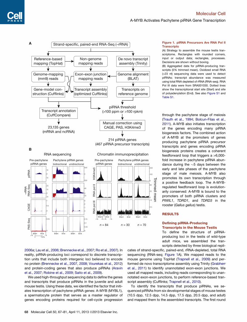
Figure 5. PachytenepiRNAsandPrecursors
Decrease in A-Myb Mutant Testes
The change in transcript or piRNA abundance per
gene in A-Myb (n = 3) and Miwi (n = 1) mutants
compared to heterozygotes in testes isolated at
14.5 and 17.5 dpp. See also Figure S5.
Molecular Cell
A-MYB Activates Pachytene piRNA Gene Transcription
supports the view that A-MYB directly regulates transcription of
pachytene piRNA genes.
A-Myb Regulates Pachytene piRNA ProductionTo test the consequences of the loss of piRNA precursor tran-
scripts, we measured piRNA abundance in the A-Myb mutant.
Like pachytene piRNA precursor transcription, pachytene piRNA
abundance significantly decreased in mutant testes. At 14.5
dpp, median piRNA abundance per pachytene gene decreased
87-fold in A-Myb homozygous mutant testes compared to
heterozygotes (p < 2.2 3 10�16; Figure 5). By 17.5 dpp, median
pachytene piRNA abundance was >9,000 times lower in the
A-Myb mutant than the heterozygotes (p < 2.2 3 10�16). In
contrast, pre-pachytene piRNA levels were essentially unal-
tered. Figure 6 presents examples of the effect at 14.5 and
17.5 dpp of the A-Myb mutant on piRNA precursor transcript
and mature piRNA abundance for one pre-pachytene and three
pachytene piRNA genes.
Our data show that A-MYB binds to the promoters of pachy-
tene piRNA genes; A-Myb, Miwi, and pachytene piRNA pre-
cursor transcription begins at 12.5 dpp; and A-Myb mutant
spermatocytes reach pachynema with subtle defects in auto-
some synapsis (Bolcun-Filas et al., 2011). Could pachytene
piRNA depletion nonetheless be an indirect consequence of
the meiotic arrest caused by the A-Myb mutant? To test this
Molecular Cell 50, 67
possibility, we sequenced small RNAs
from Spo11 mutant testes, which failed
to generate double-stranded DNA breaks
at the leptotene stage and display a
meiotic arrest (Baudat et al., 2000;
Romanienko and Camerini-Otero, 2000).
The median abundance of piRNAs from
pre-pachytene genes did not decrease
at 14.5 dpp. By 17.5 dpp, piRNA from
pachytene genes decreased just 5.9-
fold in the Spo11 mutant testes com-
pared to the heterozygotes (Figure S5).
We note that A-MYB protein abundance
is reduced in the Spo11 mutant (Bolcun-
Filas et al., 2011).
Trip13 is required to complete the
repair of double-strand DNA breaks on
fully synapsed chromosomes. Trip13
mutants display a meiotic arrest similar
to that in A-Myb mutant testes (Li and
Schimenti, 2007): pachytene arrest with
synapsed chromosomes. To further test
whether the loss of pachytene piRNA
precursor transcripts in A-Myb mutants
reflects a general effect of meiotic arrest,
we measured piRNA precursor transcript abundance in Trip13
mutant testes at 17.5 dpp. Unlike A-Myb, piRNA precursor tran-
scripts were readily detectable in the Trip13 mutant (Figure S6).
We conclude that the loss of pachytene piRNA precursor tran-
scripts and piRNAs in A-Myb mutant testes is a direct conse-
quence of the requirement for A-MYB to transcribe pachytene
piRNA genes and not a general feature of meiotic arrest at the
pachytene stage.
A-MYB Regulates Expression of piRNA BiogenesisFactorsThe A-Myb mutant more strongly affected pachytene piRNA
accumulation than it did the steady-state abundance of the cor-
responding piRNA precursor transcripts (Figure 5); the median
decrease in pachytene piRNA abundance was 2-fold greater at
14.5 dpp and 38-fold greater at 17.5 dpp than the decrease in
the steady-state abundance of pachytene precursor transcripts
(Table S1). These data suggest that A-MYB exerts a layer of
control on piRNA accumulation beyond its role in promoting
pachytene piRNA precursor transcription.
Miwi has previously been proposed to be a direct target of
A-MYB; Miwi mRNA abundance is reduced in A-MYB mutant
testes, and ChIP microarray data place A-MYB on the Miwi
promoter (Bolcun-Filas et al., 2011). Our RNA-seq data confirm
that accumulation of Miwi mRNA requires A-MYB: Miwi mRNA
–81, April 11, 2013 ª2013 Elsevier Inc. 73

Figure 6. Examples of the Effect of the A-Myb Mutation on piRNA Expression
Transcript and piRNA abundance in heterozygous (Het) and homozygous A-Myb (Mut) point-mutant testes is shown for four illustrative examples at 14.5 and
17.5 dpp. Also shown is the abundance of piRNA sequencing reads that map to the exon-exon junctions. Gene 11-qE1-9443 does not have an intron. Exons,
blue boxes; splice junctions, gaps; the last exon is compressed and not to scale. See also Figure S6.
Molecular Cell
A-MYB Activates Pachytene piRNA Gene Transcription
decreased more than 50-fold in testes isolated from A-Myb
mutant mice at 14.5 dpp compared to their heterozygous litter-
mates (Figures 7A and S7 and Table S3). Furthermore, our
ChIP data confirm that A-MYB binds the Miwi promoter in vivo
(Figures 7B, S4B, and S7). Like pachytene piRNAs, Miwi tran-
scripts first appear at 12.5 dpp (Figure 2B), and MIWI protein is
first detected in testes at 14.5 dpp (Deng and Lin, 2002). Loss
of MIWI arrests spermatogenesis at the round spermatid stage
(Deng and Lin, 2002).
A previous study reported that piRNAs fail to accumulate to
wild-type levels in Miwi mutant testes (Grivna et al., 2006b).
However, our data suggest that the overall change in piRNA
abundance caused by loss of MIWI is quite small: RNA-seq
detected no change at 14.5 dpp (change in total piRNA abun-
dance = 1.1; n = 2) and only a modest decrease at 17.5 dpp
74 Molecular Cell 50, 67–81, April 11, 2013 ª2013 Elsevier Inc.
(change in total piRNA abundance = 0.58; n = 1). piRNAs from
pachytene loci decreased just 2.7-fold at 14.5 dpp (p = 0.0046)
and 3.5-fold at 17.5 dpp (p = 1.8 3 10�6) in Miwi mutant testes
(Figure 5). By comparison, pachytene piRNAs declined 87-fold
at 14.5 dpp and 9,400-fold at 17.5 dpp in the A-Myb mutant.
Does the loss of MIWI affect piRNA precursor transcription?
We measured transcript abundance and piRNA expression in
Miwi null mutant testes at 14.5 and 17.5 dpp. In Miwi�/� testes,
pachytene piRNA precursor transcripts were present at levels
indistinguishable from Miwi heterozygotes (median change =
1.0- to 1.4-fold; q = 1; Figure 5). Thus, loss of MIWI does
not explain loss of pachytene piRNA precursor transcripts in
A-Myb mutant testes.
In addition to Miwi, ChIP-seq detected A-MYB bound to the
promoters of 12 other RNA-silencing-pathway genes (Figure 7B

A C
B
Figure 7. A-MYB Regulates Expression of mRNAs Encoding piRNA Pathway Proteins
(A) mRNA abundance in A-Mybmutant versus heterozygous testes. The 407 genes with a significant (q < 0.05) change in steady-state mRNA levels are shown as
red circles. The 203 with A-MYB peaks within 500 bp of their transcription start site are filled.
(B) A-MYB ChIP-seq signal at the transcription start sites of A-Myb and genes implicated in RNA silencing pathways. For each, the figure reports the change
in mRNA abundance between 17.5 and 10.5 dpp in wild-type testes and the mean change between A-Myb mutant and heterozygous testes at 14.5 dpp
(mean ± SD; n = 3).
(C) A model for the regulation of pachytene piRNA biogenesis by A-MYB. See also Figure S7 and Table S3.
Molecular Cell
A-MYB Activates Pachytene piRNA Gene Transcription
and Table S3). Of these, the mRNA abundance—measured
by three biologically independent RNA-seq experiments—of
Ago2, Ddx39 (uap56 in flies), Mael, Mili, Mov10l1, Tdrd9, and
Vasa did not change significantly at 14.5 dpp in A-Myb mutant
testes compared to heterozygotes (q > 0.05); except for Ago2,
all decreased significantly in the mutant at 17.5 dpp. In contrast,
Molecular Cell 50, 67–81, April 11, 2013 ª2013 Elsevier Inc. 75

Molecular Cell
A-MYB Activates Pachytene piRNA Gene Transcription
the abundance of the mRNAs encoding Tudor domain proteins
decreased significantly in A-Myb mutant testes: Tdrd6 (64-fold
decrease; q = 3.1 3 10�5) and Tdrd5 (7.5-fold decrease; q =
1.0 3 10�5). Tdrd5 is expressed in embryonic testes then
decreases around birth (Yabuta et al., 2011). TDRD5 protein re-
appears at 12 dpp, increasing throughout the pachynema (Smith
et al., 2004; Yabuta et al., 2011). Our data indicate that A-MYB
activates Tdrd5 transcription at the onset of the pachytene stage
of meiosis. Similarly, Tdrd6 mRNA can be detected at the
middle pachytene, but not the zygotene stage, and peaks after
late pachytene; TDRD6 protein can be detected at 17 dpp and
continues to increase until 21 dpp (Vasileva et al., 2009). The
findings that TDRD5 and TDRD6 colocalize with MIWI in pachy-
tene spermatocytes (Hosokawa et al., 2007; Vasileva et al., 2009;
Yabuta et al., 2011) and that TDRD6 binds MIWI (Chen et al.,
2009; Vagin et al., 2009; Vasileva et al., 2009) suggest a role
for these Tudor domain proteins in pachytene piRNA production
or function. As inMiwi�/� testes, spermatogenesis arrests at the
round spermatid stage in Tdrd5�/� and Tdrd6�/� mutant testes
(Vasileva et al., 2009; Yabuta et al., 2011). Loss of Tdrd6 expres-
sion has little effect on piRNA levels (Figure S3; Vagin et al.,
2009), perhaps because the functions of Tudor domain proteins
overlap.
Other genes encoding piRNA pathway proteins whose pro-
moters are bound by A-MYB and whose expression decreased
significantly in A-Myb mutant testes include MitoPld (Pld6;
3.9-fold decrease; q = 0.0095) and Tdrd12 (5.3-fold decrease;
q = 0.0046). MitoPld encodes an endoribonuclease implicated
in an early step in piRNA biogenesis in mice and flies (Houwing
et al., 2007; Pane et al., 2007; Haase et al., 2010; Huang et al.,
2011; Watanabe et al., 2011; Ipsaro et al., 2012; Nishimasu
et al., 2012). The function ofTdrd12 is not known, but its fly homo-
logs (Yb,Brother of Yb, andSister of Yb) are all required for piRNA
production (Handler et al., 2011). Tdrd1 decreased 3.4-fold, but
with q value = 0.015. Tdrd1 is first expressed in fetal prosperma-
togonia, then re-expressed in pachytene spermatocytes (Chuma
et al., 2006). In Tdrd1 mutant testes, spermatogenesis fails,
with no spermatocytes progressing past the round spermatid
stage (Chuma et al., 2006). TDRD1 binds MILI and MIWI (Chen
et al., 2009; Kojima et al., 2009) and colocalizes with TDRD5
and TDRD6 in the chromatoid body (Hosokawa et al., 2007).
Together, these data support the idea that at the onset of
the pachytene phase of meiosis, A-MYB coordinately activates
transcription of many genes encoding piRNA pathway proteins.
A-MYB and the Pachytene piRNA Regulatory CircuitryA number of genes encoding known and suspected piRNA
pathway proteins are bound and regulated by A-MYB (Figures
7B and S7C). Our data support a model in which A-MYB drives
both the transcription of pachytene piRNA genes and the
mRNAs encoding genes required for piRNA production includ-
ing Miwi, MitoPld, and Tdrd9. Regulation by A-MYB of both
the sources of pachytene piRNAs and the piRNA biogenesis
machinery creates a coherent feedforward loop (Figure 7C).
Feedforward loops amplify initiating signals to increase target
gene expression. Furthermore, they function as switches that
are sensitive to sustained signals; they reject transient signals
(Shen-Orr et al., 2002; Osella et al., 2011).
76 Molecular Cell 50, 67–81, April 11, 2013 ª2013 Elsevier Inc.
A-MYB also bound to the A-Myb promoter (Figure 7B), and
A-Myb transcripts decreased 4.2-fold in testes from an A-Myb
point mutant (Mybl1repro9; Figure 7B). The A-Myb mutant fails
to produce the high level of A-MYB protein observed in wild-
type testes at the late pachytene stage of meiosis (Bolcun-Filas
et al., 2011). Instead, A-MYB protein never becomes more
abundant than the level achieved in wild-type testes by the
beginning of the pachytene stage. While the lower level of
A-MYB in the A-Mybmutant may reflect instability of the mutant
protein, a simpler explanation is that mutant A-MYB cannot acti-
vate A-Myb transcription.
Feedforward Regulation of piRNA Production IsEvolutionarily ConservedIs A-MYB-mediated, feedforward control a general feature of
regulation of piRNA production among vertebrates? To test
whether A-MYB control of piRNA precursor transcription is
evolutionarily conserved, we used high-throughput sequencing
to identify piRNAs in adult rooster testes. Birds and mammals
diverged 330 million years ago (Benton and Donoghue, 2007).
After removing the sequences of identifiable miRNAs (Burnside
et al., 2008) and annotated noncoding RNAs, total small RNA
from the adult rooster testis showed peaks at both 23 and
25 nt (Figure 8A). When the RNA was oxidized before being
prepared for sequencing, only a single 25 nt peak remained,
consistent with the 25 nt small RNAs corresponding to piRNAs
containing 20-O-methyl-modified 30 termini. These longer, oxida-
tion-resistant species typically began with uracil (62%of species
and 65% of reads; Figure 8B), and we detected a significant
Ping-Pong amplification signature (Z score = 31; Figure 8C).
We conclude that the oxidation-resistant, 24–30 nt long small
RNAs correspond to rooster piRNAs. Like piRNAs generally,
rooster piRNAs are diverse, with 5,742,529 species present
among 81,121,893 genome-mapping reads. Like mouse pachy-
tene piRNAs, 70% of piRNAs from adult rooster testes mapped
to unannotated intergenic regions, 19% mapped to transpo-
sons, and 14%mapped to protein-coding genes. Of the piRNAs
that map to protein-coding genes, >95% derive from introns.
Forty-two percent of piRNA species mapped uniquely to the
Gallus gallus genome.
Using 24–30 nt piRNAs from oxidized libraries, we identified
327 rooster piRNA clusters (Figure S8). These account for 76%
of all uniquely mapping piRNAs. Of the 327 clusters, 25 over-
lapped with protein-coding genes. To begin to identify the tran-
scription start sites for the rooster piRNA clusters, we analyzed
adult rooster testes by H3K4me3 ChIP-seq. More than 81%
(268 out of 327) of the clusters contained a readily detectable
H3K4me3 peak within 1 kbp of the piRNA cluster. In contrast,
the median distance from a cluster to the nearest transcription
start site of an annotated gene was 73 kbp, suggesting that
the H3K4me3 peaks reflect the start sites for rooster piRNA
precursor transcripts.
Next, we asked where in the genome A-MYB bound in adult
rooster testes. A-MYB ChIP-seq identified 5,509 significant
peaks (FDR < 10�25). MEME analysis of the top 500 peaks with
the lowest FDR values identified a motif (E = 2.6 3 10�201; Fig-
ure 8D) similar to that found in the mouse (Figure 4A). A-MYB
is the only one of the three chicken MYB genes expressed in

A B
C D
E
Figure 8. Feedforward Regulation of piRNA
Biogenesis by A-MYB Is Conserved in
Rooster
(A) Length distributions of total rooster testis small
RNAs (black) and miRNAs (gray).
(B) Sequence logo showing the nucleotide com-
position of piRNA reads and species.
(C) The 50-50 overlap between piRNAs from
opposite strands was analyzed to determine if
rooster piRNAs display Ping-Pong amplification.
The number of pairs of piRNA reads at each
position is reported. Z score indicates that a sig-
nificant 10 nt overlap (Ping-Pong) was detected.
Z score > 1.96 corresponds to p value < 0.05.
(D) MEME-reported motif of the top 500 (by peak
score) A-MYB ChIP-seq peaks from adult rooster
testes.
(E) A-MYB, H3K4me3, and input ChIP-seq signals
at the transcription start sites of rooster PIWIL1,
TDRD1, and TDRD3. See also Figure S8.
Molecular Cell
A-MYB Activates Pachytene piRNA Gene Transcription
adult testis (X.Z.L. and P.D.Z., unpublished data), supporting the
view that these peaks correspond to A-MYB binding. The core
sequence motif associated with A-MYB binding in mouse differs
at one position (CAGTT) from that in rooster (C C/G GTT). This
difference between mammalian and chicken MYB proteins has
been noted previously (Weston, 1992; Deng et al., 1996).
To determine whether chicken A-MYB might regulate tran-
scription of some piRNA clusters in the testis, we compared
the A-MYB peak nearest to each piRNA cluster with the nearest
H3K4me3 peak. Of the 327 rooster piRNA clusters, at least 104
were occupied by A-MYB at their promoters, as defined by an
overlapping H3K4me3 peak. These 104 clusters account for
31% of uniquely mapping rooster piRNAs.
The chicken genome encodes at least two PIWI proteins:
PIWIL1 and PIWIL2. Remarkably, the promoter of Gallus gallus
PIWIL1, the homolog of mouse Miwi, contained a prominent
A-MYB peak (Figure 8E). TDRD1 and TDRD3 also showed
Molecular Cell 50, 67
A-MYB peaks (Figure 8E). Thus, as in
mice, Gallus gallus A-MYB controls the
transcription of both piRNA clusters and
genes encoding piRNA pathway proteins.
We conclude that A-MYB-mediated feed-
forward regulation of piRNA production
was likely present in the last common
ancestor of birds and mammals.
In mice, we found no piRNA-producing
genes on the sex chromosomes (Fig-
ure S1A), perhaps because mouse sex
chromosomes are silenced during the
pachytene stage (Li et al., 2009b). Birds
use a ZW rather than an XY mechanism
for sex determination, so roosters are
homogametic (ZZ), allowing the sex
chromosomes to remain transcriptionally
active in males (Namekawa and Lee,
2009; Schoenmakers et al., 2009).
Indeed, we find that 39 of the 327 rooster
piRNA clusters are on the Z chromosome, accounting for 12%
of uniquely mapping piRNAs (Figure S8). Of the 39 Z chromo-
some clusters, 18 had an A-MYB peak at their promoter.
DISCUSSION
The data presented here provide strong support for the view that
piRNAs in mammals begin as long, single-stranded precursors
generated by testis-specific, RNA Pol II transcription of indi-
vidual piRNA genes (see also Vourekas et al., 2012). Transcrip-
tion by RNA Pol II affords piRNA genes the same rich set of
transcriptional controls available to regulate mRNA expression.
Our data establish that developmentally regulated transcription
of piRNA genes determines when specific classes of piRNAs
emerge during spermatogenesis.
During mouse spermatogenesis, transcription of pachytene
piRNA genes begins at the onset of the pachytene stage of
–81, April 11, 2013 ª2013 Elsevier Inc. 77

Molecular Cell
A-MYB Activates Pachytene piRNA Gene Transcription
meiosis; pachytene piRNAs accumulate subsequently. The
presence of the MYB binding motif near the transcription start
sites of pachytene piRNA genes, the physical binding of A-MYB
to those genes, and the loss of pachytene piRNA precursor
transcripts and piRNAs in testes from A-Myb mutant mice all
argue that A-MYB regulates pachytene piRNA production.
A-MYB also drives increased expression of piRNA pathway
genes. Among these, Miwi expression shows the greatest
dependence on A-MYB, but A-MYB also drives transcription of
genes encoding other proteins in the piRNA pathway, including
MitoPld, Mael, and five genes encoding Tudor domain proteins.
For example, A-MYB increases expression of Tdrd6 more than
500-fold. Loss of A-MYB functionmore strongly depletes pachy-
tene piRNAs than loss of MIWI, in part because pachytene
piRNAs can still be loaded into MILI in Miwi mutant testes,
although MILI-loaded pachytene piRNAs do not suffice to
produce functional sperm. In the A-Myb mutant, expression of
mRNAs encoding multiple piRNA pathway proteins decreases.
We speculate that in wild-type male mice, the increased expres-
sion of these mRNAs at the onset of the pachytene stage of
meiosis ensures that sufficient piRNA-precursor-processing
and MIWI-loading factors are available to cope with the large
increase in pachytene piRNA precursor transcription.
We propose that induction of A-MYB during the early pachy-
tene stage of spermatogenesis initiates a feedforward loop
that ensures the precisely timed production of these piRNAs.
Coherent feedforward loops show delayed kinetics in order to
reject background stimuli (Mangan and Alon, 2003). Indeed,
we observed a delay from the early to middle pachytene in
the accumulation of pachytene piRNAs, despite the continued
increase in A-Myb expression (Figure 2A). Pachytene piRNA
levels increase 75-fold (median for the 100 genes) from 10.5
to 12.5 dpp, coincident with increased expression of A-Myb.
However, from 12.5 to 14.5 dpp, pachytene piRNAs increase
only 1.2-fold. Pachytene piRNAs subsequently resume their
accumulation, increasing 65-fold from 14.5 to 17.5 dpp. We
believe this delay is a consequence of a feedforward loop that
ensures the production of pachytene piRNAs only at the pachy-
tene stage of spermatogenesis. Regulation by a feedforward
loop also predicts a rapid shutdown of pachytene piRNA path-
ways at round spermatid stage VIII, when A-MYB protein levels
decrease (Horvath et al., 2009). Supporting this idea, the abun-
dance of MIWI decreases sharply by the elongated spermatid
stage of spermatogenesis (Deng and Lin, 2002). Testing this
proposal is a clear challenge for the future.
In fruit flies and zebrafish (Brennecke et al., 2007; Houwing
et al., 2007), most piRNAs map to repetitive regions, whereas
in mammals, uniquely mapping intergenic piRNAs predominate
in the adult testis. The discovery that 70% of rooster piRNA
reads map to intergenic regions suggests that the expansion of
intergenic piRNAs controlled by A-MYB feedforward regulation
arose before the divergence of birds andmammals. In the future,
detailed analysis of piRNA production across avian spermato-
genesis should provide insight into the evolutionary origins and
functions of pachytene piRNAs, a class of piRNAs thus far only
detected in mammals.
In summary, we have shown that mouse piRNA genes are
coregulated transcriptionally, establishing that A-MYB coordi-
78 Molecular Cell 50, 67–81, April 11, 2013 ª2013 Elsevier Inc.
nately regulates the biogenesis of an entire piRNA class, the
pachytene piRNAs. The discovery that a loss-of-function
A-Myb mutant, Mybl1repro9, disrupts piRNA precursor transcrip-
tion in vertebrates provides a tool to understand the transforma-
tion of long, single-stranded piRNA precursors into mature
piRNAs and to explore the functions and targets of the pachy-
tene piRNAs.
EXPERIMENTAL PROCEDURES
Mice
Mybl1repro9, Spo11tm1Sky, and Piwil1tm1Hf mice were maintained and used
according to the guidelines of the Institutional Animal Care andUseCommittee
of the University of Massachusetts Medical School and genotyped as
described (Baudat et al., 2000; Deng and Lin, 2002; Bolcun-Filas et al., 2011).
Sequencing
Small (Ghildiyal et al., 2008; Seitz et al., 2008) and long RNA-seq (Zhang et al.,
2012) and analysis (Li et al., 2009a) were as described. Reads that did not map
tomouse genomemm9weremapped to piRNA precursor transcripts to obtain
splice junction mapping small RNAs. Total small RNA libraries from different
developmental stages and from mutants were normalized to the sum of all
miRNA hairpin mapping reads. Oxidized samples were calibrated to the corre-
sponding total small RNA library via the abundance of shared, uniquely map-
ped piRNA species. piRNA expression data were grouped with Cluster 3.0.
Differential gene expression was analyzed with DESeq R (Anders and Huber,
2010); ChIP-seq reads were aligned to the genome using Bowtie version
0.12.7 (Langmead et al., 2009), and peaks were identified using MACS (Zhang
et al., 2008).
ACCESSION NUMBERS
The Gene Expression Omnibus (GEO) accession number for the RNA-seq,
ChIP-seq, and small RNA data reported in this paper is GSE44690.
SUPPLEMENTAL INFORMATION
Supplemental Information includes eight figures, three tables, and Supple-
mental Experimental Procedures and can be found with this article online at
http://dx.doi.org/10.1016/j.molcel.2013.02.016.
ACKNOWLEDGMENTS
We thank K. Chase and K. Schimenti for help collecting tissues; C. Tipping for
help with mouse husbandry; P. Johnson and B. Keagle for providing rooster
testes; G. Farley for technical assistance; H. Lin for reagents; Xi Chen, Xiaotu
Ma, Oliver Rando, and Benjamin Carone for advice on ChIP; and members of
our laboratories for critical comments on themanuscript. X.Z.L. was supported
by the Lalor Foundation and the Jane Coffin Childs Memorial Fund for Medical
Research.
Received: November 20, 2012
Revised: January 17, 2013
Accepted: February 12, 2013
Published: March 21, 2013
REFERENCES
Anders, S., and Huber,W. (2010). Differential expression analysis for sequence
count data. Genome Biol. 11, R106.
Aravin, A.A., and Hannon, G.J. (2008). Small RNA silencing pathways in germ
and stem cells. Cold Spring Harb. Symp. Quant. Biol. 73, 283–290.
Aravin, A.A., Naumova, N.M., Tulin, A.V., Vagin, V.V., Rozovsky, Y.M., and
Gvozdev, V.A. (2001). Double-stranded RNA-mediated silencing of genomic

Molecular Cell
A-MYB Activates Pachytene piRNA Gene Transcription
tandem repeats and transposable elements in the D. melanogaster germline.
Curr. Biol. 11, 1017–1027.
Aravin, A., Gaidatzis, D., Pfeffer, S., Lagos-Quintana, M., Landgraf, P., Iovino,
N., Morris, P., Brownstein, M.J., Kuramochi-Miyagawa, S., Nakano, T., et al.
(2006). A novel class of small RNAs bind to MILI protein in mouse testes.
Nature 442, 203–207.
Aravin, A.A., Sachidanandam, R., Girard, A., Fejes-Toth, K., and Hannon, G.J.
(2007). Developmentally regulated piRNAclusters implicateMILI in transposon
control. Science 316, 744–747.
Aravin, A.A., Sachidanandam, R., Bourc’his, D., Schaefer, C., Pezic, D., Toth,
K.F., Bestor, T., and Hannon, G.J. (2008). A piRNA pathway primed by indi-
vidual transposons is linked to de novo DNA methylation in mice. Mol. Cell
31, 785–799.
Ashe, A., Sapetschnig, A., Weick, E.M., Mitchell, J., Bagijn, M.P., Cording,
A.C., Doebley, A.L., Goldstein, L.D., Lehrbach, N.J., Le Pen, J., et al. (2012).
piRNAs can trigger a multigenerational epigenetic memory in the germline of
C. elegans. Cell 150, 88–99.
Bailey, T.L., and Elkan, C. (1994). Fitting a mixture model by expectation maxi-
mization to discover motifs in biopolymers. Proc. Int. Conf. Intell. Syst. Mol.
Biol. 2, 28–36.
Baudat, F., Manova, K., Yuen, J.P., Jasin, M., and Keeney, S. (2000).
Chromosome synapsis defects and sexually dimorphic meiotic progression
in mice lacking Spo11. Mol. Cell 6, 989–998.
Benton,M.J., andDonoghue, P.C. (2007). Paleontological evidence to date the
tree of life. Mol. Biol. Evol. 24, 26–53.
Bolcun-Filas, E., Bannister, L.A., Barash, A., Schimenti, K.J., Hartford, S.A.,
Eppig, J.J., Handel, M.A., Shen, L., and Schimenti, J.C. (2011). A-MYB
(MYBL1) transcription factor is a master regulator of male meiosis.
Development 138, 3319–3330.
Brennecke, J., Aravin, A.A., Stark, A., Dus, M., Kellis, M., Sachidanandam, R.,
and Hannon, G.J. (2007). Discrete small RNA-generating loci as master regu-
lators of transposon activity in Drosophila. Cell 128, 1089–1103.
Brennecke, J., Malone, C.D., Aravin, A.A., Sachidanandam, R., Stark, A., and
Hannon, G.J. (2008). An epigenetic role for maternally inherited piRNAs in
transposon silencing. Science 322, 1387–1392.
Burnside, J., Ouyang, M., Anderson, A., Bernberg, E., Lu, C., Meyers, B.C.,
Green, P.J., Markis, M., Isaacs, G., Huang, E., and Morgan, R.W. (2008).
Deep sequencing of chicken microRNAs. BMC Genomics 9, 185.
Carmell, M.A., Girard, A., van de Kant, H.J., Bourc’his, D., Bestor, T.H., de
Rooij, D.G., and Hannon, G.J. (2007). MIWI2 is essential for spermatogenesis
and repression of transposons in the mouse male germline. Dev. Cell 12,
503–514.
Cenik, E.S., and Zamore, P.D. (2011). Argonaute proteins. Curr. Biol. 21, R446–
R449.
Chen, C., Jin, J., James, D.A., Adams-Cioaba, M.A., Park, J.G., Guo, Y.,
Tenaglia, E., Xu, C., Gish, G., Min, J., and Pawson, T. (2009). Mouse Piwi
interactome identifies binding mechanism of Tdrkh Tudor domain to arginine
methylated Miwi. Proc. Natl. Acad. Sci. USA 106, 20336–20341.
Chuma, S., Hosokawa, M., Kitamura, K., Kasai, S., Fujioka, M., Hiyoshi, M.,
Takamune, K., Noce, T., and Nakatsuji, N. (2006). Tdrd1/Mtr-1, a tudor-related
gene, is essential for male germ-cell differentiation and nuage/germinal
granule formation in mice. Proc. Natl. Acad. Sci. USA 103, 15894–15899.
Deng, W., and Lin, H. (2002).miwi, a murine homolog of piwi, encodes a cyto-
plasmic protein essential for spermatogenesis. Dev. Cell 2, 819–830.
Deng, Q.L., Ishii, S., and Sarai, A. (1996). Binding site analysis of c-Myb:
screening of potential binding sites by using the mutation matrix derived
from systematic binding affinity measurements. Nucleic Acids Res. 24,
766–774.
Farazi, T.A., Juranek, S.A., and Tuschl, T. (2008). The growing catalog of small
RNAs and their association with distinct Argonaute/Piwi family members.
Development 135, 1201–1214.
Gan, H., Lin, X., Zhang, Z., Zhang, W., Liao, S., Wang, L., and Han, C. (2011).
piRNA profiling during specific stages of mouse spermatogenesis. RNA 17,
1191–1203.
Ghildiyal, M., Seitz, H., Horwich, M.D., Li, C., Du, T., Lee, S., Xu, J., Kittler, E.L.,
Zapp, M.L., Weng, Z., and Zamore, P.D. (2008). Endogenous siRNAs derived
from transposons andmRNAs inDrosophila somatic cells. Science 320, 1077–
1081.
Girard, A., Sachidanandam, R., Hannon, G.J., and Carmell, M.A. (2006).
A germline-specific class of small RNAs binds mammalian Piwi proteins.
Nature 442, 199–202.
Grabherr, M.G., Haas, B.J., Yassour, M., Levin, J.Z., Thompson, D.A., Amit, I.,
Adiconis, X., Fan, L., Raychowdhury, R., Zeng, Q., et al. (2011). Full-length
transcriptome assembly from RNA-Seq data without a reference genome.
Nat. Biotechnol. 29, 644–652.
Grivna, S.T., Beyret, E., Wang, Z., and Lin, H. (2006a). A novel class of small
RNAs in mouse spermatogenic cells. Genes Dev. 20, 1709–1714.
Grivna, S.T., Pyhtila, B., and Lin, H. (2006b). MIWI associates with translational
machinery and PIWI-interacting RNAs (piRNAs) in regulating spermatogen-
esis. Proc. Natl. Acad. Sci. USA 103, 13415–13420.
Gu, W., Lee, H.C., Chaves, D., Youngman, E.M., Pazour, G.J., Conte, D., Jr.,
and Mello, C.C. (2012). CapSeq and CIP-TAP identify Pol II start sites and
reveal capped small RNAs as C. elegans piRNA precursors. Cell 151, 1488–
1500.
Guenther, M.G., Levine, S.S., Boyer, L.A., Jaenisch, R., and Young, R.A.
(2007). A chromatin landmark and transcription initiation at most promoters
in human cells. Cell 130, 77–88.
Gupta, S., Stamatoyannopoulos, J.A., Bailey, T.L., and Noble, W.S. (2007).
Quantifying similarity between motifs. Genome Biol. 8, R24.
Haase, A.D., Fenoglio, S., Muerdter, F., Guzzardo, P.M., Czech, B., Pappin,
D.J., Chen, C., Gordon, A., and Hannon, G.J. (2010). Probing the initiation
and effector phases of the somatic piRNA pathway in Drosophila. Genes
Dev. 24, 2499–2504.
Handler, D., Olivieri, D., Novatchkova, M., Gruber, F.S., Meixner, K., Mechtler,
K., Stark, A., Sachidanandam, R., and Brennecke, J. (2011). A systematic anal-
ysis of Drosophila TUDOR domain-containing proteins identifies Vreteno and
the Tdrd12 family as essential primary piRNA pathway factors. EMBO J. 30,
3977–3993.
Hartig, J.V., Tomari, Y., and Forstemann, K. (2007). piRNAs—the ancient
hunters of genome invaders. Genes Dev. 21, 1707–1713.
Horvath, G.C., Kistler, M.K., and Kistler, W.S. (2009). RFX2 is a candidate
downstream amplifier of A-MYB regulation in mouse spermatogenesis. BMC
Dev. Biol. 9, 63.
Hosokawa, M., Shoji, M., Kitamura, K., Tanaka, T., Noce, T., Chuma, S., and
Nakatsuji, N. (2007). Tudor-related proteins TDRD1/MTR-1, TDRD6 and
TDRD7/TRAP: domain composition, intracellular localization, and function in
male germ cells in mice. Dev. Biol. 301, 38–52.
Houwing, S., Kamminga, L.M., Berezikov, E., Cronembold, D., Girard, A., van
den Elst, H., Filippov, D.V., Blaser, H., Raz, E., Moens, C.B., et al. (2007). A role
for Piwi and piRNAs in germ cell maintenance and transposon silencing in
Zebrafish. Cell 129, 69–82.
Huang, H., Gao, Q., Peng, X., Choi, S.Y., Sarma, K., Ren, H., Morris, A.J., and
Frohman, M.A. (2011). piRNA-associated germline nuage formation and
spermatogenesis require MitoPLD profusogenic mitochondrial-surface lipid
signaling. Dev. Cell 20, 376–387.
Ipsaro, J.J., Haase, A.D., Knott, S.R., Joshua-Tor, L., and Hannon, G.J. (2012).
The structural biochemistry of Zucchini implicates it as a nuclease in piRNA
biogenesis. Nature 491, 279–283.
Kawaoka, S., Hara, K., Shoji, K., Kobayashi, M., Shimada, T., Sugano, S.,
Tomari, Y., Suzuki, Y., and Katsuma, S. (2013). The comprehensive epigenome
map of piRNA clusters. Nucleic Acids Res. 41, 1581–1590.
Kim, V.N., Han, J., and Siomi, M.C. (2009). Biogenesis of small RNAs in
animals. Nat. Rev. Mol. Cell Biol. 10, 126–139.
Molecular Cell 50, 67–81, April 11, 2013 ª2013 Elsevier Inc. 79

Molecular Cell
A-MYB Activates Pachytene piRNA Gene Transcription
Kojima, K., Kuramochi-Miyagawa, S., Chuma, S., Tanaka, T., Nakatsuji, N.,
Kimura, T., and Nakano, T. (2009). Associations between PIWI proteins and
TDRD1/MTR-1 are critical for integrated subcellular localization in murine
male germ cells. Genes Cells 14, 1155–1165.
Kumar, M., and Carmichael, G.G. (1998). Antisense RNA: function and fate of
duplex RNA in cells of higher eukaryotes. Microbiol. Mol. Biol. Rev. 62, 1415–
1434.
Kuramochi-Miyagawa, S., Kimura, T., Ijiri, T.W., Isobe, T., Asada, N., Fujita, Y.,
Ikawa, M., Iwai, N., Okabe, M., Deng, W., et al. (2004). Mili, a mammalian
member of piwi family gene, is essential for spermatogenesis. Development
131, 839–849.
Kuramochi-Miyagawa, S., Watanabe, T., Gotoh, K., Totoki, Y., Toyoda, A.,
Ikawa, M., Asada, N., Kojima, K., Yamaguchi, Y., Ijiri, T.W., et al. (2008).
DNA methylation of retrotransposon genes is regulated by Piwi family
members MILI and MIWI2 in murine fetal testes. Genes Dev. 22, 908–917.
Kutter, C., Brown, G.D., Goncalves, A., Wilson, M.D., Watt, S., Brazma, A.,
White, R.J., and Odom, D.T. (2011). Pol III binding in six mammals shows
conservation among amino acid isotypes despite divergence among tRNA
genes. Nat. Genet. 43, 948–955.
Langmead, B., Trapnell, C., Pop, M., and Salzberg, S.L. (2009). Ultrafast and
memory-efficient alignment of short DNA sequences to the human genome.
Genome Biol. 10, R25.
Latham, K.E., Litvin, J., Orth, J.M., Patel, B., Mettus, R., and Reddy, E.P.
(1996). Temporal patterns of A-myb and B-myb gene expression during testis
development. Oncogene 13, 1161–1168.
Lau, N.C., Seto, A.G., Kim, J., Kuramochi-Miyagawa, S., Nakano, T., Bartel,
D.P., and Kingston, R.E. (2006). Characterization of the piRNA complex from
rat testes. Science 313, 363–367.
Lee, H.C., Gu, W., Shirayama, M., Youngman, E., Conte, D.J., Jr., and Mello,
C.C. (2012). C. elegans piRNAs mediate the genome-wide surveillance of
germline transcripts. Cell 150, 78–87.
Li, X.C., and Schimenti, J.C. (2007). Mouse pachytene checkpoint 2 (trip13) is
required for completing meiotic recombination but not synapsis. PLoS Genet.
3, e130.
Li, C., Vagin, V.V., Lee, S., Xu, J., Ma, S., Xi, H., Seitz, H., Horwich, M.D.,
Syrzycka, M., Honda, B.M., et al. (2009a). Collapse of germline piRNAs in
the absence of Argonaute3 reveals somatic piRNAs in flies. Cell 137, 509–521.
Li, X.C., Barringer, B.C., andBarbash, D.A. (2009b). The pachytene checkpoint
and its relationship to evolutionary patterns of polyploidization and hybrid
sterility. Heredity (Edinb) 102, 24–30.
Mangan, S., and Alon, U. (2003). Structure and function of the feed-forward
loop network motif. Proc. Natl. Acad. Sci. USA 100, 11980–11985.
Mettus, R.V., Litvin, J.,Wali, A., Toscani, A., Latham, K., Hatton, K., andReddy,
E.P. (1994). Murine A-myb: evidence for differential splicing and tissue-
specific expression. Oncogene 9, 3077–3086.
Modzelewski, A.J., Holmes, R.J., Hilz, S., Grimson, A., and Cohen, P.E. (2012).
AGO4 regulates entry into meiosis and influences silencing of sex chromo-
somes in the male mouse germline. Dev. Cell 23, 251–264.
Montgomery, M.K., Xu, S., and Fire, A. (1998). RNA as a target of double-
stranded RNA-mediated genetic interference in Caenorhabditis elegans.
Proc. Natl. Acad. Sci. USA 95, 15502–15507.
Namekawa, S.H., and Lee, J.T. (2009). XY and ZW: ismeiotic sex chromosome
inactivation the rule in evolution? PLoS Genet. 5, e1000493.
Nebel, B.R., Amarose, A.P., and Hacket, E.M. (1961). Calendar of gameto-
genic development in the prepuberal male mouse. Science 134, 832–833.
Newburger, D.E., and Bulyk, M.L. (2009). UniPROBE: an online database of
protein binding microarray data on protein-DNA interactions. Nucleic Acids
Res. 37(Database issue), D77–D82.
Nishimasu, H., Ishizu, H., Saito, K., Fukuhara, S., Kamatani, M.K., Bonnefond,
L., Matsumoto, N., Nishizawa, T., Nakanaga, K., Aoki, J., et al. (2012).
Structure and function of Zucchini endoribonuclease in piRNA biogenesis.
Nature 491, 284–287.
80 Molecular Cell 50, 67–81, April 11, 2013 ª2013 Elsevier Inc.
Oh, I.H., and Reddy, E.P. (1999). The myb gene family in cell growth, differen-
tiation and apoptosis. Oncogene 18, 3017–3033.
Osella, M., Bosia, C., Cora, D., and Caselle, M. (2011). The role of incoherent
microRNA-mediated feedforward loops in noise buffering. PLoS Comput. Biol.
7, e1001101.
Pane, A., Wehr, K., and Schupbach, T. (2007). zucchini and squash encode
two putative nucleases required for rasiRNA production in the Drosophila
germline. Dev. Cell 12, 851–862.
Reuter, M., Chuma, S., Tanaka, T., Franz, T., Stark, A., and Pillai, R.S. (2009).
Loss of theMili-interacting Tudor domain-containing protein-1 activates trans-
posons and alters the Mili-associated small RNA profile. Nat. Struct. Mol. Biol.
16, 639–646.
Reuter, M., Berninger, P., Chuma, S., Shah, H., Hosokawa, M., Funaya, C.,
Antony, C., Sachidanandam, R., and Pillai, R.S. (2011). Miwi catalysis is
required for piRNA amplification-independent LINE1 transposon silencing.
Nature 480, 264–267.
Ro, S., Park, C., Song, R., Nguyen, D., Jin, J., Sanders, K.M., McCarrey, J.R.,
and Yan, W. (2007). Cloning and expression profiling of testis-expressed
piRNA-like RNAs. RNA 13, 1693–1702.
Robine, N., Lau, N.C., Balla, S., Jin, Z., Okamura, K., Kuramochi-Miyagawa,
S., Blower, M.D., and Lai, E.C. (2009). A broadly conserved pathway generates
3’UTR-directed primary piRNAs. Curr. Biol. 19, 2066–2076.
Romanienko, P.J., and Camerini-Otero, R.D. (2000). Themouse Spo11 gene is
required for meiotic chromosome synapsis. Mol. Cell 6, 975–987.
Saito, K., Inagaki, S., Mituyama, T., Kawamura, Y., Ono, Y., Sakota, E., Kotani,
H., Asai, K., Siomi, H., and Siomi, M.C. (2009). A regulatory circuit for piwi by
the large Maf gene traffic jam in Drosophila. Nature 461, 1296–1299.
Schoenmakers, S., Wassenaar, E., Hoogerbrugge, J.W., Laven, J.S.,
Grootegoed, J.A., and Baarends, W.M. (2009). Female meiotic sex chromo-
some inactivation in chicken. PLoS Genet. 5, e1000466.
Seitz, H., Ghildiyal, M., and Zamore, P.D. (2008). Argonaute loading improves
the 50 precision of bothMicroRNAs and their miRNA* strands in flies. Curr. Biol.
18, 147–151.
Shen-Orr, S.S., Milo, R., Mangan, S., and Alon, U. (2002). Network motifs in the
transcriptional regulation network of Escherichia coli. Nat. Genet. 31, 64–68.
Shirayama, M., Seth, M., Lee, H.C., Gu, W., Ishidate, T., Conte, D.J., Jr., and
Mello, C.C. (2012). piRNAs initiate an epigenetic memory of nonself RNA in
the C. elegans germline. Cell 150, 65–77.
Smith, J.M., Bowles, J., Wilson, M., Teasdale, R.D., and Koopman, P. (2004).
Expression of the tudor-related gene Tdrd5 during development of the male
germline in mice. Gene Expr. Patterns 4, 701–705.
Thomson, T., and Lin, H. (2009). The biogenesis and function of PIWI proteins
and piRNAs: progress and prospect. Annu. Rev. Cell Dev. Biol. 25, 355–376.
Toscani, A., Mettus, R.V., Coupland, R., Simpkins, H., Litvin, J., Orth, J.,
Hatton, K.S., and Reddy, E.P. (1997). Arrest of spermatogenesis and defective
breast development in mice lacking A-myb. Nature 386, 713–717.
Trapnell, C., Pachter, L., and Salzberg, S.L. (2009). TopHat: discovering splice
junctions with RNA-Seq. Bioinformatics 25, 1105–1111.
Trapnell, C., Williams, B.A., Pertea, G., Mortazavi, A., Kwan, G., van Baren,
M.J., Salzberg, S.L., Wold, B.J., and Pachter, L. (2010). Transcript assembly
and quantification by RNA-Seq reveals unannotated transcripts and isoform
switching during cell differentiation. Nat. Biotechnol. 28, 511–515.
Trauth, K., Mutschler, B., Jenkins, N.A., Gilbert, D.J., Copeland, N.G., and
Klempnauer, K.H. (1994). Mouse A-myb encodes a trans-activator and is ex-
pressed in mitotically active cells of the developing central nervous system,
adult testis and B lymphocytes. EMBO J. 13, 5994–6005.
Vagin, V.V., Klenov, M.S., Kalmykova, A.I., Stolyarenko, A.D., Kotelnikov, R.N.,
and Gvozdev, V.A. (2004). The RNA interference proteins and vasa locus are
involved in the silencing of retrotransposons in the female germline of
Drosophila melanogaster. RNA Biol. 1, 54–58.
Molecular Cell
A-MYB Activates Pachytene piRNA Gene Transcription
Vagin, V.V., Sigova, A., Li, C., Seitz, H., Gvozdev, V., and Zamore, P.D. (2006).
A distinct small RNA pathway silences selfish genetic elements in the germline.
Science 313, 320–324.
Vagin, V.V., Wohlschlegel, J., Qu, J., Jonsson, Z., Huang, X., Chuma, S.,
Girard, A., Sachidanandam, R., Hannon, G.J., and Aravin, A.A. (2009).
Proteomic analysis of murine Piwi proteins reveals a role for arginine methyla-
tion in specifying interaction with Tudor family members. Genes Dev. 23,
1749–1762.
Vasileva, A., Tiedau, D., Firooznia, A., Muller-Reichert, T., and Jessberger, R.
(2009). Tdrd6 is required for spermiogenesis, chromatoid body architecture,
and regulation of miRNA expression. Curr. Biol. 19, 630–639.
Vourekas, A., Zheng, Q., Alexiou, P., Maragkakis, M., Kirino, Y., Gregory, B.D.,
and Mourelatos, Z. (2012). Mili and Miwi target RNA repertoire reveals piRNA
biogenesis and function of Miwi in spermiogenesis. Nat. Struct. Mol. Biol. 19,
773–781.
Watanabe, T., Chuma, S., Yamamoto, Y., Kuramochi-Miyagawa, S., Totoki, Y.,
Toyoda, A., Hoki, Y., Fujiyama, A., Shibata, T., Sado, T., et al. (2011). MITOPLD
is a mitochondrial protein essential for nuage formation and piRNA biogenesis
in the mouse germline. Dev. Cell 20, 364–375.
Weston, K. (1992). Extension of the DNA binding consensus of the chicken
c-Myb and v-Myb proteins. Nucleic Acids Res. 20, 3043–3049.
Yabuta, Y., Ohta, H., Abe, T., Kurimoto, K., Chuma, S., and Saitou, M. (2011).
TDRD5 is required for retrotransposon silencing, chromatoid body assembly,
and spermiogenesis in mice. J. Cell Biol. 192, 781–795.
Zhang, Y., Liu, T., Meyer, C.A., Eeckhoute, J., Johnson, D.S., Bernstein, B.E.,
Nusbaum, C., Myers, R.M., Brown, M., Li, W., and Liu, X.S. (2008). Model-
based analysis of ChIP-Seq (MACS). Genome Biol. 9, R137.
Zhang, Z., Theurkauf, W.E., Weng, Z., and Zamore, P.D. (2012). Strand-
specific libraries for high throughput RNA sequencing (RNA-Seq) prepared
without poly(A) selection. Silence 3, 9.
Molecular Cell 50, 67–81, April 11, 2013 ª2013 Elsevier Inc. 81