Embed Size (px)
Citation preview
Molecular Cell
Article
Acetylation Targets the M2 Isoform of PyruvateKinase for Degradation through Chaperone-MediatedAutophagy and Promotes Tumor GrowthLei Lv,1,3,4 Dong Li,5,10 Di Zhao,3,4,10 Ruiting Lin,3,4 Yajing Chu,6 Heng Zhang,1,3,4 Zhengyu Zha,3 Ying Liu,2 Zi Li,7
Yanping Xu,3,4 Gang Wang,6 Yiran Huang,5 Yue Xiong,3,4,8 Kun-Liang Guan,1,3,9 and Qun-Ying Lei1,3,*1Key Laboratory of Molecular Medicine, Ministry of Education, Department of Biochemistry and Molecular Biology2Department of PathologyShanghai Medical College, Fudan University, Shanghai 200032, China3Molecular and Cell Biology Lab, Institutes of Biomedical Sciences, Fudan University, Shanghai 200032, China4School of Life Science, Fudan University, Shanghai 200433, China5Department of Urology, Ren Ji Hospital, Shanghai Jiao Tong University, Shanghai 200127, China6Institute of Biochemistry and Cell Biology7Institute for Nutritional Sciences
Shanghai Institutes for Biological Sciences, Chinese Academy of Sciences, Shanghai 200031, China8Department of Biochemistry and Biophysics, Lineberger Comprehensive Cancer Center, University of North Carolina at Chapel Hill,Chapel Hill, NC 27599, USA9Department of Pharmacology and Moores Cancer Center, University of California at San Diego, La Jolla, CA 92093-0815, USA10These authors contributed equally to this work
*Correspondence: [email protected] 10.1016/j.molcel.2011.04.025
SUMMARY
Most tumor cells take up more glucose than normalcells but metabolize glucose via glycolysis even inthe presence of normal levels of oxygen, a phenom-enon known as the Warburg effect. Tumor cellscommonly express the embryonic M2 isoform ofpyruvate kinase (PKM2) that may contribute to themetabolism shift from oxidative phosphorylation toaerobic glycolysis and tumorigenesis. Here weshow that PKM2 is acetylated on lysine 305 andthat this acetylation is stimulated by high glucoseconcentration. PKM2 K305 acetylation decreasesPKM2 enzyme activity and promotes its lysosomal-dependent degradation via chaperone-mediatedautophagy (CMA). Acetylation increases PKM2 inter-action with HSC70, a chaperone for CMA, and asso-ciation with lysosomes. Ectopic expression of anacetylation mimetic K305Q mutant accumulatesglycolytic intermediates and promotes cell prolifera-tion and tumor growth. These results reveal an acet-ylation regulation of pyruvate kinase and the linkbetween lysine acetylation and CMA.
INTRODUCTION
It was first noted byOttoWarburg that cancer cells rely mainly on
aerobic glycolysis to generate ATP instead ofmore efficientmito-
chondrial oxidative phosphorylation, resulting in the increased
rate of glucose uptake and lactate production even in the pres-
M
ence of sufficient oxygen supply (Warburg, 1956). Based on the
dramatically increased glucose consumption in cancer cells,
positron emission tomography (PET) of 2-(18F)-fluoro-2-deoxy-
D-glucose (FDG) has been developed as a diagnostic technique
to detect cancer cells in clinics (Funes et al., 2007). Activation of
oncogenes or loss of tumor suppressor genes, such asmutations
in Ras (Dang and Semenza, 1999; Ramanathan et al., 2005), AKT
(Manning and Cantley, 2007), Myc (Gordan et al., 2007a, 2007b),
and p53 (Bensaad et al., 2006; Matoba et al., 2006) increase
glucose uptake and lactate production. These observations
rekindle attention to Warburg effect and cancer metabolism.
A key glycolytic enzyme consistently altered in expression
during tumorigenesis is pyruvate kinase (E.C. 2.7.1.40) (Alten-
berg and Greulich, 2004; Majumder et al., 2004), which catalyzes
the transfer of phosphate from phosphoenolpyruvate (PEP) to
ADP, resulting in the formation of pyruvate and ATP. There are
four pyruvate kinase isoforms in mammals: L, R, M1, and M2.
The L and R isoforms are specifically expressed in liver and
red blood cells, respectively (Mazurek et al., 2005). PKM1 is
expressed in most adult tissues, while PKM2 is exclusively ex-
pressed during embryonic development. Notably, most tumor
cells re-express PKM2 (Dombrauckas et al., 2005; Mazurek
et al., 2005), suggesting that the switch from PKM1 to PKM2
expression may be beneficial to tumor cells. Indeed, switching
from PKM2 to PKM1 reverses aerobic glycolysis, providing the
selective growth advantage of PKM2 expression for tumor cells
in vivo (Christofk et al., 2008a). Recently, the PKM1-to-PKM2
switch was found to be regulated by the Myc oncogene (David
et al., 2009), providing further evidence linking the re-expression
of the M2 isoform to the tumorigenesis.
The benefit of expressing PKM2 isoform to the rapidly growing
embryonic and tumorigenic cells is believed to result from a
decreased PK activity, which would lead to accumulation of
olecular Cell 42, 719–730, June 24, 2011 ª2011 Elsevier Inc. 719
A
NAM
- + +
-Ac
Flag-PKM2
TSA 16h
Flag-PKM2 + +
-
- 16h 16h
16h8h 12h -
-
IP
:-F
la
g
B
C D
K305Ac
Flag-PKM2
IP
:-F
la
g
Flag-PKM2
Flag-PKM2K305R
- +
+ -
K305Ac
TSA+NAM
PKM2
- +
+-NAM + TSA
2
1
0
Re
la
tive
A
ce
tyl-P
KM
2 le
vel
E
Flag-PKM2
0
0.4
1.2
0.8
PKM2 K62Q K62R K305Q K305R
Re
lative P
KM
2 activ
ity
G
ADP concentration (mM)
500
0
1000
1500
PEP concentration (mM)
12
500
0
1000
1500
96300 1 2 3 4
Re
lative P
KM
2 activ
ity
Re
lative P
KM
2 activ
ity
Flag-PKM2
0
1
3
5
6
7
2
4
- + - + - +NAM+TSA
WT K305Q K305R
Re
lative P
KM
2 activ
ity
F
Flag-PKM2
WT
Pan-Ac
62R 62Q 305R 305Q Vector
0.95 0.99 0.55 0.49 1
IP
:-F
la
g
αα
αααα
αα
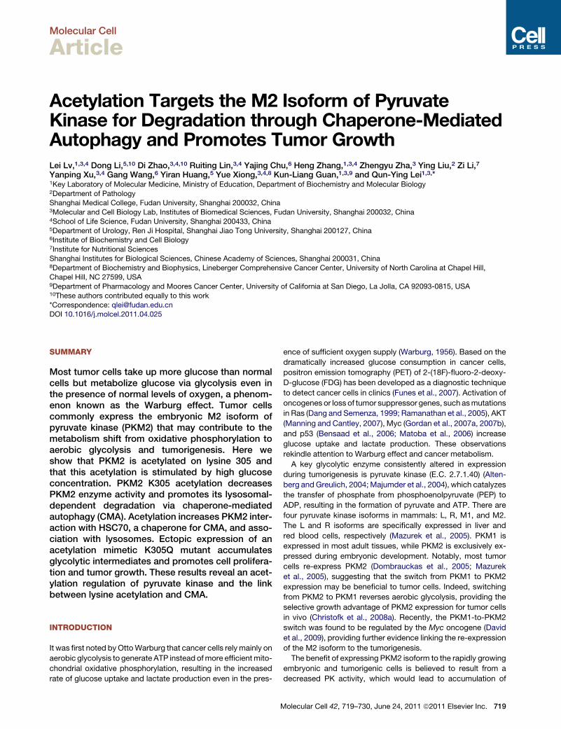
Figure 1. Acetylation at K305 Decreases PKM2 Enzyme Activity
(A) PKM2 is acetylated. Flag-PKM2 was transfected into 293T cells followed by treatment with TSA and NAM for the indicated time, and PKM2 acetylation and
protein levels were analyzed by western blot with indicated antibody, respectively.
(B) Mutation of K305 decreases PKM2 acetylation. The indicated plasmids were cotransfected into 293T cells, and protein was immunoprecipitated (IP) for
acetylation analysis. Acetylation levels were normalized against b-actin.
(C) Characterization of acetyl-PKM2 (K305) antibody. The indicated plasmids were transfected into 293T cells, and acetylation level of IPed Flag-PKM2 was
measured by the site-specific K305 acetylation antibody.
(D) Endogenous PKM2 is acetylated at K305. 293T cells were treated with TSA and NAM. Endogenous PKM2 was immunoprecipitated, and protein levels and
acetylation of K305 were determined by western blot with indicated antibodies (left panel). Relative PKM2K305 acetylation over protein level was quantified (right
panel). Error bars represent ± SD for triplicate experiments.
(E) K305Q mutant decreases PKM2 enzyme activity. Flag-tagged wild-type and mutant PKM2 protein were expressed in 293T cells and purified by IP.
The enzyme activity wasmeasured and normalized against protein level. Mean values of relative enzyme activity of triplicate experiments with standard deviation
(±SD) are presented.
Molecular Cell
PKM2 Acetylation Promotes Cancer Cell Metabolism
720 Molecular Cell 42, 719–730, June 24, 2011 ª2011 Elsevier Inc.
Molecular Cell
PKM2 Acetylation Promotes Cancer Cell Metabolism
various glycolytic metabolites for macromolecular biosynthesis
to support cell growth. According to this notion, a regulation
that decreases and increases PK activity could favor active
dividing and quiescent cells, respectively. Unlike PKM1, full
activity of PKM2 requires allosteric activation by fructose 1, 6-bi-
sphosphate (F-1, 6-BP). One such regulation is the binding of
PKM2, but not PKM1, to phosphotyrosine, and this binding
releases the allosteric activator F-1,6-BP from PKM2, leading
to a decreased PKM2 activity and shifting catabolism from
energy production to anabolic processes, leading to increased
cell proliferation and tumor growth (Christofk et al., 2008b).
Protein acetylation has recently emerged as a broadly used
modification in the regulation of a wide range of cellular
processes (Choudhary et al., 2009; Kim et al., 2006; Zhao et al.,
2010). In particular, we have found that most of the intermediate
metabolic enzymes are acetylated and that acetylation can
directly affect enzyme function (Zhao et al., 2010). Notably, acet-
ylation of metabolic enzymes is regulated by extracellular cues,
such as the nutrient availability. These findings indicate a broad
role of acetylation in the coordination between the extracellular
nutrients and intracellular metabolic pathways. In this paper, we
report that PKM2 activity and protein stability are regulated by
lysine acetylation. Specifically, acetylation of lysine K305 inhibits
PKM2 activity and promotes lysosome-dependent degradation
of PKM2 via CMA. Our study reveals an acetylation regulation
of pyruvate kinase and the link between acetylation and CMA.
RESULTS
PKM2 Is Acetylated at K305Protein acetylation has long been known to play a key role in
regulation of chromatin structure and gene transcription through
modification of histones and nuclear transcription regulators
(Soutoglou et al., 2000). We and others have recently discovered
that a large number of nonnuclear proteins involved in a wide
range of cellular pathways are also acetylated in human and
mouse liver and leukemia cells (Choudhary et al., 2009; Kim
et al., 2006; Zhao et al., 2010). To further explore acetylation of
nonnuclear protein in other tissues, we fractioned cell extracts
of both human prostate cancer cell line LnCAP and cancer
tissues into nuclear, mitochondrial, and cytosolic fractions.
Cytosolic fractions were digested with trypsin, and acetylated
peptides were immunoprecipitated with an antibody specific to
acetyllysine. The immunopurified peptides were analyzed by
tandem liquid chromatography-tandem mass spectrometry
(LC-MS/MS). These analyses identified 113 acetylated proteins
(data not shown), including several enzymes involved in glycol-
ysis pathway. PKM2 was identified to be acetylated by the
mass spectrometric analyses in samples from both cultured
LnCAP cells and primary prostate cancer tissues. Identified
acetylated PKM2 peptides are shown in Table S1, available
online. To confirm its acetylation, Flag-tagged PKM2 was ectop-
(F) NAM and TSA treatment decreases PKM2 wild-type but not mutant enzyme ac
cells and treatedwith or without NAM and TSA, then purified by IP. The PKM2 enzy
relative enzyme activity of triplicate experiments with standard deviation (±SD) a
(G) K305Qmutation decreases the binding affinity toward PEP. The activities of wi
or PEP as indicated. Error bars represent ±SD for triplicate experiments.
M
ically expressed into HEK293T cells and immunoprecipitated.
Western blotting with anti-acetyllysine antibody confirmed that
PKM2 was indeed acetylated and its acetylation was enhanced
approximately 3-fold after treatment with trichostatin A (TSA, an
inhibitor of histone deacetylase HDAC I and II) (Ekwall et al.,
1997; Furumai et al., 2001) and nicotinamide (NAM, an inhibitor
of the SIRT family deacetylases) (Avalos et al., 2005) (Figure 1A).
Two acetylation sites were identified in PKM2, K62 and K305
(Table S1). K62 is not conserved, while K305 is conserved from
C. elegans to mammals (Figure S1A). We generated Gln and
Arg substitution mutants of both sites (K62Q, K62R, K305Q,
and K305R) and transfected 293T cells with these mutants
individually, followed by direct western blotting with anti-acetyl-
lysine antibody. Mutation of K62 only slightly reduced the acety-
lation of PKM2, while mutation of K305 reduced the PKM2
acetylation by 45% (Figure 1B), and double mutant of K62 and
K305 comparably reduced PKM2 acetylation level as K305
single mutant (Figure S1B), indicating that under this condition
K305 is a major acetylation site of ectopically expressed
PKM2. There is still residual acetylation in the double mutants,
suggesting additional acetylation sites in PKM2. To determine
if K305 is acetylated in vivo, we generated antibody specific to
acetylated K305. To characterize the specificity of this antibody,
we performed dot blot assay and found that PKM2 acetyl K305
antibody preferentially detected the acetylated peptide, but not
the unmodified peptide (Figure S2A). Western blotting using
this antibody detected strong signal of ectopically expressed
wild-type PKM2, but not K305R mutant (Figure 1C). Moreover,
PKM2 knockdown significantly decreased the signal detected
by the acetyl K305 antibody (Figure S2B). We also found that
the antibody recognition of PKM2 was competed by acetyla-
tion-modified peptide, but not by the unmodified peptide (Fig-
ure S2C), confirming the specificity of the antibody. Furthermore,
western blotting of whole-cell extract from 293T cells detected
a band that has a molecular weight of expected as PKM2 and
whose intensity was substantially increased upon treatment of
cells with TSA and NAM prior to the lysis (Figure 1D). Similarly,
NAM and TSA treatment also increased PKM2 acetylation in
LnCAP cells (Figure S3A). As shown in Figure S1C, both NAM
and TSA increased endogenous PKM2 acetylation at K305.
Moreover, IHC staining showed that PKM2 was indeed acety-
lated at K305 in human prostate cancer tissues (Figure S3B
and Table S2). These results demonstrate an in vivo acetylation
of PKM2 at Lys305.
Acetylation Mimetic Mutation at K305 DecreasesPKM2 ActivityTo determine the effect of acetylation at these two sites on PKM2
enzyme activity, we transfected 293T cells with individual PKM2
mutants and assayed for the PKM2 enzyme activity. Mutation of
Lys62 to either Gln or Arg reduced the activity of PKM2 by 28%
and 40%, respectively, while mutation of Lys305 to Arg reduces
tivity. Flag-tagged wild-type andmutant PKM2 protein were expressed in 293T
me activity wasmeasured and normalized against protein level. Mean values of
re presented.
ld-type andmutant PKM2were assayed with increasing concentrations of ADP
olecular Cell 42, 719–730, June 24, 2011 ª2011 Elsevier Inc. 721
Molecular Cell
PKM2 Acetylation Promotes Cancer Cell Metabolism
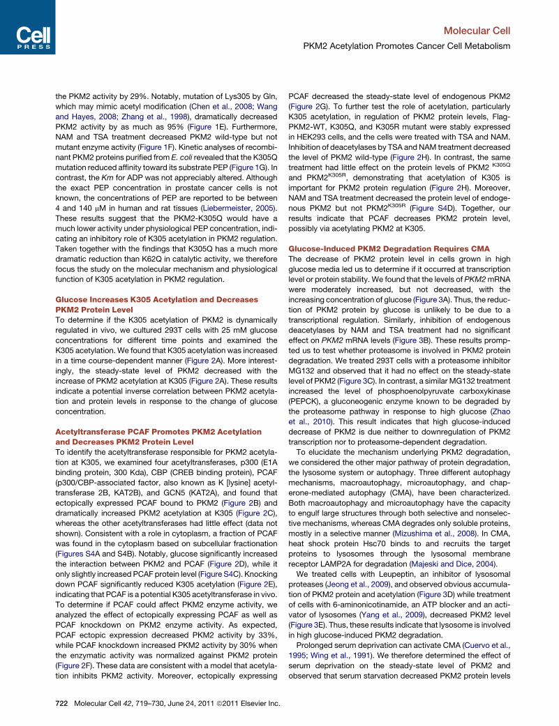
the PKM2 activity by 29%. Notably, mutation of Lys305 by Gln,
which may mimic acetyl modification (Chen et al., 2008; Wang
and Hayes, 2008; Zhang et al., 1998), dramatically decreased
PKM2 activity by as much as 95% (Figure 1E). Furthermore,
NAM and TSA treatment decreased PKM2 wild-type but not
mutant enzyme activity (Figure 1F). Kinetic analyses of recombi-
nant PKM2 proteins purified from E. coli revealed that the K305Q
mutation reduced affinity toward its substrate PEP (Figure 1G). In
contrast, the Km for ADP was not appreciably altered. Although
the exact PEP concentration in prostate cancer cells is not
known, the concentrations of PEP are reported to be between
4 and 140 mM in human and rat tissues (Liebermeister, 2005).
These results suggest that the PKM2-K305Q would have a
much lower activity under physiological PEP concentration, indi-
cating an inhibitory role of K305 acetylation in PKM2 regulation.
Taken together with the findings that K305Q has a much more
dramatic reduction than K62Q in catalytic activity, we therefore
focus the study on the molecular mechanism and physiological
function of K305 acetylation in PKM2 regulation.
Glucose Increases K305 Acetylation and DecreasesPKM2 Protein LevelTo determine if the K305 acetylation of PKM2 is dynamically
regulated in vivo, we cultured 293T cells with 25 mM glucose
concentrations for different time points and examined the
K305 acetylation. We found that K305 acetylation was increased
in a time course-dependent manner (Figure 2A). More interest-
ingly, the steady-state level of PKM2 decreased with the
increase of PKM2 acetylation at K305 (Figure 2A). These results
indicate a potential inverse correlation between PKM2 acetyla-
tion and protein levels in response to the change of glucose
concentration.
Acetyltransferase PCAF Promotes PKM2 Acetylationand Decreases PKM2 Protein LevelTo identify the acetyltransferase responsible for PKM2 acetyla-
tion at K305, we examined four acetyltransferases, p300 (E1A
binding protein, 300 Kda), CBP (CREB binding protein), PCAF
(p300/CBP-associated factor, also known as K [lysine] acetyl-
transferase 2B, KAT2B), and GCN5 (KAT2A), and found that
ectopically expressed PCAF bound to PKM2 (Figure 2B) and
dramatically increased PKM2 acetylation at K305 (Figure 2C),
whereas the other acetyltransferases had little effect (data not
shown). Consistent with a role in cytoplasm, a fraction of PCAF
was found in the cytoplasm based on subcellular fractionation
(Figures S4A and S4B). Notably, glucose significantly increased
the interaction between PKM2 and PCAF (Figure 2D), while it
only slightly increased PCAF protein level (Figure S4C). Knocking
down PCAF significantly reduced K305 acetylation (Figure 2E),
indicating that PCAF is a potential K305 acetyltransferase in vivo.
To determine if PCAF could affect PKM2 enzyme activity, we
analyzed the effect of ectopically expressing PCAF as well as
PCAF knockdown on PKM2 enzyme activity. As expected,
PCAF ectopic expression decreased PKM2 activity by 33%,
while PCAF knockdown increased PKM2 activity by 30% when
the enzymatic activity was normalized against PKM2 protein
(Figure 2F). These data are consistent with a model that acetyla-
tion inhibits PKM2 activity. Moreover, ectopically expressing
722 Molecular Cell 42, 719–730, June 24, 2011 ª2011 Elsevier Inc.
PCAF decreased the steady-state level of endogenous PKM2
(Figure 2G). To further test the role of acetylation, particularly
K305 acetylation, in regulation of PKM2 protein levels, Flag-
PKM2-WT, K305Q, and K305R mutant were stably expressed
in HEK293 cells, and the cells were treated with TSA and NAM.
Inhibition of deacetylases by TSA and NAM treatment decreased
the level of PKM2 wild-type (Figure 2H). In contrast, the same
treatment had little effect on the protein levels of PKM2 K305Q
and PKM2K305R, demonstrating that acetylation of K305 is
important for PKM2 protein regulation (Figure 2H). Moreover,
NAM and TSA treatment decreased the protein level of endoge-
nous PKM2 but not PKM2K305R (Figure S4D). Together, our
results indicate that PCAF decreases PKM2 protein level,
possibly via acetylating PKM2 at K305.
Glucose-Induced PKM2 Degradation Requires CMAThe decrease of PKM2 protein level in cells grown in high
glucose media led us to determine if it occurred at transcription
level or protein stability. We found that the levels of PKM2mRNA
were moderately increased, but not decreased, with the
increasing concentration of glucose (Figure 3A). Thus, the reduc-
tion of PKM2 protein by glucose is unlikely to be due to a
transcriptional regulation. Similarly, inhibition of endogenous
deacetylases by NAM and TSA treatment had no significant
effect on PKM2 mRNA levels (Figure 3B). These results promp-
ted us to test whether proteasome is involved in PKM2 protein
degradation. We treated 293T cells with a proteasome inhibitor
MG132 and observed that it had no effect on the steady-state
level of PKM2 (Figure 3C). In contrast, a similar MG132 treatment
increased the level of phosphoenolpyruvate carboxykinase
(PEPCK), a gluconeogenic enzyme known to be degraded by
the proteasome pathway in response to high glucose (Zhao
et al., 2010). This result indicates that high glucose-induced
decrease of PKM2 is due neither to downregulation of PKM2
transcription nor to proteasome-dependent degradation.
To elucidate the mechanism underlying PKM2 degradation,
we considered the other major pathway of protein degradation,
the lysosome system or autophagy. Three different autophagy
mechanisms, macroautophagy, microautophagy, and chap-
erone-mediated autophagy (CMA), have been characterized.
Both macroautophagy and microautophagy have the capacity
to engulf large structures through both selective and nonselec-
tive mechanisms, whereas CMA degrades only soluble proteins,
mostly in a selective manner (Mizushima et al., 2008). In CMA,
heat shock protein Hsc70 binds to and recruits the target
proteins to lysosomes through the lysosomal membrane
receptor LAMP2A for degradation (Majeski and Dice, 2004).
We treated cells with Leupeptin, an inhibitor of lysosomal
proteases (Jeong et al., 2009), and observed obvious accumula-
tion of PKM2 protein and acetylation (Figure 3D) while treatment
of cells with 6-aminonicotinamide, an ATP blocker and an acti-
vator of lysosomes (Yang et al., 2009), decreased PKM2 level
(Figure 3E). Thus, these results indicate that lysosome is involved
in high glucose-induced PKM2 degradation.
Prolonged serum deprivation can activate CMA (Cuervo et al.,
1995; Wing et al., 1991). We therefore determined the effect of
serum deprivation on the steady-state level of PKM2 and
observed that serum starvation decreased PKM2 protein levels
Flag-PCAF
HA-PKM2
- +
+ +
HA-PKM2
IP
:αα-
Fla
g
Flag-PCAF
HA-PKM2
In
pu
t
Flag-PCAF
Flag-PCAF- +
PKM2
Flag-PCAF
ββ-actin
A B
C D
K305Ac
PKM2
ββ-actin
PCAF
PCAF siRNA - +
Flag-PKM2
PCAF
K305Ac
PKM2
PCAF
PCAF siRNA
+
-
-
+ +
-
- +
+
F
IP
: αα-
Fla
g
Flag- PCAF
Flag-PKM2
- +
+ +
K305Ac
Flag-PCAF
we
ste
rn
Flag-PKM2
IP: α-Flag
H
ββ-actin
Flag-PKM2
NAM+TSA - + - + - +
H1299 stable
1 0.40 1 0.89 1 0.92
WT K305R K305Q
Ratio
K305Ac/actin Ratio
Glucose 25mM,hr 0 2
PKM2
ββ-actin
4 6 8 10
K305Ac
1 1.27 1.83 1.88 2.2 2.28
1 0.82 0.8 0.68 0.55 0.45PKM2/actin Ratio
E
G
Glucose(mM)
HA-PKM2
Flag-PCAF+
+
- +
++
3 25 3
HA-PKM2
Flag-PCAF
HA-PKM2
Flag-PCAF
IP
:αα-
Fla
g
In
pu
t
Re
la
tiv
e P
KM
2 A
ctiv
ity
0
0.5
1.5
1
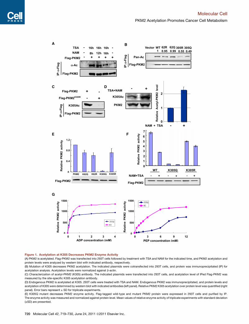
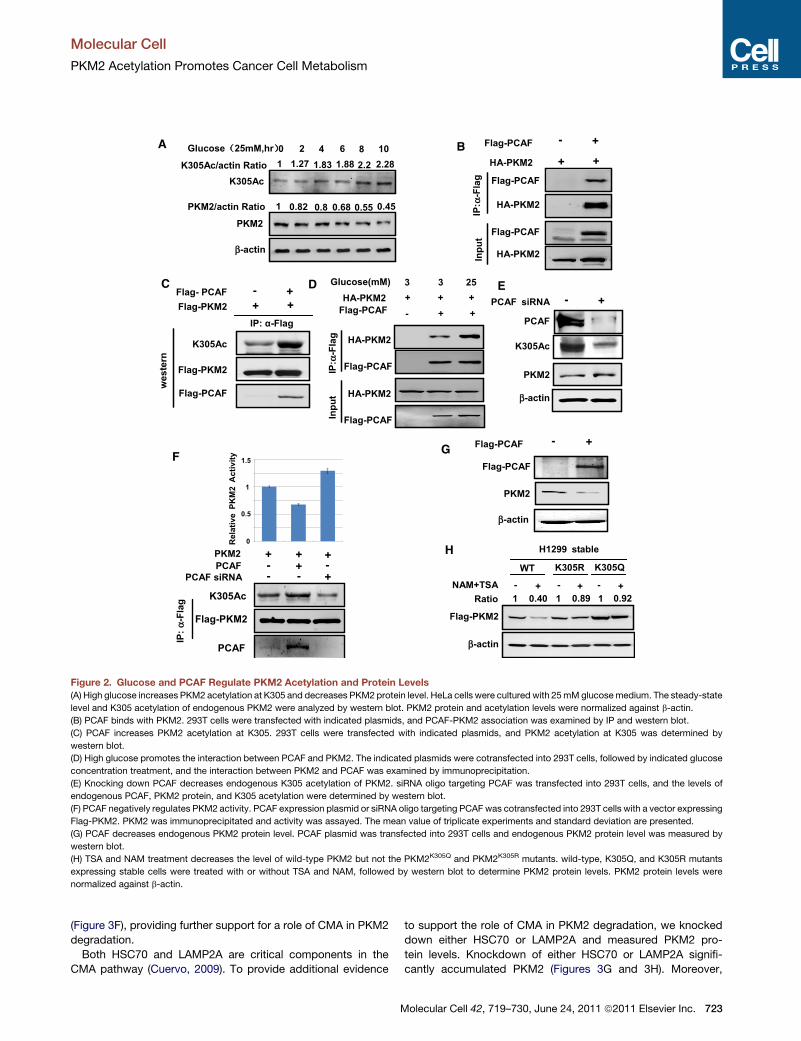
Figure 2. Glucose and PCAF Regulate PKM2 Acetylation and Protein Levels
(A) High glucose increases PKM2 acetylation at K305 and decreases PKM2 protein level. HeLa cells were cultured with 25mMglucosemedium. The steady-state
level and K305 acetylation of endogenous PKM2 were analyzed by western blot. PKM2 protein and acetylation levels were normalized against b-actin.
(B) PCAF binds with PKM2. 293T cells were transfected with indicated plasmids, and PCAF-PKM2 association was examined by IP and western blot.
(C) PCAF increases PKM2 acetylation at K305. 293T cells were transfected with indicated plasmids, and PKM2 acetylation at K305 was determined by
western blot.
(D) High glucose promotes the interaction between PCAF and PKM2. The indicated plasmids were cotransfected into 293T cells, followed by indicated glucose
concentration treatment, and the interaction between PKM2 and PCAF was examined by immunoprecipitation.
(E) Knocking down PCAF decreases endogenous K305 acetylation of PKM2. siRNA oligo targeting PCAF was transfected into 293T cells, and the levels of
endogenous PCAF, PKM2 protein, and K305 acetylation were determined by western blot.
(F) PCAF negatively regulates PKM2 activity. PCAF expression plasmid or siRNA oligo targeting PCAF was cotransfected into 293T cells with a vector expressing
Flag-PKM2. PKM2 was immunoprecipitated and activity was assayed. The mean value of triplicate experiments and standard deviation are presented.
(G) PCAF decreases endogenous PKM2 protein level. PCAF plasmid was transfected into 293T cells and endogenous PKM2 protein level was measured by
western blot.
(H) TSA and NAM treatment decreases the level of wild-type PKM2 but not the PKM2K305Q and PKM2K305R mutants. wild-type, K305Q, and K305R mutants
expressing stable cells were treated with or without TSA and NAM, followed by western blot to determine PKM2 protein levels. PKM2 protein levels were
normalized against b-actin.
Molecular Cell
PKM2 Acetylation Promotes Cancer Cell Metabolism
(Figure 3F), providing further support for a role of CMA in PKM2
degradation.
Both HSC70 and LAMP2A are critical components in the
CMA pathway (Cuervo, 2009). To provide additional evidence
M
to support the role of CMA in PKM2 degradation, we knocked
down either HSC70 or LAMP2A and measured PKM2 pro-
tein levels. Knockdown of either HSC70 or LAMP2A signifi-
cantly accumulated PKM2 (Figures 3G and 3H). Moreover,
olecular Cell 42, 719–730, June 24, 2011 ª2011 Elsevier Inc. 723
C D
F
PKM2
ββ-actin
MG132 (h) 0 42
PEPCK
6
I
PKM2
LAMP2A
β-actin
β-actin
β-actin
Con. shLAMP2A
E
G
H
A B
4 NAM+TSA (h)
0.4
0
1.2
Re
la
tive
PKM2 m
RN
A level
0.8
1.6
0 6 8
6-AN (h) 0
PKM2
β-actin
12 24 36
shLAMP2A Control
3.125 6.25 12.5 25 3.125 6.25 12.5 25
PKM2
Glucose
(mM, 10 h)
PKM2
β-actin
12h 36h 72h
+ - + - + -
Serum
deprivation
Glucose (mM)
0
0.4
1.2
0.8
1.6
Re
la
tive
PKM2 m
RN
A level
3.125 6.25 12.5 25
K305Ac
Leupeptin(mM) - 0.8
PKM2
β-actin
0.4
β-actin
PKM2
HSC70
- +Antisence-HSC70
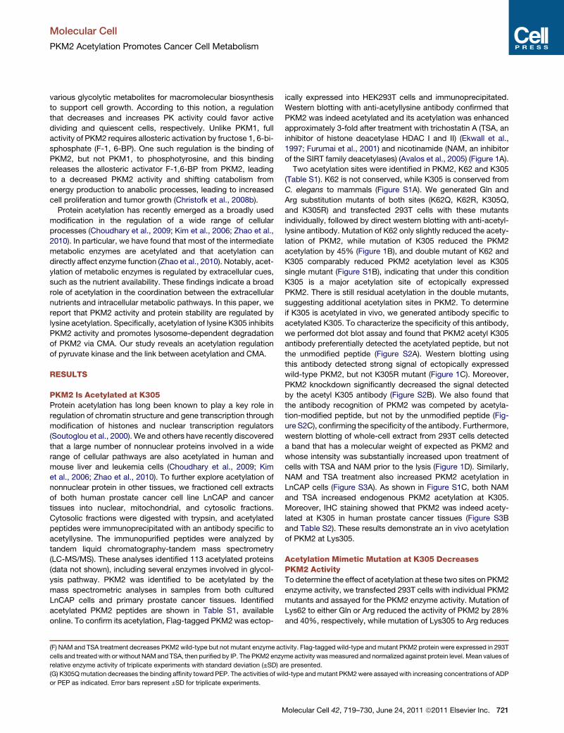
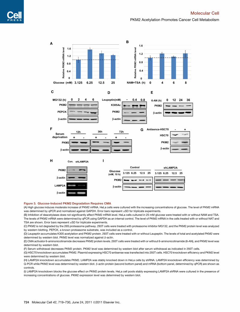
Figure 3. Glucose-Induced PKM2 Degradation Requires CMA
(A) High glucose induces moderate increase of PKM2 mRNA. HeLa cells were cultured with the increasing concentrations of glucose. The level of PKM2 mRNA
was determined by qPCR and normalized against GAPDH. Error bars represent ±SD for triplicate experiments.
(B) Inhibition of deacetylases does not significantly affect PKM2 mRNA level. HeLa cells cultured in 25 mM glucose were treated with or without NAM and TSA.
The levels of PKM2 mRNA were determined by qPCR using GAPDH as an internal control. The level of PKM2 mRNA in the cells treated with or without NAT and
TSA are shown. Error bars represent ±SD for triplicate experiments.
(C) PKM2 is not degraded by the 26S proteasome pathway. 293T cells were treated with proteasome inhibitor MG132, and the PKM2 protein level was analyzed
by western blotting. PEPCK, a known proteasome substrate, was included as a control.
(D) Leupeptin accumulates K305 acetylation and PKM2 protein. 293T cells were treated with or without Leupeptin. The levels of total and acetylated PKM2 were
determined by western blot. PKM2 level was normalized against b-actin.
(E) CMA activator 6-aminonicotinamide decreases PKM2 protein levels. 293T cells were treatedwith or without 6-aminonicotinamide (6-AN), and PKM2 level was
determined by western blot.
(F) Serum withdrawal decreases PKM2 protein. PKM2 level was determined by western blot after serum withdrawal as indicated in 293T cells.
(G) HSC70 knockdown accumulates PKM2. Plasmid expressing HSC70 antisense was transfected into 293T cells. HSC70 knockdown efficiency and PKM2 level
were determined by western blot.
(H) LAMP2A knockdown accumulates PKM2. LAMP2A was stably knocked down in HeLa cells by shRNA. LAMP2A knockdown efficiency was determined by
Q-PCR while PKM2 level was determined by western blot. b-actin protein (second bottom panel) and mRNA (bottom panel, determined by qPCR) are shown as
controls.
(I) LAMP2A knockdown blocks the glucose effect on PKM2 protein levels. HeLa cell pools stably expressing LAMP2A shRNA were cultured in the presence of
increasing concentrations of glucose. PKM2 expression level was determined by western blot.
Molecular Cell
PKM2 Acetylation Promotes Cancer Cell Metabolism
724 Molecular Cell 42, 719–730, June 24, 2011 ª2011 Elsevier Inc.
Molecular Cell
PKM2 Acetylation Promotes Cancer Cell Metabolism
glucose-induced PKM2 degradation was blocked in LAMP2A
knockdown cells (Figure 3I). We conclude that high glucose-
induced PKM2 reduction is achieved through CMA-mediated
protein degradation.
PKM2 Acetylation Promotes Its Binding with HSC70and Uptake by LysosomesThe apparent inverse correlation between PKM2 acetylation and
protein levels led us to explore a potential role of K305 acetylation
in PKM2 degradation. In CMA, HSC70 functions to recruit target
proteins to lysosomes for degradation. We first employed acetyl
mimetic mutant to examine if PKM2K305Q exhibits a binding with
HSC70. Coimmunoprecipitation showed that K305Q mutant
displayed a much stronger interaction with HSC70 than wild-
type PKM2 (Figure 4A). Consistently, treatment of cells with de-
acetylase inhibitors TSA and NAM significantly increased the
binding between PKM2 and HSC70 (Figure 4B), suggesting
that PKM2 acetylation may increase its interaction with HSC70.
Furthermore, coexpression of HSC70 dramatically decreased
the level of K305-acetylated PKM2 (Figure 4C), consistent with
a notion that HSC70 destabilizes the acetylated PKM2. The
PKM2 immunoprecipitated by HSC70 had enriched K305-acety-
lated PKM2 (Figure 4D). The interaction between endogenous
PKM2 and HSC70 was confirmed by coimmunoprecipitation
(Figure 4E). These data demonstrate that HSC70 preferentially
associates with the K305-acetylated PKM2. Importantly, the
PKM2-HSC70 association was substantially increased with the
increase of glucose concentrations (Figure 4F). Similarly, glucose
treatment increased the interaction between endogenous PKM2
and HSC70 (Figure S4E). These results indicate that K305 acety-
lation promotes PKM2 degradation by enhancing its interaction
with HSC70 in response to glucose.
Proteins undergoing CMA-mediated lysosomal degradation
often contain a loosely defined KFERQ motif important for
HSC70 binding (Cuervo, 2009). The KFERQ motif usually
contains five residues, including a critical glutamine (Q) residue
that is preceded or followed by four amino acids consisting of
a basic (R or K), an acidic (E or D), or a bulky hydrophobic residue
(I, L, V, or F) (Dice, 1990). Inspection of PKM2 amino sequence
reveals three potential HSC70 binding motifs, 184QVKQK188,223EKDIQDLKF231, and 393QLFEE398. We replaced Q184V185,
Q223D224, and Q393L394 by alanines and examined the inter-
action of each mutant with HSC70. While neither Q184A/
V185A nor Q223A/D224A mutation decreased the interaction
with HSC70, Q393A/L394A mutation significantly reduced its
interaction with HSC70 (Figure 4G), providing an additional
support for a CMA-mediated degradation of PKM2. Finally, gel
filtration analysis showed that coexpression of PCAF led to a shift
of a fraction of PKM2 to higher molecular complex and the
acetylated PKM2was exclusively present in the highermolecular
weight fractions (Figure 4H). These observations are consistent
with an idea that acetylation promotes the interaction of PKM2
and HSC70.
To determine if PKM2 can be uptaken by lysosomes, we incu-
bated the immunopurified PKM2with isolated lysosomes in vitro.
The lysosomes were repurified and the associated PKM2 was
detected by western blotting to determine PKM2 binding.
When the above experiment was performed in the presence of
M
lysosomal protease inhibitors, this assay would show the total
PKM2 that was bound to and taken up by the lysososomes.
We found that PKM2 bound to lysosomes. Moreover, the
PKM2 isolated from TSA and NAM-treated cells showed more
lysosomal binding and uptake (Figure 4I), suggesting that the
acetylated PKM2 has higher affinity to lysosomes. Furthermore,
the PKM2-Q393A/L394A mutant, which has mutations of the
putative HSC70 recognition motif, significantly decreased lyso-
somal binding and uptake (Figure 4J). These results demonstrate
that PKM2 can bind to and be uptaken by the lysosomes and that
acetylation enhances these processes.
Acetylation Mimetic Mutant K305Q AccumulatesGlycolytic IntermediatesAccumulation of glycolytic intermediates resulting from
decreased PKM2 activity by cellular signals, such as binding
with phosphotyrosine or phosphorylation by FGFR1 (Christofk
et al., 2008a; Hitosugi et al., 2009), has been suggested to pro-
mote cell and tumor growth. We wanted to determine the phys-
iological function of PKM2 acetylation. To this end, we generated
stable H1299 cell lines in which the endogenous PKM2 was
knocked down by short hairpin RNA (shRNA) and either wild-
type (referred to as H1299/PKM2), K305Q mutant (referred to
as H1299/PKM2K305Q), or K305R mutant (referred to as H1299/
PKM2K305R) was stably expressed. Various glycolytic metabo-
lites were measured by LC-MS/MS analysis. We found that the
H1299/PKM2K305Q cells accumulated more fructose-6-phos-
phate (F-6P), glucose-6-phosphate (G-6P), and F-1,6-BP, but
produced less lactate and pyruvate than H1299/PKM2 cells
when these cells were cultured in the high glucose media (Fig-
ure 5A). The increase of F-6P, G-6P and F-1, 6-BP and decrease
of lactate were statistically significant. In contrast, the H1299/
PKM2K305R cells accumulated less F-6P and G-6P, but pro-
duced more lactate than the H1299/PKM2 cells under the
same culture conditions (Figure S5A). The decrease of F-6P
and G-6P and increase of lactate were statistically significant.
Interestingly, we found that the glycolysis rate in K305R cells
was indeed higher compared to wild-type and K305Q cells, sug-
gesting that acetylation inhibits PKM2, and thus accumulates
glycolytic intermediates for biosynthesis (Figure S5B). Further-
more, we also observed a significant increase and decrease of
NADPH in H1299 cells expressing PKM2K305Q (Figure 5B) and
PKM2K305R (Figure S5C), respectively. Together, these data
show that expression of PKM2K305Q mutant accumulates glyco-
lytic intermediates and NADPH.
PKM2 Mutant Promotes Cell Proliferationand Tumor GrowthGiven the high frequency of PKM2 expression in tumor cells, we
next examined the effect of mutation at K305 of PKM2 on cell
proliferation and tumor growth. We first verified the knocking
down of endogenous PKM2 and stable ectopic expression of
both wild-type and K305Q mutant PKM2 (Figure 6A). We found
that H1299/PKM2K305Q cells proliferated twice as fast as the
H1299/PKM2 cells (Figure 6B), indicating a growth advantage
conferred by the acetyl mimetic substitution at K305.
To determine whether PKM2K305Q mutant also rendered
growth advantage to tumor cells in vivo, we performed xenograft
olecular Cell 42, 719–730, June 24, 2011 ª2011 Elsevier Inc. 725
A B
In
pu
t
Glucose mM3.125 6.25 12.5 253.125
Flag-HSC70
HA-PKM2
Flag-HSC70
HA-PKM2
IP
:α-
Fla
g
HA-PKM2 +
-
+ +
Flag-HSC70 + +
+ +
+ +
C
-- -- --
Flag-HSC70
HA-PKM2
Flag-HSC70
HA-PKM2
IP
:α-
Fla
g
In
pu
t
HA-Q393A/L394A ++
-- -- ++HA-Q223A/D224A --
-- ++ --HA-Q184A/V185A --
++ -- --HA-305Q --
+- +- +-Flag-HSC70 +-
E
H
G
HA-PKM2
K305Ac
Input IPD
F
++
K305Ac
HA-PKM2
-Flag-HSC70 +
HA-PKM2
Flag-HSC70
HA-PKM2
+-
WT K305Q
+-
Flag-HSC70
HA-PKM2
IP
:α-
Fla
g
In
pu
t
Flag-HSC70
HA-PKM2
HA-PKM2 +
-
+
-
+
-
Flag-HSC70
NAM + TSA
+ +
+
Flag-HSC70
HA-PKM2
IP
:α-
Fla
g
In
pu
t
Flag-HSC70
HA-PKM2
Flag-PKM2
Flag-PKM2
K305Ac
K305Ac
Fraction 18 19 20 21 22 23 24 25 26 27 28 29 30 31 32 33
Flag-PKM2
Flag-PKM2
+ PCAF
669 440 158 75 kDa2000 MW
Flag-PKM2Q393A/L394A
Flag-PKM2
LAMP2A
Flag-PKM2
Lysosome
Input
+Flag-PKM2
Lysosome
Cocktail
+ -
- - +
- + +
+ + +Flag-PKM2
LAMP2A
K305Ac
Flag-PKM2
Ly
so
so
me
In
pu
t
++ +
+- +
+--
-- -
Flag-PKM2
TSA+NAM
Lysosome
Cocktail ++ +
++ +
+- +
+--
I J
PKM2 antibody
HSC70
+-
PKM2
HSC70
PKM2
IP
:α-
PK
M2
In
pu
t
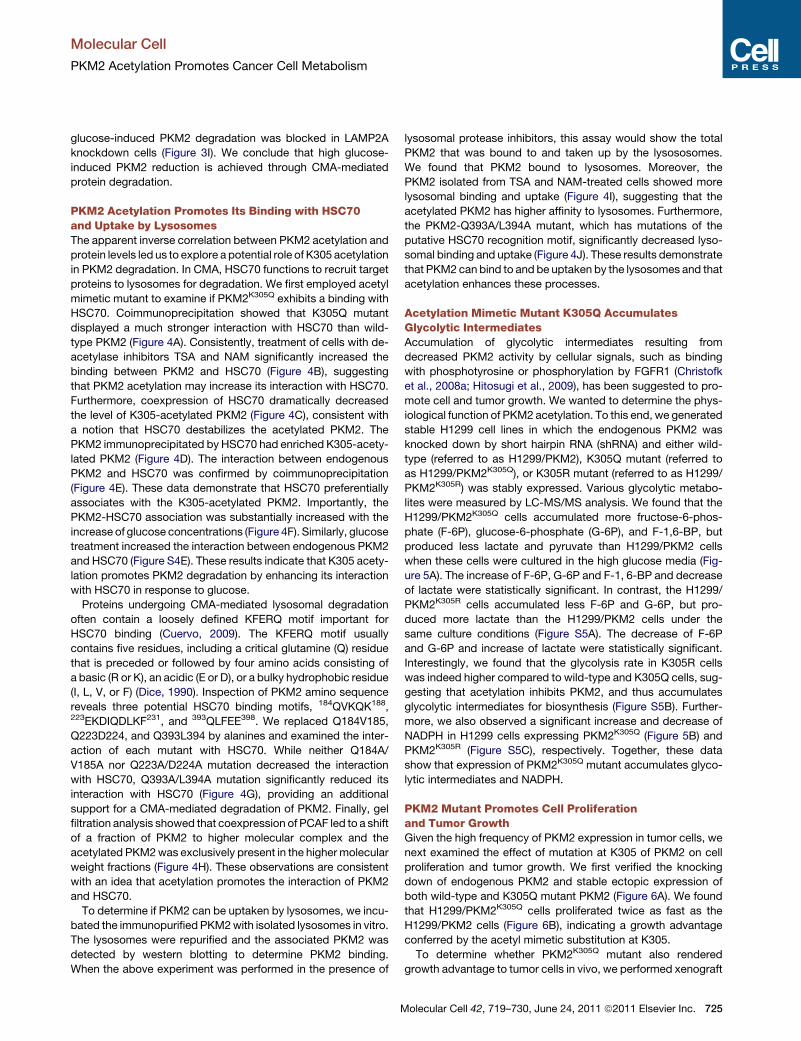
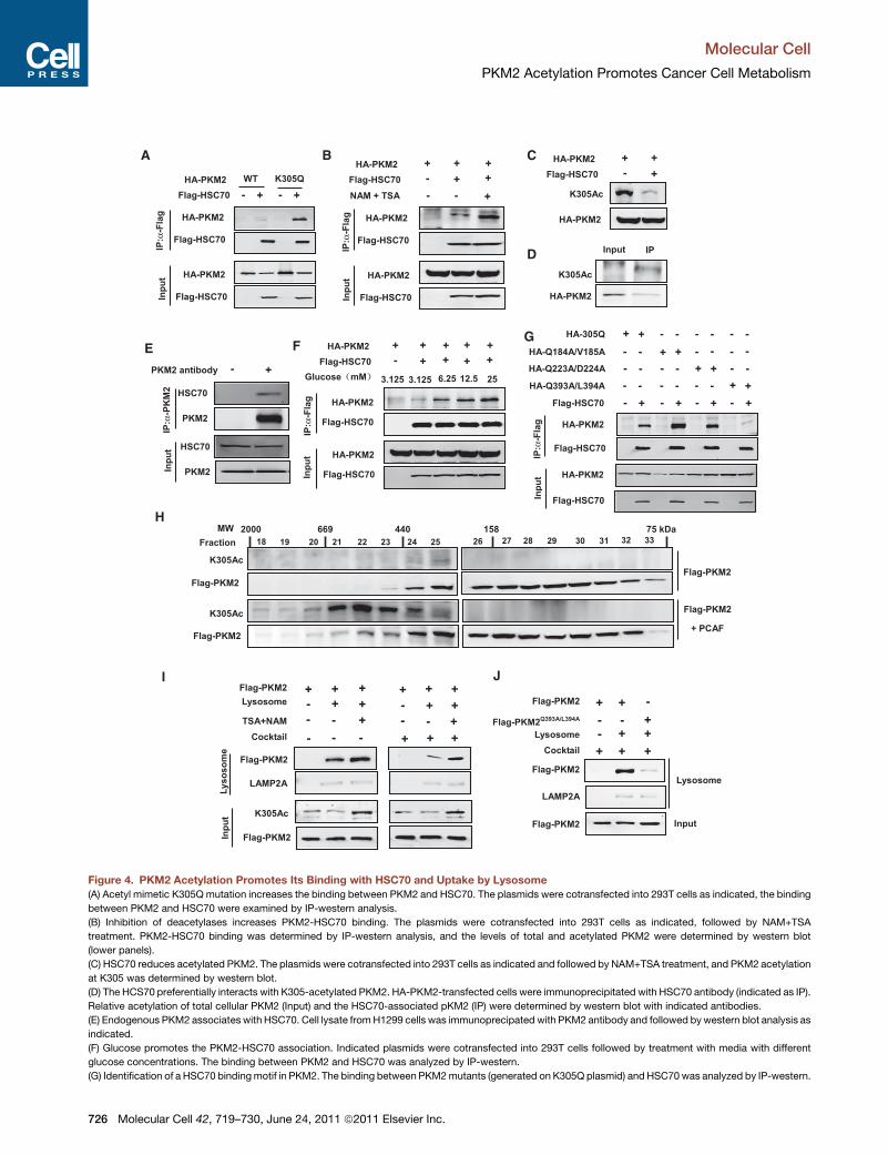
Figure 4. PKM2 Acetylation Promotes Its Binding with HSC70 and Uptake by Lysosome
(A) Acetyl mimetic K305Q mutation increases the binding between PKM2 and HSC70. The plasmids were cotransfected into 293T cells as indicated, the binding
between PKM2 and HSC70 were examined by IP-western analysis.
(B) Inhibition of deacetylases increases PKM2-HSC70 binding. The plasmids were cotransfected into 293T cells as indicated, followed by NAM+TSA
treatment. PKM2-HSC70 binding was determined by IP-western analysis, and the levels of total and acetylated PKM2 were determined by western blot
(lower panels).
(C) HSC70 reduces acetylated PKM2. The plasmids were cotransfected into 293T cells as indicated and followed by NAM+TSA treatment, and PKM2 acetylation
at K305 was determined by western blot.
(D) The HCS70 preferentially interacts with K305-acetylated PKM2. HA-PKM2-transfected cells were immunoprecipitated with HSC70 antibody (indicated as IP).
Relative acetylation of total cellular PKM2 (Input) and the HSC70-associated pKM2 (IP) were determined by western blot with indicated antibodies.
(E) Endogenous PKM2 associates with HSC70. Cell lysate fromH1299 cells was immunoprecipated with PKM2 antibody and followed bywestern blot analysis as
indicated.
(F) Glucose promotes the PKM2-HSC70 association. Indicated plasmids were cotransfected into 293T cells followed by treatment with media with different
glucose concentrations. The binding between PKM2 and HSC70 was analyzed by IP-western.
(G) Identification of a HSC70 bindingmotif in PKM2. The binding between PKM2mutants (generated on K305Q plasmid) and HSC70 was analyzed by IP-western.
Molecular Cell
PKM2 Acetylation Promotes Cancer Cell Metabolism
726 Molecular Cell 42, 719–730, June 24, 2011 ª2011 Elsevier Inc.
*
p = 0.002
A
B
0
0.2
0.4
0.6
0.8
1
1.2
1.4
1.6
K305Q
Relative am
ou
nt o
f N
AD
PH
PKM2
*
Ratio
(K
305Q
/W
T)
*
Lactate Pyruvate GAPF-1, 6-BPF-6P G-6P
0
0.5
1
2
2.5
1.5
3
p = 0.0002 **
*p = 0.102
p = 0.033
p = 0.009
p = 0.001
p = 0.014
Flag-PKM2
Endog. PKM2
Flag PKM2
shPKM2
Flag-PKM2
- + + +
- - WT K305Q
ββ-actin
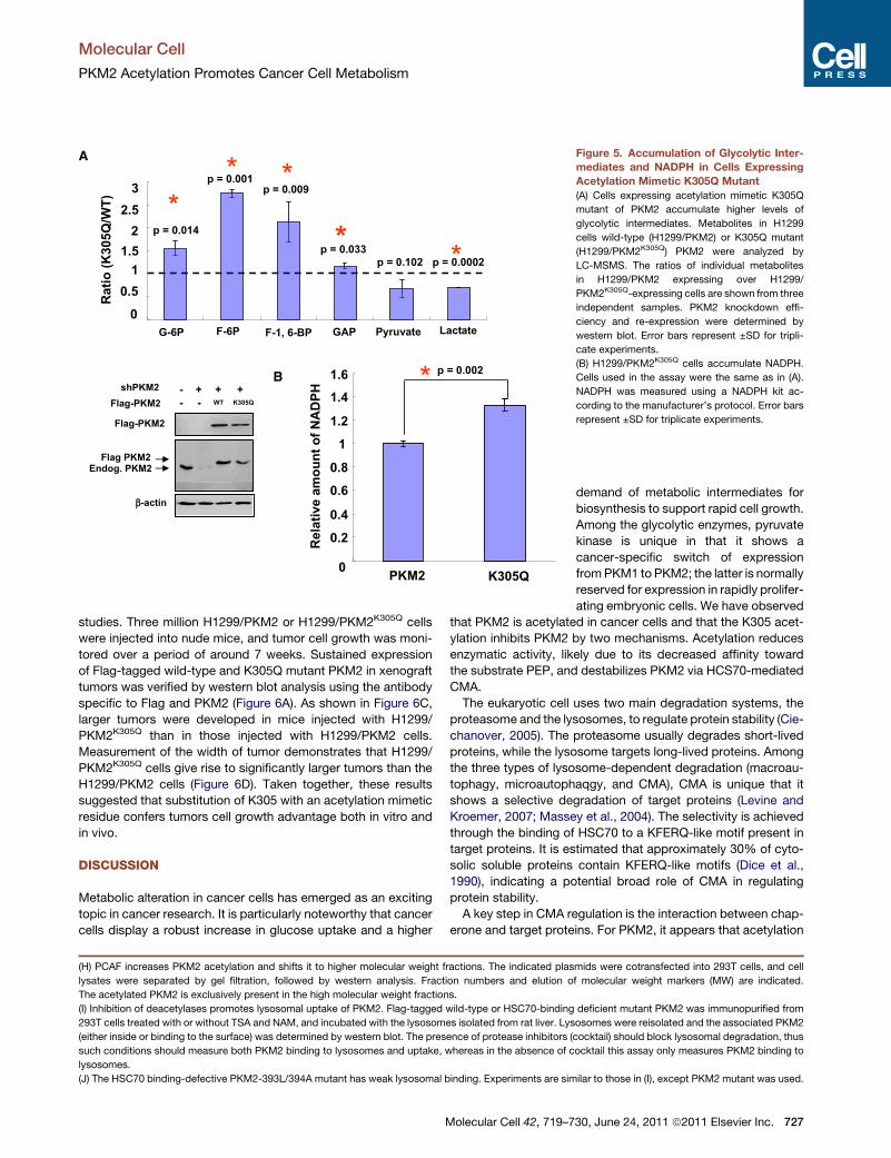
Figure 5. Accumulation of Glycolytic Inter-
mediates and NADPH in Cells Expressing
Acetylation Mimetic K305Q Mutant
(A) Cells expressing acetylation mimetic K305Q
mutant of PKM2 accumulate higher levels of
glycolytic intermediates. Metabolites in H1299
cells wild-type (H1299/PKM2) or K305Q mutant
(H1299/PKM2K305Q) PKM2 were analyzed by
LC-MSMS. The ratios of individual metabolites
in H1299/PKM2 expressing over H1299/
PKM2K305Q-expressing cells are shown from three
independent samples. PKM2 knockdown effi-
ciency and re-expression were determined by
western blot. Error bars represent ±SD for tripli-
cate experiments.
(B) H1299/PKM2K305Q cells accumulate NADPH.
Cells used in the assay were the same as in (A).
NADPH was measured using a NADPH kit ac-
cording to the manufacturer’s protocol. Error bars
represent ±SD for triplicate experiments.
Molecular Cell
PKM2 Acetylation Promotes Cancer Cell Metabolism
studies. Three million H1299/PKM2 or H1299/PKM2K305Q cells
were injected into nude mice, and tumor cell growth was moni-
tored over a period of around 7 weeks. Sustained expression
of Flag-tagged wild-type and K305Q mutant PKM2 in xenograft
tumors was verified by western blot analysis using the antibody
specific to Flag and PKM2 (Figure 6A). As shown in Figure 6C,
larger tumors were developed in mice injected with H1299/
PKM2K305Q than in those injected with H1299/PKM2 cells.
Measurement of the width of tumor demonstrates that H1299/
PKM2K305Q cells give rise to significantly larger tumors than the
H1299/PKM2 cells (Figure 6D). Taken together, these results
suggested that substitution of K305 with an acetylation mimetic
residue confers tumors cell growth advantage both in vitro and
in vivo.
DISCUSSION
Metabolic alteration in cancer cells has emerged as an exciting
topic in cancer research. It is particularly noteworthy that cancer
cells display a robust increase in glucose uptake and a higher
(H) PCAF increases PKM2 acetylation and shifts it to higher molecular weight fractions. The indicated plas
lysates were separated by gel filtration, followed by western analysis. Fraction numbers and elution o
The acetylated PKM2 is exclusively present in the high molecular weight fractions.
(I) Inhibition of deacetylases promotes lysosomal uptake of PKM2. Flag-tagged wild-type or HSC70-binding
293T cells treated with or without TSA and NAM, and incubated with the lysosomes isolated from rat liver. Lys
(either inside or binding to the surface) was determined by western blot. The presence of protease inhibitors (
such conditions should measure both PKM2 binding to lysosomes and uptake, whereas in the absence of c
lysosomes.
(J) The HSC70 binding-defective PKM2-393L/394A mutant has weak lysosomal binding. Experiments are sim
Molecular Cell 42, 719–7
demand of metabolic intermediates for
biosynthesis to support rapid cell growth.
Among the glycolytic enzymes, pyruvate
kinase is unique in that it shows a
cancer-specific switch of expression
fromPKM1 to PKM2; the latter is normally
reserved for expression in rapidly prolifer-
ating embryonic cells. We have observed
that PKM2 is acetylated in cancer cells and that the K305 acet-
ylation inhibits PKM2 by two mechanisms. Acetylation reduces
enzymatic activity, likely due to its decreased affinity toward
the substrate PEP, and destabilizes PKM2 via HCS70-mediated
CMA.
The eukaryotic cell uses two main degradation systems, the
proteasome and the lysosomes, to regulate protein stability (Cie-
chanover, 2005). The proteasome usually degrades short-lived
proteins, while the lysosome targets long-lived proteins. Among
the three types of lysosome-dependent degradation (macroau-
tophagy, microautophaqgy, and CMA), CMA is unique that it
shows a selective degradation of target proteins (Levine and
Kroemer, 2007; Massey et al., 2004). The selectivity is achieved
through the binding of HSC70 to a KFERQ-like motif present in
target proteins. It is estimated that approximately 30% of cyto-
solic soluble proteins contain KFERQ-like motifs (Dice et al.,
1990), indicating a potential broad role of CMA in regulating
protein stability.
A key step in CMA regulation is the interaction between chap-
erone and target proteins. For PKM2, it appears that acetylation
mids were cotransfected into 293T cells, and cell
f molecular weight markers (MW) are indicated.
deficient mutant PKM2 was immunopurified from
osomes were reisolated and the associated PKM2
cocktail) should block lysosomal degradation, thus
ocktail this assay only measures PKM2 binding to
ilar to those in (I), except PKM2 mutant was used.
30, June 24, 2011 ª2011 Elsevier Inc. 727
Time (day)
0 1 2 3 4C
ell co
un
ts (x 10
5
)
0
1
2
3
4
5
6
7
8
H1299 / PKM2
H1299 / PKM2K305Q
Flag-PKM2
K305Q
WT
β-actin
D
B
C
H1299/PKM2K305Q
H1299/PKM2
A
+
H1299 xenograft
Flag-PKM2
β-actin
Endog. PKM2
Flag PKM2
shPKM2
Flag-PKM2
- + + +
- - WT K305Q
+
K305QWT
**
**
Days after injection
20 25 32 38 45
WT
K305Q
0
5
10
15
20
25
Wid
th
o
f tu
mo
ur (m
m)
P=0.0000
P=0.0009
P=0.0106
P=0.0432
P=0.1919
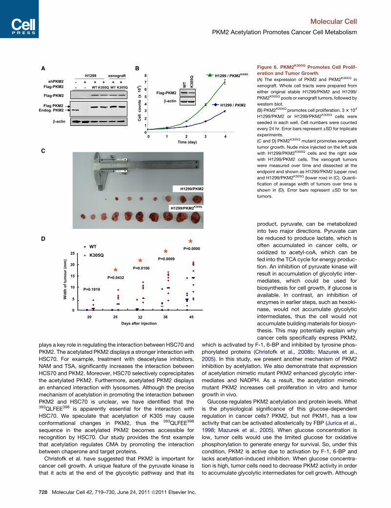
Figure 6. PKM2K305Q Promotes Cell Prolif-
eration and Tumor Growth
(A) The expression of PKM2 and PKM2K305Q in
xenograft. Whole cell tracts were prepared from
either original stable H1299/PKM2 and H1299/
PKM2K305Q pools or xenograft tumors, followed by
western blot.
(B) PKM2K305Q promotes cell proliferation. 33 104
H1299/PKM2 or H1299/PKM2K305Q cells were
seeded in each well. Cell numbers were counted
every 24 hr. Error bars represent ±SD for triplicate
experiments.
(C and D) PKM2K305Q mutant promotes xenograft
tumor growth. Nude mice injected on the left side
with H1299/PKM2K305Q cells and the right side
with H1299/PKM2 cells. The xenograft tumors
were measured over time and dissected at the
endpoint and shown as H1299/PKM2 (upper row)
and H1299/PKM2K305Q (lower row) in (C). Quanti-
fication of average width of tumors over time is
shown in (D). Error bars represent ±SD for ten
tumors.
Molecular Cell
PKM2 Acetylation Promotes Cancer Cell Metabolism
plays a key role in regulating the interaction between HSC70 and
PKM2. The acetylated PKM2 displays a stronger interaction with
HSC70. For example, treatment with deacetylase inhibitors,
NAM and TSA, significantly increases the interaction between
HCS70 and PKM2. Moreover, HSC70 selectively coprecipitates
the acetylated PKM2. Furthermore, acetylated PKM2 displays
an enhanced interaction with lysosomes. Although the precise
mechanism of acetylation in promoting the interaction between
PKM2 and HSC70 is unclear, we have identified that the393QLFEE398 is apparently essential for the interaction with
HSC70. We speculate that acetylation of K305 may cause
conformational changes in PKM2, thus the 393QLFEE398
sequence in the acetylated PKM2 becomes accessible for
recognition by HSC70. Our study provides the first example
that acetylation regulates CMA by promoting the interaction
between chaperone and target proteins.
Christofk et al. have suggested that PKM2 is important for
cancer cell growth. A unique feature of the pyruvate kinase is
that it acts at the end of the glycolytic pathway and that its
728 Molecular Cell 42, 719–730, June 24, 2011 ª2011 Elsevier Inc.
product, pyruvate, can be metabolized
into two major directions. Pyruvate can
be reduced to produce lactate, which is
often accumulated in cancer cells, or
oxidized to acetyl-coA, which can be
fed into the TCA cycle for energy produc-
tion. An inhibition of pyruvate kinase will
result in accumulation of glycolytic inter-
mediates, which could be used for
biosynthesis for cell growth, if glucose is
available. In contrast, an inhibition of
enzymes in earlier steps, such as hexoki-
nase, would not accumulate glycolytic
intermediates, thus the cell would not
accumulate building materials for biosyn-
thesis. This may potentially explain why
cancer cells specifically express PKM2,
which is activated by F-1, 6-BP and inhibited by tyrosine phos-
phorylated proteins (Christofk et al., 2008b; Mazurek et al.,
2005). In this study, we present another mechanism of PKM2
inhibition by acetylation. We also demonstrate that expression
of acetylation mimetic mutant PKM2 enhanced glycolytic inter-
mediates and NADPH. As a result, the acetylation mimetic
mutant PKM2 increases cell proliferation in vitro and tumor
growth in vivo.
Glucose regulates PKM2 acetylation and protein levels. What
is the physiological significance of this glucose-dependent
regulation in cancer cells? PKM2, but not PKM1, has a low
activity that can be activated allosterically by FBP (Jurica et al.,
1998; Mazurek et al., 2005). When glucose concentration is
low, tumor cells would use the limited glucose for oxidative
phosphorylation to generate energy for survival. So, under this
condition, PKM2 is active due to activation by F-1, 6-BP and
lacks acetylation-induced inhibition. When glucose concentra-
tion is high, tumor cells need to decrease PKM2 activity in order
to accumulate glycolytic intermediates for cell growth. Although
Molecular Cell
PKM2 Acetylation Promotes Cancer Cell Metabolism
high glucose results in a high F-1, 6-BP, PKM2 activity is in check
because high glucose induces PKM2 acetylation, which directly
decreases PKM2 activity and indirectly stimulates the CMA-
dependent degradation. Therefore, together with activation by
F-1, 6-BP, PKM2 regulation by glucose-induced acetylation
would provide an advantage for tumor cell growth under glucose
sufficiency. One may speculate that drugs modulating PKM2
activity or acetylation could have potential value for cancer
treatment.
EXPERIMENTAL PROCEDURES
Cell Lysis and Immunological Procedures
Cells were lysed in a NP-40 buffer, and western blot analysis was carried out
according to standard methods. Antibodies specific to Flag (Sigma), HSC70
(Abcam), Lamp2A (Abcam), HA (Santa Cruz), actin (Sigma), and PSA (Bio-
world) were purchased commercially. Antibodies to pyruvate kinase and
acetyl-PKM2(K305) were generated by immunizing rabbits with antigen
peptides at Shanghai Genomic Inc.
Measurement of Pyruvate Kinase Activity
Pyruvate kinase activity was measured by a continuous assay coupled to
lactate dehydrogenase (LDH). The change in absorbance resulting from
NADH oxidation wasmeasured using a F-4600 Fluorescence Spectrophotom-
eter (HITACHI). Assays for PK activity were carried out as previously described
(Christofk et al., 2008a).
Lysosome Binding and Uptake Assay
Lysosomes were isolated by following previously described procedures
(Cuervo et al., 1995). Both lysosome binding and uptake assays were carried
out as described previously (Cuervo et al., 2004).
Short Hairpin RNA Constructs and Retroviral Production
A shRNA retrovirus targeting pyruvate kinase was constructed and retrovi-
ruses were produced using a two-plasmid packaging system as previously
described (Christofk et al., 2008a).
Putting Back
Flag-tagged human PKM2 and PKM2K305Q containing two silent nucleotide
substitutions in the sequence corresponding to the shRNA-targeted region
were cloned into the retroviral vector (pQCXIH) and were cotransfected into
293T cells together with vectors expressing the gag and vsvg genes. Retroviral
supernatant was harvested 36 hr after initial plasmid transfection and mixed
with polybrene (8 mg/ml) to increase the infection efficiency. H1299 cells
were infected with retrovirus and selected in hygromycin (350 mg/ml) for
2 weeks.
Cell Proliferation Analysis
33 104 cells were seeded in triplicate in 6-well plates, and cell numbers were
counted every 24 hr over a 5 day period.
Measurement of Metabolites
To extract metabolites, 23 107 cells was directly chilled into 2ml ice-cold 80%
ethanol containing 0.1% formic acid, followed by a centrifugation at
10,000 rpm for 20 min at 4�C. The supernatant was collected and dried in
Concentrator 5301 (Eppendorf). The dried extract was dissolved in 200 ml
20% acetonitrile, and the insoluble material was removed by centrifugation
(10,000 rpm for 20 min at 4�C). Of the supernatant, 20 ml was used to for
LC-MS analysis.
NADPH Assay
The assays were carried out according to the protocol of NADP/NADPH Assay
Kit (BioVision).
M
Xenograft Studies
Nude mice (nu/nu, male 6- to 8-week-old) were injected subcutaneously with
33 106H1299 cells. Around 7weeks after injection, the tumorswere dissected
and weighed.
Immunohistochemistry
Prostate samples were acquired from Affiliated Renji Hospital of Jiaotong
University. A physician obtained informed consent from the patients. The
procedures related to human subjects were approved by Ethic Committee
of the Institutes of Biomedical Sciences (IBS), Fudan University. IHC was
performed as previously described (Lei et al., 2006).
Measurement of Glycolysis Rate
Glycolysis rate wasmeasured bymonitoring the conversion of 5-3H-glucose to3H2O, as described previously (Liang et al., 1997).
SUPPLEMENTAL INFORMATION
Supplemental Information includes five figures and two tables and can be
found with this article online at doi:10.1016/j.molcel.2011.04.025.
ACKNOWLEDGMENTS
We thank the members of the Fudan MCB laboratory for discussions
throughout this study. PcDNA/Zeo(�)-antisense-Hsc70 was kindly provided
by Drs. Janice S. Blum and Zixu Mao. This work was supported by 973 (grant
numbers 2009CB918401, 2011CB910600), NCET (grant number 09-0315),
and NSFC (grant numbers 30600112, 30871255, 31071192). This work was
also supported by the 985 Program and Shanghai key project (grant number
09JC1402300); 100 Talents Programme of Shanghai Health; the Shanghai
Leading Academic Discipline Project, project number B110; and National
Institutes of Health (NIH) grants (Y. Xiong and K.-L.G.).
Received: September 24, 2010
Revised: April 5, 2011
Accepted: April 21, 2011
Published: June 23, 2011
REFERENCES
Altenberg, B., and Greulich, K.O. (2004). Genes of glycolysis are ubiquitously
overexpressed in 24 cancer classes. Genomics 84, 1014–1020.
Avalos, J.L., Bever, K.M., and Wolberger, C. (2005). Mechanism of sirtuin
inhibition by nicotinamide: altering the NAD(+) cosubstrate specificity of
a Sir2 enzyme. Mol. Cell 17, 855–868.
Bensaad, K., Tsuruta, A., Selak, M.A., Vidal, M.N., Nakano, K., Bartrons, R.,
Gottlieb, E., and Vousden, K.H. (2006). TIGAR, a p53-inducible regulator of
glycolysis and apoptosis. Cell 126, 107–120.
Chen, C.C., Carson, J.J., Feser, J., Tamburini, B., Zabaronick, S., Linger, J.,
and Tyler, J.K. (2008). Acetylated lysine 56 on histone H3 drives chromatin
assembly after repair and signals for the completion of repair. Cell 134,
231–243.
Choudhary, C., Kumar, C., Gnad, F., Nielsen, M.L., Rehman, M., Walther, T.C.,
Olsen, J.V., andMann,M. (2009). Lysine acetylation targets protein complexes
and co-regulates major cellular functions. Science 325, 834–840.
Christofk, H.R., Vander Heiden, M.G., Harris, M.H., Ramanathan, A., Gerszten,
R.E.,Wei, R., Fleming,M.D., Schreiber, S.L., andCantley, L.C. (2008a). TheM2
splice isoform of pyruvate kinase is important for cancer metabolism and
tumour growth. Nature 452, 230–233.
Christofk, H.R., Vander Heiden, M.G., Wu, N., Asara, J.M., and Cantley, L.C.
(2008b). Pyruvate kinase M2 is a phosphotyrosine-binding protein. Nature
452, 181–186.
Ciechanover, A. (2005). Intracellular protein degradation: from a vague idea
thru the lysosome and the ubiquitin-proteasome system and onto human
diseases and drug targeting. Cell Death Differ. 12, 1178–1190.
olecular Cell 42, 719–730, June 24, 2011 ª2011 Elsevier Inc. 729
Molecular Cell
PKM2 Acetylation Promotes Cancer Cell Metabolism
Cuervo, A.M. (2009). Chaperone-mediated autophagy: selectivity pays off.
Trends Endocrinol. Metab. 21, 142–150.
Cuervo, A.M., Knecht, E., Terlecky, S.R., and Dice, J.F. (1995). Activation of
a selective pathway of lysosomal proteolysis in rat liver by prolonged starva-
tion. Am. J. Physiol. 269, C1200–C1208.
Cuervo, A.M., Stefanis, L., Fredenburg, R., Lansbury, P.T., and Sulzer, D.
(2004). Impaired degradation of mutant alpha-synuclein by chaperone-
mediated autophagy. Science 305, 1292–1295.
Dang, C.V., and Semenza, G.L. (1999). Oncogenic alterations of metabolism.
Trends Biochem. Sci. 24, 68–72.
David, C.J., Chen,M., Assanah,M., Canoll, P., andManley, J.L. (2009). HnRNP
proteins controlled by c-Myc deregulate pyruvate kinase mRNA splicing in
cancer. Nature 463, 364–368.
Dice, J.F. (1990). Peptide sequences that target cytosolic proteins for lyso-
somal proteolysis. Trends Biochem. Sci. 15, 305–309.
Dice, J.F., Terlecky, S.R., Chiang, H.L., Olson, T.S., Isenman, L.D.,
Short-Russell, S.R., Freundlieb, S., and Terlecky, L.J. (1990). A selective
pathway for degradation of cytosolic proteins by lysosomes. Semin. Cell
Biol. 1, 449–455.
Dombrauckas, J.D., Santarsiero, B.D., and Mesecar, A.D. (2005). Structural
basis for tumor pyruvate kinase M2 allosteric regulation and catalysis.
Biochemistry 44, 9417–9429.
Ekwall, K., Olsson, T., Turner, B.M., Cranston, G., and Allshire, R.C. (1997).
Transient inhibition of histone deacetylation alters the structural and functional
imprint at fission yeast centromeres. Cell 91, 1021–1032.
Funes, J.M., Quintero, M., Henderson, S., Martinez, D., Qureshi, U.,
Westwood, C., Clements, M.O., Bourboulia, D., Pedley, R.B., Moncada, S.,
and Boshoff, C. (2007). Transformation of human mesenchymal stem cells
increases their dependency on oxidative phosphorylation for energy produc-
tion. Proc. Natl. Acad. Sci. USA 104, 6223–6228.
Furumai, R., Komatsu, Y., Nishino, N., Khochbin, S., Yoshida, M., and
Horinouchi, S. (2001). Potent histone deacetylase inhibitors built from trichos-
tatin A and cyclic tetrapeptide antibiotics including trapoxin. Proc. Natl. Acad.
Sci. USA 98, 87–92.
Gordan, J.D., Bertout, J.A., Hu, C.J., Diehl, J.A., and Simon, M.C. (2007a).
HIF-2alpha promotes hypoxic cell proliferation by enhancing c-myc transcrip-
tional activity. Cancer Cell 11, 335–347.
Gordan, J.D., Thompson, C.B., and Simon, M.C. (2007b). HIF and c-Myc:
sibling rivals for control of cancer cell metabolism and proliferation. Cancer
Cell 12, 108–113.
Hitosugi, T., Kang, S., Vander Heiden, M.G., Chung, T.W., Elf, S., Lythgoe, K.,
Dong, S., Lonial, S., Wang, X., Chen, G.Z., et al. (2009). Tyrosine phosphory-
lation inhibits PKM2 to promote the Warburg effect and tumor growth. Sci.
Signal. 2, ra73. 10.1126/scisignal.2000431.
Jeong, H., Then, F., Melia, T.J., Jr., Mazzulli, J.R., Cui, L., Savas, J.N., Voisine,
C., Paganetti, P., Tanese, N., Hart, A.C., et al. (2009). Acetylation targets
mutant huntingtin to autophagosomes for degradation. Cell 137, 60–72.
Jurica, M.S., Mesecar, A., Heath, P.J., Shi, W., Nowak, T., and Stoddard, B.L.
(1998). The allosteric regulation of pyruvate kinase by fructose-1,6-bisphos-
phate. Structure 6, 195–210.
Kim, S.C., Sprung, R., Chen, Y., Xu, Y., Ball, H., Pei, J., Cheng, T., Kho, Y.,
Xiao, H., Xiao, L., et al. (2006). Substrate and functional diversity of lysine acet-
ylation revealed by a proteomics survey. Mol. Cell 23, 607–618.
730 Molecular Cell 42, 719–730, June 24, 2011 ª2011 Elsevier Inc.
Lei, Q., Jiao, J., Xin, L., Chang, C.J., Wang, S., Gao, J., Gleave, M.E., Witte,
O.N., Liu, X., andWu, H. (2006). NKX3.1 stabilizes p53, inhibits AKT activation,
and blocks prostate cancer initiation caused by PTEN loss. Cancer Cell 9,
367–378.
Levine, B., and Kroemer, G. (2007). Autophagy in the pathogenesis of disease.
Cell 132, 27–42.
Liang, Y., Buettger, C., Berner, D.K., and Matschinsky, F.M. (1997). Chronic
effect of fatty acids on insulin release is not through the alteration of glucose
metabolism in a pancreatic beta-cell line (beta HC9). Diabetologia 40, 1018–
1027.
Liebermeister, W. (2005). Predicting physiological concentrations of metabo-
lites from their molecular structure. J. Comput. Biol. 12, 1307–1315.
Majeski, A.E., and Dice, J.F. (2004). Mechanisms of chaperone-mediated
autophagy. Int. J. Biochem. Cell Biol. 36, 2435–2444.
Majumder, P.K., Febbo, P.G., Bikoff, R., Berger, R., Xue, Q., McMahon, L.M.,
Manola, J., Brugarolas, J., McDonnell, T.J., Golub, T.R., et al. (2004). mTOR
inhibition reverses Akt-dependent prostate intraepithelial neoplasia through
regulation of apoptotic and HIF-1-dependent pathways. Nat. Med. 10,
594–601.
Manning, B.D., and Cantley, L.C. (2007). AKT/PKB signaling: navigating
downstream. Cell 129, 1261–1274.
Massey, A., Kiffin, R., and Cuervo, A.M. (2004). Pathophysiology of chap-
erone-mediated autophagy. Int. J. Biochem. Cell Biol. 36, 2420–2434.
Matoba, S., Kang, J.G., Patino, W.D., Wragg, A., Boehm, M., Gavrilova, O.,
Hurley, P.J., Bunz, F., and Hwang, P.M. (2006). p53 regulates mitochondrial
respiration. Science 312, 1650–1653.
Mazurek, S., Boschek, C.B., Hugo, F., and Eigenbrodt, E. (2005). Pyruvate
kinase type M2 and its role in tumor growth and spreading. Semin. Cancer
Biol. 15, 300–308.
Mizushima, N., Levine, B., Cuervo, A.M., and Klionsky, D.J. (2008). Autophagy
fights disease through cellular self-digestion. Nature 451, 1069–1075.
Ramanathan, A., Wang, C., and Schreiber, S.L. (2005). Perturbational profiling
of a cell-line model of tumorigenesis by using metabolic measurements. Proc.
Natl. Acad. Sci. USA 102, 5992–5997.
Soutoglou, E., Katrakili, N., and Talianidis, I. (2000). Acetylation regulates
transcription factor activity at multiple levels. Mol. Cell 5, 745–751.
Wang, X., and Hayes, J.J. (2008). Acetylation mimics within individual core
histone tail domains indicate distinct roles in regulating the stability of
higher-order chromatin structure. Mol. Cell. Biol. 28, 227–236.
Warburg, O. (1956). On the origin of cancer cells. Science 123, 309–314.
Wing, S., Chiang, H.L., Goldberg, A.L., and Dice, J.F. (1991). Proteins contain-
ing peptide sequences related to KFERQ are selectively depleted in liver and
heart, but notskeletal muscle, of fasted rats. Biochem. J. 275, 165–169.
Yang, Q., She, H., Gearing, M., Colla, E., Lee, M., Shacka, J.J., and Mao, Z.
(2009). Regulation of neuronal survival factor MEF2D by chaperone-mediated
autophagy. Science 323, 124–127.
Zhang, W., Bone, J.R., Edmondson, D.G., Turner, B.M., and Roth, S.Y. (1998).
Essential and redundant functions of histone acetylation revealed by mutation
of target lysines and loss of the Gcn5p acetyltransferase. EMBO J. 17, 3155–
3167.
Zhao, S., Xu, W., Jiang, W., Yu, W., Lin, Y., Zhang, T., Yao, J., Zhou, L., Zeng,
Y., Li, H., et al. (2010). Regulation of cellular metabolism by protein lysine
acetylation. Science 327, 1000–1004.