Embed Size (px)
Citation preview

Molecular biology of partial D and weak D
Implications for Blood Bank PracticeQuote source, if using these slides.
Willy A. FlegelDept. Transfusion Medicine, University of Ulm
DRK Blutspendedienst Baden-Württemberg - HessenUlm, Germany
http://www.uni-ulm.de/~wflegel/RH/ 2002
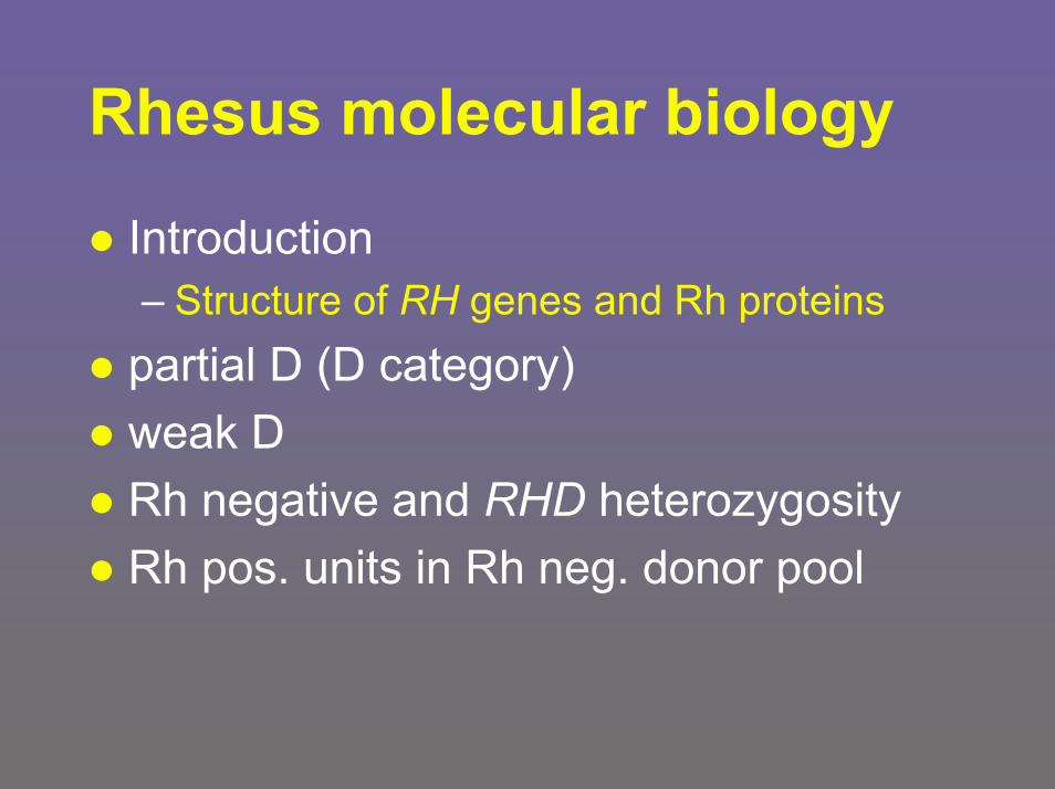
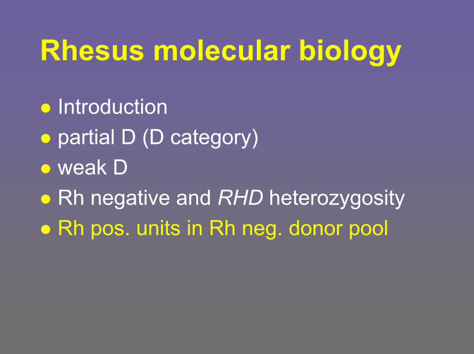
Rhesus molecular biology
Introduction– Structure of RH genes and Rh proteins
partial D (D category)weak DRh negative and RHD heterozygosityRh pos. units in Rh neg. donor pool
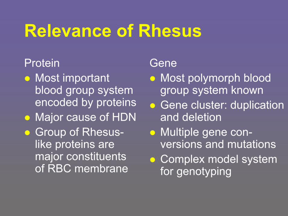
Relevance of RhesusProtein
Most important blood group system encoded by proteinsMajor cause of HDNGroup of Rhesus-like proteins are major constituents of RBC membrane
GeneMost polymorph blood group system knownGene cluster: duplication and deletionMultiple gene con-versions and mutationsComplex model system for genotyping
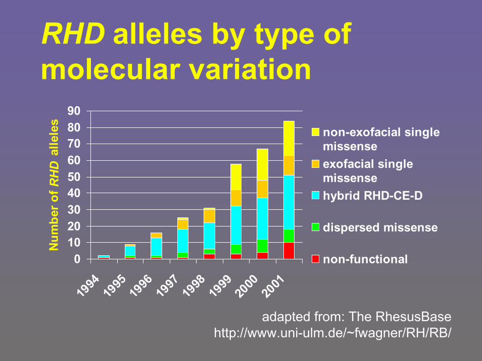
RHD alleles by type of molecular variation
0102030405060708090
1994
1995
1996
1997
1998
1999
2000
2001
Num
ber o
f RH
D a
llele
s
non-exofacial singlemissenseexofacial singlemissensehybrid RHD-CE-D
dispersed missense
non-functional
adapted from: The RhesusBasehttp://www.uni-ulm.de/~fwagner/RH/RB/
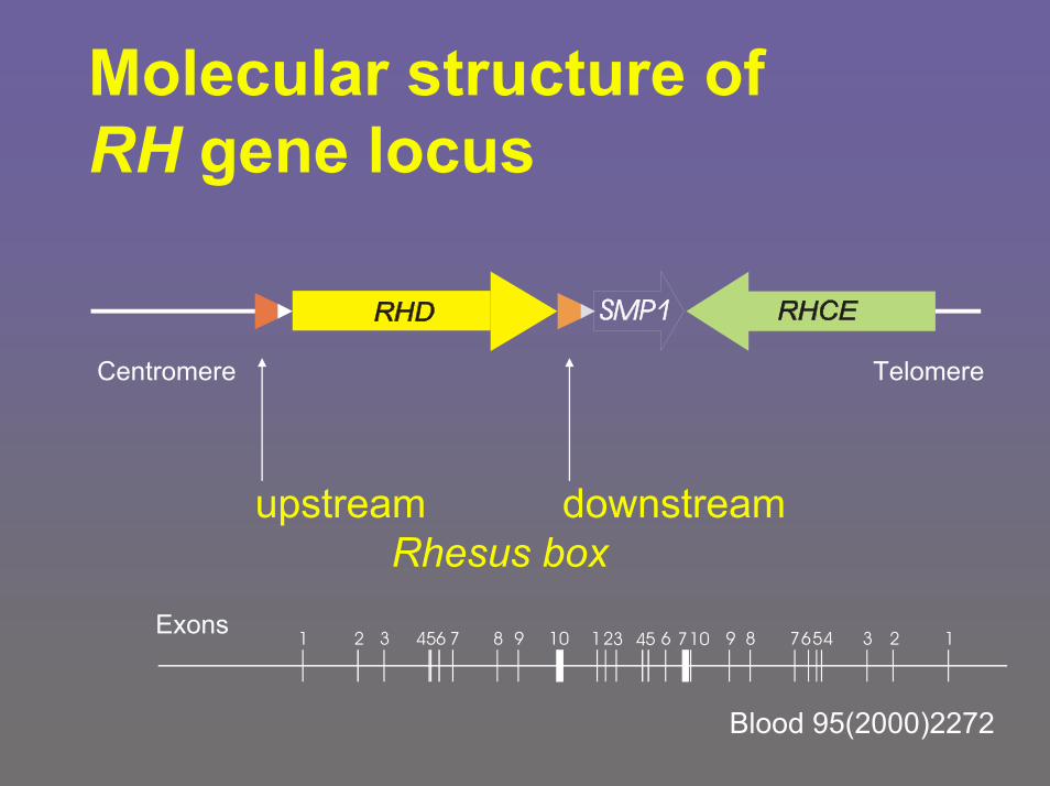
Molecular structure of RH gene locus
b
upstream downstreamRhesus box
1 2 3 456 7 8 9 10 123 4 65 710 9 8 765 2 14 3
Centromere Telomere
Exons
Blood 95(2000)2272
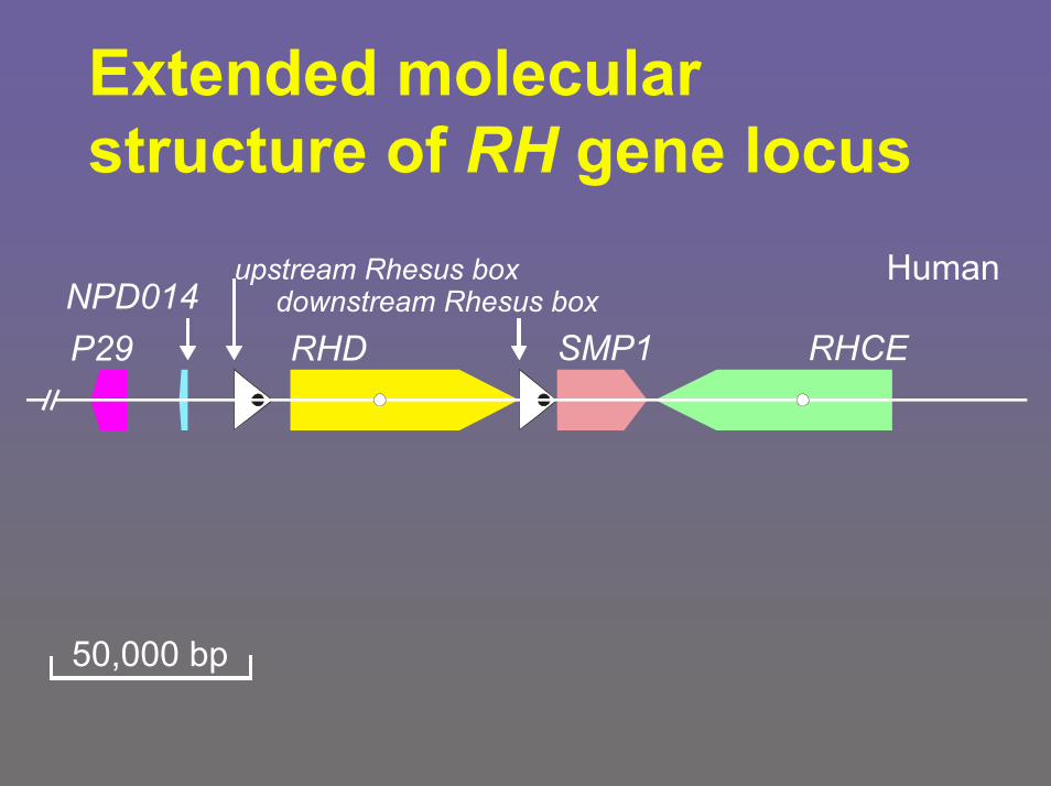
Extended molecularstructure of RH gene locus
P29 RHDdownstream Rhesus box
SMP1
upstream Rhesus box HumanNPD014
RHCE
50,000 bp
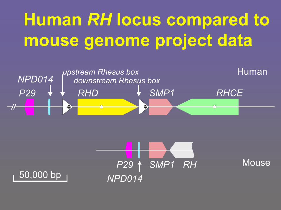
Human RH locus compared tomouse genome project data
RHSMP1
P29 RHDdownstream Rhesus box
SMP1
upstream Rhesus box HumanNPD014
RHCE
MouseP2950,000 bp NPD014
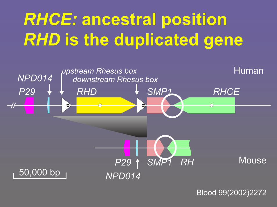
RHCE: ancestral positionRHD is the duplicated gene
RHSMP1
P29 RHDdownstream Rhesus box
SMP1
upstream Rhesus box HumanNPD014
RHCE
Mouse
Blood 99(2002)2272
P2950,000 bp NPD014
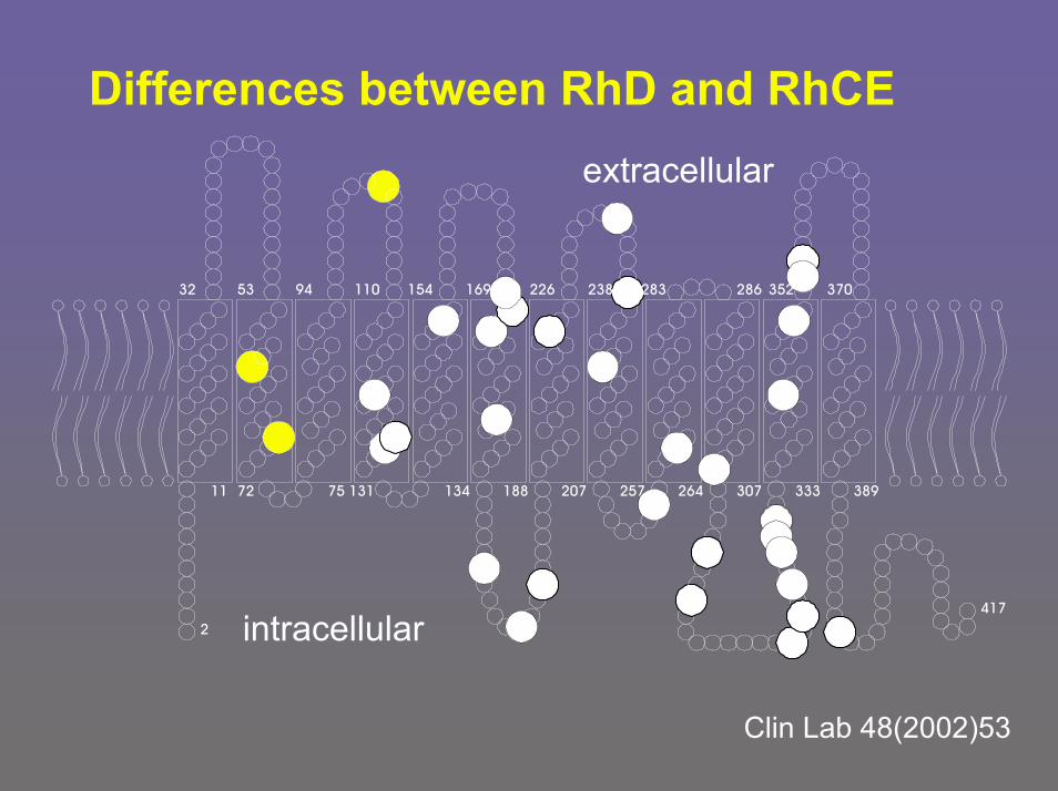
Differences between RhD and RhCE
11 72 75 131 134 188 207 257 264 307 333 389
169 226 238 283 370
extracellular
32 53 94 110 154 286 352
417
intracellular2
Clin Lab 48(2002)53
Rhesus molecular biologyIntroductionpartial D (D category)– RHD/RHCE hybrid alleles
Typically causing „D categories“ – subset of partial D
weak DRh negative and RHD heterozygosityRh pos. units in Rh neg. donor pool
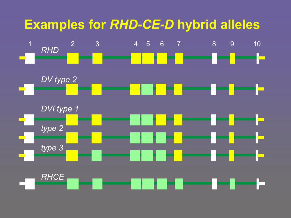
Examples for RHD-CE-D hybrid alleles1 2 3 4 5 6 7 8 9 10
RHD
DV type 2
DVI type 1
type 2
type 3
RHCE
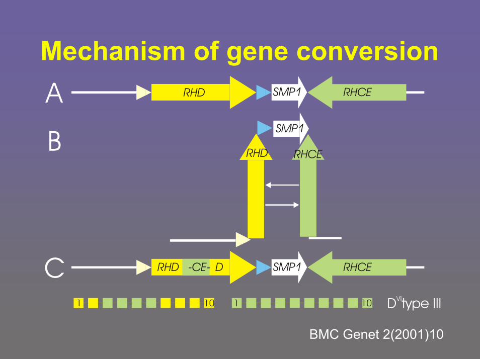
Mechanism of gene conversion
B RHD
SMP1
RHCE
A RHDb SMP1 RHCE
C RHD RHDb SMP1 RHCED-CE-
D type IIIVI1 110 10
BMC Genet 2(2001)10
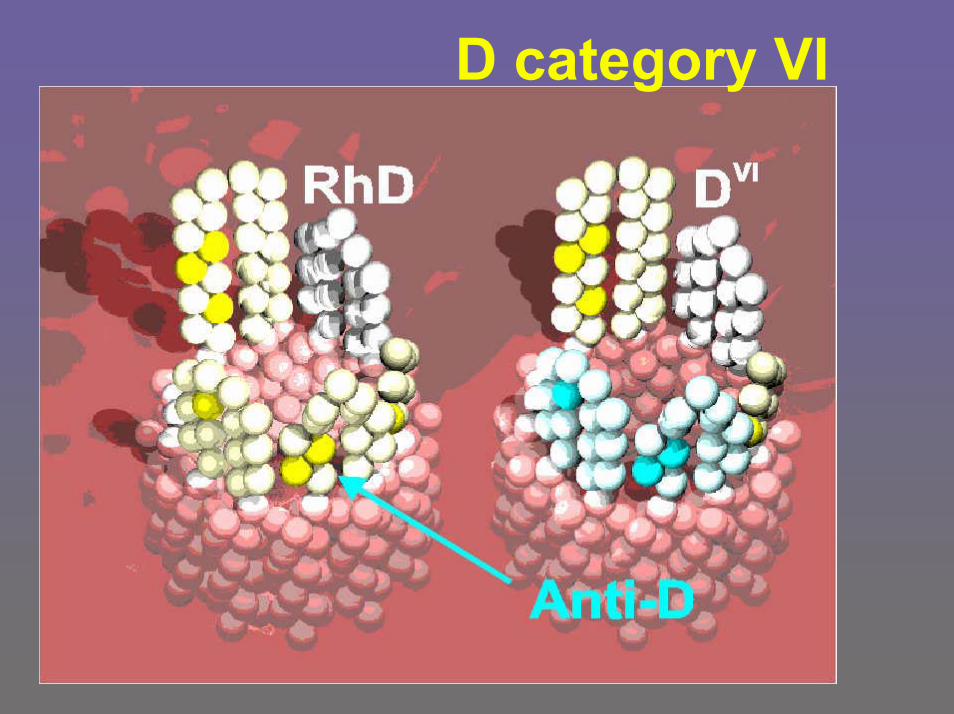
D category VI
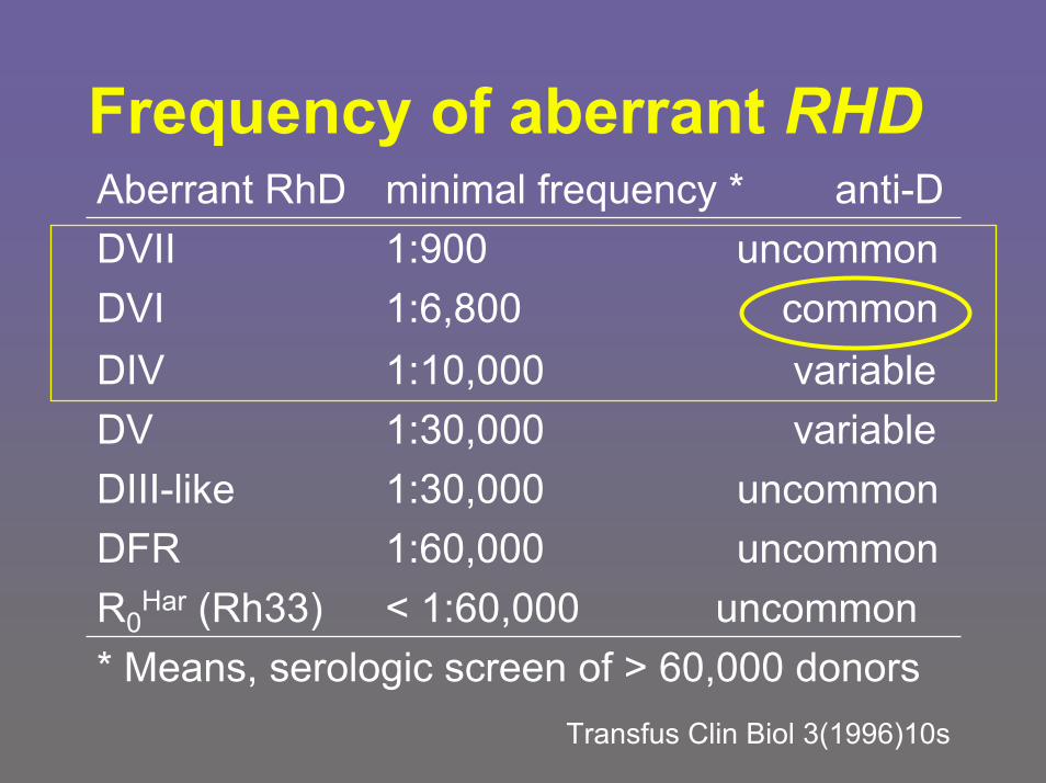
Frequency of aberrant RHD
* Means, serologic screen of > 60,000 donorsTransfus Clin Biol 3(1996)10s
< 1:60,000 uncommonR0Har (Rh33)
1:60,000 uncommon DFR1:30,000 uncommonDIII-like1:30,000 variableDV1:10,000 variableDIV1:6,800 commonDVI1:900 uncommonDVIIminimal frequency * anti-DAberrant RhD
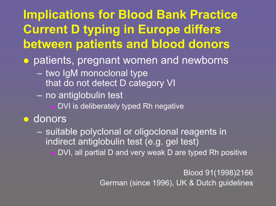
Implications for Blood Bank PracticeCurrent D typing in Europe differsbetween patients and blood donors
patients, pregnant women and newborns– two IgM monoclonal type
that do not detect D category VI – no antiglobulin test
DVI is deliberately typed Rh negative
donors– suitable polyclonal or oligoclonal reagents in
indirect antiglobulin test (e.g. gel test)DVI, all partial D and very weak D are typed Rh positive
Blood 91(1998)2166German (since 1996), UK & Dutch guidelines
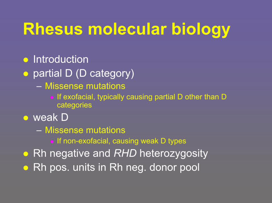
Rhesus molecular biologyIntroductionpartial D (D category)– Missense mutations
If exofacial, typically causing partial D other than Dcategories
weak D– Missense mutations
If non-exofacial, causing weak D types
Rh negative and RHD heterozygosityRh pos. units in Rh neg. donor pool
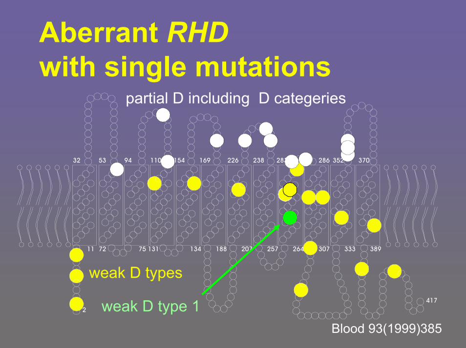
Aberrant RHDwith single mutations
11
2
72 75 131 134 188 207 257 264 307 333 389
94 110 154 283 370
partial D including D categeries
32 53 169 226 238 286 352
weak D types
weak D type 1 417
Blood 93(1999)385
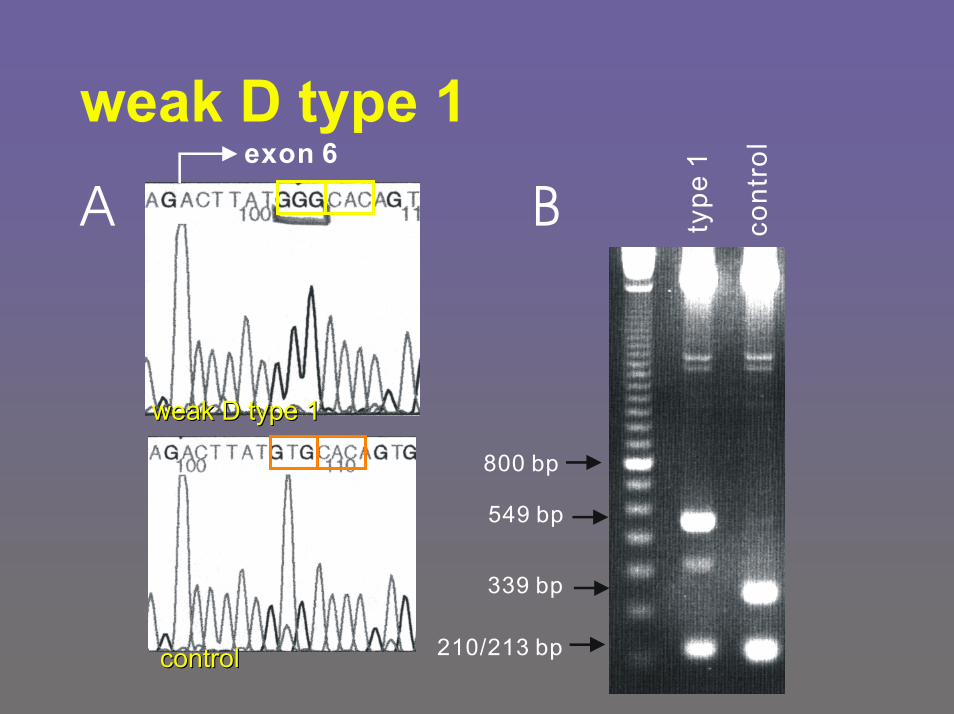
weak D type 1
type
1
cont
rol
800 bp
549 bp
339 bp
210/213 bp
exon 6
A B
controlcontrol
weakweak D D type type 11
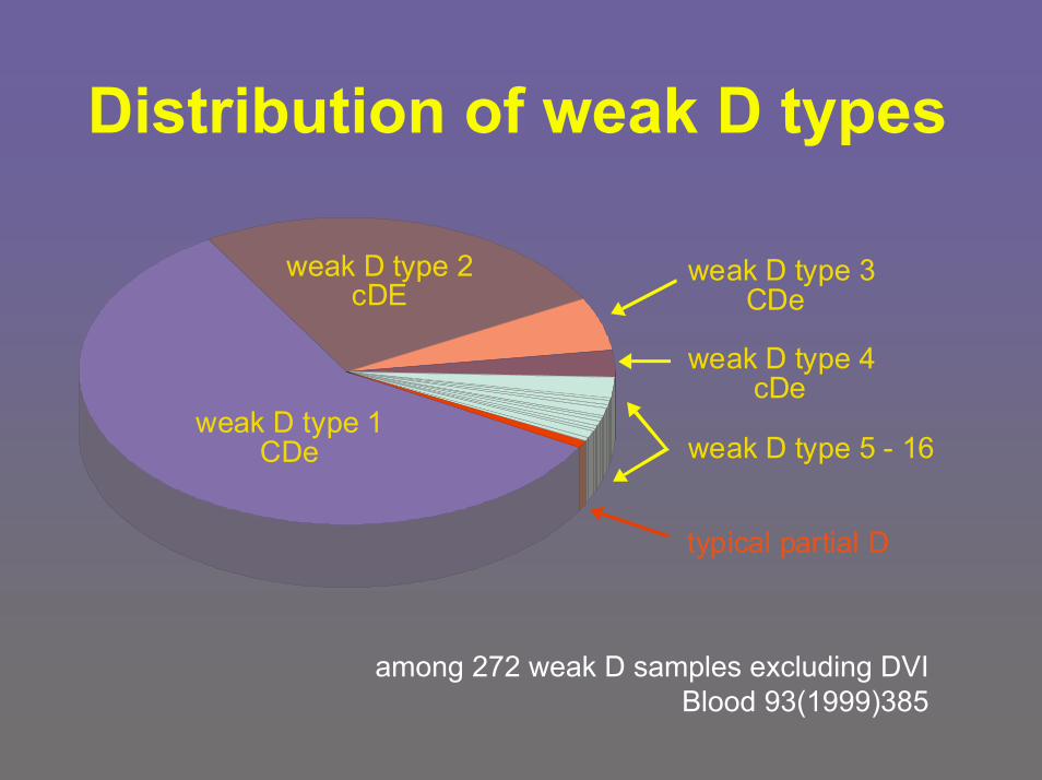
Distribution of weak D types
weak D type 1CDe
weak D type 2cDE
typical partial D
weak D type 3 CDe
weak D type 4 cDe
weak D type 5 - 16
among 272 weak D samples excluding DVIBlood 93(1999)385
Qualityassurance
D pos. proband with anti-DRegistry since 199860 submissions confirmed13 international submissionsSeveral new partial D, like DNB, DOL, DAU-320 weak D samples:– Allo-anti-D among weak D type 4.2 (DAR) & type
15 only– No allo-anti-D among prevalent weak D types:
they all carried auto-anti-Dhttp://www.uni-ulm.de/~wflegel/RH/RIR/ accessed Oct 2002
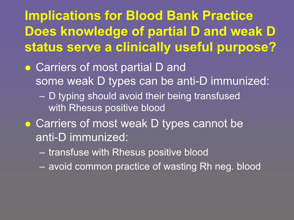
Implications for Blood Bank PracticeDoes knowledge of partial D and weak D status serve a clinically useful purpose?
Carriers of most partial D and some weak D types can be anti-D immunized:– D typing should avoid their being transfused
with Rhesus positive bloodCarriers of most weak D types cannot be anti-D immunized:– transfuse with Rhesus positive blood– avoid common practice of wasting Rh neg. blood
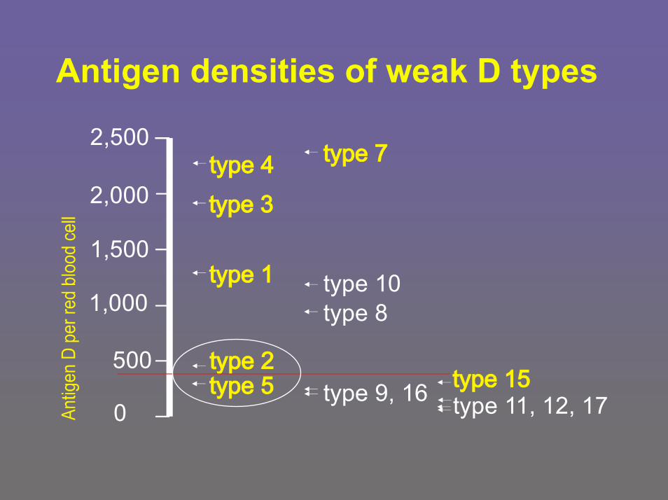
Antigen densities of weak D types
2,500
2,000
1,500
1,000
500
0
type 10
type 11, 12, 17
type 8
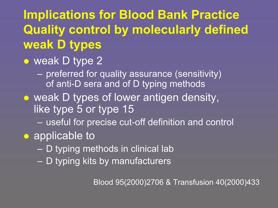
Implications for Blood Bank Practice Quality control by molecularly defined weak D types
weak D type 2– preferred for quality assurance (sensitivity)
of anti-D sera and of D typing methodsweak D types of lower antigen density, like type 5 or type 15– useful for precise cut-off definition and control
applicable to– D typing methods in clinical lab– D typing kits by manufacturers
Blood 95(2000)2706 & Transfusion 40(2000)433
Rhesus molecular biology
Introductionpartial D (D category)weak DRh negative and RHD heterozygosityRh pos. units in Rh neg. donor pool
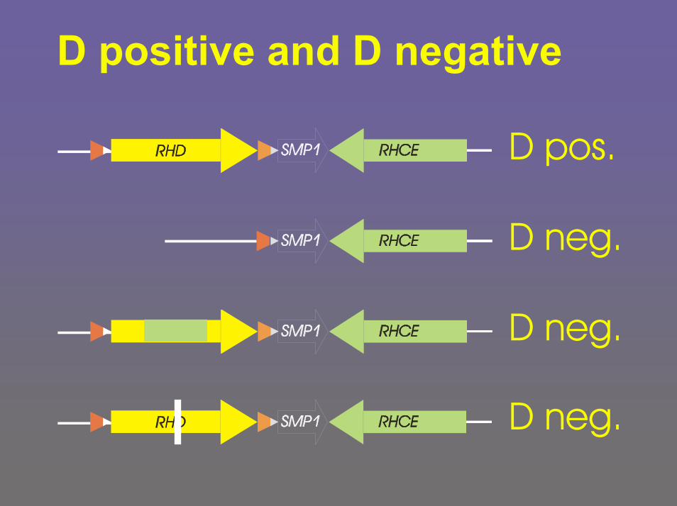
D positive and D negative
D pos.
D neg.
D neg.
D neg.
b
b
b
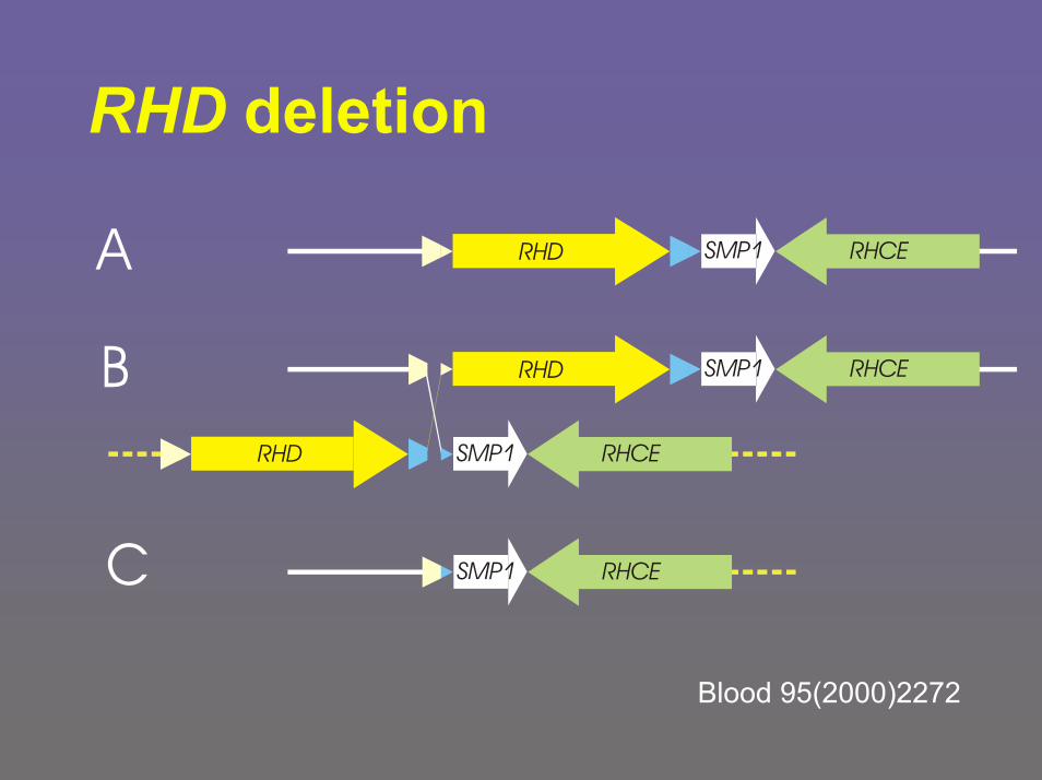
RHD deletion
A
B
C
RHD
RHD
RHD
b
b
SMP1
SMP1
SMP1
SMP1
RHCE
RHCE
RHCE
RHCE
Blood 95(2000)2272
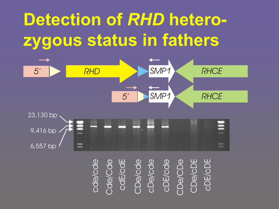
Detection of RHD hetero-zygous status in fathers
cd
e/c
de
Cd
e/C
de
cd
E/c
dE
CD
e/c
de
cD
e/c
de
cDE/
cd
e
CD
e/C
De
CD
e/c
DE
cDE/
cD
E
9,416 bp
6,557 bp
23,130 bp
RHD SMP1
SMP1
RHCE
RHCE
5’
5’
Rhesus molecular biology
Introductionpartial D (D category)weak DRh negative and RHD heterozygosityRh pos. units in Rh neg. donor pool
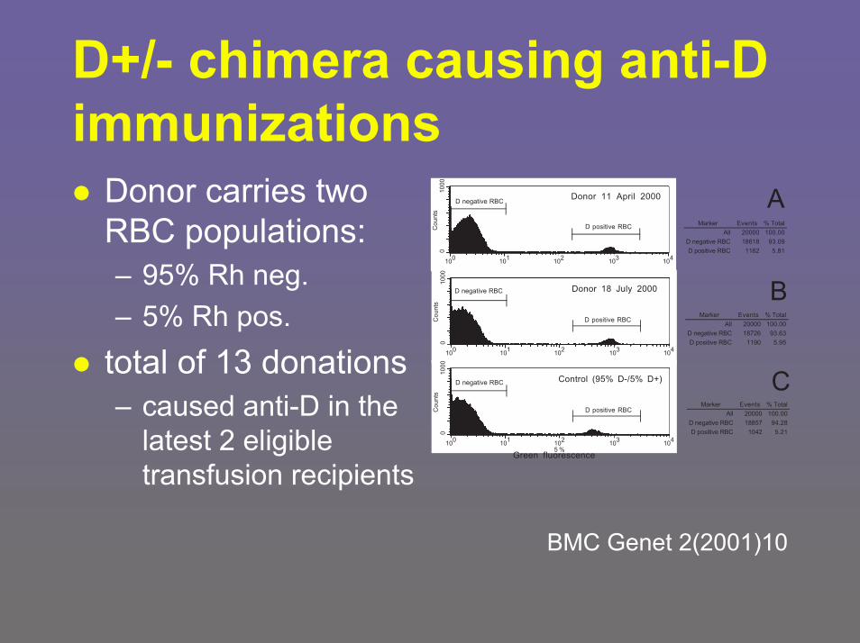
D+/- chimera causing anti-Dimmunizations
Donor carries twoRBC populations:– 95% Rh neg.– 5% Rh pos.
total of 13 donations– caused anti-D in the
latest 2 eligible transfusion recipients
01000
Counts
100
101
102
103
104
01000
Counts
01000
Counts
BMC Genet 2(2001)10
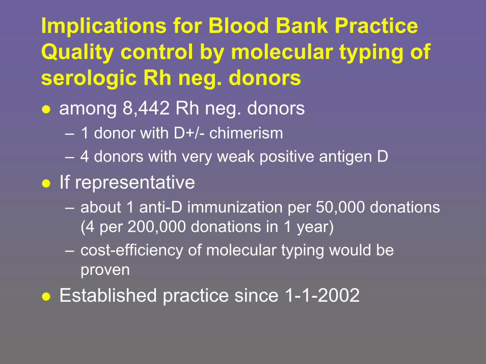
Implications for Blood Bank Practice Quality control by molecular typing of serologic Rh neg. donors
among 8,442 Rh neg. donors– 1 donor with D+/- chimerism– 4 donors with very weak positive antigen D
If representative– about 1 anti-D immunization per 50,000 donations
(4 per 200,000 donations in 1 year)– cost-efficiency of molecular typing would be
provenEstablished practice since 1-1-2002
Implications for Blood Bank Practice Why RHD genotyping?
Superior sensitivity– uncover (many?) weak D in the “Rhesus
negative“ donor pool of blood centersSuperior specificity– as genotyping becomes routine, clinical
implications of known and new RHD alleleswill be recognized
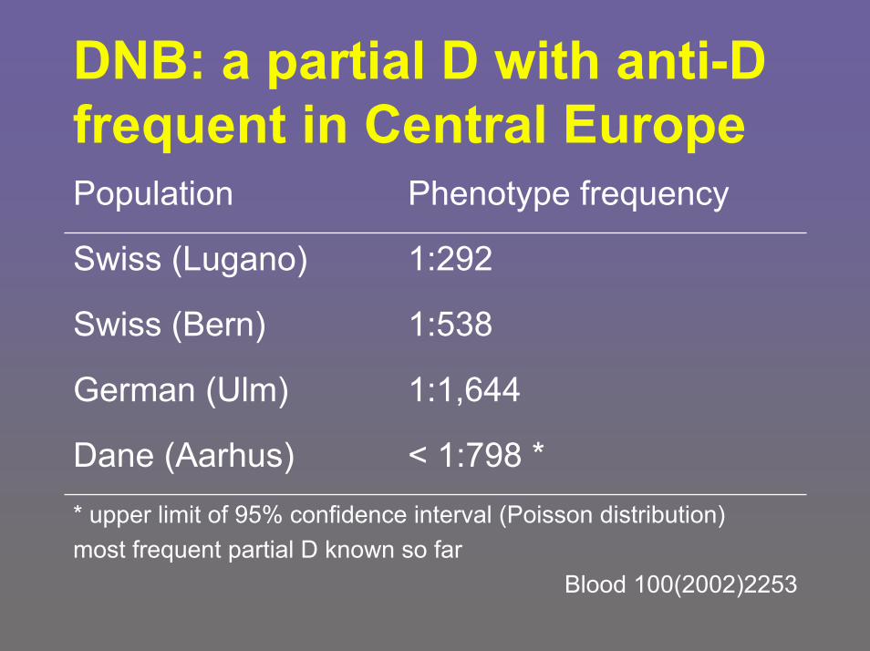
DNB: a partial D with anti-D frequent in Central Europe
* upper limit of 95% confidence interval (Poisson distribution)most frequent partial D known so far
Blood 100(2002)2253
< 1:798 *Dane (Aarhus)
1:1,644German (Ulm)
1:538Swiss (Bern)
1:292Swiss (Lugano)
Phenotype frequencyPopulation
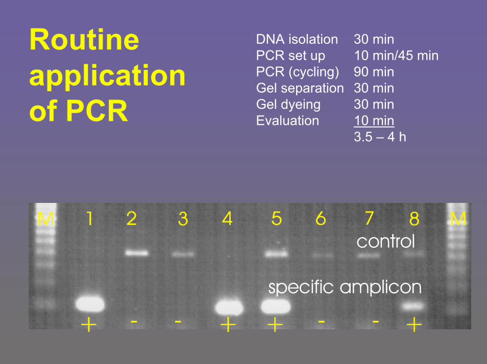
Routineapplication of PCR
DNA isolation 30 minPCR set up 10 min/45 minPCR (cycling) 90 minGel separation 30 minGel dyeing 30 minEvaluation 10 min
3.5 – 4 h
specific amplicon
control
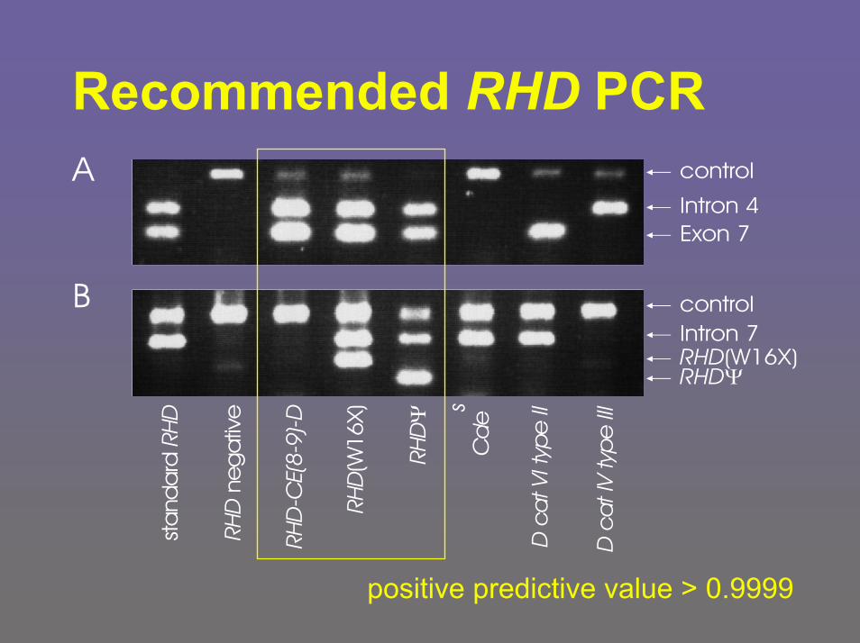
Recommended RHD PCR
B
A control
Intron 4 Exon 7
controlIntron 7 RHD(W16X)RHDΨ
RHD
neg
ativ
e
D c
at V
I typ
e II
RHD
-CE(
8-9)
-D
RHD
(W16
X)
RHDΨ
Cd
es
D c
at I
V ty
pe II
I
sta
nda
rd R
HD
positive predictive value > 0.9999
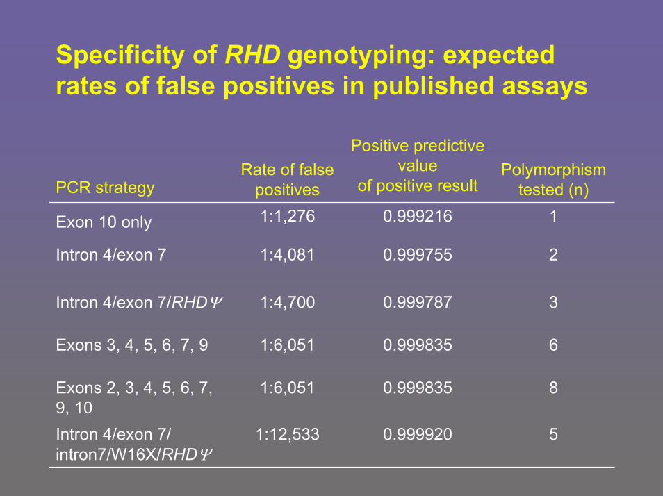
Specificity of RHD genotyping: expected rates of false positives in published assays
Polymorphism tested (n)
Positive predictive value
of positive resultRate of false
positivesPCR strategy
60.9998351:6,051Exons 3, 4, 5, 6, 7, 9
50.9999201:12,533Intron 4/exon 7/intron7/W16X/RHDΨ
80.9998351:6,051Exons 2, 3, 4, 5, 6, 7, 9, 10
30.9997871:4,700Intron 4/exon 7/RHDΨ
20.9997551:4,081Intron 4/exon 7
10.9992161:1,276Exon 10 only
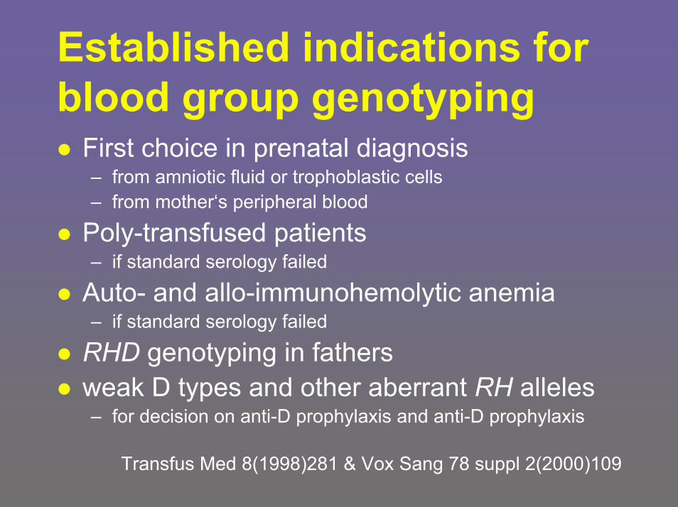
Established indications for blood group genotyping
First choice in prenatal diagnosis– from amniotic fluid or trophoblastic cells– from mother‘s peripheral blood
Poly-transfused patients– if standard serology failed
Auto- and allo-immunohemolytic anemia– if standard serology failed
RHD genotyping in fathersweak D types and other aberrant RH alleles– for decision on anti-D prophylaxis and anti-D prophylaxis
Transfus Med 8(1998)281 & Vox Sang 78 suppl 2(2000)109
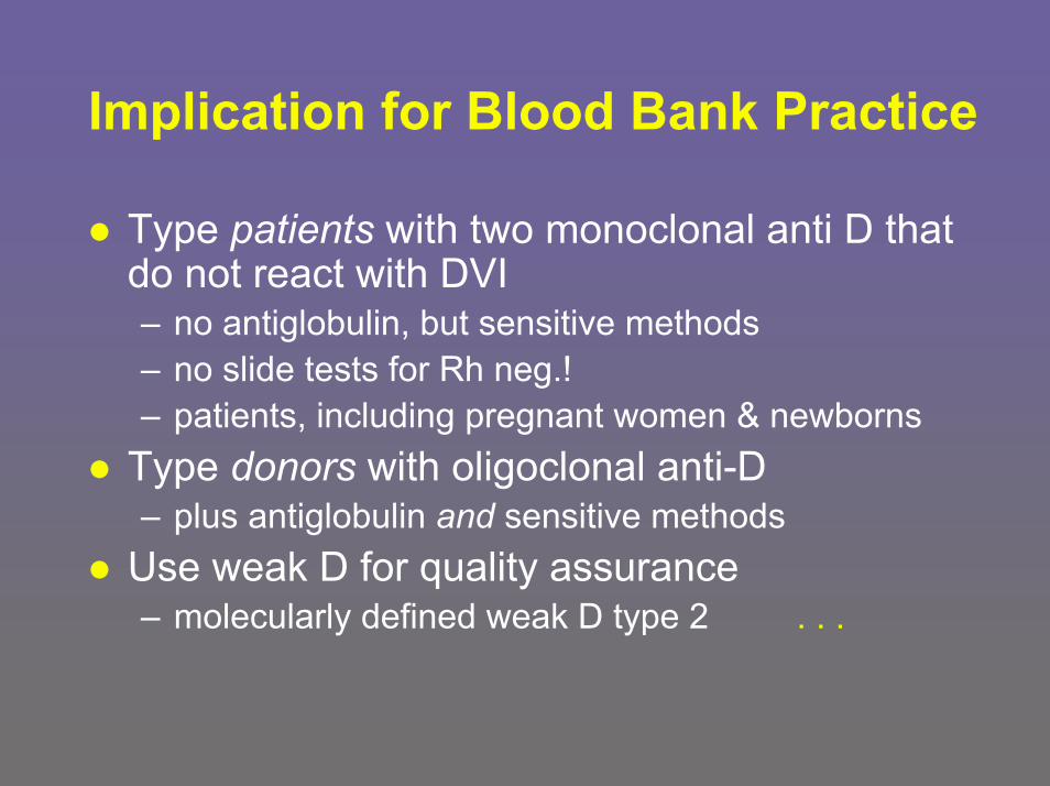
Implication for Blood Bank Practice
Type patients with two monoclonal anti D that do not react with DVI– no antiglobulin, but sensitive methods– no slide tests for Rh neg.!– patients, including pregnant women & newborns
Type donors with oligoclonal anti-D– plus antiglobulin and sensitive methods
Use weak D for quality assurance – molecularly defined weak D type 2 . . .
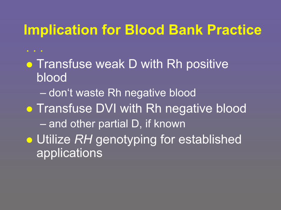
. . .Transfuse weak D with Rh positiveblood– don‘t waste Rh negative blood
Transfuse DVI with Rh negative blood– and other partial D, if known
Utilize RH genotyping for established applications
Implication for Blood Bank Practice
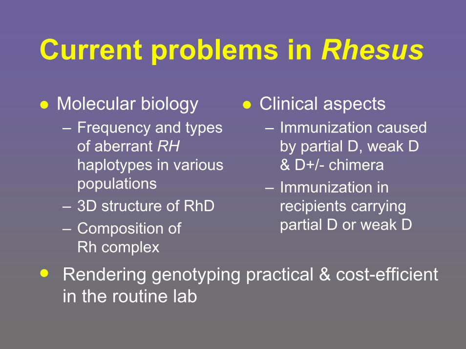
Current problems in Rhesus
Molecular biology– Frequency and types
of aberrant RHhaplotypes in various populations
– 3D structure of RhD– Composition of
Rh complex
Clinical aspects– Immunization caused
by partial D, weak D & D+/- chimera
– Immunization in recipients carryingpartial D or weak D
Rendering genotyping practical & cost-efficient in the routine lab

Universität Ulm


![arXiv:1302.5147v1 [cond-mat.mes-hall] 20 Feb 2013 · Partial-measurement back-action and non-classical weak values in a superconducting circuit J. P. Groen, 1 D. Riste, 1 L. Tornberg,](https://img.pdfslide.us/doc/110x75/5f0805bf7e708231d41ff0c6/arxiv13025147v1-cond-matmes-hall-20-feb-2013-partial-measurement-back-action.jpg)