Embed Size (px)
Citation preview
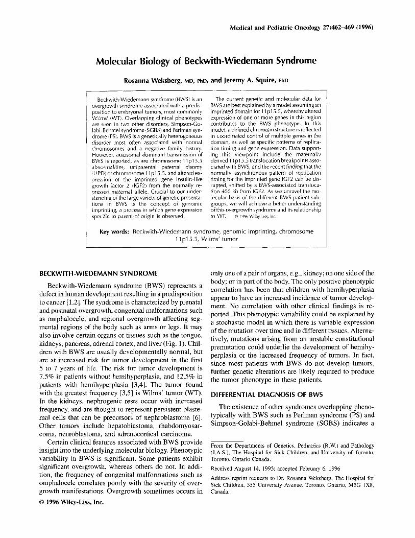
Medical and Pediatric Oncology 27:462-469 (1996)
Molecular Biology of Beckwith-Wiedemann Syndrome
Rosanna Weksberg, MD, PhD, and Jeremy A. Squire, PhD
Beckwith-Wiedemann syndrome (BWS) is an overgrowth syndrome associated with a predis- position to embryonal tumors, most commonly Wilms’ (WT). Overlapping clinical phenotypes are seen in two other disorders, Simpson-Go- labi-Behmel syndrome (SGBS) and Perlman syn- drome (PS). BWS is a genetically heterogeneous disorder most often associated with normal chromosomes and a negative family history. However, autosomal dominant transmission of BWS is reported, as are chromosome 11 pl5.5 abnormalities, uniparental paternal disomy (UPD) of chromosome 11 pl5.5, and altered ex- pression of the imprinted gene insulin-like growth factor 2 (IGF2j from the normally re- pressed maternal allele. Crucial to our under- standing of the large variety of genetic presenta- tions in BWS is the concept of genomic imprinting, a process in which gene expression specific to parent-of-origin is observed.
The current genetic and molecular data for BWS are best explained by a model assuming an imprinted domain for 11 pl5.5, whereby altered expression of one or more genes in this region contributes to the BWS phenotype. In this model, a defined chromatin structure is reflected in coordinated control of multiple genes in the domain, as well as specific patterns of replica- tion timing and gene expression. Data support- ing this viewpoint include the maternally derived 1 1 p15.5 translocation breakpoints asso- ciated with BWS, and the recent finding that the normally asynchronous pattern of replication timing for the imprinted gene IGF2 can be dis- rupted, shifted by a BWS-associated transloca- tion 400 kb from IGF2. As we unravel the mo- lecular basis of the different BWS patient sub- groups, we will achieve a better understanding of this overgrowth syndrome and its relationship to WT. 0 1996 Wiiey-Liss, inc.
Key words: Beckwith-Wiedemann syndrome, genomic imprinting, chromosome 11 pl5.5, Wilms’ tumor
BECKWITH-WIEDEMAN N SYNDROME
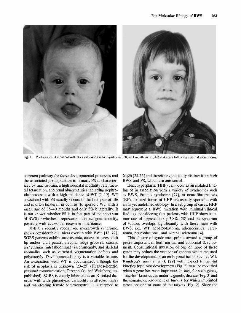
Beckwith-Wiedemann syndrome (B WS) represents a defect in human development resulting in a predisposition to cancer [ 1,2]. The syndrome is characterized by prenatal and postnatal overgrowth, congenital malformations such as omphalocele, and regional overgrowth affecting seg- mental regions of the body such as arms or legs. It may also involve certain organs or tissues such as the tongue, kidneys, pancreas, adrenal cortex, and liver (Fig. 1). Chil- dren with BWS are usually developmentally normal, but are at increased risk for tumor development in the first 5 to 7 years of life. The risk for tumor development is 7.5% in patients without hemihyperplasia, and 12.5% in patients with hemihyperplasia [3,4]. The tumor found with the greatest frequency [3,5] is Wilms’ tumor (WT). In the kidneys, nephrogenic rests occur with increased frequency, and are thought to represent persistent blaste- ma1 cells that can be precursors of nephroblastoma [6]. Other tumors include hepatoblastoma, rhabdomyosar- coma, neuroblastoma, and adrenocortical carcinoma.
Certain clinical features associated with BWS provide insight into the underlying molecular biology. Phenotypic variability in BWS is significant. Some patients exhibit significant overgrowth, whereas others do not. In addi- tion, the frequency of congenital malformations such as omphalocele correlates poorly with the severity of over- growth manifestations. Overgrowth sometimes occurs in 0 1996 Wiley-Liss, Inc.
only one of a pair of organs, e.g., kidney; on one side of the body; or in part of the body. The only positive phenotypic correlation has been that children with hemihyperplasia appear to have an increased incidence of tumor develop- ment. No correlation with other clinical findings is re- ported. This phenotypic variability could be explained by a stochastic model in which there is variable expression of the mutation over time and in different tissues. Alterna- tively, mutations arising from an unstable constitutional premutation could underlie the development of hemihy- perplasia or the increased frequency of tumors. In fact, since most patients with BWS do not develop tumors, further genetic alterations are likely required to produce the tumor phenotype in these patients.
DIFFERENTIAL DIAGNOSIS OF BWS
The existence of other syndromes overlapping pheno- typically with BWS such as Perlman syndrome (PS) and Simpson-Golabi-Behmel syndrome (SGBS) indicates a
From the Departments of Genetics, Pediatrics (R.W.) and Pathology (J.A.S.), The Hospital for Sick Children, and University of Toronto, Toronto, Ontario Canada.
Received August 14, 1995; accepted February 6, 1996
Address reprint requests to Dr. Rosanna Weksberg, The Hospital for Sick Children, 555 University Avenue, Toronto, Ontario, M5G 1x8, Canada.
The Molecular Biology of BWS 463
Fig. 1. Photographs of a patient with Beckwith-Wiedemann syndrome (left) at 1 month and (right) at 4 years following a partial glossectomy.
common pathway for these developmental processes and the associated predisposition to tumors. PS is character- ized by macrosomia, a high neonatal mortality rate, men- tal retardation, and renal abnormalities including nephro- blastomatosis with a high incidence of WT [7-121. WT associated with PS usually occurs in the first year of life and is often bilateral, in contrast to sporadic WT with a mean age of 35-40 months and only 5% bilaterality. It is not known whether PS is in fact part of the spectrum of BWS or whether it represents a distinct genetic entity, possibly with autosomal recessive inheritance.
SGBS, a recently recognized overgrowth syndrome, shows considerable clinical overlap with BWS [ 13-22]. SGBS patients exhibit macrosomia, coarse features, cleft lip and/or cleft palate, alveolar ridge grooves, cardiac arrhythmias, intraabdominal visceromegaly, and skeletal anomalies such as vertebral segmentation defects and polydactyly. Developmental delay is a variable feature. An association with WT is documented, although the risk of neoplasia is unknown [23-251 (Hughes-Benzie, personal communication; Terespolsky and Weksberg, un- published). SGBS is clearly inherited as an X-linked dis- order with wide phenotypic variability in affected males and manifesting female heterozygotes. It is mapped to
Xq26 [24,26] and therefore genetically distinct from both BWS and PS, which are autosomal.
Hemihyperplasia (HHP) can occur as an isolated find- ing or in association with a variety of syndromes such as BWS, Proteus syndrome [27], or neurofibromatosis (NF). Isolated forms of HHP are usually sporadic, with an as yet undefined etiology. In a subgroup of cases, HHP may represent a BWS mutation with minimal clinical findings, considering that patients with HHP show a tu- mor rate of approximately 3.8% [28] and the spectrum of tumors overlaps significantly with those seen with BWS, i.e., WT, hepatoblastoma, adrenocortical carci- noma, neuroblastoma, and adrenal adenoma [4].
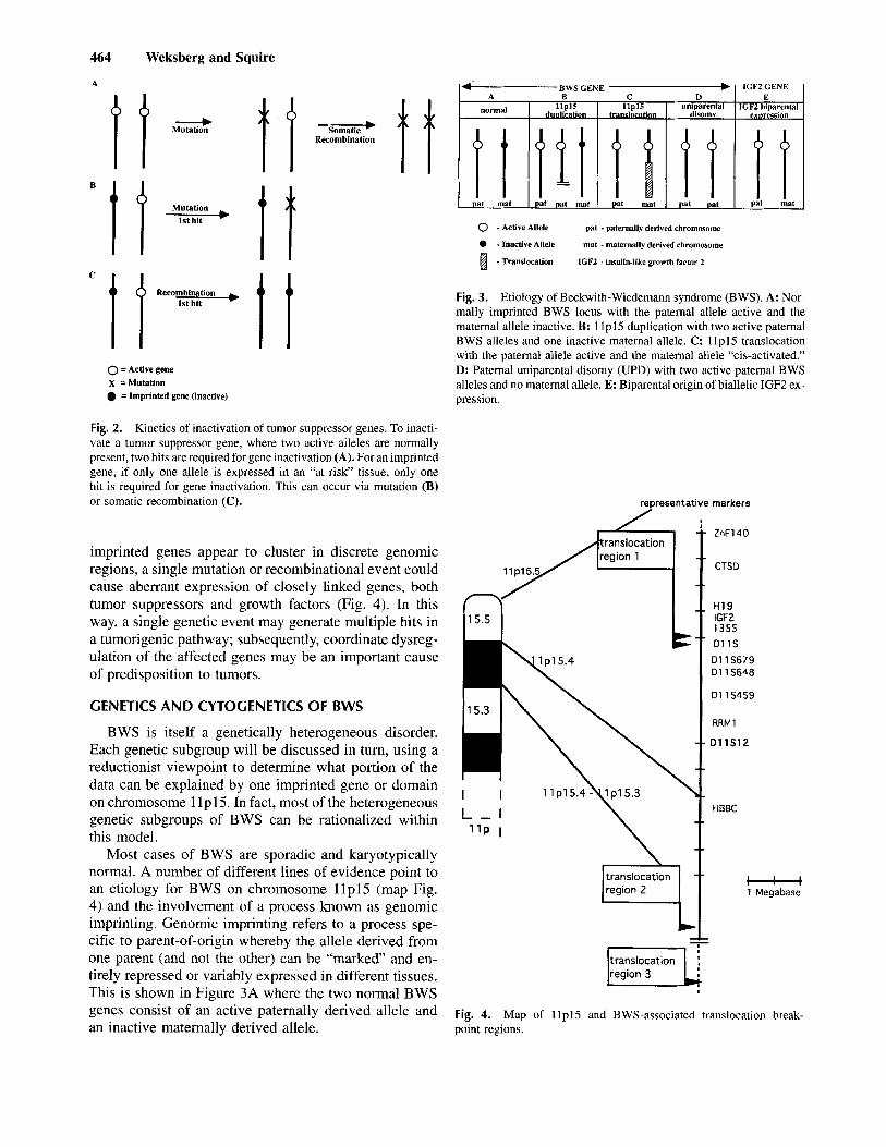
This cluster of syndromes points toward a group of genes important in both normal and abnormal develop- ment. Constitutional mutation of one or more of these genes may reduce the number of genetic events required for the development of an embryonal tumor such as WT. Knudson’s seminal work [29] with respect to two-hit kinetics for tumor development (Fig. 2) must be modified when a gene has been imprinted. In fact, for such genes, “one-hit’’ kinetics can underlie genetic disease (Fig. 3) and the somatic development of tumors for which imprinted genes are one or more of the targets (Fig. 2). Since the
464 Weksberg and Squire
A 1 1 M* 1 1 Recombination Somatic . / / B
0 -Active Allele pat - paternally derived chromosome
- Inactive Allele mat - maternally derived chromosome
1 - Translocation IGFZ - insulin-like growth factor 2
0 =Active gene X =Mutation
= Imprinted gene (inactive)
Fig. 2. Kinetics of inactivation of tumor suppressor genes. To inacti- vate a tumor suppressor gene, where two active alleles are normally present, two hits are required for gene inactivation (A). For an imprinted gene, if only one allele is expressed in an “at risk” tissue, only one hit is required for gene inactivation. This can occur via mutation (B) or somatic recombination (C).
imprinted genes appear to cluster in discrete genomic regions, a single mutation or recombinational event could cause aberrant expression of closely linked genes, both tumor suppressors and growth factors (Fig. 4). In this way, a single genetic event may generate multiple hits in a tumorigenic pathway; subsequently, coordinate dysreg- ulation of the affected genes may be an important cause of predisposition to tumors.
GENETICS AND CYTOGENETICS OF BWS
BWS is itself a genetically heterogeneous disorder. Each genetic subgroup will be discussed in turn, using a reductionist viewpoint to determine what portion of the data can be explained by one imprinted gene or domain on chromosome 1 lp1.5. In fact, most of the heterogeneous genetic subgroups of BWS can be rationalized within this model.
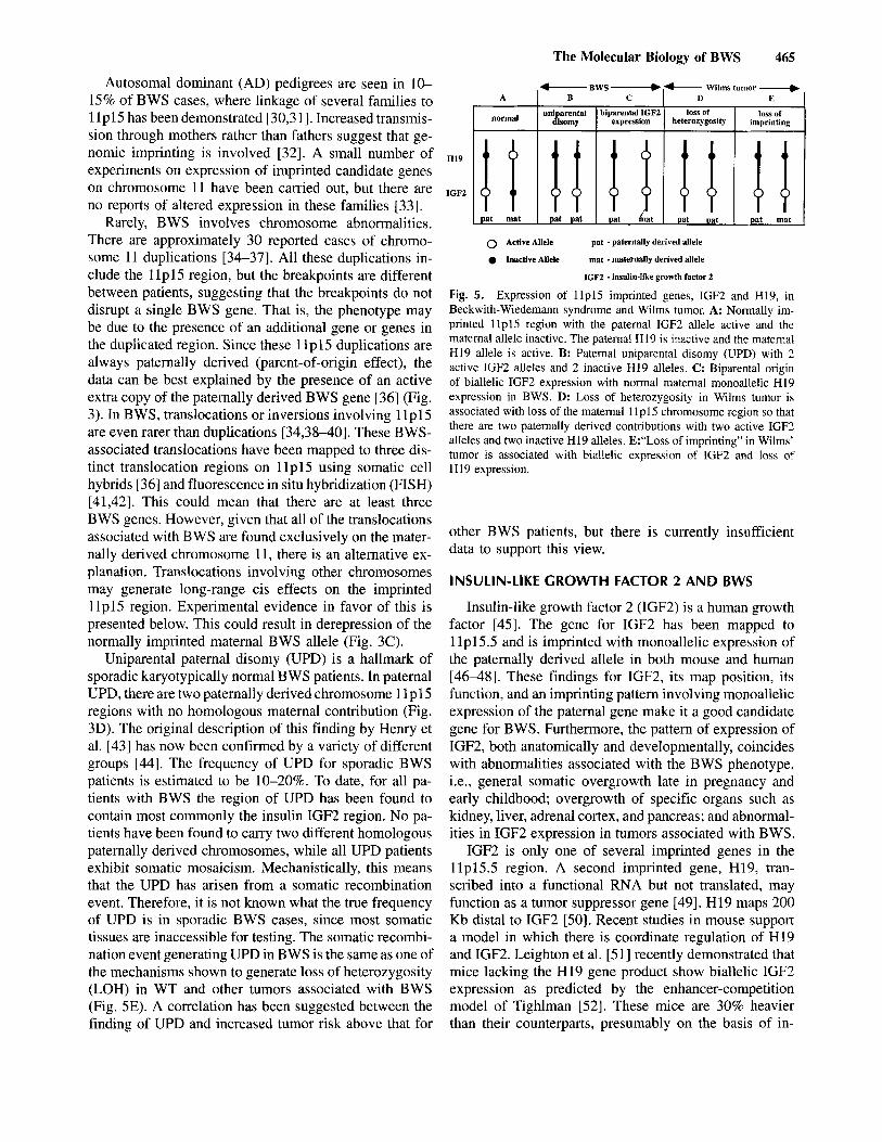
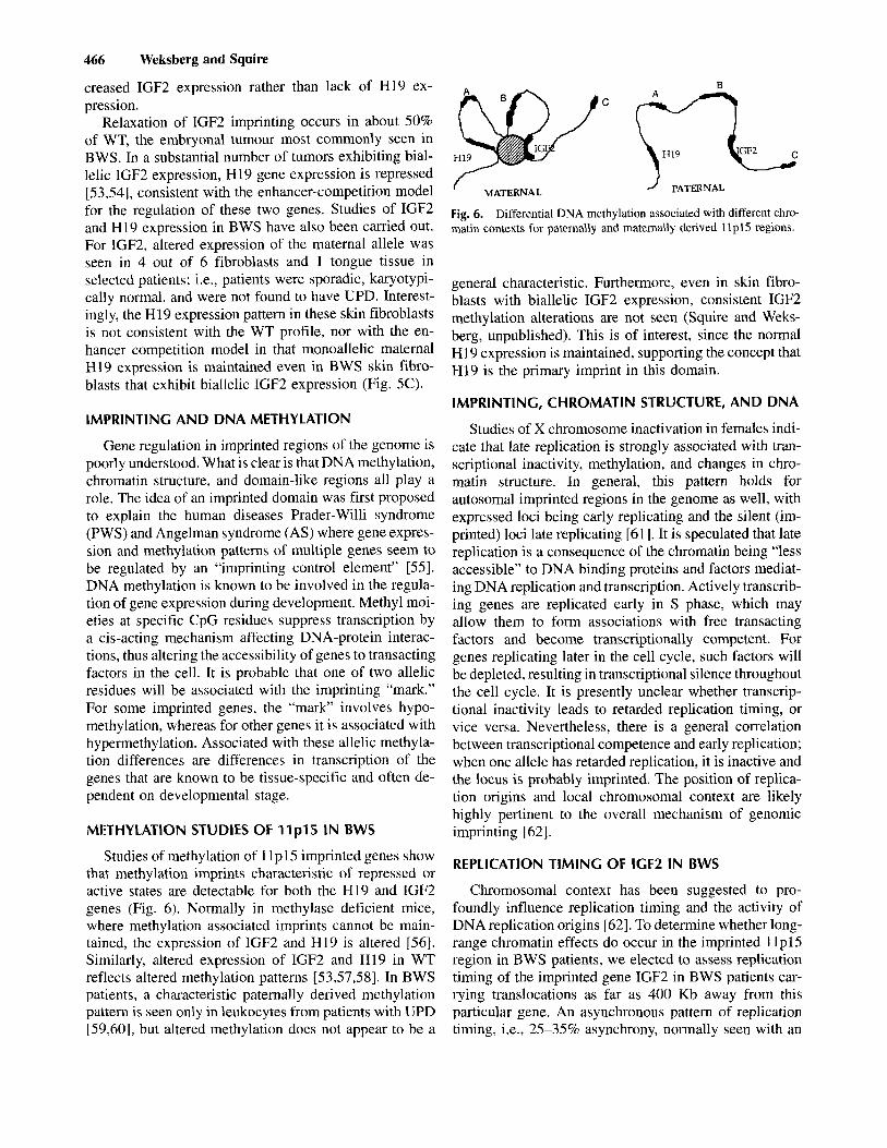
Most cases of BWS are sporadic and karyotypically normal. A number of different lines of evidence point to an etiology for BWS on chromosome 1 1 ~ 1 5 (map Fig. 4) and the involvement of a process known as genomic imprinting. Genomic imprinting refers to a process spe- cific to parent-of-origin whereby the allele derived from one parent (and not the other) can be “marked” and en- tirely repressed or variably expressed in different tissues. This is shown in Figure 3A where the two normal BWS
Fig. 3. Etiology of Beckwith-Wiedemann syndrome (BWS). A: Nor- mally imprinted BWS locus with the paternal allele active and the maternal allele inactive. B: l lp15 duplication with two active paternal BWS alleles and one inactive maternal allele. C: l lp15 translocation with the paternal allele active and the maternal allele “cis-activated.’’ D: Paternal uniparental disomy (UPD) with two active paternal BWS alleles and no maternal allele. E: Biparental origin of biallelic IGF2 ex- pression.
representative markers
translocation
4 N l p 1 5
\ .3 I;
region 3 I
ZnFl40
CTSD
H19 IGF2 1 3 5 5 D11S
D11S679 Dl 1 S648
D11S459
RRM 1
D l l S 1 2
HBBC
- 1 Megabase
genes consist Of an active paternally derived allele and an inactive maternally derived allele.
Fig. 4. point regions.
Map of l lp15 and BWS-associated translocation break-
The Molecular Biology of BWS 465
normal
Autosomal dominant (AD) pedigrees are seen in 10- 15% of BWS cases, where linkage of several families to llp15 has been demonstrated [30,31]. Increased transmis- sion through mothers rather than fathers suggest that ge- nomic imprinting is involved [32]. A small number of experiments on expression of imprinted candidate genes on chromosome 11 have been carried out, but there are no reports of altered expression in these families [33].
Rarely, BWS involves chromosome abnormalities. There are approximately 30 reported cases of chromo- some 11 duplications [34-371. All these duplications in- clude the llp15 region, but the breakpoints are different between patients, suggesting that the breakpoints do not disrupt a single BWS gene. That is, the phenotype may be due to the presence of an additional gene or genes in the duplicated region. Since these 1 lp15 duplications are always paternally derived (parent-of-origin effect), the data can be best explained by the presence of an active extra copy of the paternally derived BWS gene [36] (Fig. 3). In BWS, translocations or inversions involving llp15 are even rarer than duplications [34,38-40]. These BWS- associated translocations have been mapped to three dis- tinct translocation regions on llp15 using somatic cell hybrids [36] and fluorescence in situ hybridization (FISH) [41,42]. This could mean that there are at least three BWS genes. However, given that all of the translocations associated with BWS are found exclusively on the mater- nally derived chromosome 11, there is an alternative ex- planation. Translocations involving other chromosomes may generate long-range cis effects on the imprinted llp15 region. Experimental evidence in favor of this is presented below. This could result in derepression of the normally imprinted maternal BWS allele (Fig. 3C).
Uniparental paternal disomy (UPD) is a hallmark of sporadic karyotypically normal B WS patients. In paternal UPD, there are two paternally derived chromosome 1 lp15 regions with no homologous maternal contribution (Fig. 3D). The original description of this finding by Henry et al. [43] has now been confirmed by a variety of different groups [44]. The frequency of UPD for sporadic BWS patients is estimated to be 10-20%. To date, for all pa- tients with BWS the region of UPD has been found to contain most commonly the insulin IGF2 region. No pa- tients have been found to carry two different homologous paternally derived chromosomes, while all UPD patients exhibit somatic mosaicism. Mechanistically, this means that the UPD has arisen from a somatic recombination event. Therefore, it is not known what the true frequency of UPD is in sporadic BWS cases, since most somatic tissues are inaccessible for testing. The somatic recombi- nation event generating UPD in BWS is the same as one of the mechanisms shown to generate loss of heterozygosity (LOH) in WT and other tumors associated with BWS (Fig. 5E). A correlation has been suggested between the finding of UPD and increased tumor risk above that for
loss of uniparentsl biparental IGF2 loss of disomy expression heterozygosily imprinting
IGF2
pat -paternally derived allele 0 Active Allele
Inactive Allele mat - maternally derived allele
IGF2 - insdin-like growth factor 2
Fig. 5. Expression of llp15 imprinted genes, IGF2 and H19, in Beckwith-Wiedemann syndrome and Wilms tumor. A: Normally im- printed l lp15 region with the paternal IGF2 allele active and the maternal allele inactive. The paternal H19 is inactive and the maternal H19 allele is active. B: Paternal uniparental disomy (UPD) with 2 active IGF2 alleles and 2 inactive H19 alleles. C: Biparental origin of biallelic IGF2 expression with normal maternal monoallelic HI9 expression in BWS. D: Loss of heterozygosity in Wilms tumor is associated with loss of the maternal l lp15 chromosome region so that there are two paternally derived contributions with two active IGF2 alleles and two inactive H19 alleles. E:“Loss of imprinting” in Wilms’ tumor is associated with biallelic expression of IGF2 and loss of H19 expression.
other BWS patients, but there is currently insufficient data to support this view.
INSULIN-LIKE GROWTH FACTOR 2 AND BWS
Insulin-like growth factor 2 (IGF2) is a human growth factor [45]. The gene for IGF2 has been mapped to llp15.5 and is imprinted with monoallelic expression of the patemally derived allele in both mouse and human [4648] . These findings for IGF2, its map position, its function, and an imprinting pattern involving monoallelic expression of the paternal gene make it a good candidate gene for BWS. Furthermore, the pattern of expression of IGF2, both anatomically and developmentally, coincides with abnormalities associated with the BWS phenotype, i.e., general somatic overgrowth late in pregnancy and early childhood; overgrowth of specific organs such as kidney, liver, adrenal cortex, and pancreas; and abnormal- ities in IGF2 expression in tumors associated with BWS.
IGF2 is only one of several imprinted genes in the 1 lp15.5 region. A second imprinted gene, H19, tran- scribed into a functional RNA but not translated, may function as a tumor suppressor gene [49]. H19 maps 200 Kb distal to IGF2 [50]. Recent studies in mouse support a model in which there is coordinate regulation of H19 and IGF2. Leighton et al. [5 11 recently demonstrated that mice lacking the H19 gene product show biallelic IGF2 expression as predicted by the enhancer-competition model of Tighlman [52]. These mice are 30% heavier than their counterparts, presumably on the basis of in-
466 Weksberg and Squire
creased IGF2 expression rather than lack of H19 ex- pression.
Relaxation of IGF2 imprinting occurs in about SO% of WT, the embryonal tumour most commonly seen in BWS. In a substantial number of tumors exhibiting bial- lelic IGF2 expression, H19 gene expression is repressed [53,S4], consistent with the enhancer-competition model for the regulation of these two genes. Studies of IGF2 and H19 expression in BWS have also been carried out. For IGF2, altered expression of the maternal allele was seen in 4 out of 6 fibroblasts and 1 tongue tissue in selected patients; i.e., patients were sporadic, karyotypi- cally normal, and were not found to have UPD. Interest- ingly, the H19 expression pattern in these skin fibroblasts is not consistent with the WT profile, nor with the en- hancer competition model in that monoallelic maternal H19 expression is maintained even in BWS skin fibro- blasts that exhibit biallelic IGF2 expression (Fig. 5C).
IMPRINTING AND DNA METHYLATION
Gene regulation in imprinted regions of the genome is poorly understood. What is clear is that DNA methylation, chromatin structure, and domain-like regions all play a role. The idea of an imprinted domain was first proposed to explain the human diseases Prader-Willi syndrome (PWS) and Angelman syndrome (AS) where gene expres- sion and methylation patterns of multiple genes seem to be regulated by an “imprinting control element” [S5]. DNA methylation is known to be involved in the regula- tion of gene expression during development. Methyl moi- eties at specific CpG residues suppress transcription by a cis-acting mechanism affecting DNA-protein interac- tions, thus altering the accessibility of genes to transacting factors in the cell. It is probable that one of two allelic residues will be associated with the imprinting “mark.” For some imprinted genes, the “mark” involves hypo- methylation, whereas for other genes it is associated with hypermethylation. Associated with these allelic methyla- tion differences are differences in transcription of the genes that are known to be tissue-specific and often de- pendent on developmental stage.
METHYLATION STUDIES OF l l p 1 5 IN BWS
Studies of methylation of 1 lp15 imprinted genes show that methylation imprints characteristic of repressed or active states are detectable for both the H19 and IGF2 genes (Fig. 6). Normally in methylase deficient mice, where methylation associated imprints cannot be main- tained, the expression of IGF2 and H19 is altered [56]. Similarly, altered expression of IGF2 and H19 in WT reflects altered methylation patterns [53,57,58]. In BWS patients, a characteristic paternally derived methylation pattern is seen only in leukocytes from patients with UPD 159,601, but altered methylation does not appear to be a
‘ MATERNAL PATERNAL
Fig. 6. matin contexts for paternally and maternally derived 1 l p l 5 regions.
Differential DNA methylation associated with different chro-
general characteristic. Furthermore, even in skin fibro- blasts with biallelic IGF2 expression, consistent IGF2 methylation alterations are not seen (Squire and Weks- berg, unpublished). This is of interest, since the normal H19 expression is maintained, supporting the concept that H19 is the primary imprint in this domain.
IMPRINTING, CHROMATIN STRUCTURE, AND DNA
Studies of X chromosome inactivation in females indi- cate that late replication is strongly associated with tran- scriptional inactivity, methylation, and changes in chro- matin structure. In general, this pattern holds for autosomal imprinted regions in the genome as well, with expressed loci being early replicating and the silent (im- printed) loci late replicating [61]. It is speculated that late replication is a consequence of the chromatin being “less accessible” to DNA binding proteins and factors mediat- ing DNA replication and transcription. Actively transcrib- ing genes are replicated early in S phase, which may allow them to form associations with free transacting factors and become transcriptionally competent. For genes replicating later in the cell cycle, such factors will be depleted, resulting in transcriptional silence throughout the cell cycle. It is presently unclear whether transcrip- tional inactivity leads to retarded replication timing, or vice versa. Nevertheless, there is a general correlation between transcriptional competence and early replication; when one allele has retarded replication, it is inactive and the locus is probably imprinted. The position of replica- tion origins and local chromosomal context are likely highly pertinent to the overall mechanism of genomic imprinting [62].
REPLICATION TIMING OF ICF2 IN BWS
Chromosomal context has been suggested to pro- foundly influence replication timing and the activity of DNA replication origins [62]. To determine whether long- range chromatin effects do occur in the imprinted 1 1 ~ 1 5 region in BWS patients, we elected to assess replication timing of the imprinted gene IGF2 in BWS patients car- rying translocations as far as 400 Kb away from this particular gene. An asynchronous pattern of replication timing, i.e., 25-35% asynchrony, normally seen with an
The Molecular Biology of BWS 467
was normal [64]. This difference in phenotype is due to opposite skewing of X inactivation: in the affected twin the X chromosome carrying the normal DMD allele is almost exclusively inactivated, where in the normal twin the X chromosome carrying the DMD mutation is almost exclusively inactivated. For this reason, and because the X-linked overgrowth syndrome SGBS overlaps signifi- cantly with BWS, we assessed patterns of X-inactivation in fibroblasts of two MZ female twin pairs discordant for BWS to determine whether they had differential skewing of the X chromosome. In fact, in one female twin pair, both showed skewed X-inactivation, but in the same di- rection, while in another twin pair very little skewing was observed. Thus, differences in X inactivation patterns between MZ twins is not a general explanation for the occurrence of such twins. Other mechanisms that may explain the BWS discordance of MZ twins include as yet unidentified X chromosome or autosomal differences between the twins, or differential gene expressiodim- printing of loci in early development, when the rate of development of females lags behind that of males.
To determine whether X inactivation plays a role in the etiology of sporadic BWS, Bird et al. [65] have exam- ined a population of BWS individuals to determine whether they have skewed X inactivation different from that of the general population. Their positive findings support the concept that the X chromosome is involved in the expression of the BWS phenotype, at least in some patients, either directly, via the X-linked SGBS, or via other genes which may control imprinting of autosomal loci.
imprinted gene such as IGF2, was in fact seen in the controls. However, the translocation 11;22 mapping 400 kb away from IGF2 [41] resulted in partial loss of asyn- chrony of IGF2 replication, indicating a long-range effect on the imprinted domain as measured by replication tim- ing. The BWS patient with the 11;22 translocation showed a trend towards loss of asynchronous replication when compared with controls. This trend has been found to be statistically significant in two separate experiments. Since the 11;22 BWS translocation contains 400 kb of DNA between the translocation and IGF2, loss of asynchronous replication might occur because long segments of normal chromatin structure are required for normal regulation and transcription in this region. That is, the introduction of non-chromosome 11 DNA into this region might cause the translocated region to be affected by the cis effects of chromosome 22, which may cause changes in confor- mation and thereby affect the timing of replication. Since there are approximately 10 such llp15 translocations spanning over 1 megabase of DNA and involving many other chromosomes [42], we propose that DNA re- arrangements that place llp15.5 near other regions of the genome may disrupt putative chromatin control elements.
TWINS DISCORDANT FOR BWS
Among the intriguing observations of clinical pheno- types associated with BWS are reports of approximately 20 monozygotic (MZ) twin pairs, most of whom are female and discordant for BWS. In addition, one concor- dant female pair has been described, one concordant male pair and two discordant male pairs. Of particular interest in this regard is the possibility of a postzygotic somatic recombination to explain the discordance in MZ twins [63]. Slun fibroblasts derived from three such twin pairs were examined, since the blood of MZ twins often under- goes mixing in utero and may not reflect the genetic constitution of only one twin. All pairs were tested for monozygosity at multiple loci and found to have a >99.9% probability of being MZ. However, restriction fragment length polymorphism (RFLP) studies to com- pare llp15 contributions in BWS discordant twin pairs show no differences between these three twin pairs. In this way, paternal isodisomy for the IGF2hnsulin region was shown to be an unlikely explanation for the dis- cordance, since somatic mosaicism cannot be entirely excluded via a complete tissue survey in these twins.
An alternate explanation of the unusual female MZ twin discordance in BWS relates to possible differences in X chromosome inactivation between twins. This type of discrepancy has been described for X-linked disorders such as Duchenne muscular dystrophy where two MZ female twins both carrying the Duchenne muscular dys- trophy (DMD) mutation showed opposite phenotypes; i.e., one expressed the DMD phenotype and the other
CONCLUSIONS
Recent results suggest the existence of chromosome domains containing clusters of imprinted genes that may be controlled in a regional manner. Therefore, it seems likely that the regulation of imprinted genes is complex, utilizing multiple control mechanisms and most certainly involving both local and regional effectors of expression. Our knowledge of the basis of BWS and its associated tumors is still rudimentary, but it is clear that a number of powerful new experimental tools have become avail- able to clarify the basis of overgrowth syndromes and the associated tumors in the near future.
REFERENCES
Beckwith JB: Macroglossia, omphalocele, adrenal cytomegaly, gigantism, and hyperplastic visceromegaly. Birth Defects: Original Articles Series V 188-196, 1969. Wiedemann HR: Complexe malformatif familial avec hernia om- bilicale et macroglossie-un syndrome nouveau? J Geneti Hum
Wiedemann HR: Tumours and hemihypertrophy associated with Wiedemann-Beckwith syndrome. Euro J Pediatrics 141:129,1983.
13~223-232, 1964.
468 Weksberg and Squire
4. Gorlin RJ, Cohen MM, Jr, Levin LS: “Syndromes of the Head and Neck.” Oxford, 1990. pp 323416, 1990.
5 . Sotelo-Avila C, Gooch WM: Neoplasms associated with the Beck- with-Wiedemann syndrome. Persp Pediatric Pathol 344:255- 272, 1976.
6. Beckwith JB, Kiviat NB, Bonadio JF: (1990) Nephrogenic rests,
7
8
9
10
11
12
13
14
15
16
17
18
19
20
21.
22
23.
24.
2s
26.
nephroblastomatosis and the pathogenesis of Wilms’ tumor. Ped Pathol 10: 1-36, 1990. Liban E, Kozenitzky IL: Metanephric hamartomas and neph- roblastosis in siblings. Cancer 25:885-888, 1970. Perlman M, Goldberg GM, Bar-Ziv J, Danovitch G: Renal hamarto- mas and nephroblastomatosis with fetal gigantism: A familial syn- drome. J Pediatrics 83:414418, 1973. Perlman M, Levin M, Wittels B: Syndrome of fetal gigantism, renal hamartomas, and nephroblastomatosis with Wilms’ tumor. Cancer 35:1212-1217, 1975. Perlman M: Letter to the Editor: Perlman syndrome: Familial renal dysplasia with Wilms’ tumor, fetal gigantism and multiple congenital anomalies. Am J Med Genet 25:793-795, 1986. Greenberg F, Copeland K, Gresik MV: Expanding the spectrum of the Perlman syndrome. Am J Med Genet 29773-776, 1988. Grundy RG, Pritchard J, Baraitser M, Risdon A, Robards M: Perlman and Wiedemann-Beckwith syndromes: Two distinct con- ditions associated with Wilms tumour. Europ J Pediatrics 15 12395- 898, 1992. Simpson JL, Landey S , New M, German J: A previously unrecog- nized X-linked syndrome of dysmorphia. Birth Defects: Original Article Series XI: 18-24, 1975. Golabi M, Rosen L: A new X-linked mental retardation overgrowth syndrome. Am J Med Genet 17:345-358, 1984. Opitz JM, Hemnann J, Gilbert EFN, Matalon R: Simpson-Golabi- Behmel syndrome: Follow-up of their Michigan family. Am J Med Genet 30:301-308, 1988. Opitz JM: The Golabi-Rosen syndrome-report of a second family. Am J Med Gen 17:359-366, 1984. Behmel A, Plochl E, Rosenkranz W: A new X-linked dysplasia gigantism syndrome: Identical with the Simpson dysplasia syn- drome’? Hum Genet 67:409413, 1984. Behmel A, Plochl E, Rosenkranz W A new X-linked dysplasia gigantism syndrome: Follow-up in the first family and a report on a second Austrian family. Am J Med Genet 30:275-285, 1988. Neri G, Marini R, Cappa M, Borrelli P, Opitz JM: Simpson- Golabi-Behmel syndrome: An X-linked encephalo-tropho-schisis syndrome. Am J Med Genet 30:287-299, 1988. Gurrieri F, Cappa M, Neri G: Further delineation of the Simpson- Golabi-Behmel (SGB) syndrome. Am J Med Genet 44:136-137, 1992. Garganta CL, Bodurtha JN: Report of another family with Simp- son-Golabi-Behmel syndrome and a review of the literature. Am J Med Genet 44:129-135, 1992. Chen E, Johnson JP, Cox VA, Golabi M: Simpson-Golabi-Behmel syndrome: Congenital diaphragmatic hernia and radiological find- ings in two patients and follow-up of a previously reported case. Am J Med Genet 46574-578, 1993. Hughes-Benzie RM, Hunter AGW, Allanson JE, MacKenzie AE: Simpson-Golabi-BehmeI syndrome associated with renal dysplasia and embryonal tumor: Localization of the gene Xqcen-q21. Am J Med Gen 43:428-435, 1992. Xuan JY, Besner A, Ireland M, Hughes-Benzie R, MacKenzie A: Mapping of Simpson-Golabi-BehmeI syndrome to Xq25-q27. Hum Mol Genet 3:133-137, 1994. Verloes A, Massart B, Dehalleux I, Langhendries J-P, Koulischer L: Clinical overlap of Beckwith-Wiedemann, Perlmann and Simpson- Golabi-Behmel syndromes: A diagnostic pitfall. Clin Genet
Orth U, Guirrieri F, Behmel A, Genuardi M, Cremer M, Gal A, 47r2.57-262, 1995.
27.
28.
29.
30.
31.
32.
33 .
34.
35.
36.
37.
38.
39.
40.
41.
42.
43.
Neri G: Gene for Simpson-Golabi-Behniel syndrome is linked to HPRT in Xq26 in two European families. Am J Med Genet
Cohen MM, Jr: Proteus syndrome: Clinical evidence for somatic mosaicism and selective review. Am J Med Genet 645-652, 1993. Hoyme HE: The incidence of neoplasia in children with isolated congenital hemihypertophy. David Smith Meeting on malforma- tions and Morphogenesis. Burlington, Vermont, USA. Knudson AG, Strong LC: Mutation and cancer: A model for Wilms’ tumor of the kidney. J Nat Cancer Tnst 48:313-324, 1972. Koufos A, Grundy P, Morgan K, Aleck KA, Hadro T, Lampkin BC, Kalbakji A, Cavenee WK: Familial Wiedemann-Beckwith syndrome and a second Wilms’ tumor locus both map to llp15.5. Am J Hum Genet 44:711-179, 1989. Ping AJ, Reeve AE, Law DJ, Young MR, Boehnke M, Feinberg AP: Genetic linkage of Beckwith-Wiedemann syndrome to 1 lp15. Am J Hum Genet 44:720-723, 1989. Moutou C, Junien C, Henry I, Bonaiti-Pellie C: Beckwith-Wiede- mann syndrome: A demonstration of the mechanisms responsible for the excess of transmitting females. J Med Genet 29:217- 220, 1992. Nystrom A, Hedborg F, Ohlsson R: Insulin-like growth factor 2 cannot be linked to a familial form of Beckwith-Wiedemann syndrome. Europ J Pediatrics 153574-580, 1994. Waziri M, Patil SR, Hanson JW, Bartley JA: Abnormality of chro- mosome I1 in patients with features of Beckwith-Wiedemann syndrome. J Pediatrics 102373-876, 1983. Ogawa 0, Eccles MR, Szeto J, McNoe LA, Yu K, Mae MA, Smith PJ, Reeve AE: Relaxation of insulin-like growth factor I1 gene imprinting implicated in Wilms’ tumour. Nature 362:749- 751, 1993. Weksberg R, Teshima I, Williams BRG, Greenberg CR, Pueschel SM, Chemos JE, Fowlow SB, Hoyme E, Anderson IJ, Whiteman DAH, Fisher N, Squire J: Molecular characterizationof cytogenetic alterations associated with the Beckwith-Wiedemann syndrome (BWS) phenotype refines the localization and suggests the gene for BWS is imprinted. Hum Molec Genet 2549-556, 1993. Henry I, Jeanpierre M, Barichard F, Serre JL, Mallet J, Turleau C de, GJ, Junien C: Duplication of HRAS I , INS, and IGF2 is not a common event in Beckwith-Wiedemann syndrome. Ann Genet
Pueschel SM, Padre-Mendoza T: Chromsome I 1 and Beckwith- Wiedemann syndrome. J Pediatrics 104:484485, 1984. Turleau C, de Grouchy J, Chavin-Colin F, Martelli H, Voyer M, Charlas R: Trisomy 1 lp15 and Beckwith-Wiedemann syndrome: A report of two cases. Hum Genet 67:219-221, 1984. Okano Y, Osasa Y, Yamamoto H, Hase Y, Tsuruhara T, Fujita H: An infant with Beckwith-Wiedemann syndrome and chromosomal duplication 1 lpl3-pter: Correlation of symptoms between I lp tri- somy and Beckwith-Wiedemann syndrome. Jap J Hum Genet
Mannens M, Hoovers JMN, Redeker E, Verjaal M, Feinberg AP, Little P, Boavida M, Coad N, Steenman N, Bilek J, Niikawa N, Tonoki H, Nakamura Y, de Boer EG, Slater RM, John R, Cowell JK, Junien C, Henry I, Tommerup N, Weksberg R, Pueschel SM, Leschot NJ, Westerveld A: Parental imprinting of human chromo- some region 1 Ipl5.3-pter involved in the Beckwith-Wiedemann syndrome and various human neoplasia. Europ J Hum Genet 2:3- 23, 1994. Redeker E, Alders M, Hoovers JMN, Richard CW 111, Westerveld A, Mannens M: Physical mapping of 3 candidate tumor suppressor genes relative to Beckwith-Wiedemann syndrome-associated chro- mosomal breakpoints at I Ip15.3. Cytogenet Cell Genet 68:222- 225, 1995. Henry I, Bonaiti-Pellie C, Chehensse V, Beldjord C, Schwartz C,
501388-390, 1994.
31:216-20, 1988.
31 ~365-372, 1986.
The Molecular Biology of BWS 469
58. Rainier S, Reinberg A: Genomic imprinting, DNA methylation and cancer. J Nat Cancer Instit 86:753-759, 1994.
59. Schneid H, Seurin D, Varquez M-P, Gourmelen M, Cabrol S, Le Bouc Y Parental allele specific methylation of the human insulin- like growth factor I1 gene and Beckwith-Wiedemann syndrome. J Med Genet 30:353-362, 1993.
60. Reik W, Brown KW, Slatter RE, Sartori P, Elliott M, Maher ER: Allelic methylation of H19 and IGF2 in the Beckwith-Wiedemann syndrome. Hum Molec Genet 3:1297-1301, 1994.
61. Kitsberg D, Selig S, Brandeis M, Simon I, Keshet I, Driscoll DJ, Nicholls RD, Cedar H: Allele-specific replication timing of imprinted gene regions. Nature 364:459-463, 1993.
62. Knoll JHM, Cheng S-D, Lalande M: Allele specificity of DNA replication timing in the AngelmadPrader-Willi syndrome im- printed chromosomal region. Nature Genet 6:4146, 1994.
63. Cote GB, and Gyftodimou J: Twinning and mitotic crossing-over: Some possibilities and their implications. Am J Human Gent 49:120-130, 1991.
64. Richards CS, Watkins SC, Hoffman EP, Schneider NR, Milsark IW, Katz KS, Cook JD, Kunkel LM, Cortada JM: Skewed X-inactivation in a female MZ twin results in Duchenne muscular dystrophy. Am J Hum Genet 46:672-681, 1990.
65. Bird L, Naumova A, Feinberg A, Grundy P, Henry I, Junien C, Newsham I, Weksberg R, Sapienza C: Skewed X-inactivation in sporadic Beckwith-Wiedemann patients. International Conference on Molecular and Clinical Genetics of Childhood Renal Tumors. Philadelphia, PA U.S.A, 1995.
Utermann G, Junien C: Uniparental paternal disomy in a genetic cancer-predisposing syndrome. Nature 35 1 :665-667.
44. Nystrom A, Cheetham JE, Engstmm W, Schofield PN: Molecular analysis of patients with Wiedemann-Beckwith syndrome. 11. Pa- ternally derived disomies of chromosome l l . Europ J Pediatrics 151:511-514, 1992.
45. Humbel R: Insulin-like growth factors I and 11. Europ J Biochem 190:445462, 1990.
46. Giannoukakis N, Deal C, Paquette J, Goodyer CG, Polychronakos C: Parental genomic imprinting of the human IGF2 gene. Nature Genet 4:98-101, 1993.
47. Ohlsson R, Hedborg F, Holmgren L, Walsh C, Ekstrom TJ: Over- lapping patterns of IGF2 and H19 expression during human devel- opment: Biallelic IGF2 expression correlates with a lack of HI9 expression. Development 120:361-368, 1994.
48. Zhang S, Shapiro DN, Helman LJ: Activation of an imprinted allele of the insulin-like growth factor I1 gene implicated in rhabdo- myosarcoma. J Clin Invest 94:445448, 1994.
49. Hao Y, Crenshaw T, Moulton T, Newcomb E, Tycko B: Tumor suppressor activity of H19 RNA. Nature 365:764-767, 1993.
50. Zemel S, Bartolomei MS, Tilghman SM: Physical linkage of two mammalian imprinted genes, HI 9 and insulin-like growth factor 2. Nature Genet 2:61-65, 1992.
5 1. Leighton PA, Ingram RS, Eggenschwiler J, Efstratiadis A, Tilgh- man SM: Disruption of imprinting causing by deletion of the H19 gene region in mice. Nature 375:34-39, 1995.
52. Tilghman SM, Bartolomei MS, Webber AL, Brunkow ME, Saam J, Leighton PA, Pfeifer K, Zemel S: Parental imprinting of the H19 and IGF2 genes in the mouse. Cold Spring Harbor Symposia on Quantitative Biology LVIII:287-295, 1993.
53. Moulton T, Crenshaw T, Hao Y, Moosikasuwan J, Lin N, Dembitzer F, Hensle T, Weiss L, McMorrow L, Loew T, Kraus W, Gerald W, Tycko B: Epigenetic lesions at the HI9 locus in Wilms’ tumour patients. Nature Genet 7:440447, 1994.
54. Rainier S, Johnson LA, Dobry CJ, Ping AJ, Gmndy PE, Feinberg AP: Relaxation of imprinted genes in human cancer. Nature 362:747-749, 1993.
55. Nicholls RD: New insights reveal complex mechanisms involved in genomic imprinting. Am J Hum Gent 54:733-740, 1994.
56. Ferguson-Smith A, Sasaki H, Cattanach B, Surani A: Parental- origin-specific epigenetic modification of the mouse H19 gene. Nature 362:751-755, 1993.
57. Zhang Y, Shields T, Crenshaw T Imprinting of human H19: Allele- specific CpG methylation, loss of the active allele in Wilms tumor, and potential for somatic allele switching. Am J Hum Genet 53:113-124. 1993.
COMMENTARY
Weksberg and Squire discuss the genetics, cytogenetics and molecular genetics of Beckwith-Wiedemann syn- drome (BWS). The BWS gene is thought to be localized on chromosome llp15, a locus also believed to harbor the second Wilms’ tumor suppressor gene, WT2. With this in mind, the authors indicate that the available data for BWS are best explained by a model assuming an imprinted domain for chromosome llp15, whereby al- tered expression of one or more genes in this region contribute to the BWS phenotype. For an explanation of the term, “imprinting,” see the manuscript by Moulton et al., in this issue.


![[Clarinet_Institute] Wiedemann Staccato.pdf](https://img.pdfslide.us/doc/110x75/5695d0061a28ab9b02909c61/clarinetinstitute-wiedemann-staccatopdf.jpg)