Embed Size (px)
Citation preview
1
Application of molecularbiology methods in
hematology and oncology
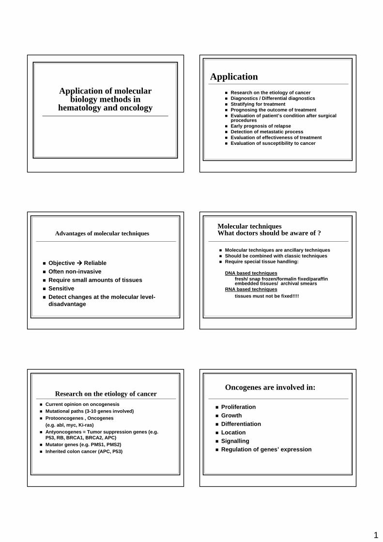
� Research on the etiology of cancer� Diagnostics / Differential diagnostics� Stratifying for treatment� Prognosing the outcome of treatment� Evaluation of patient’s condition after surgical
procedures� Early prognosis of relapse� Detection of metastatic process� Evaluation of effectiveness of treatment� Evaluation of susceptibility to cancer
Application
Advantages of molecular techniques
� Objective ���� Reliable� Often non-invasive� Require small amounts of tissues� Sensitive� Detect changes at the molecular level-
disadvantage
Molecular techniquesWhat doctors should be aware of ?
� Molecular techniques are ancillary techniques� Should be combined with classic techniques� Require special tissue handling:
DNA based techniquesfresh/ snap frozen/formalin fixed/paraffinembedded tissues/ archival smears
RNA based techniquestissues must not be fixed!!!!
Research on the etiology of cancer� Current opinion on oncogenesis� Mutational paths (3-10 genes involved)� Protooncogenes , Oncogenes
(e.g. abl, myc, Ki-ras)� Antyoncogenes = Tumor suppression genes (e.g.
P53, RB, BRCA1, BRCA2, APC)� Mutator genes (e.g. PMS1, PMS2)� Inherited colon cancer ( APC, P53)
Oncogenes are involved in:
� Proliferation� Growth� Differentiation� Location� Signalling� Regulation of genes’ expression
2
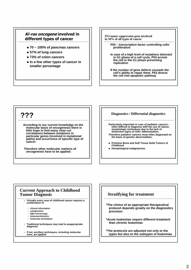
� 70 – 100% of pancreas cancers� 57% of lung cancers� 73% of colon cancers� In a few other types of cancer in
smaller percentage
Ki-ras oncogene involved indifferent types of cancer
P53 – transcription factor controlling cells’ proliferation
In case of a high level of mutations detectedin G1 phase of a cell cycle, P53 arreststhe cell in the G1 phase preventingreplication
If the number of gene defects exceeds thecell’s ability to repair them, P53 directsthe cell into apoptotic pathway
P53 tumor suppression gene involvedin 50% of all types of cancer
???According to our current knowledge on the
molecular basis of oncogenesis there islittle hope to find many clear-cutcorrelations between mutations inparticular genes (involved in mutationalpaths) and occurrence of specific type ofcancer.
Therefore other molecular markers ofoncogenesis have to be applied.
Diagnostics / Differential diagnostics
Particularly important in case of pediatric cancers –often difficult to diagnose with the use of classicmorphologic techniques due to the lack ofdistinctive signs of cells’ differentiation.
Therefore pediatric cancers most often diagnosed on the basis of genetic abnormalities.
� Primitive Bone and Soft Tissue Solid Tumors ofChildhood
� Hematological malignancies
Current Approach to ChildhoodTumor Diagnosis
� Virtually every case of childhood cancer requires a combination of
� clinical information� cytogenetics� light microscopy� immunochemistry� molecular techniques
� Traditional techniques may lead to anappropriatediagnosis
� If not, ancillary techniques, including molecularones, are applied
Stratifying for treatment
*The choice of an appropriate therapeuticalprotocol depends greatly on the diagnosticsprecision
*Acute leukemias require different treatmentthan chronic leukemias
*The protocols are adjusted not only to thetypes but also to the subtypes of leukemias
3
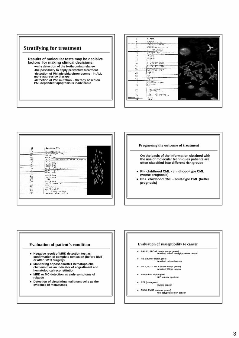
Results of molecular tests may be decisivefactors for making clinical decisions:
-early detection of the forthcoming relapse-the possibility to apply preventive treatment-detection of Philadelphia chromosome in ALL more aggressive therapy-detection of P53 mutation - therapy based on P53-dependent apoptosis is inadvisable
Stratifying for treatment
Prognosing the outcome of treatment
On the basis of the information obtained withthe use of molecular techniques patients areoften classified into different risk groups:
� Ph- childhood CML - childhood-type CML (worse prognosis)
� Ph+ childhood CML - adult-type CML (betterprognosis)
Evaluation of patient’s condition
� Negative result of MRD detection test as confirmation of complete remission (before BMT or after BMT/ surgery)
� Monitoring of post-alloBMT hematopoieticchimerism as an indicator of engraftment andhematological reconstitution
� MRD or MC detection as early symptoms ofrelapse
� Detection of circulating malignant cells as theevidence of metastases
Evaluation of susceptibility to cancer
� BRCA1, BRCA2 (tumor suppr.genes) inherited breast /ovary/ prostate cancer
� RB 1 (tumor suppr.gene) inherited retinoblastoma
� WT 1, WT 2, WT 3 (tumor suppr.genes) inherited Wilms tumour
� P53 (tumor suppr.gene) Li-Fraumeni syndrom
� RET (oncogene)thyroid cancer
� PMS1, PMS2 (mutator genes) non-polyposis colon cancer

4
Molecular techniques most widely used inhematology & oncology:
PCR-techniques
� conventional PCR� RT-PCR� competitive PCR� semi-quantitative PCR� PCR with
fluorescently labelledprimers
� PCR with radioactivelylabelled primers
FISH-techniques
� one-colour FISH� two-colour FISH� multicolour FISH� spectral karyotyping� FISH detecting mRNA
PCR (Polymerase Chain Reaction) A procedure that produces millions of copies of a
short segment of DNA through repeatedcycles of:
� denaturation (950C)� annealing (37-650C)� elongation (720C)A very common procedure in molecular genetics
that may be used to: � generate a sufficient quantity of DNA to
perform a test (e.g., sequence analysis, mutation scanning)
� may be modified e.g. to study geneexpression or for quantitative study
� PCR-reagents: water, PCR-buffer, dNTPs, primers, Taq polymerase, template DNA
� Approximately 30 cycles, each consistingof 3 steps (denat./anneal./elong.)
� sensitive, reliable, rapid� requires only small amount of DNA/RNA� requires the knowledge on the sequences
of the regions flanking the gene ofinterest
� sensitive to contamination� requires positive and negative controls
PCR (Polymerase Chain Reaction) RT-PCR (Reverse TranscriptasePCR)� A variation of the PCR technique in which
cDNA (complementary DNA) is made fromRNA via reverse transcription. The resultantcDNA is then amplified using standard PCR protocols.
� A method for assessing gene expression , detecting even low copy number mRNAtranscripts, and generating complementaryDNAs (cDNAs) for further study.
RT-PCR (Reverse TranscriptasePCR)
� RT-PCR-reagents: water, buffer, dNTPs, random primers, specific primers, reverse transcriptase, Taqpolymerase, RNA
� optimum temp. for RT � 370C (cDNA synthesis)
� ~ 550C (annealing of specific primers to the cDNA)
� 720C (extension of primers by Taqpolymerase)
RQ-PCR (Real-Time Quantitative PCR) � A variation of the PCR technique used for quantitative
study, e.g. � quantitative assessment of MRD � quantitative gene expression study
� The reaction requires additional reagent, which is a dual-dye labelled fluorescent probe specific for a site internal to the target sequence
� The amount of fluorescence and the number of cyclesrequired for its appearance are a measure of theamount of target DNA present in the sample

5
QC-PCR (quantitativecompetitive PCR)
In QC-PCR a known amount of a control sequence (competitor) similar enough to the target DNA to compete for the same reagents but different enough to be distinguished is added to the sample.
Both, the target sequence and the competitor are amplifiedsimultaneously. The quantity of the target sequence in thesample is calculated from its ratio with the competitor (T:C).
This is more accurate than measuring the total amount of thetarget sequence, since the inevitable errors in the PCR process affect both the target sequence and the controlsequence equally.
QC-PCR is labour � rarely in routine medical use
Semi-quantitative PCR
� A technique used for assessing the approximate amount ofsequence of interest, e.g. assessment of MYCN gene amplification in NB
� Involves several separate PCRs in which different DNA templates are used, e.g.�DNA of the patient examined�DNA of a healthy individual (negative control)�DNA isolated from a cell line expressing the gene amplification(positive control)
� The comparison of the amounts of electrophoreticaly resolvedPCR products enables the sample of interest to be quantitativelyassessed. (NOT a precise quantitative technique!!!)
PCR with fluorescently labelledprimers
� A modification of the PCR technique used for quantitativestudy, e.g. monitoring of post-alloBMT hematopoieticchimerism.
� The use of fluorescently labelled primers enables PCR-products to be quantitatively assessed duringelectrophoresis (in PAA gel) performed in an apparatusequipped with a laser .
� As fluorescent PCR-products pass through the laser beam itexcites their fluorescence which is detected and used for determination of length and amount of PCR-products.
PCR with radioactively labelledprimers
� The use of radioactively labelled primers enablesPCR-products to be visualised via autoradiography performed afterelectrophoresis in PAA gel.
� Autoradiography = Photographic film is exposed to theradioactivity of the gel for several hours and then developed.
� The stains on the film reflect the positions of PCR-products andprovide information on their length .This is NOT a quantitative technique!!!
� Applied e.g. In molecular study of hematopoietic chimerism in caseof sex-mis-matched alloBMT
FISH (Fluorescent in situ Hybridisation)
Used to identify the specific chromosomes or chromosomal regionsthrough hybridization (attachment) of fluorescently-labelled DNA probes to denatured chromosomal DNA.
Examination under fluorescent lighting detects thepresence/absence of the hybridized fluorescent signal andhence the presence/absence of the target chromosomal region.
� one-colour FISH e.g. � detection of geneamplification
� detection of deletion� two-colour FISH e.g. � detection of fusion genes
resulting from chromosomaltranslocations
� multicolour FISH � spectral karyotyping

6
FISHADVANTAGES DISADVANTAGES
� metaphase nuclei �possibility to detect evenminor chromosomalabnormalities
� interphase nuclei �no need for cells culturing
� fresh / fixed material� FISH for detection of
chromosomal aberrations� FISH for gene expression
study� FISH as quantitative
technique
� requires the knowledge on the sequences of interestto design oligonucleotideprobes
� unlike classic cytogeneticsFISH excludes thepossibility to detect novelgenetic abnormalities
� expensive in comparisonwith classic cytogenetics
SKY (Spectral karyotyping)
A novel molecular cytogenetic FISH-based technique (analternative to classic black-and-white banding pattern) thatallows colour karyotyping of human and mousechromosomes.
SKY permits the visualisation of all chromosomes at one time , ‘painting’ each pair of chromosomes at differentfluorescent colour.
Applied to a variety of human malignancies and mouse model systems.
Highly effective in deciphering many complex karyotypicrearrangements.
SKY (Spectral Karyotyping)
Traditional black andwhite banding pattern
Spectral karyotypingmulticolour image
Detection of MRD
� MRD - condition of a patient (leukemia or other malignant disease) whoachieved a state of complete remission, but is still at risk of relapse dueto the presence of residual malignant cells (�managed to survive thetherapy and may initiate the relapse).
� MRD – often below the sensitivity threshold of light microscopy or evenimmunohistochemical methods; detectable with the use of moleculartechniques such as PCR-derived techniques.
� MRD detection - crucial for the outcome of treatment � enables therelapse to be prevented by applying additional treatment
Diagnostic application of detection of specificchromosomal translocations
� t (9;22) � BCR-ABL Ph+ (~95% of CML)BCR – function unknownABL – protein kinase
� t (8;14) � IgH-cMYC (~90% of Burkitt’slymphoma)
cMYC – oncogene, transcription factorIgH – heavy chain of immunoglobulins
(very strong promotor region!!! �cMYC overexpression)
Detection of MRD in ALL
Quantitative Real-Time PCR (RQ-PCR) analysis of clone-specific rearrangements of genes encoding for immunoglobulins and T-cell receptors (TCR).
The rearrangements occur between the families of genesand are a part of lymphocytes maturation process.
The rearrangements are often called clonality markers as particular rearrangement is specific for particular clone ofleukemic cells in particular patient.
� ALL of B-lineage origin � IgH rearr. � ALL of T-lineage origin � TCR rearr.

7
Detection of MRD in ALL
IgH genes contain~70 genes within the V-genes family~30 genes in D-genes family~ 6 genes in J-genes family
Rearrangements between genes families� different combinations of genes� diversity of possible immunoglobulins’ structure� diversity of immunological responses
This diversity is even greater due to the fact that the double DNA breaksproduced during the rearrangement process are joined by an enzyme addingrandom nucleotides. The regions of junction between rearranged genes arehighly specific for leukemic lymphocytes in particular patient.
Differential diagnostics of leukemias on thebasis of IgH & TCR genes rearrangements ?
Specific kinds of rearrangements occur at given stages of
lymphocytes maturation
-the rearrangement between D & J family of genes is earlier thanthe V & D rearrangement
Having identified particular type of rearrangement in leukemic blasts we
may determine their stage of growth (the stage of growth at which they
became malignant) and on that basis we should be able to classify theleukemia into an appropriate subtype
This type of classification would be much more accurate than that based
on cytomorphology or immunophenotyping.
M V1 V2 V3 V4 V5 V6 V1 V2 V3 V4 V5 V6
Monitoring of hematopoieticchimerism after alloBMT
� mixed chimerism – coexistence of hematopoietic cells ofdonor’s and recipient’s origin
� complete donor’s chimerism – only the cells of donor’sorigin detectable
� complete recipient’s chimerism - only the cells ofrecipient’s origin detectable
� recipient’s-growing MC - an early prognostic indicator ofthe forthcoming relapseThe detection of mixed chimerism itself has no prognostic value - regular quantitative study is requiredto establish the D:R ratio in given time-points afteralloBMT

8
Monitoring of hematopoietic chimerismafter alloBMT
Most widely used technique for quantitative study ofhematopoietic chimerism:
*amplification of microsatellite markers with the use offluorescently labelled primers followed by automated DNA sizing and quantification
*Microsatellites = Short Tandem Repeats (STR) arepolymorphic DNA sequences distributed evenly along thegenome
*Each STR locus consists of 5-100 repeats of the coresequence (2-6 nt)
*Individuals may be distinguished on the basis of thedifference in length of given STR loci (informative STR markers)
Marker Gene Ruler 50 bp DNA Ladder (Fermentas)
VirusVirusVirusVirus genotyping as evidence for genotyping as evidence for genotyping as evidence for genotyping as evidence for
horizontal transmission horizontal transmission horizontal transmission horizontal transmission
VirusVirusVirusVirus detectiondetectiondetectiondetection andandandand quantificationquantificationquantificationquantification
Neuroblastoma (NB)
NB - develops from the peripheral nervous system NB - one of SRCTs (a group of difficult-to-diagnose child hood
tumors). Unlike other SRCTs NB is relatively easy to diag nosebecause of the neural phenotype of the tumor cells.
NB - characterized by clinical heterogeneity:���� tumors regress spontaneously���� become benign tumors���� tumors progress despite intensive therapyTherefore there has been an intensive search for reliable p redictors
of prognosis in NB.
Prognosticmarkers
� age
� stage
� localization
� histopatology
� ferritin, LDH, neuronospecificenolase, VMA, dopamine
� molecular markers� MYCN amplification
� Loss of 1p36, 11q23, 14q32
� 17q gain
� chemoresistance
Genetic markers in neuroblastomaSeveral genetic abnormalities have been identified in
NB: � MYCN gene amplification� deletion of fragment of chromosome 1 (1p)� deviations from diploid DNA content� chromosomal regions of faint band’s pattern
(heterogenously staining regions; hsr)� presence of additional mini-chromosomes
(double minute chromosomes; d-min) as a resultof MYCN gene amplification
NB should be divided into three subsets of differentprognosis.
The amplification of MYCN gene (over 10 copies) is
regarded the most important prognostic factor .

9
MYCN gene amplification
� MYCN amplification may be detected in cancer cells by Southern blot, but preferably by semi-quantitative PCR FISH or quantitative PCR.
� cancer tissue obtained by surgical resection or biopsy
� circulating MYCN DNA may be a powerful and non-invasive prognostic marker at the time of diagnosis
� it may be also used in the non-invasive follow-upexamination of patients at high risk of relapse.
NeuroblastomaMYCN amplification
FISH showing N-myc gene amplification in interphase cells. The N-myc DNA Probe (Oncor) is used to identify normal and amplified N-mycgenomic sequences.
Differential diagnostics ofEwing’s Family of Tumors (EFTs)
� EFTs – classified into SRCTs
� differential diagnostics of EFTs is based on thedetection of specificchromosomal translocations � fusion genes
� fusion genes detected with the use of
� FISH
� PCR / RT-PCR
� classic cytogenetics (not all fusionsdetectable)
EFTs are characterized by the EWS-ETS fusion genes family . Detection of a given fusion gene (given translocation) enablesthese tumors to be classified:
� t (11;22) � EWS-FLI1 � t (7;22) � EWS-EVI1� t (21;22) � EWS-ERG
EWS – function unknownETS – family of transcription factorsEWS-ETS genes encode for the altered transcription
factors !
Differential diagnostics ofEwing’s Family of Tumors (EFTs)
10
Telomeres were first Telomeres were first Telomeres were first Telomeres were first
discovered bydiscovered bydiscovered bydiscovered by cancer cancer cancer cancer
researchers in 1961.researchers in 1961.researchers in 1961.researchers in 1961.
Each time a cell divides, part of Each time a cell divides, part of Each time a cell divides, part of Each time a cell divides, part of
iiiitstststs telomeres are lost or destroyed.telomeres are lost or destroyed.telomeres are lost or destroyed.telomeres are lost or destroyed.
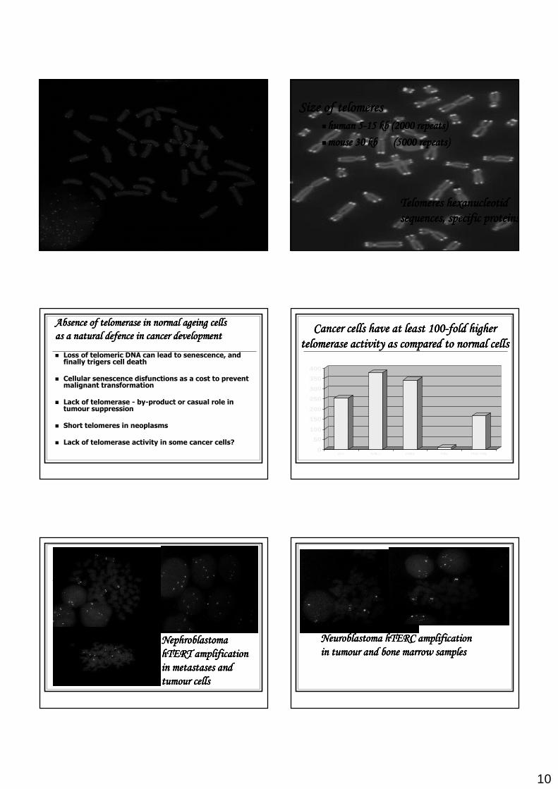
SizeSizeSizeSize ofofofof telomerestelomerestelomerestelomeres
�humanhumanhumanhuman 5555----15 15 15 15 kbkbkbkb (2000 (2000 (2000 (2000 repeatsrepeatsrepeatsrepeats))))
�mousemousemousemouse 33330 0 0 0 kbkbkbkb (5000 (5000 (5000 (5000 repeatsrepeatsrepeatsrepeats))))
TelomeresTelomeresTelomeresTelomeres hexanucleotidhexanucleotidhexanucleotidhexanucleotid
sequencessequencessequencessequences, , , , specificspecificspecificspecific proteinsproteinsproteinsproteins
AbsenceAbsenceAbsenceAbsence ofofofof telomerasetelomerasetelomerasetelomerase inininin normalnormalnormalnormal ageingageingageingageing cellscellscellscells
as a as a as a as a naturalnaturalnaturalnatural defencedefencedefencedefence inininin cancercancercancercancer developmentdevelopmentdevelopmentdevelopment
� Loss of telomeric DNA can lead to senescence, andfinally trigers cell death
� Cellular senescence disfunctions as a cost to preventmalignant transformation
� Lack of telomerase - by-product or casual role intumour suppression
� Short telomeres in neoplasms
� Lack of telomerase activity in some cancer cells? 0
50
100
150
200
250
300
350
400
ALL ANLL K562 PBL PHA-PBL
CancerCancerCancerCancer cells have at least 100cells have at least 100cells have at least 100cells have at least 100----fold higher fold higher fold higher fold higher
telomerase activity telomerase activity telomerase activity telomerase activity asasasas comparcomparcomparcomparedededed to normal cellsto normal cellsto normal cellsto normal cells
NephroblastomaNephroblastomaNephroblastomaNephroblastoma
hTERThTERThTERThTERT amplificationamplificationamplificationamplification
inininin metastasesmetastasesmetastasesmetastases andandandand
tumourtumourtumourtumour cellscellscellscells
NeuroblastomaNeuroblastomaNeuroblastomaNeuroblastoma hTERChTERChTERChTERC amplificationamplificationamplificationamplification
inininin tumourtumourtumourtumour andandandand bonebonebonebone marrowmarrowmarrowmarrow samplessamplessamplessamples
11
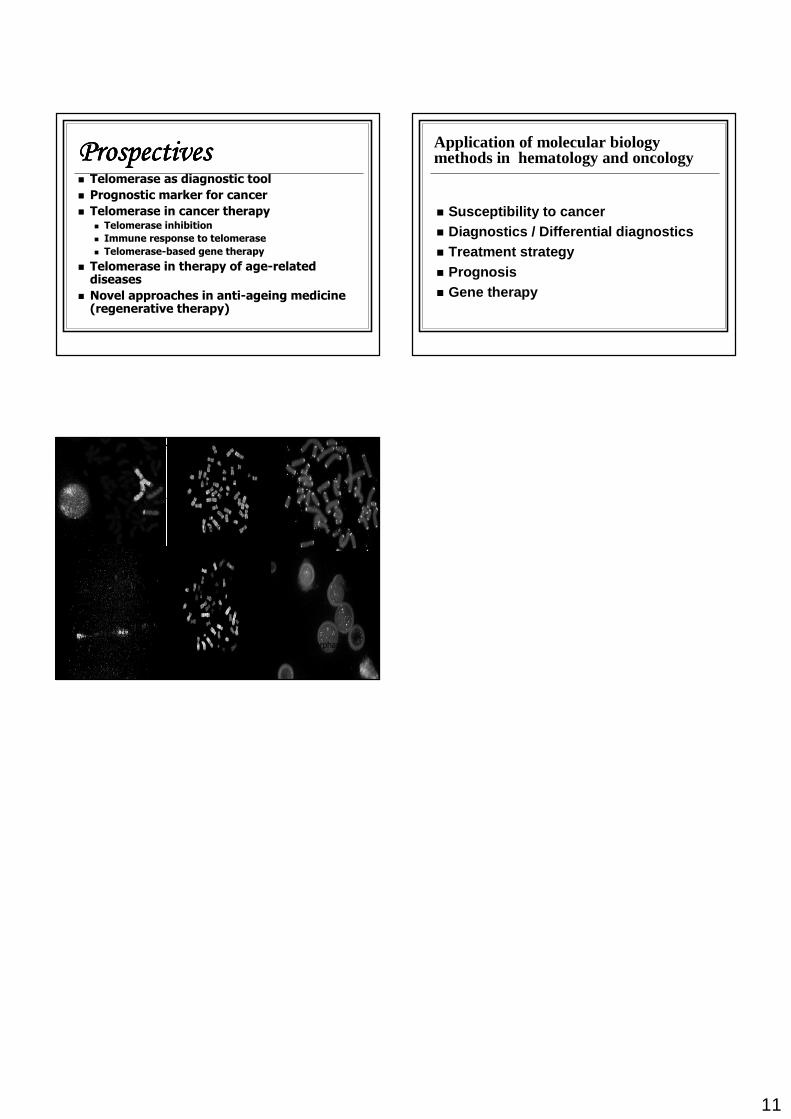
ProspectivesProspectivesProspectivesProspectives� Telomerase as diagnostic tool
� Prognostic marker for cancer
� Telomerase in cancer therapy� Telomerase inhibition
� Immune response to telomerase
� Telomerase-based gene therapy
� Telomerase in therapy of age-relateddiseases
� Novel approaches in anti-ageing medicine(regenerative therapy)
Application of molecular biologymethods in hematology and oncology
� Susceptibility to cancer� Diagnostics / Differential diagnostics� Treatment strategy� Prognosis� Gene therapy
FISH t (1;2)FISH - telomery
Chromatide fiber FISH FISH to interphasenuclei
CGH (ca.rectum)
M-FISH