Embed Size (px)
Citation preview
Molecular BiologyFourth Edition
Chapter 3
An Introduction to Gene Function
Lecture PowerPoint to accompany
Robert F. Weaver
Copyright © The McGraw-Hill Companies, Inc. Permission required for reproduction or display.
3-2
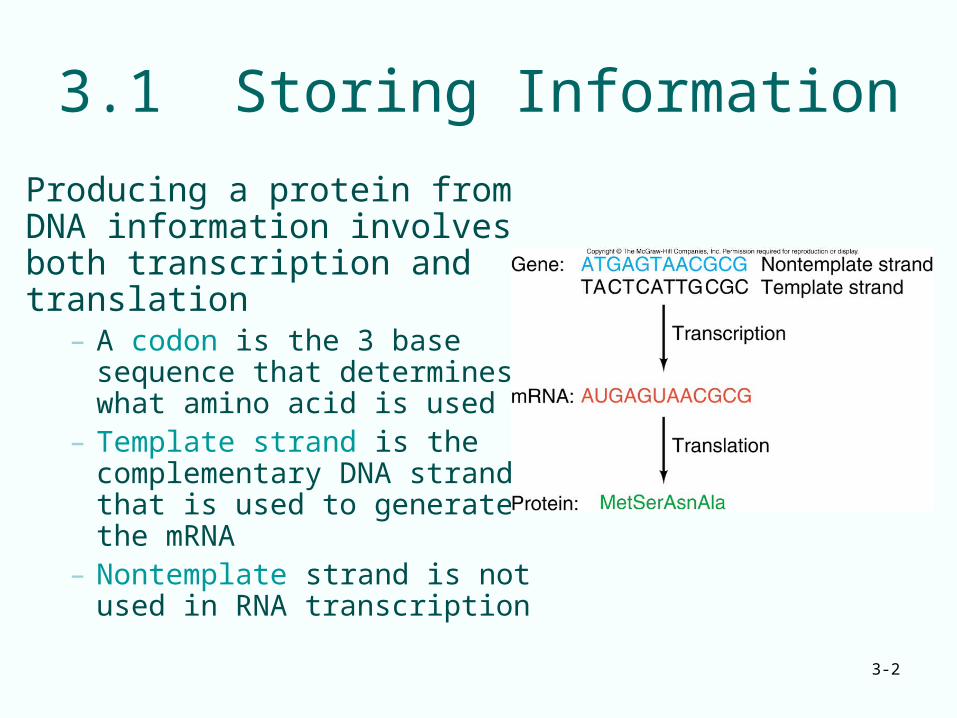
3.1 Storing Information
Producing a protein from DNA information involves both transcription and translation
– A codon is the 3 base sequence that determines what amino acid is used
– Template strand is the complementary DNA strand that is used to generate the mRNA
– Nontemplate strand is not used in RNA transcription
3-3
Protein Structure
Proteins are chain-like polymers of small subunits, amino acids
– DNA has 4 different nucleotides– Proteins have 20 different amino acids with:
• An amino group• A hydroxyl group• A hydrogen atom• A specific side chain
3-4
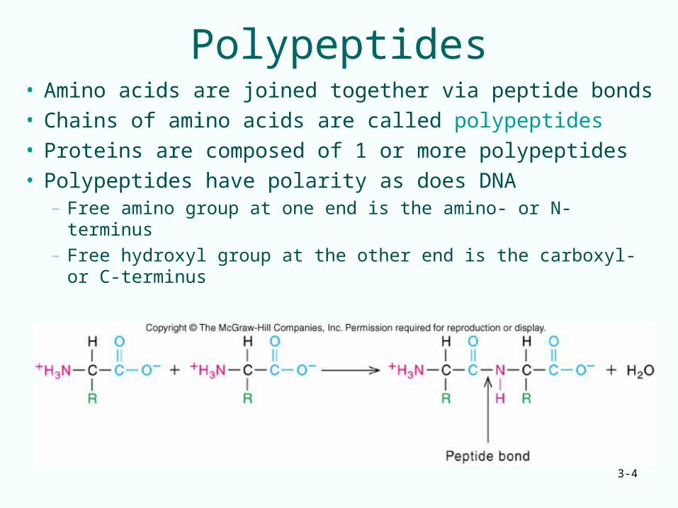
Polypeptides• Amino acids are joined together via peptide bonds• Chains of amino acids are called polypeptides• Proteins are composed of 1 or more polypeptides• Polypeptides have polarity as does DNA
– Free amino group at one end is the amino- or N-terminus– Free hydroxyl group at the other end is the carboxyl- or
C-terminus
3-5
Types of Protein Structure
• Linear order of amino acids is a protein’s primary structure
• Interaction of the amino acids’ amino and carboxyl groups gives rise to the secondary structure of a protein– Secondary structure is the result of amino acid and
carboxyl group hydrogen bonding among near neighbors
– Common types of secondary structure: helix sheet
3-6
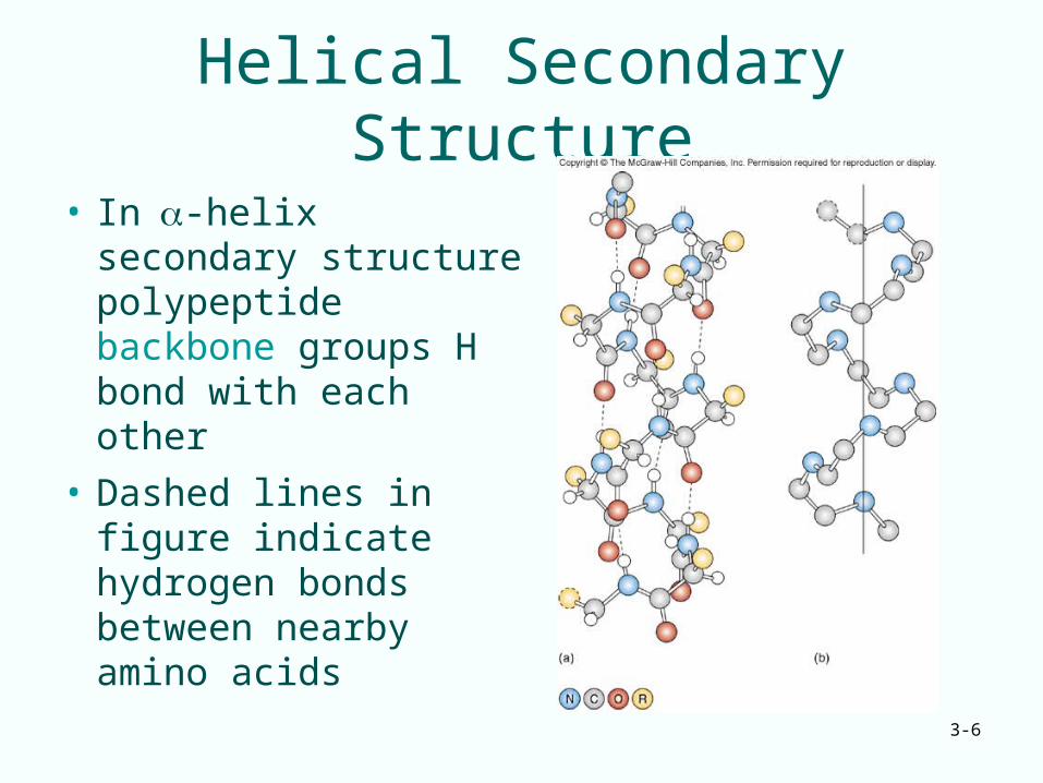
Helical Secondary Structure
• In-helix secondary structure polypeptide backbone groups H bond with each other
• Dashed lines in figure indicate hydrogen bonds between nearby amino acids

3-7
Sheet Secondary Structure
• The -sheet pattern of 2° structure also occurs when polypeptide backbone groups form H bonds
• In the sheet configuration, extended polypeptide chains are packed side by side
• This side-by-side packing creates a sheet appearance
3-8
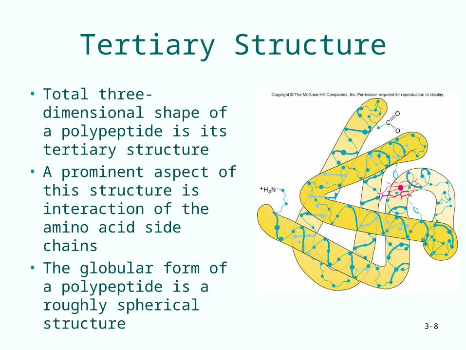
Tertiary Structure
• Total three-dimensional shape of a polypeptide is its tertiary structure
• A prominent aspect of this structure is interaction of the amino acid side chains
• The globular form of a polypeptide is a roughly spherical structure
3-9
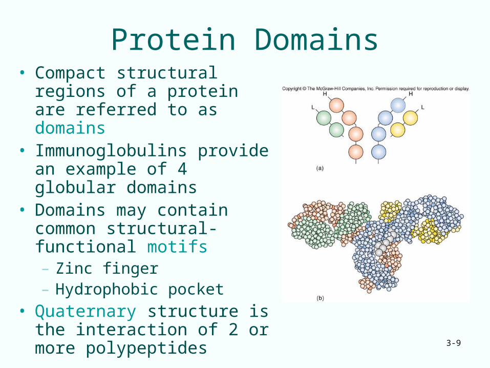
Protein Domains• Compact structural regions of
a protein are referred to as domains
• Immunoglobulins provide an example of 4 globular domains
• Domains may contain common structural-functional motifs – Zinc finger– Hydrophobic pocket
• Quaternary structure is the interaction of 2 or more polypeptides
3-10
Summary• Proteins are polymers of amino acids
linked through peptide bonds
• Sequence of amino acids in a polypeptide (primary structure) gives rise to that molecule’s: – Local shape (secondary structure)– Overall shape (tertiary structure)– Interaction with other polypeptides
(quaternary structure)
3-11
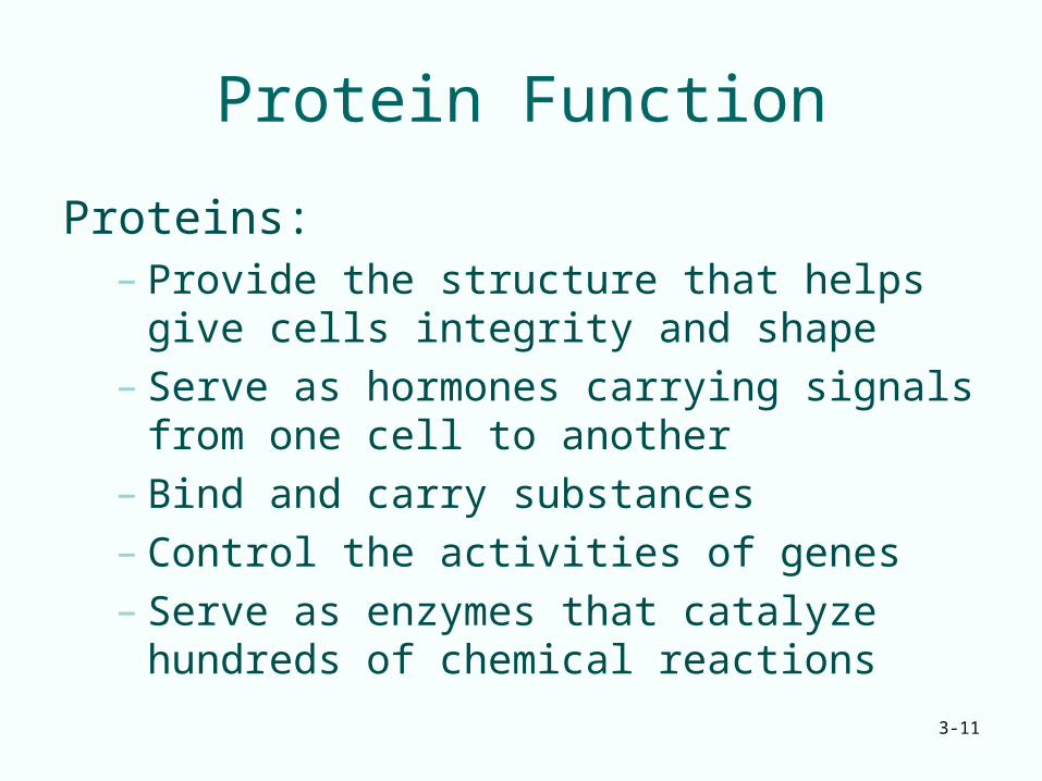
Protein Function
Proteins:– Provide the structure that helps give cells
integrity and shape– Serve as hormones carrying signals from one
cell to another– Bind and carry substances– Control the activities of genes– Serve as enzymes that catalyze hundreds of
chemical reactions
3-12
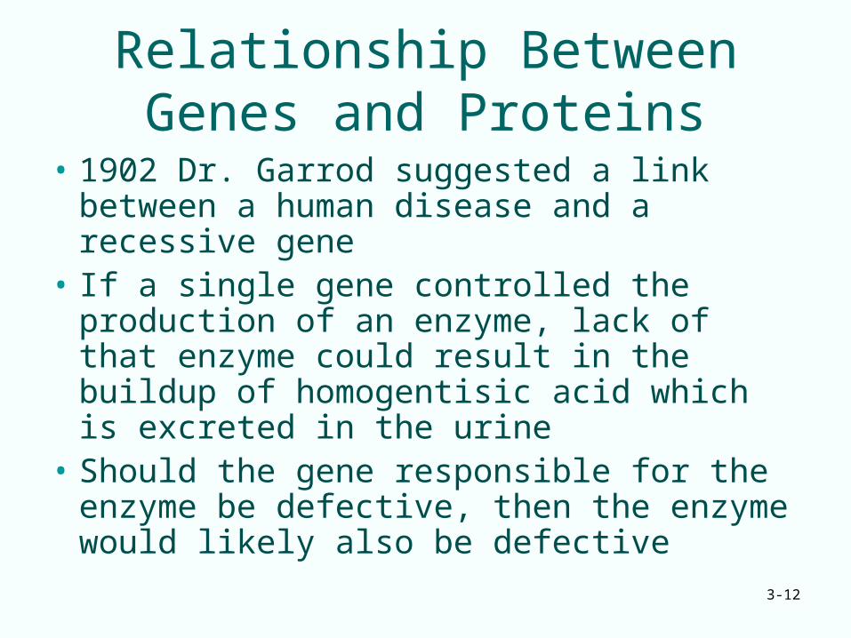
Relationship Between Genes and Proteins
• 1902 Dr. Garrod suggested a link between a human disease and a recessive gene
• If a single gene controlled the production of an enzyme, lack of that enzyme could result in the buildup of homogentisic acid which is excreted in the urine
• Should the gene responsible for the enzyme be defective, then the enzyme would likely also be defective
3-13
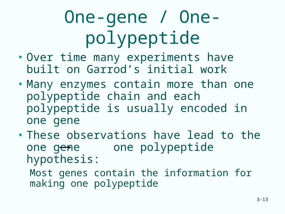
One-gene / One-polypeptide
• Over time many experiments have built on Garrod’s initial work
• Many enzymes contain more than one polypeptide chain and each polypeptide is usually encoded in one gene
• These observations have lead to the one gene one polypeptide hypothesis:Most genes contain the information for making one polypeptide
3-14
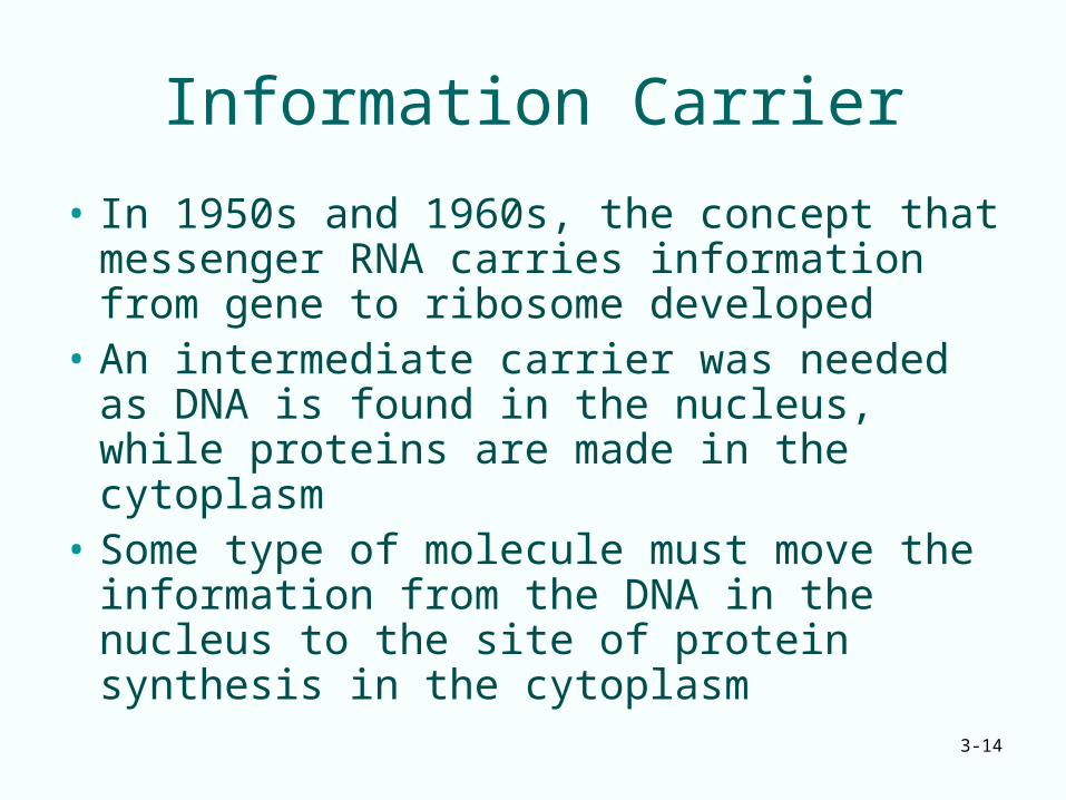
Information Carrier
• In 1950s and 1960s, the concept that messenger RNA carries information from gene to ribosome developed
• An intermediate carrier was needed as DNA is found in the nucleus, while proteins are made in the cytoplasm
• Some type of molecule must move the information from the DNA in the nucleus to the site of protein synthesis in the cytoplasm
3-15
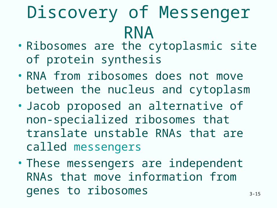
Discovery of Messenger RNA• Ribosomes are the cytoplasmic site of
protein synthesis
• RNA from ribosomes does not move between the nucleus and cytoplasm
• Jacob proposed an alternative of non-specialized ribosomes that translate unstable RNAs that are called messengers
• These messengers are independent RNAs that move information from genes to ribosomes
3-16
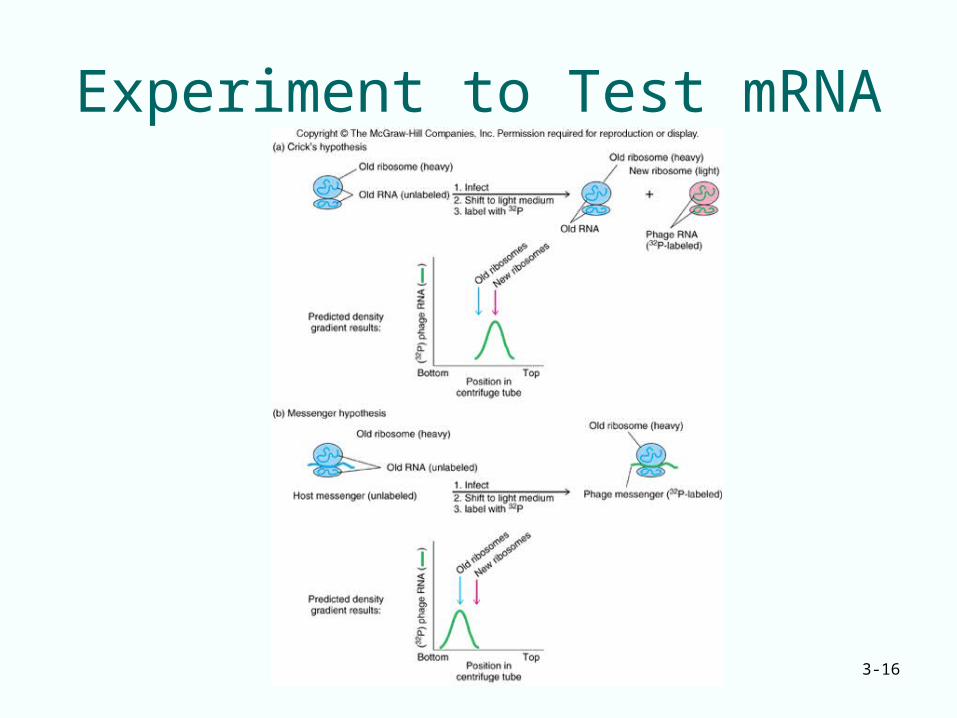
Experiment to Test mRNA
3-17
Crick and Jacob Experiments
• Radio-labeled phage RNA in experiments was found to be associated with old ribosomes whose rRNA was made before infection
• rRNA doesn’t carry information from DNA
• A different class of unstable RNAs associate transiently with ribosomes
3-18
Summary
Messenger RNAs carry the genetic information from the genes to the ribosomes, which then synthesize polypeptides
3-19
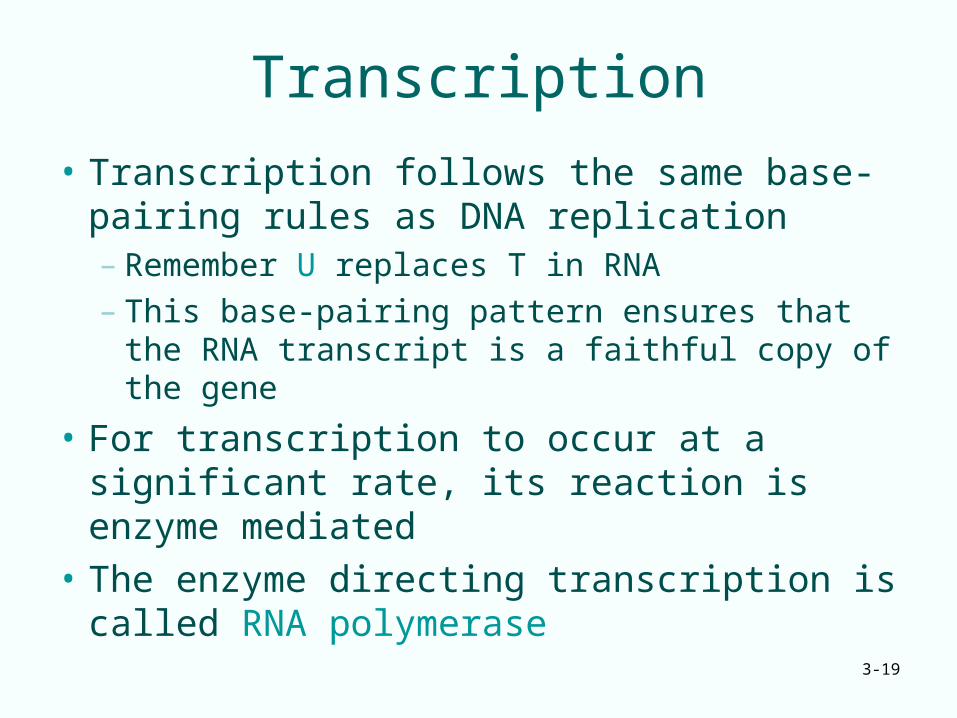
Transcription
• Transcription follows the same base-pairing rules as DNA replication– Remember U replaces T in RNA– This base-pairing pattern ensures that the
RNA transcript is a faithful copy of the gene
• For transcription to occur at a significant rate, its reaction is enzyme mediated
• The enzyme directing transcription is called RNA polymerase
3-20
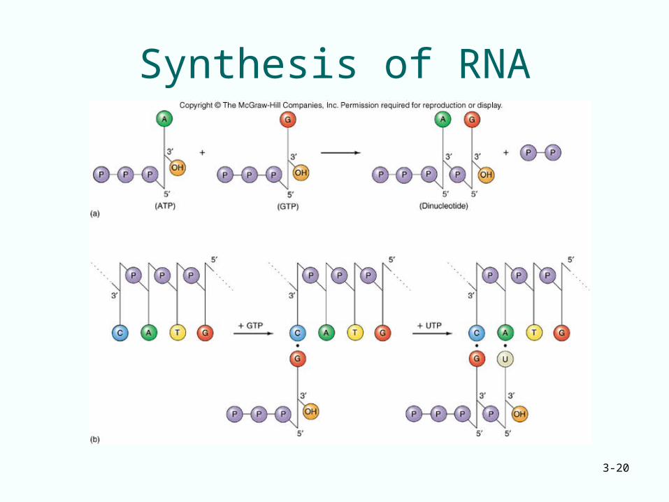
Synthesis of RNA
3-21
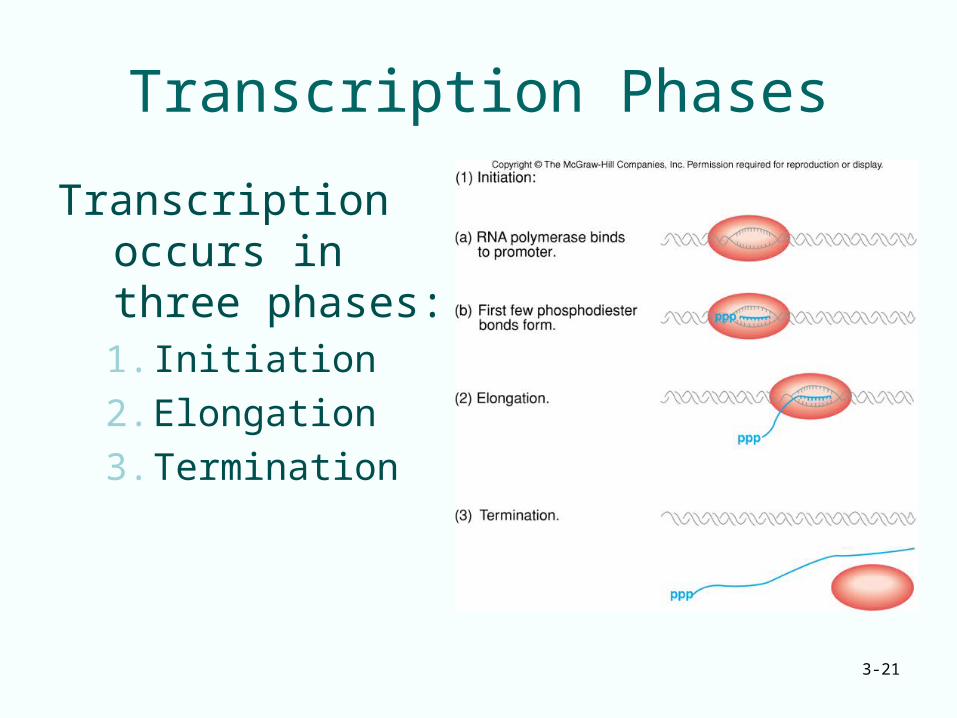
Transcription Phases
Transcription occurs in three phases:1. Initiation
2. Elongation
3. Termination
3-22
Initiation
• RNA polymerase recognizes a region, the promoter, which lies just upstream of gene
• Polymerase binds tightly to promoter causing localized separation of the two DNA strands
• Polymerase starts building the RNA chain adding ribonucleotides
• After several ribonucleotides are joined together the enzyme leaves the promoter and elongation begins
3-23
Elongation
• RNA polymerase directs binding of ribonucleotides in the 5’ to 3’ direction
• Movement of the polymerase along the DNA template causes the “bubble” of separated DNA strands to move also
• As DNA transcription passes, the two DNA strands reform the double helix
3-24
Differences Between Transcription and DNA Replication
There are two fundamental differences between transcription and DNA replication
1. RNA polymerase only makes one RNA strand during transcription, it copies only one DNA strand in a given gene– This makes transcription asymmetrical– Replication is semiconservative
2. DNA melting is limited and transient during transcription, but the separation is permanent in replication
3-25
Termination
• Analogous to the initiating activity of promoters, there are regions at the other end of genes that serve to terminate transcription
• These terminators work with the RNA polymerase to loosen the association between RNA product and DNA template
• As a result, the RNA dissociates from the RNA polymerase and the DNA and transcription stops
3-26
Transcription Landmarks• RNA sequences are written 5’ to 3’, left to
right
• Translation occurs 5’ to 3’ with ribosomes reading the message 5’ to 3’
• Genes are written so that transcription proceeds from left to right
• The gene’s promoter area lies just before the start area, said to be upstream of transcription
• Genes are therefore said to lie downstream of their promoters
3-27
Summary• Transcription takes place in three stages:
– Initiation– Elongation– Termination
• Initiation involves binding RNA polymerase to the promoter, local melting and forming the first few phosphodiester bonds
• During elongation, the RNA polymerase links together ribonucleotides in the 5’ to 3’ direction to make the rest of the RNA
• In termination, the polymerase and RNA product dissociate from the DNA template
3-28
Translation - Ribosomes
• Ribosomes are the protein synthesizing machines– Ribosome subunits are designated with
numbers such as 50S or 30S– Number is the sedimentation coefficient - a
measure of speed with which the particles sediment through a solution spun in an ultracentrifuge
• Each ribosomal subunit contains RNA and protein
3-29
Ribosomal RNA
• The two ribosomal subunits both contain ribosomal RNA (rRNA) molecules and a variety of proteins
• rRNAs participate in protein synthesis but do NOT code for proteins
• No translation of rRNA occurs
3-30
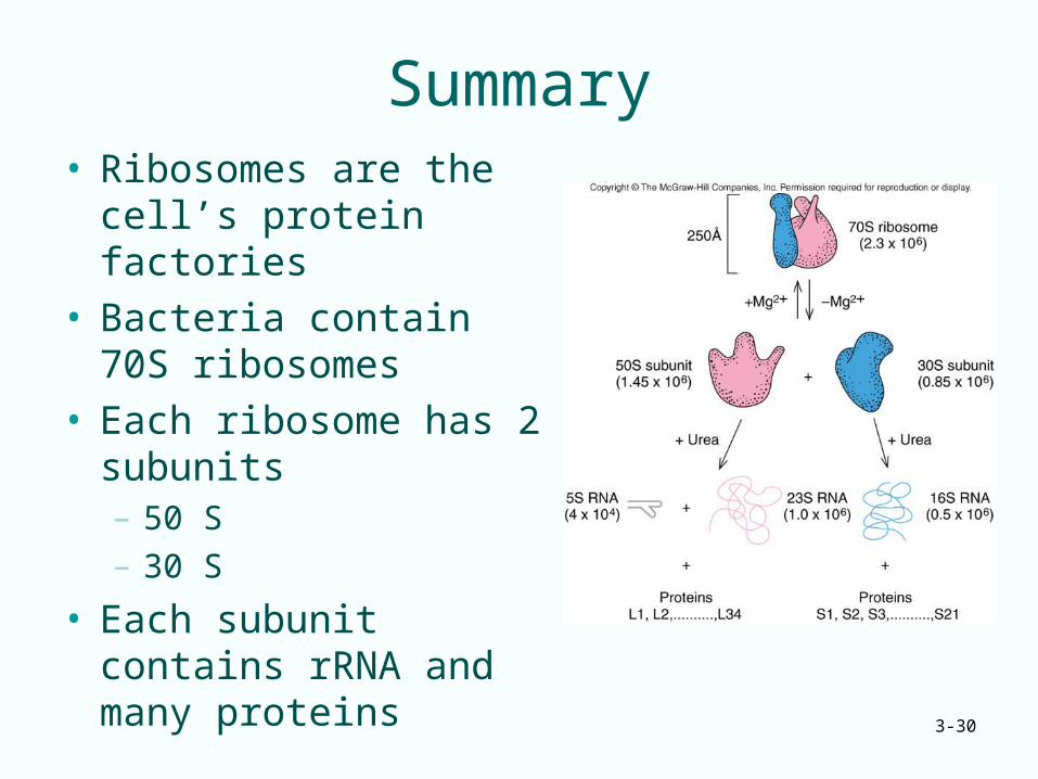
Summary• Ribosomes are the cell’s
protein factories• Bacteria contain 70S
ribosomes• Each ribosome has 2
subunits– 50 S– 30 S
• Each subunit contains rRNA and many proteins
3-31
Translation Adapter Molecule
• Generating protein from ribosomes requires change from the nucleic acid to amino acid
• This change is described as translation from the nucleic acid base pair language to the amino acid language
• Crick proposed that some type of adapter molecule was needed to provide the bridge for translation, perhaps a small RNA
3-32
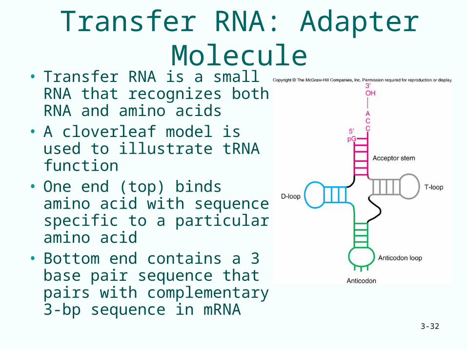
Transfer RNA: Adapter Molecule• Transfer RNA is a small
RNA that recognizes both RNA and amino acids
• A cloverleaf model is used to illustrate tRNA function
• One end (top) binds amino acid with sequence specific to a particular amino acid
• Bottom end contains a 3 base pair sequence that pairs with complementary 3-bp sequence in mRNA
3-33
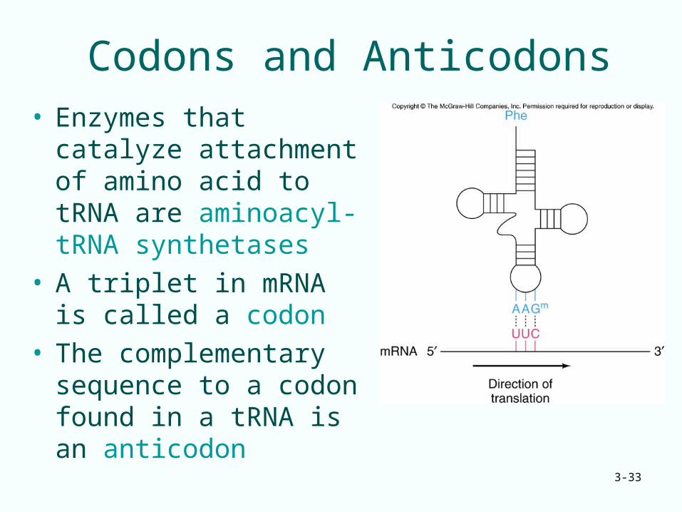
Codons and Anticodons• Enzymes that catalyze
attachment of amino acid to tRNA are aminoacyl-tRNA synthetases
• A triplet in mRNA is called a codon
• The complementary sequence to a codon found in a tRNA is an anticodon
3-34
Summary
• Two important sites on tRNAs allow them to recognize both amino acids and nucleic acids
• One site binds covalently to an amino acid• The site contains an anticodon the base-
pairs with a 3-bp codon in mRNA• The tRNAs are capable of serving the
adapter role postulated by Crick and are the key to the mechanism of translation
3-35
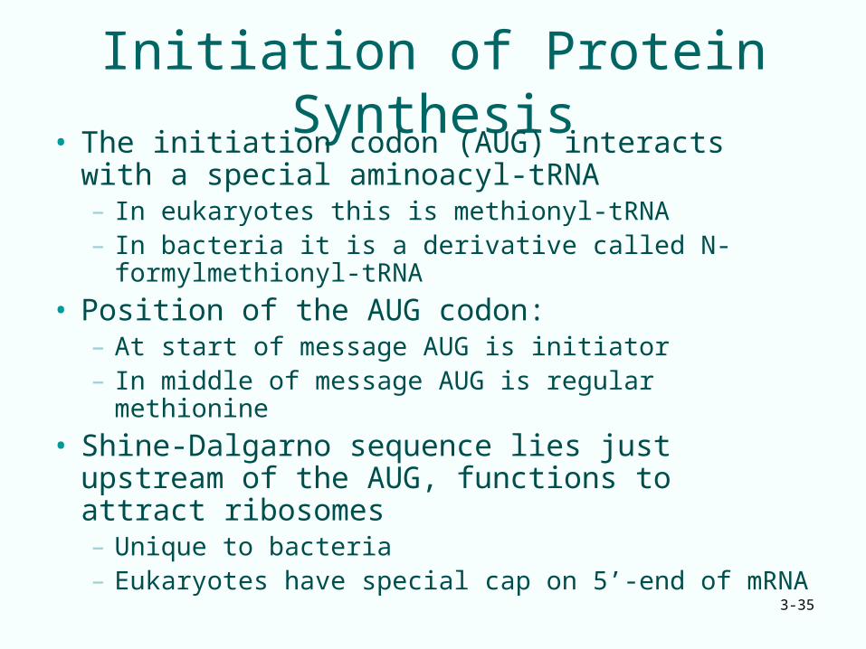
Initiation of Protein Synthesis• The initiation codon (AUG) interacts with a
special aminoacyl-tRNA– In eukaryotes this is methionyl-tRNA– In bacteria it is a derivative called N-formylmethionyl-
tRNA
• Position of the AUG codon:– At start of message AUG is initiator– In middle of message AUG is regular methionine
• Shine-Dalgarno sequence lies just upstream of the AUG, functions to attract ribosomes– Unique to bacteria– Eukaryotes have special cap on 5’-end of mRNA
3-36
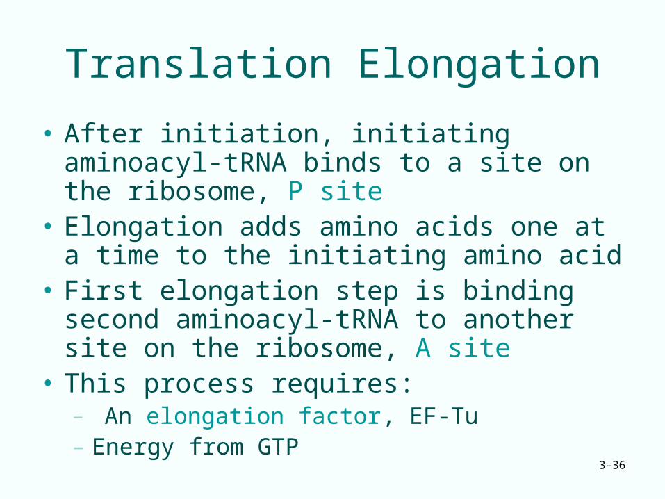
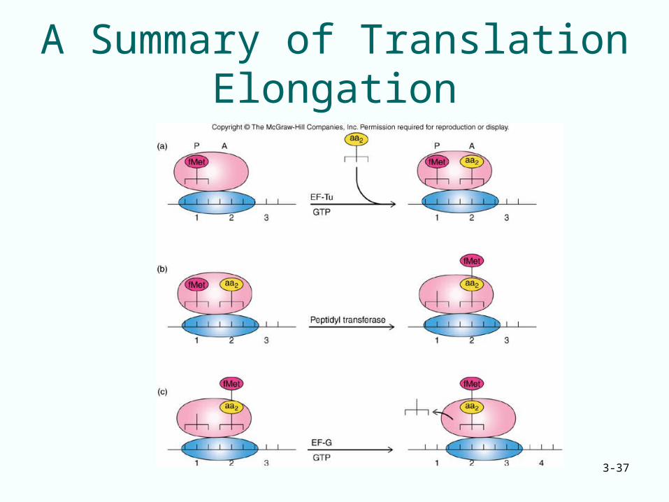
Translation Elongation
• After initiation, initiating aminoacyl-tRNA binds to a site on the ribosome, P site
• Elongation adds amino acids one at a time to the initiating amino acid
• First elongation step is binding second aminoacyl-tRNA to another site on the ribosome, A site
• This process requires:– An elongation factor, EF-Tu– Energy from GTP
3-37
A Summary of Translation Elongation
3-38
Termination of Translation and mRNA Structure
• Three different codons (UAG, UAA, UGA) cause translation termination
• Proteins called release factors recognize these stop codons causing– Translation to stop– Release of the polypeptide chain
• Initiation codon and termination codon at the ends define an open reading frame (ORF)
3-39
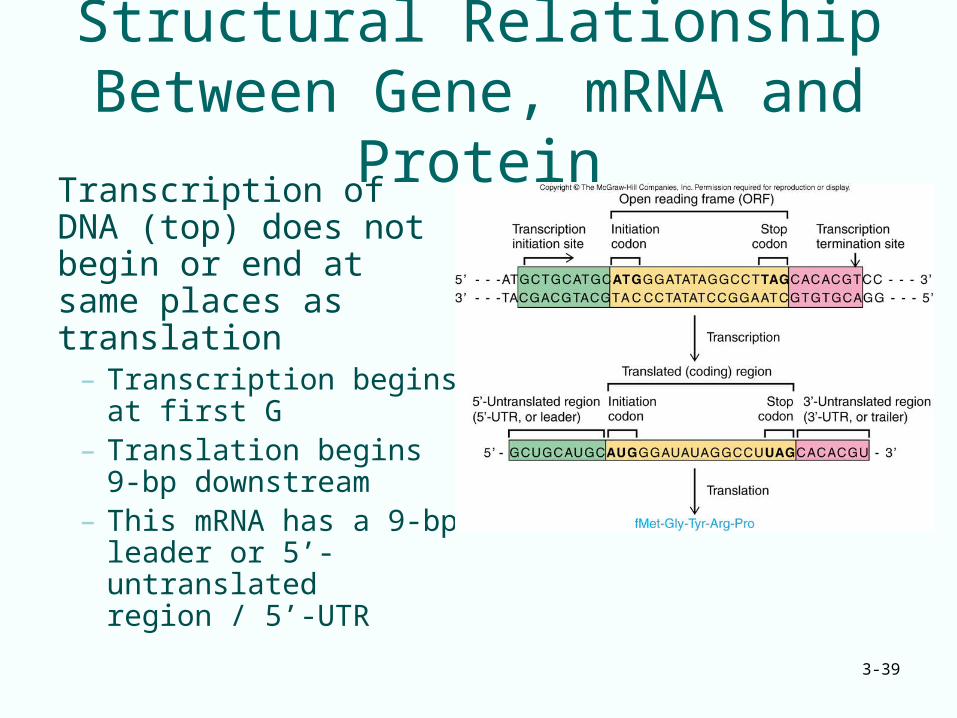
Structural Relationship Between Gene, mRNA and Protein
Transcription of DNA (top) does not begin or end at same places as translation
– Transcription begins at first G
– Translation begins 9-bp downstream
– This mRNA has a 9-bp leader or 5’-untranslated region / 5’-UTR
3-40
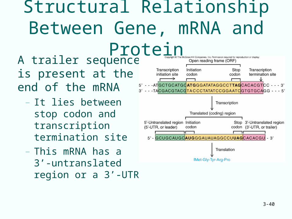
Structural Relationship Between Gene, mRNA and Protein
A trailer sequence is present at the end of the mRNA
– It lies between stop codon and transcription termination site
– This mRNA has a 3’-untranslated region or a 3’-UTR
3-41
3.2 Replication
• Genes replicate faithfully
• Semiconservative replication produces new DNA with each daughter double helix having 1 parental strand and one new strand
3-42
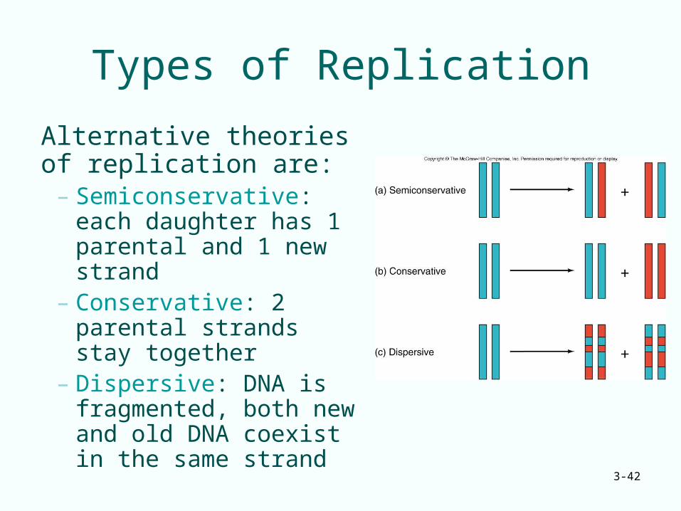
Types of Replication
Alternative theories of replication are:
– Semiconservative: each daughter has 1 parental and 1 new strand
– Conservative: 2 parental strands stay together
– Dispersive: DNA is fragmented, both new and old DNA coexist in the same strand
3-43
3.3 Mutations
• Genes accumulate changes or mutations• Mutation is essential for evolution• If a nucleotide in a gene changes, likely a
corresponding change will occur in an amino acid of that gene’s protein product– If a mutation results in a different codon for
the same amino acid it is a silent mutation– Often a new amino acid is structurally similar
to the old and the change is conservative
3-44
Sickle Cell Disease• Sickle cell disease is a genetic disorder• The disease results from a single base
change in the gene for -globin– Altered base causes insertion an incorrect
amino acid into one position of the -globin protein
– Altered protein results in distortion of red blood cells under low-oxygen conditions
• This disease illustrates that a change in a gene can cause corresponding change in the protein product of the gene
3-45
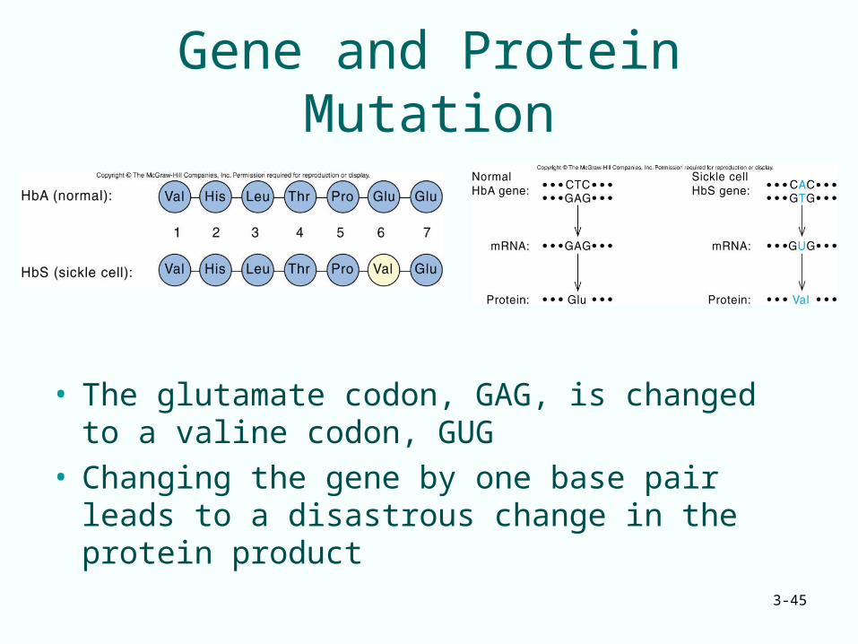
Gene and Protein Mutation
• The glutamate codon, GAG, is changed to a valine codon, GUG
• Changing the gene by one base pair leads to a disastrous change in the protein product