Embed Size (px)
Citation preview
TrendsPrecisely controlling the dose of MBsdelivered into live cells, which allowsquantitative analysis at single-cell reso-lution, is becoming increasingly feasible.
The design of MBs is moving towardsincreased sensitivity and longer-lastingresistance to nucleases and otherenzymes.
The ‘specificity’ parameter is particu-larly important for prognostic bio-chipsand in vivo imaging. MB designs thataddress single-nucleotide mutations
ReviewMolecular BeaconNano-Sensors for ProbingLiving Cancer CellsTairong Kuang,1,2,z Lingqian Chang,3,z,* Xiangfang Peng,1
Xianglong Hu,4,* and Daniel Gallego-Perez3,5,6
Heterogeneities and oncogenesis essentially result from proteomic disordersorchestrated by changes in DNA and/or cytoplasmic mRNA. These geneticfluctuations, however, cannot be decoded through conventional label-freemethods (e.g., patch clamps, electrochemical cellular biosensors, etc.) or mor-phological characterization. Molecular beacons (MBs) have recently emergedas efficient probes for interrogating biomarkers in live cancer cells. MBs hybrid-ize with their intracellular targets (e.g., mRNAs, DNAs, or proteins), emitting afluorescent signal that can be quantified and correlated with the expressionlevels of their targets. In this reviewwe discussMBprobeswith different deliveryplatforms for intracellular probing as well as novel MB designs for detecting avariety of targets in living cancer cells. Finally, we describe current trends inMB-based intracellular biosensors.
and that can detect multiple targetsare becoming popular.
1National Engineering Research Centerof Novel Equipment for PolymerProcessing, The Key Laboratory ofPolymer Processing Engineering ofMinistry of Education, South ChinaUniversity of Technology, Guangzhou,510640, China2Department of Chemical andBiomolecular Engineering, The OhioState University, Columbus, OH43210, USA3Department of BiomedicalEngineering, The Ohio StateUniversity, Columbus, OH 43210, USA4Ministry of Education Key Laboratoryof Laser Life Science and Institute ofLaser Life Science, College ofBiophotonics, South China NormalUniversity, Guangzhou 510631, China5Department of Surgery, The OhioState University, Columbus, OH43210, USA6Mission CAIRRS (Consortium forAdvancements and Innovations inRestorative and RegenerativeStrategies), Center for RegenerativeMedicine and Cell-Based Therapies,
Challenges in Probing Living Cancer CellsThe living cell contains [62_TD$DIFF] a sophisticated machinery that is responsive to both intracellular andextracellular stimuli. Cellular functions, modulated by nuclear DNA, cytosolic mRNA, andproteins, play a major role in determining cellular proliferation, differentiation, oncogenesis,apoptosis, etc. Decoding genetic information is crucial for gene therapy (e.g., gene editingand adoptive immunotherapy) and cancer diagnosis (e.g., gene mutation and intracellularheterogeneities of cancer stem-like cells) [1–7]. Over the past few decades efforts have beendevoted to probing intracellular components (e.g., DNA, mRNA, and proteins). Well-knowntechniques such as PCR, DNA microarrays, western blotting, and ELISA have demonstratedhigh resolution for quantitatively analyzing the exact copy number of genes [8–12]. However,these methods are not compatible with live-cell bio-interrogation, and [11_TD$DIFF] therefore have limitedimpact in early cancer detection (e.g., for circulating tumor cells) or in precision/personalizedmedicine. Label-free biosensors are among the commonest tools for monitoring live cells. Inparticular, advances in the fields of micro-/nanofabrication (top-down) and biomolecularsynthesis (bottom-up) have remarkably boosted the sensitivity and specificity of cellularsensors [13]. However, their implementation for gene-based probing remains challenging[14].
MBs have emerged as promising tools for live-cell interrogation with single-molecule resolution[15–17]. MBs are single-stranded (ss) oligonucleotides that form a hairpin structure that confinesa fluorophore and quencher in close proximity such that no fluorescence signal is emitted. Oncethey are delivered into the cytosol, the MBs seek their target molecules (e.g., mRNAs), hybridize(via the complementary binding of the MB on the coding sequence (CDS) of mRNA), and emit afluorescence signal via the controlled separation of the quencher and fluorophore. Precisetitration of MB delivery, for example, can quantify mRNA expression based on fluorescence
Trends in Biotechnology, April 2017, Vol. 35, No. 4 http://dx.doi.org/10.1016/j.tibtech.2016.09.003 347© 2016 Elsevier Ltd. All rights reserved.
The Ohio State University, Columbus,OH 43210, USAzThese authors contributed equally tothis work.
*Correspondence: [email protected](L. Chang) and [email protected] (X.Hu).
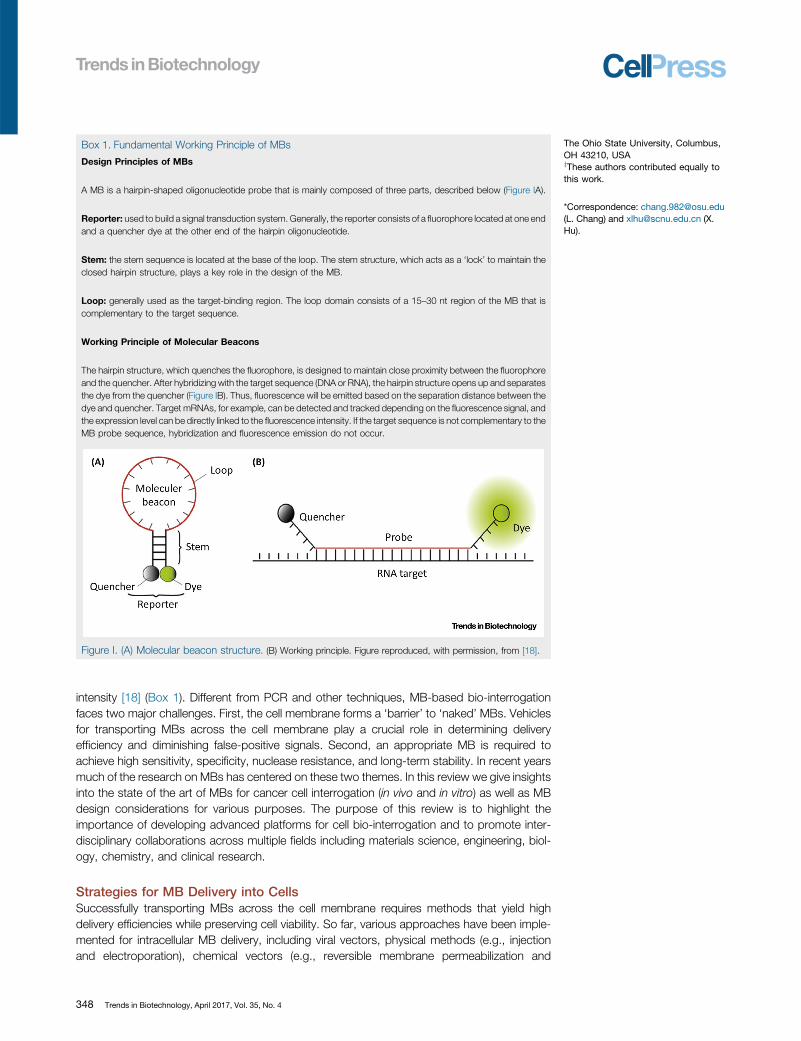
Box 1. Fundamental Working [47_TD$DIFF]Principle of MBs
Design Principles of MBs
[48_TD$DIFF]A MB is a [2_TD$DIFF] hairpin-shaped oligonucleotide probe that is mainly composed of three parts, described below (Figure IA).
Reporter: used to build a signal transduction system. Generally, the reporter consists of a fluorophore located at one endand a quencher dye at the other end of the hairpin oligonucleotide.
Stem: [49_TD$DIFF]the stem sequence[50_TD$DIFF] is located at the base of the loop. The stem structure, which acts as a ‘lock’ to maintain theclosed hairpin structure, plays a key role in the design of the MB.
Loop: generally used as the target-binding region. The loop domain consists of a 15–30 nt region of the MB that iscomplementary to the target sequence.
Working Principle of Molecular Beacons
The hairpin structure, which quenches the fluorophore, is designed [51_TD$DIFF]to maintain close proximity between the fluorophoreand the quencher. After hybridizing with the target sequence (DNA or RNA), the hairpin structure opens up and separatesthe [4_TD$DIFF] dye from the quencher [5_TD$DIFF] (Figure IB). Thus, fluorescence will be emitted [52_TD$DIFF]based [53_TD$DIFF]on the [54_TD$DIFF] separation distance between the [4_TD$DIFF]dye and quencher[5_TD$DIFF]. [55_TD$DIFF]Target [56_TD$DIFF]mRNAs, [57_TD$DIFF]for example, can be detected and tracked depending on the fluorescence signal [6_TD$DIFF], andthe expression level [7_TD$DIFF] can be [58_TD$DIFF]directly [59_TD$DIFF]linked [60_TD$DIFF]to the [1_TD$DIFF] fluorescence intensity. If the target[8_TD$DIFF] sequence is not [9_TD$DIFF] complementary to theMB probe sequence, hybridization and fluorescence [61_TD$DIFF] emission do not occur.
Figure I. (A) Molecular beacon structure. (B) [39_TD$DIFF]Working principle. Figure reproduced, with permission, from [18].
intensity [18] (Box 1). Different from PCR and other techniques, MB-based bio-interrogationfaces two major challenges. First, the cell membrane forms a ‘barrier’ to ‘naked’ MBs. Vehiclesfor transporting MBs across the cell membrane play a crucial role in determining deliveryefficiency and diminishing false-positive signals. Second, an appropriate MB is required toachieve high sensitivity, specificity, nuclease resistance, and long-term stability. In recent yearsmuch of the research on MBs has centered on these two themes. In this review we give insightsinto the state of the art of MBs for cancer cell interrogation (in vivo and in vitro) as well as MBdesign considerations for various purposes. The purpose of this review is to highlight theimportance of developing advanced platforms for cell bio-interrogation and to promote inter-disciplinary collaborations across multiple fields including materials science, engineering, biol-ogy, chemistry, and clinical research.
Strategies for MB Delivery into CellsSuccessfully transporting MBs across the cell membrane requires methods that yield highdelivery efficiencies while preserving cell viability. So far, various approaches have been imple-mented for intracellular MB delivery, including viral vectors, physical methods (e.g., injectionand electroporation), chemical vectors (e.g., reversible membrane permeabilization and
348 Trends in Biotechnology, April 2017, Vol. 35, No. 4
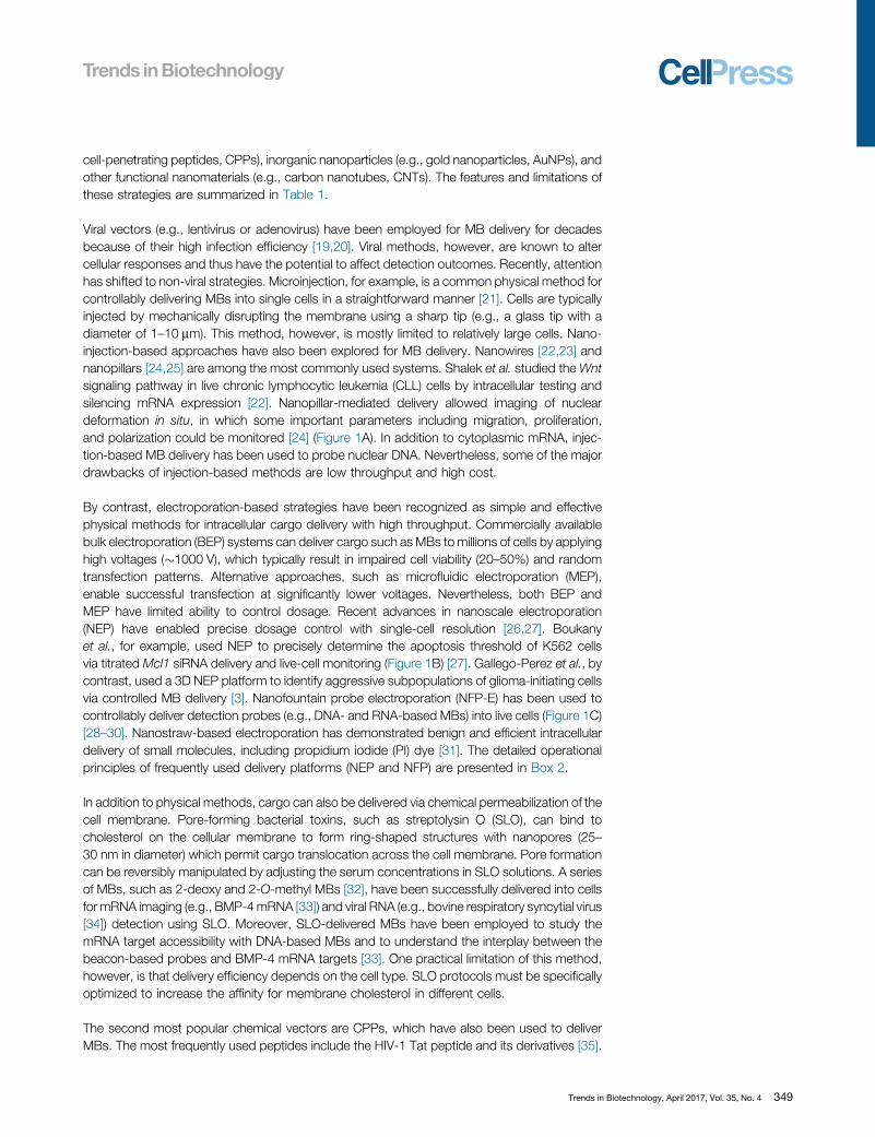
cell-penetrating peptides, CPPs), inorganic nanoparticles (e.g., gold nanoparticles, AuNPs), andother functional nanomaterials (e.g., carbon nanotubes, CNTs). The features and limitations ofthese strategies are summarized in Table 1.
Viral vectors (e.g., lentivirus or adenovirus) have been employed for MB delivery for decadesbecause of their high infection efficiency [19,20]. Viral methods, however, are known to altercellular responses and thus have the potential to affect detection outcomes. Recently, attentionhas shifted to non-viral strategies. Microinjection, for example, is a common physical method forcontrollably delivering MBs into single cells in a straightforward manner [21]. Cells are typicallyinjected by mechanically disrupting the membrane using a sharp tip (e.g., a glass tip with adiameter of 1–10 mm). This method, however, is mostly limited to relatively large cells. Nano-injection-based approaches have also been explored for MB delivery. Nanowires [22,23] andnanopillars [24,25] are among the most commonly used systems. Shalek et al. studied theWntsignaling pathway in live chronic lymphocytic leukemia (CLL) cells by intracellular testing andsilencing mRNA expression [22]. Nanopillar-mediated delivery allowed imaging of nucleardeformation in situ, in which some important parameters including migration, proliferation,and polarization could be monitored [24] (Figure 1A). In addition to cytoplasmic mRNA, injec-tion-based MB delivery has been used to probe nuclear DNA. Nevertheless, some of the majordrawbacks of injection-based methods are low throughput and high cost.
By contrast, electroporation-based strategies have been recognized as simple and effectivephysical methods for intracellular cargo delivery with high throughput. Commercially availablebulk electroporation (BEP) systems can deliver cargo such as MBs tomillions of cells by applyinghigh voltages (�1000 V), which typically result in impaired cell viability (20–50%) and randomtransfection patterns. Alternative approaches, such as microfluidic electroporation (MEP),enable successful transfection at significantly lower voltages. Nevertheless, both BEP andMEP have limited ability to control dosage. Recent advances in nanoscale electroporation(NEP) have enabled precise dosage control with single-cell resolution [26,27]. Boukanyet al., for example, used NEP to precisely determine the apoptosis threshold of K562 cellsvia titrated Mcl1 siRNA delivery and live-cell monitoring (Figure 1B) [27]. Gallego-Perez et al., [63_TD$DIFF]by[64_TD$DIFF]contrast, used a 3D NEP platform to identify aggressive subpopulations of glioma-initiating cellsvia controlled MB delivery [3] [13_TD$DIFF]. Nanofountain probe electroporation (NFP-E) has been used tocontrollably deliver detection probes (e.g., DNA- and RNA-based MBs) into live cells (Figure 1C)[28–30]. Nanostraw-based electroporation has demonstrated benign and efficient intracellulardelivery of small molecules, including propidium iodide (PI) dye [31]. The detailed operationalprinciples of frequently used delivery platforms (NEP and NFP) are presented in Box 2.
In addition to physical methods, cargo can also be delivered via [65_TD$DIFF]chemical [66_TD$DIFF]permeabilization of thecell membrane. Pore-forming bacterial toxins, such as streptolysin O (SLO), can bind tocholesterol on the cellular membrane to form ring-shaped structures with nanopores (25–30 nm in diameter) which permit cargo translocation across the cell membrane. Pore formationcan be reversibly manipulated by adjusting the serum concentrations in SLO solutions. A seriesof MBs, such as 2-deoxy and 2-O-methyl MBs [32], have been successfully delivered into cellsfor mRNA imaging (e.g., BMP-4mRNA [33]) and viral RNA (e.g., bovine respiratory syncytial virus[34]) detection using SLO. Moreover, SLO-delivered MBs have been employed to study themRNA target accessibility with DNA-based MBs and to understand the interplay between thebeacon-based probes and BMP-4 mRNA targets [33]. One practical limitation of this method,however, is that delivery efficiency depends on the cell type. SLO protocols must be specificallyoptimized to increase the affinity for membrane cholesterol in different cells.
The second most popular chemical vectors are CPPs, which have also been used to deliverMBs. The most frequently used peptides include the HIV-1 Tat peptide and its derivatives [35].
Trends in Biotechnology, April 2017, Vol. 35, No. 4 349
Table 1. Currently Available Techniques for MB Deliverya,b
Delivery Platform Typical [40_TD$DIFF]Platform Detected [41_TD$DIFF]Cells Efficiencyc[40_TD$DIFF] Viability Throughput Features Limitations Refs
Viral vectors AAV2 HeLa High High High High transductionefficiency
Oncogenesis; safetyconcern; side-effectsin vivo
[19,20]
Physicalvectors
Injection Microinjection Micropipettes PtK2 High Medium Low Direct method; dosecontrol; destination control
Costly equipment;low throughput
[21]
Nanoinjection [42_TD$DIFF]Nanowires;Nanopillars
HeLa, MCF-7,fibroblast
High High Low Direct injection; benignpenetration; dose control
Costly equipment;low throughput
[22–25]
Electroporation Bulkelectroporation
BEP N/A N/A Low High Simple; straightforward Harsh environment;low viability; randomtransfection
[26,27]
Microporation MEP NIH/3T3 Medium High N/A Benign environment Random delivery; limiteddose control; limited inability to deliver largecargo/molecules
[26,27]
Nanoporation NEP KG1a, A549 High High Low (2D NEP);high (3D NEP)
Instantaneous delivery;benign transfection;dose control
Complicated on-[43_TD$DIFF]chipcell manipulation
[3,26,27]
NFP-E HeLa, HT1080 High High Low Benign environment Low throughput [28–30]
Chemicalvectors
Reversiblepermeabilization
SLO HDF, HeLa High High High Convenient; less damageto cells; targeted delivery(nucleus)
Limited to[44_TD$DIFF] certain cell[45_TD$DIFF]types; long incubationtime
[32–34]
Cell-penetratingpeptides
HIV-1 Tat peptide HeLa, fibroblast High N/A High Simplicity and low cost;fast delivery
Complex process(covalent conjugation);toxicity/side-effects forin vivo applications
[35,36]
Liposome Oligofectamine H460, fibroblast,HEK293T
Medium High High High-throughput; benigncarrier
Slow delivery process;MB degradation in[46_TD$DIFF]endosomes
[37,38]
Dendrimer Superfect SK-Hep1 Medium High High High-throughput;benign carrier
[39]
350Trend
sin
Biotechnology,A
pril2017,V
ol.35,No.4
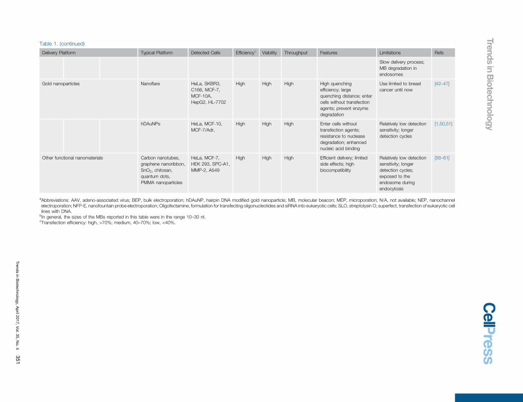
Table 1. (continued)
Delivery Platform Typical [40_TD$DIFF]Platform Detected [41_TD$DIFF]Cells Efficiencyc[40_TD$DIFF] Viability Throughput Features Limitations Refs
Slow delivery process;MB degradation in[46_TD$DIFF]endosomes
Gold nanoparticles Nanoflare HeLa, SKBR3,C166, MCF-7,MCF-10A,HepG2, HL-7702
High High High High quenchingefficiency; largequenching distance; entercells without transfectionagents; prevent enzymedegradation
Use limited to breastcancer until now
[42–47]
hDAuNPs HeLa, MCF-10,MCF-7/Adr,
High High High Enter cells withouttransfection agents;resistance to nucleasedegradation; enhancednucleic acid binding
Relatively low detectionsensitivity; longerdetection cycles
[1,50,51]
Other functional nanomaterials Carbon nanotubes,graphene nanoribbon,SnO2, chitosan,quantum dots,PMMA nanoparticles
HeLa, MCF-7,HEK 293, SPC-A1,MMP-2, A549
High High High Efficient delivery; limitedside effects; highbiocompatibility
Relatively low detectionsensitivity; longerdetection cycles;exposed to theendosome duringendocytosis
[56–61]
aAbbreviations: AAV, adeno-associated virus; BEP, bulk electroporation; hDAuNP, hairpin DNA modified gold nanoparticle; MB, molecular beacon; MEP, microporation; N/A, not available; NEP, nanochannelelectroporation; NFP-E, nanofountain probe electroporation; Oligofectamine, formulation for transfecting oligonucleotides and siRNA into eukaryotic cells; SLO, streptolysin O; superfect, transfection of eukaryotic celllines with DNA.
bIn general, the sizes of the MBs reported in this table were in the range 10–30 nt.cTransfection efficiency: high, >70%; medium, 40–70%; low, <40%.
Trendsin
Biotechnology,A
pril2017,V
ol.35,No.4
351
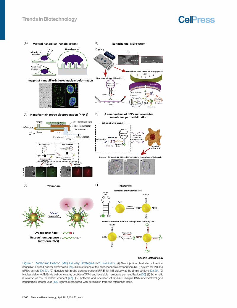
Figure 1. Molecular Beacon (MB) Delivery Strategies into Live Cells. (A) Nanoinjection: illustration of verticalnanopillar-induced nuclear deformation [24]. (B) Illustrations of the nanochannel electroporation (NEP) system for MB andsiRNA delivery [26,27]. (C) Nanofountain probe electroporation (NFP-E) for MB delivery at the single-cell level [28,29]. (D)Nuclear delivery of MBs via cell-penetrating peptides (CPPs) and reversible membrane permeabilization [36]. (E) Schematicillustration of the ‘nanoflare’ concept [47]. (F) Synthesis and operation of hDAuNP (hairpin DNA-functionalized goldnanoparticle)-based MBs [49]. Figures reproduced with permission from the references listed.
352 Trends in Biotechnology, April 2017, Vol. 35, No. 4
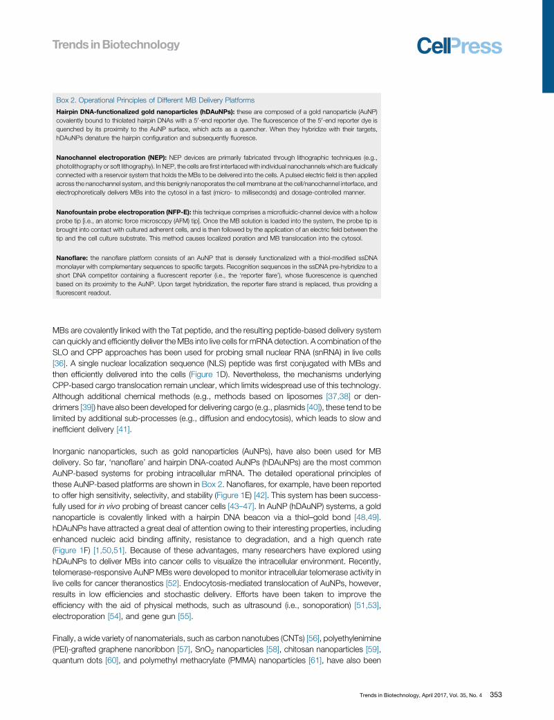
Box 2. Operational Principles of Different MB Delivery Platforms
Hairpin DNA-functionalized gold nanoparticles (hDAuNPs): these are composed of a gold nanoparticle (AuNP)covalently bound to thiolated hairpin DNAs with a 50-end reporter dye. The fluorescence of the 50-end reporter dye isquenched by its proximity to the AuNP surface, which acts as a quencher. When they hybridize with their targets,hDAuNPs denature the hairpin configuration and subsequently fluoresce.
Nanochannel electroporation (NEP): NEP devices are primarily fabricated through lithographic techniques (e.g.,photolithography or soft lithography). In NEP, the cells are first interfacedwith individual nanochannels which are fluidicallyconnected with a reservoir system that holds the MBs to be delivered into the cells. A pulsed electric field is then appliedacross the nanochannel system, and this benignly nanoporates the cell membrane at the cell/nanochannel interface, andelectrophoretically delivers MBs into the cytosol in a fast (micro- to milliseconds) and dosage-controlled manner.
Nanofountain probe electroporation (NFP-E): this technique comprises a microfluidic-channel device with a hollowprobe tip [i.e., an atomic force microscopy (AFM) tip]. Once the MB solution is loaded into the system, the probe tip isbrought into contact with cultured adherent cells, and is then followed by the application of an electric field between thetip and the cell culture substrate. This method causes localized poration and MB translocation into the cytosol.
Nanoflare: the nanoflare platform consists of an AuNP that is densely functionalized with a thiol-modified ssDNAmonolayer with complementary sequences to specific targets. Recognition sequences in the ssDNA pre-hybridize to ashort DNA competitor containing a fluorescent reporter (i.e., the ‘reporter flare’), whose fluorescence is quenchedbased on its proximity to the [10_TD$DIFF] AuNP. Upon target hybridization, the reporter flare strand is replaced, thus providing afluorescent readout.
MBs are covalently linked with the Tat peptide, and the resulting peptide-based delivery systemcan quickly and efficiently deliver theMBs into live cells for mRNA detection. A combination of theSLO and CPP approaches has been used for probing small nuclear RNA (snRNA) in live cells[36]. A single nuclear localization sequence (NLS) peptide was first conjugated with MBs andthen efficiently delivered into the cells (Figure 1D). Nevertheless, the mechanisms underlyingCPP-based cargo translocation remain unclear, [67_TD$DIFF]which limits [15_TD$DIFF]widespread use of this technology.Although additional chemical methods (e.g., methods based on liposomes [37,38] or den-drimers [39]) have also been developed for delivering cargo (e.g., plasmids [40]), these tend to belimited by additional sub-processes (e.g., diffusion and endocytosis), which leads to slow andinefficient delivery [41].
Inorganic nanoparticles, such as gold nanoparticles (AuNPs), have also been used for MBdelivery. So far, ‘nanoflare’ and hairpin DNA-coated AuNPs (hDAuNPs) are the most commonAuNP-based systems for probing intracellular mRNA. The detailed operational principles ofthese AuNP-based platforms are shown in Box 2. Nanoflares, for example, have been reportedto offer high sensitivity, selectivity, and stability (Figure 1E) [42]. This system has been success-fully used for[15_TD$DIFF] in vivo probing of breast cancer cells [43–47]. In AuNP (hDAuNP) systems, a goldnanoparticle is covalently linked with a hairpin DNA beacon via a thiol–gold bond [48,49].hDAuNPs have attracted a great deal of attention owing to their interesting properties, includingenhanced nucleic acid binding affinity, resistance to degradation, and a high quench rate(Figure 1F) [1,50,51]. Because of these advantages, many researchers have explored usinghDAuNPs to deliver MBs into cancer cells to visualize the intracellular environment. Recently,telomerase-responsive AuNPMBs were developed to monitor intracellular telomerase activity inlive cells for cancer theranostics [52]. Endocytosis-mediated translocation of AuNPs, however,results in low efficiencies and stochastic delivery. Efforts have been taken to improve theefficiency with the aid of physical methods, such as ultrasound (i.e., sonoporation) [51,53],electroporation [54], and gene gun [55].
Finally, a wide variety of nanomaterials, such as carbon nanotubes (CNTs) [56], polyethylenimine(PEI)-grafted graphene nanoribbon [57], SnO2 nanoparticles [58], chitosan nanoparticles [59],quantum dots [60], and polymethyl methacrylate (PMMA) nanoparticles [61], have also been
Trends in Biotechnology, April 2017, Vol. 35, No. 4 353
reported for MB delivery into live cancer cells including HeLa cells (probe: miR-21) [58], lungcancer cells (probe: miR-155) [59], and MDA-MB-231 cells (probe: MMP-2 MBs) [60].
MB Designs for Intracellular Bio-Interrogation of Cancer CellsDesigning MBs to accurately probe biomarkers in cancer cells presents equally challengingopportunities. [68_TD$DIFF]Conventional [69_TD$DIFF]assays [70_TD$DIFF](e.g., [71_TD$DIFF]morphological [72_TD$DIFF]analysis) often [73_TD$DIFF]lead to low accuracy anduncertainty [74_TD$DIFF], and fail to fully document the highly heterogeneous nature of cancer cells. [75_TD$DIFF]Tumors[76_TD$DIFF]exhibit [77_TD$DIFF]specific [78_TD$DIFF]cellular [79_TD$DIFF]and [80_TD$DIFF]microenvironmental [81_TD$DIFF]characteristics, including [57_TD$DIFF]unique mRNAs, spe-cial tumor [82_TD$DIFF]biomarkers, [83_TD$DIFF] a reductive cytosol milieu, and low pH, among others, [84_TD$DIFF] which provide [85_TD$DIFF] ahost of possibilities for detecting and sensing [86_TD$DIFF]cancerous cells. MBs have thus gained a greatdeal of attention recently as powerful tools for thoroughly probing tumor-related biomarkers(Figure 2A). We summarize here the latest advances in MB designs for cancer research.
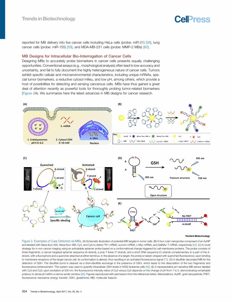
Figure 2. Examples of Cues [38_TD$DIFF]Detected via MBs. (A) Schematic illustration of potential MB targets in tumor cells. (B) A four-color nanoprobe composed of an AuNPand labeled with Alexa fluor 405, Alexa fluor 488, Cy3, and Cy5 to detect TK1 mRNA, survivin mRNA, c-Myc mRNA, and GalNAc-T mRNA, respectively [64]. (C) A novelstrategy for in vivo cancer imaging using an activatable aptamer probe based on a conformational change triggered by cell-membrane proteins. The probe consists ofthree fragments: a cancer-targeted aptamer sequence (A-strand), a poly-T linker (T-strand), and a short DNA sequence (C-strand) complementary to a part of the A-strand, with a fluorophore and a quencher attached at either terminus. In the absence of a target, the probe is hairpin-shaped with quenched fluorescence; upon bindingto membrane receptors of the target cancer cell, its conformation is altered, thus resulting in an activated fluorescence signal [73]. (D) A disulfide-decorated MB for thedetection of GSH. The disulfide bond is cleaved via a thiol–disulfide exchange in the presence of GSH, which leads to the dissociation of the two fragments andfluorescence enhancement. This system was used to quantify intracellular GSH levels in K562 leukemia cells [82]. (E) A representative pH-sensitive MB sensor labeledwith Cy3 and Cy5; upon excitation at 530 nm, the fluorescence intensity ratios of Cy3 versus Cy5 depends on the change of pH from 7 to 3, demonstrating remarkablepotency to sense pH within a narrow acidic window [86]. Figures reproduced with permission from the references listed. Abbreviations: AuNP, gold nanoparticle; FRET,fluorescence resonance energy transfer; GSH, glutathione; MB, molecular beacon.
354 Trends in Biotechnology, April 2017, Vol. 35, No. 4
MBs have been primarily designed to target cytosolic mRNAs in cancer cells. [87_TD$DIFF] TumormRNAs canoften represent specific markers for therapeutic, diagnostic, and prognostic purposes. Recentstudies have highlighted the relevance of these probes in early detection efforts. Tang et al.developed an effective approach to detect multiple tumor mRNAs [62]. A bimolecular beacon(bi-MB) termed AuNP/bi-MB was self-assembled from gold nanoparticles. This functional hybridMB could simultaneously target two types of tumor [88_TD$DIFF]mRNAs, cyclin D1 mRNA and survivinmRNA, in breast cancer cells. This system has been shown to effectively avoid false-positiveresults and yield more comprehensive and dependable information for the early detection ofcancer. The same group recently described the in vitro detection of up to three tumor-relatedmRNAs (c-MycmRNA, GalNAc-T mRNA, and TK1mRNA) in [89_TD$DIFF]different cells. Themodified AuNP-MB probes show higher accuracy than the single-marker [90_TD$DIFF]assays, as well as higher resistanceagainst nucleases, and they have been shown to differentiate tumor cells from normal cells usingbreast and liver cancer models [63].
To minimize the rate of false positives, Tang et al. proposed a four-color nanoprobe that cansimultaneously detect up to four types of mRNAs in live cells (Figure 2B) [64]. The nanoprobeconsisted of AuNPs and a dense corona of MBs that can discern multiple intracellular mRNAtranscripts, which increased the accuracy in cancer cell identification compared to single MBs. Inparticular, the probe was labeled with Alexa fluor 405, Alexa fluor 488, Cy3, and Cy5 to detectTK1 mRNA, survivin mRNA, c-Myc mRNA, and GalNAc-T mRNA, respectively. Subsequentwork has focused on detecting and imaging tumor mRNA in vivo [48,50,65–67].
MBs have also been reported to identify DNA mutations with high specificity, sensitivity, andspeed [68]. In an early report Yang et al. proposed a [91_TD$DIFF]method to detect the expression levels ofmultiple genes in both fixed and viable cells using MB-based imaging [69]. Simultaneous deliveryof MBs with two different DNA targets (i.e., survivin and cyclin D1) successfully differentiatedcancer cells from normal/healthy cells in a breast cancer model. Both cyclin D1 and survivingMBs only generate fluorescent signals [92_TD$DIFF]upon [93_TD$DIFF]hybridization with their specific DNA target.Recently, Wu and coworkers reported a primer-integrated label-free multifunctional MB (LMMB)to detect specific DNA sequences via integrating target recognition sequences, polymerizationprimers, templates, and G-quadruplex structures (also known as G4-DNA, nucleic acids that arerich in guanine [94_TD$DIFF] and tend to form tetramers via hydrogen bonds) into a single system [70]. Such asystem is compatible with target DNA amplification and detection with SYBR green I. Moreover,the LMMB technology [95_TD$DIFF]can be adapted for amplifying target DNA without an extra DNA probe.The polymerization reaction is thought to generate double-stranded fragments suitable for theintercalation of SYBR green I to detect hybridization between the LMMB and target DNA. Byusing a G-quadruplex instead of conventional hairpin, the target DNA can induce furtherconformational changes in the LMMB structure, permitting the detection of single-nucleotidemutations. In recent years, MBs have been reported to detect tumor markers (e.g., p53) [71] andcirculating DNA from serum samples in a breast cancer model [72].
MBs can also be designed to detect specific tumor proteins. Aptamer probes have been used tosuccessfully target particular membrane proteins on cancer cells for contrast-enhanced tumorvisualization in mice (Figure 2C) [73]. Conformational alterations upon binding to a target proteincan result in fluorescence, which is then used to conduct in vitro and in vivo studies with CCRF-CEM cancer cells (a T cell line associated with human acute lymphoblastic leukemia) and aspecific aptamer, sgc8. The activated fluorescence signals show high sensitivity and highspecificity for identifying CCRF-CEM cells in vivo.
Glutathione (GSH) is an important peptide responsible for modulating a host of biologicalprocesses. Tumor cells, in particular, exhibit about a 100- to 1000-fold increase in intracellularGSH levels compared to the extracellular microenvironment [74–76], and this hasmade the GSH
Trends in Biotechnology, April 2017, Vol. 35, No. 4 355
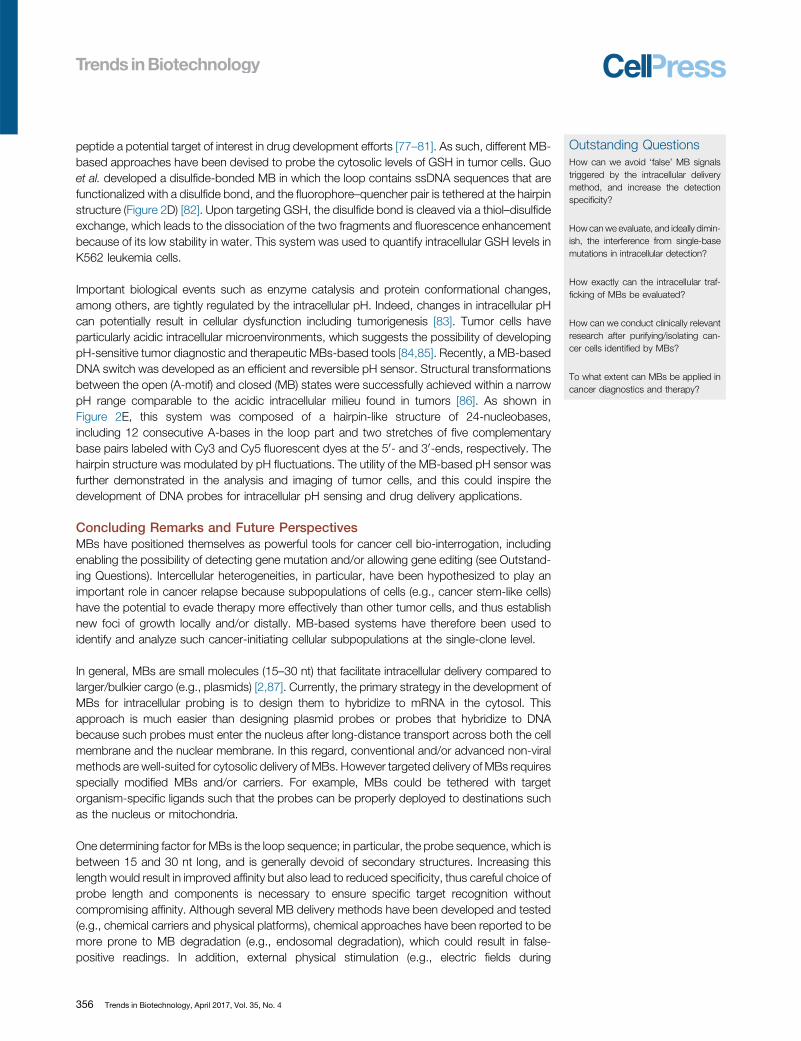
Outstanding QuestionsHow can we avoid ‘false’ MB signals[130_TD$DIFF]triggered [131_TD$DIFF]by the intracellular delivery[132_TD$DIFF]method, [133_TD$DIFF]and increase the detectionspecificity?
How can we evaluate, and ideally dimin-ish, the interference from single-basemutations in intracellular detection?
How exactly can the intracellular traf-ficking of MBs be evaluated?
How can we [134_TD$DIFF]conduct clinically [135_TD$DIFF]relevant[136_TD$DIFF]research after purifying[137_TD$DIFF]/isolating can-cer cells identified by MBs?
To what extent can MBs be applied incancer diagnostics and therapy?
peptide a potential target of interest in drug development efforts [77–81]. As such, different MB-based approaches have been devised to probe the cytosolic levels of GSH in tumor cells. Guoet al. developed a disulfide-bonded MB in which the loop contains ssDNA sequences that arefunctionalized with a disulfide bond, and the fluorophore–quencher pair is tethered at the hairpinstructure (Figure 2D) [82]. Upon targeting GSH, the disulfide bond is cleaved via a thiol–disulfideexchange, which leads to the dissociation of the two fragments and fluorescence enhancementbecause of its low stability in water. This system was used to quantify intracellular GSH levels inK562 leukemia cells.
Important biological events such as enzyme catalysis and protein conformational changes,among others, are tightly regulated by the intracellular pH. Indeed, changes in intracellular pHcan potentially result in cellular dysfunction including tumorigenesis [83]. Tumor cells haveparticularly acidic intracellular microenvironments [20_TD$DIFF], which suggests the possibility of developingpH-sensitive tumor diagnostic and therapeutic MBs-based tools [84,85]. Recently, a MB-basedDNA switch was developed as an efficient and reversible pH sensor. [96_TD$DIFF]Structural [97_TD$DIFF]transformationsbetween the open (A-motif) and closed (MB) states [98_TD$DIFF]were [99_TD$DIFF]successfully [100_TD$DIFF]achieved within a narrowpH range comparable to the acidic intracellular milieu found in tumors [86]. As shown inFigure 2E, this system was composed of a hairpin-like structure of 24[101_TD$DIFF]-nucleobases,[102_TD$DIFF]including 12 consecutive A [103_TD$DIFF]-bases in the loop [104_TD$DIFF] part and two stretches of five complementary[105_TD$DIFF]base pairs labeled with Cy3 and Cy5 fluorescent dyes at the 50- and 30-ends, respectively. The [22_TD$DIFF]hairpin structure was modulated by pH fluctuations. The utility of the MB-based pH sensor wasfurther demonstrated in the analysis and imaging of tumor cells, and this could inspire thedevelopment of DNA probes for intracellular pH sensing and drug delivery applications.
Concluding Remarks and Future PerspectivesMBs have positioned themselves as powerful tools for cancer cell bio-interrogation, including[106_TD$DIFF]enabling the [107_TD$DIFF]possibility of [108_TD$DIFF] detecting gene mutation and[109_TD$DIFF]/or allowing gene editing (see Outstand-ing Questions). Intercellular heterogeneities, in particular, have been hypothesized to play animportant role in cancer relapse because subpopulations of cells (e.g., cancer stem-like cells)have the potential to evade therapy more effectively than other tumor cells, and thus establishnew foci of growth locally and/or distally. MB-based systems have therefore been used toidentify and analyze such cancer-initiating cellular subpopulations at the single-clone level.
In general, MBs are small molecules (15–30 nt) that facilitate intracellular delivery [110_TD$DIFF]compared [111_TD$DIFF]tolarger/bulkier cargo (e.g., plasmids) [2,87]. Currently, the primary strategy in the development ofMBs for intracellular probing is to design them to hybridize to mRNA in the cytosol. Thisapproach is much easier than designing plasmid probes or probes that hybridize to DNAbecause such probes must enter the nucleus after long-distance transport across both the cellmembrane and the nuclear membrane. In this regard, [112_TD$DIFF]conventional [113_TD$DIFF]and/or advanced non-viralmethods are [114_TD$DIFF]well-suited [24_TD$DIFF] for [115_TD$DIFF]cytosolic delivery of [116_TD$DIFF]MBs. However [26_TD$DIFF] targeted delivery of MBs [117_TD$DIFF]requiresspecially modified MBs [118_TD$DIFF]and/or [119_TD$DIFF]carriers. For example, MBs could be tethered with targetorganism-specific ligands such that the probes can [120_TD$DIFF] be properly [121_TD$DIFF]deployed to destinations suchas the nucleus or mitochondria.
One determining factor for MBs is the loop sequence; in particular, [29_TD$DIFF] the probe sequence [122_TD$DIFF], [123_TD$DIFF]which [124_TD$DIFF]isbetween 15 and 30 nt [125_TD$DIFF]long, and is generally devoid of secondary [126_TD$DIFF]structures. Increasing thislength would result in improved affinity but also lead to reduced specificity, thus careful choice ofprobe length and components is necessary to ensure specific [127_TD$DIFF]target [128_TD$DIFF]recognition withoutcompromising affinity. Although several MB delivery methods have been developed and tested(e.g., chemical carriers and physical platforms), chemical approaches have been reported to bemore prone to MB degradation (e.g., endosomal degradation), which could result in false-positive readings. In addition, external physical stimulation [129_TD$DIFF] (e.g., electric fields during
356 Trends in Biotechnology, April 2017, Vol. 35, No. 4
electroporation) may also alter the function of the MBs [32_TD$DIFF]. Multiple studies have therefore usedsupplementary techniques (e.g., PCR, transcriptomemicroarray) to verify the reliability of the MBsignal [3,26]. Co-delivery of multiple MBs for a single target has also been shown to be anefficient approach to reduce the incidence of false positives [88,89]. Co-delivery of MBs couldalso enable the development of advanced multiplexed analytic platforms to monitor, in real time,the intracellular levels of multiple targets at the same time. Such targets, however, couldpotentially be found in different intracellular compartments (e.g., the nucleus vs the cytosol),and this may require the development of novel delivery approaches and/or MB designs thatincorporate specific localization sequences compatible with compartmentalized delivery. Inaddition, further work concerning the trafficking and final fate of MBs within cells is alsoimperative for the design [33_TD$DIFF] of MBs.
Finally, for in vitro early cancer detection, single-nucleotide [34_TD$DIFF]mutations [35_TD$DIFF] remain challenging for MB-based platforms. The ability of MBs to recognize the genetic heterogeneity associated withsingle-nucleotide mutations is currently limited. In the future, incorporating the PCR technique toadd an altered base to the mutated base of the primer may reduce the risk of inducing single-nucleotide mutations and increase the accuracy of MBs in cancer detection. In addition, morestudies need to be conducted to better understand themechanisms underlying the hybridizationbetween MBs and their targets, and any potential downstream influence on cell behavior. Suchefforts could play a pivotal role in developing optimized MB designs and/or delivery methods,and these could have a significant impact not only on disease diagnosis and prognosis but alsotherapeutics for precision medicine.
AcknowledgmentsThe authors acknowledges the support of the National Natural Science Foundation of China (grants 51573063, 21174044,
21674040, and 51403042 [36_TD$DIFF]), the Guangdong Science and Technology Planning Project (2014B010104004 and
2013B090600126), the Natural Science Foundation for Distinguished Young Scholars of Guangdong Province
(2016A030306013), the Guangdong Program for Support of Top-Notch Young Professionals (2015TQ01R604), the
Guangzhou Science and Technology Planning Project (201604010013),[138_TD$DIFF] and the Scientific Research Projects of Guangz-
hou (201607010328 [37_TD$DIFF]).
References
1. Zheng, J. et al. (2015) Rationally designed molecular beacons forbioanalytical and biomedical applications. Chem. Soc. Rev. 44,3036–3055
2. Chang, L.Q. et al. (2016) Nanoscale bio-platforms for living cellinterrogation: current status and future perspectives.Nanoscale 8,3181–3206
3. Gallego-Perez, D. et al. (2016) On-chip clonal analysis of gliomastem cell motility and therapy resistance.Nano Lett. 16, 5326–5332
4. Rau, K. et al. (2016) CRISPR/Cas9 – a new tool for RNA imaging inlive cells. Chembiochem. 17, 1–4
5. Nelles, D.A. et al. (2016) Programmable RNA tracking in live cellswith CRISPR/Cas9. Cell 165, 488–496
6. Gao, F. et al. (2016) DNA-guided genome editing using the Natro-nobacterium gregoryi Argonaute. Nat. Biotechnol. 34, 768–773
7. Dai, Y.L. et al. (2013) In vivo multimodality imaging and cancertherapy by near-infrared light-triggered trans-platinum pro-drug-conjugated upconverison nanoparticles. J. Am. Chem. Soc. 135,18920–18929
8. Ding, C.M. (2007) ‘Other’ applications nucleotide of single poly-morphisms. Trends Biotechnol. 25, 279–283
9. Zare, R.N. et al. (2010) Microfluidic platforms for single-cell analy-sis. Annu. Rev. Biomed. Eng. 12, 187–201
10. Collins, B.C. et al. (2013) Quantifying protein interaction dynamicsby SWATH mass spectrometry: application to the 14-3-3 system.Nat. Methods 10, 1246–1253
11. Xiao, H.H. et al. (2012) A prodrug strategy to deliver cisplatin(IV)and paclitaxel in nanomicelles to improve efficacy and tolerance.Biomaterials 33, 6507–6519
12. Qi, R.G. et al. (2012) Biodegradable copolymers with identicalcationic segments and their performance in siRNA delivery. J.Control. Release 159, 251–260
13. Zhang, A.Q. et al. (2016) Nano-bioelectronics. Chem. Rev. 116,215–257
14. Liu, J. et al. (2015) Voyage inside the cell: microsystems andnanoengineering for intracellular measurement and manipulation.Microsystems & Nanoengineering 1, 15020
15. Xie, N.L. et al. (2016) A DNA tetrahedron-based molecular beaconfor tumor-related mRNA detection in living cells. Chem. Commun.52, 2346–2349
16. Santangelo, P.J. (2010) Molecular beacons and related probesfor intracellular RNA imaging. Wires. Nanomed. Nanobi. 2,11–19
17. Santangelo, P.J. et al. (2012) Probes for intracellular RNA imagingin live cells. Method Enzymol. 505, 383–399
18. Wile, B.M. et al. (2014) Molecular beacon-enabled purification ofliving cells by targeting cell type-specific mRNAs. Nat. Protoc. 9,2411–2424
19. Pai, P. et al. (2013) Prospects of miRNA-based therapy for pan-creatic cancer. Curr. Drug Targets 14, 1101–1109
20. Li, C.W. et al. (2005) Adeno-associated virus vectors: potentialapplications for cancer gene therapy. Cancer Gene Ther. 12,913–925
21. Kim, K.H. et al. (2015) Rapid, high-throughput, and direct molec-ular beacon delivery to human cancer cells using a nanowire-incorporated and pneumatic pressure-driven microdevice. Small11, 6215–6224
Trends in Biotechnology, April 2017, Vol. 35, No. 4 357
22. Shalek, A.K. et al. (2012) Nanowire-mediated delivery enablesfunctional interrogation of primary immune cells: application tothe analysis of chronic lymphocytic leukemia. Nano Lett. 12,6498–6504
23. Yan, R.X. et al. (2012) Nanowire-based single-cell endoscopy.Nat. Nanotechnol. 7, 191–196
24. Hanson, L. et al. (2015) Vertical nanopillars for in situ probingof nuclear mechanics in adherent cells. Nat. Nanotechnol. 10,554–562
25. Xie, C. et al. (2012) Intracellular recording of action potentials bynanopillar electroporation. Nat. Nanotechnol. 7, 185–190
26. Gao, K.L. et al. (2016) Induced apoptosis investigation in wild-type and FLT3-ITD acute myeloid leukemia cells by nanochannelelectroporation and single-cell qRT-PCR. Mol. Ther. 24, 956–964
27. Boukany, P.E. et al. (2011) Nanochannel electroporation deliversprecise amounts of biomolecules into living cells. Nat. Nanotech-nol. 6, 747–754
28. Kang, W.M. et al. (2013) Nanofountain probe electroporation(NFP-E) of single cells. Nano Lett. 13, 2448–2457
29. Giraldo-Vela, J.P. et al. (2015) Single-cell detection of mRNAexpression using nanofountain-probe electroporated molecularbeacons. Small 11, 2386–2391
30. Kang, W.M. et al. (2014) Microfluidic parallel patterning and cellulardelivery of molecules with a nanofountain probe. J. Lab. Autom.19, 100–109
31. Xie, X. et al. (2013) Nanostraw-electroporation system for highlyefficient intracellular delivery and transfection. ACS Nano 7,4351–4358
32. Nitin, N. et al. (2009) Translation inhibition reveals interaction of 2-deoxy and 2-O-methyl molecular beacons with mRNA targets inliving cells. Nucleic Acids Res. 37, 4977–4986
33. Rhee, W.J. et al. (2008) Target accessibility and signal specificity inlive-cell detection of BMP-4 mRNA using molecular beacons.Nucleic Acids Res. 36, e30
34. Santangelo, P. et al. (2006) Live-cell characterization and analysisof a clinical isolate of bovine respiratory syncytial virus, usingmolecular beacons. J. Virol. 80, 682–688
35. Sawant, R. et al. (2010) Intracellular transduction using cell-pene-trating peptides. Mol. Biosyst. 6, 628–640
36. Nitin, N. et al. (2008) NLS peptide conjugated molecular beaconsfor visualizing nuclear RNA in living cells. Bioconjugate Chem. 19,2205–2211
37. Barton, G.M. et al. (2002) Retroviral delivery of small interferingRNA into primary cells. Proc. Natl. Acad. Sci. U.S.A. 99, 14943–14945
38. Jo, S.M. et al. (2013) Rapid detection of exon 2-deleted AIMP2mutation as a potential biomarker for lung cancer by molecularbeacons. Biosens. Bioelectron. 46, 142–149
39. Zhang, Y. et al. (2012) Efficient siRNA delivery using a polyamido-amine dendrimer with a modified pentaerythritol core. Pharm. Res.29, 1627–1636
40. Yin, H. et al. (2014) Non-viral vectors for gene-based therapy. Nat.Rev. Genet. 15, 541–555
41. Chang, L.Q. et al. (2016) 3D nanochannel electroporation for high-throughput cell transfection with high uniformity and dosage con-trol. Nanoscale 8, 243–252
42. Seferos, D.S. et al. (2007) Nano-flares: probes for transfection andmRNA detection in living cells. J. Am. Chem. Soc. 129, 15477–15479
43. Prigodich, A.E. et al. (2012) Multiplexed nanoflares: mRNA detec-tion in live cells. Anal. Chem. 84, 2062–2066
44. Prigodich, A.E. et al. (2009) Nano-flares for mRNA regulation anddetection. ACS Nano 3, 2147–2152
45. Zheng, D. et al. (2009) Aptamer nano-flares for molecular detec-tion in living cells. Nano Lett. 9, 3258–3261
46. Li, N. et al. (2012) A multicolor nanoprobe for detection andimaging of tumor-related mRNAs in living cells. Angew. Chem.Int. Edit. 51, 7426–7430
47. Halo, T.L. et al. (2014) NanoFlares for the detection, isolation, andculture of live tumor cells from human blood. Proc. Natl. Acad. Sci.U.S.A. 111, 17104–17109
358 Trends in Biotechnology, April 2017, Vol. 35, No. 4
48. Chinen, A.B. et al. (2015) Nanoparticle probes for the detection ofcancer biomarkers, cells, and tissues by fluorescence.Chem. Rev.115, 10530–10574
49. Deng, D.W. et al. (2015) Gold nanoparticle-based beacon todetect STAT5b mRNA expression in living cells: a case optimizedby bioinformatics screen. Int. J. Nanomed. 10, 3231–3244
50. Pan, W. et al. (2013) Dual-targeted nanocarrier based on cellsurface receptor and intracellular mRNA: an effective strategyfor cancer cell imaging and therapy. Anal. Chem. 85, 6930–6935
51. Zhou, Q.M. et al. (2016) Optimized ultrasound conditions forenhanced sensitivity of molecular beacons in the detection ofMDR1 mRNA in living cells. Anal. Chem. 88, 2808–2816
52. Ma, Y. et al. (2016) A telomerase-specific doxorubicin-releasingmolecular beacon for cancer theranostics.Angew. Chem. Int. Edit.55, 3304–3308
53. Ding, B.H. et al. (2015) Recording the dynamic endocytosis ofsingle gold nanoparticles by AFM-based force tracing. Nanoscale7, 7545–7549
54. West, D.L. et al. (2014) Assessment and optimization of electro-poration-assisted tumoral nanoparticle uptake in a nude mousemodel of pancreatic ductal adenocarcinoma. Int. J. Nanomed. 9,4169–4175
55. Zhang, D.W. et al. (2014) Microneedle assisted micro-particledelivery from gene guns: experiments using skin-mimicking aga-rose gel. J. Pharm. Sci. 103, 613–627
56. Liang, H. et al. (2014) Functional DNA-containing nanomaterials:cellular applications in biosensing, imaging, and targeted therapy.Accounts Chem. Res. 47, 1891–1901
57. Dong, H.F. et al. (2011) The use of polyethylenimine-grafted gra-phene nanoribbon for cellular delivery of locked nucleic acid mod-ified molecular beacon for recognition of microRNA. Biomaterials32, 3875–3882
58. Dong, H.F. et al. (2012) Target-cell-specific delivery, imaging, anddetection of intracellular microRNA with a multifunctional SnO2
nanoprobe. Angew. Chem. Int. Edit. 51, 4607–4612
59. Zhu, H.Z. et al. (2014) Chitosan combined with molecular beaconfor Mir-155 detection and imaging in lung cancer. Molecules 19,14710–14722
60. Li, X. et al. (2014) Quantum dots based molecular beacons for invitro and in vivo detection of MMP-2 on tumor. Biosens. Bioelec-tron. 61, 512–518
61. Barbara, A. et al. (2016) Molecular beacon-decorated polymethyl-methacrylate core-shell fluorescent nanoparticles for the detectionof survivin mRNA in human cancer cells. Biosens. Bioelectron.http://dx.doi.org/10.1016/j.bios.2016.05.102
62. Qiao, G. et al. (2011) Simultaneous detection of intracellular tumormRNA with bi-color imaging based on a gold nanoparticle/molec-ular beacon. Chem. Eur. J. 17, 11210–11215
63. Li, N. et al. (2012) A multicolor nanoprobe for detection andimaging of tumor-related mRNAs in living cells. Angew. Chem.Inter. Edit. 51, 7426–7430
64. Pan, W. et al. (2013) Multiplexed detection and Imaging of intra-cellular mRNAs using a four-color nanoprobe. Anal. Chem. 85,10581–10588
65. Lee, D.S. et al. (2016) Cellular processing and destinies of artificialDNA nanostructures. Chem. Soc. Rev. 45, 4199–4255
66. Li, X-L. et al. (2016) Oriented assembly of invisible probes: towardssingle mRNA imaging in living cells. Chem. Sci. 7, 3256–3263
67. Xue, J.P. et al. (2013) Visual detection of STAT5B gene expressionin living cell using the hairpin DNA modified gold nanoparticlebeacon. Biosens. Bioelectron. 41, 71–77
68. Wang, Q. et al. (2013) Molecular beacons of xeno-nucleic acid fordetecting nucleic acid. Theranostics 3, 395–408
69. Peng, X-H. et al. (2005) Real-time detection of gene expression incancer cells using molecular beacon imaging: new strategies forcancer research. Cancer Res. 65, 1909–1917
70. Dong, H. et al. (2016) A biofunctional molecular beacon for detect-ing single base mutations in cancer cells.Mol. Ther. Nucleic Acids5, e302
71. Li, F. et al. (2016) Highly sensitive detection of cancer-relatedgenes based on complete fluorescence restoration of a molecularbeacon with a functional overhang. Analyst 141, 4417–4423
72. Gui, Z. et al. (2016) Direct detection of circulating free DNAextracted from serum samples of breast cancer using lockednucleic acid molecular beacon. Talanta 154, 520–525
73. Shi, H. et al. (2011) Activatable aptamer probe for contrast-enhanced in vivo cancer imaging based on cell membrane pro-tein-triggered conformation alteration. Proc. Natl. Acad. Sci. U.S.A. 108, 3900–3905
74. Kuppusamy, P. et al. (2002) Noninvasive imaging of tumor redoxstatus and its modification by tissue glutathione levels. CancerRes. 62, 307–312
75. Kuppusamy, P. et al. (1998) In vivo electron paramagnetic reso-nance imaging of tumor heterogeneity and oxygenation in amurinemodel. Cancer Res. 58, 1562–1568
76. Cheng, R. et al. (2013) Dual and multi-stimuli responsive polymericnanoparticles for programmed site-specific drug delivery. Bioma-terials 34, 3647–3657
77. Dai, J. et al. (2011) Interlayer-crosslinked micelle with partiallyhydrated core showing reduction and pH dual sensitivity for pin-pointed intracellular drug release. Angew. Chem. Int. Edit. 50,9404–9408
78. Han, H.S. et al. (2015) Bioreducible core-crosslinked hyaluronicacid micelle for targeted cancer therapy. J. Control. Release 200,158–166
79. Hu, X. et al. (2013) Polyprodrug amphiphiles: hierarchical assem-blies for shape-regulated cellular internalization, trafficking, anddrug delivery. J. Am. Chem. Soc. 135, 17617–17629
80. Hu, X. et al. (2015) Cell-penetrating hyperbranched polyprodrugamphiphiles for synergistic reductive milieu-triggered drug releaseand enhanced magnetic resonance signals. J. Am. Chem. Soc.137, 362–368
81. Xiao, H.H. et al. (2012) The use of polymeric platinum(IV) pro-drugs to deliver multinuclear platinum(II) drugs with reducedsystemic toxicity and enhanced antitumor efficacy. Biomaterials33, 8657–8669
82. Guo, Y.S. et al. (2012) A disulfide bound-molecular beacon as afluorescent probe for the detection of reduced glutathione and itsapplication in cells. Chem. Commun. 48, 3221–3223
83. Wang, Y.G. et al. (2014) A nanoparticle-based strategy for theimaging of a broad range of tumours by nonlinear amplification ofmicroenvironment signals. Nat. Mater. 13, 204–212
84. Song, L. et al. (2013) Efficient, pH-triggered drug delivery using apH-responsive DNA-conjugated gold nanoparticle. Adv. Healthc.Mater. 2, 275–280
85. Zhang, W. et al. (2016) pH and near-infrared light dual-stimuliresponsive drug delivery using DNA-conjugated gold nanorodsfor effective treatment of multidrug resistant cancer cells. J. Con-trol. Release 232, 9–19
86. Narayanaswamy, N. et al. (2016) A molecular beacon-based DNAswitch for reversible pH sensing in vesicles and live cells. Chem.Commun. 52, 8741–8744
87. Gonzalez, F. et al. (2011) Methods for making induced pluripotentstem cells: reprogramming a la carte. Nat. Rev. Genet. 12,231–242
88. Perez-Pinera, P. et al. (2013) Synergistic and tunable human geneactivation by combinations of synthetic transcription factors. Nat.Methods 10, 239–242
89. Lu, Y. et al. (2015) Highly multiplexed profiling of single-cell effectorfunctions reveals deep functional heterogeneity in response topathogenic ligands. Proc. Natl. Acad. Sci. U.S.A. 112, E607–E615
Trends in Biotechnology, April 2017, Vol. 35, No. 4 359