Embed Size (px)
Citation preview
Molecular basis for chromatin bindingand regulation of MLL5Muzaffar Alia,1, Héctor Rincón-Aranob,1, Wei Zhaoc, Scott B. Rothbartd, Qiong Tonga, Susan M. Parkhurstb,Brian D. Strahld, Lih-Wen Dengc, Mark Groudineb,e,2, and Tatiana G. Kutateladzea,2
aDepartment of Pharmacology, University of Colorado School of Medicine, Aurora, CO 80045; bBasic Science Division, Fred Hutchinson Cancer ResearchCenter, Seattle, WA 98109; cDepartment of Biochemistry, Yong Loo Lin School of Medicine, National University of Singapore, Singapore 117597; dDepartmentof Biochemistry and Biophysics and the Lineberger Comprehensive Cancer Center, University of North Carolina School of Medicine, Chapel Hill, NC 27599; andeDepartment of Radiation Oncology, University of Washington School of Medicine, Seattle, WA 98109
Contributed by Mark Groudine, May 29, 2013 (sent for review May 15, 2013)
The human mixed-lineage leukemia 5 (MLL5) protein mediateshematopoietic cell homeostasis, cell cycle, and survival; however,the molecular basis underlying MLL5 activities remains unknown.Here, we show that MLL5 is recruited to gene-rich euchromaticregions via the interaction of its plant homeodomain finger withthe histone mark H3K4me3. The 1.48-Å resolution crystal structureof MLL5 plant homeodomain in complex with the H3K4me3 pep-tide reveals a noncanonical binding mechanism, whereby K4me3 isrecognized through a single aromatic residue and an aspartate. Thebinding induces a unique His–Asp swapping rearrangement medi-ated by a C-terminal α-helix. Phosphorylation of H3T3 and H3T6abrogates the association with H3K4me3 in vitro and in vivo, re-leasing MLL5 from chromatin in mitosis. This regulatory switch isconserved in the Drosophila ortholog of MLL5, UpSET, and suggeststhe developmental control for targeting of H3K4me3. Together, ourfindings provide first insights into the molecular basis for the re-cruitment, exclusion, and regulation of MLL5 at chromatin.
The mixed-lineage leukemia 5 (MLL5) protein was initiallyidentified as a candidate tumor suppressor and more recently
has been shown to play a critical role in hematopoiesis and lym-phopoiesis (1). TheMLL5 gene-containing region of chromosome 7is frequently deleted in patients with hematological disorders, in-cluding acute myeloid and therapy-induced leukemias and myeloiddysplastic syndrome (MDS). Depletion of Mll5 in mice causes mildpostnatal lethality, with some of the surviving animals showing re-tarded growth, male sterility, and decreased size of thymus, spleen,and lymph nodes (2–4). Genetic analyses of these Mll5 deficienciesreveals a 30% decrease in the number of hematopoietic stem cells(HSC) and progenitors, defects in HSC self-renewal mechanisms,and impaired myeloid differentiation (2–4). In addition to being anessential mediator of HSC homeostasis, MLL5 has been implicatedin cytokinesis, the DNA damage response, and genome mainte-nance (5–7). Overexpression and knockdown of MLL5 both inducecell cycle arrest at various phases, suggesting a versatile function ofMLL5 throughout the cell cycle (5).MLL5 belongs to the MLL family of methyltransferases that
regulate gene expression during developmental processes. Theseenzymes catalyze the addition of methyl groups to the e-aminomoiety of lysine and are highly specific for lysine 4 of histone H3.Along with MLL5, also known as a lysine methyltransferase 2E(KMT2E), the MLL family contains MLL1-4, SET1A, andSET1B (KMT2A-KMT2D, KMT2F, and KMT2G, respectively)(8). Full-length MLL5 is ∼200 kDa and is evolutionarily distantfrom the more canonical and better-characterized members ofthis family. Unlike the other multimodular MLL proteins, MLL5consists of only two conserved motifs near the N terminus, a planthomeodomain (PHD) finger, followed by a catalytic Su(var)3–9,enhancer of zeste, trithorax (SET) domain. A long, ∼1,000-residueC-terminal region of MLL5 displays no apparent homology to anyknown structural domain and is truncated in a shorter ∼75-kDaisoform, MLL5 short (MLL5s). Furthermore, MLL5 has no in-trinsic histone lysine methyltransferase (HKMT) activity (7);however, once posttranslationally modified through glycosylationat Thr440 in the SET domain, the short isoform MLL5s is capable
of generating mono- and dimethylated H3K4 marks (9). TheHKMT activity of MLL5s stimulates expression of retinoic acidreceptor α target genes and facilitates retinoic acid-dependentgranulopoiesis in human promyelocytes (9). The glycosylation-induced catalytic function of MLL5s appears to be unique tomammals. The yeast and drosophila orthologs of MLL5 (Set3/4and UpSET, respectively) lack the HKMT activity and instead arefound in histone deacetylase complexes (10). One of the mostdiscernible features of MLL5 is that, in contrast to other MLLs thatharbor multiple PHD finger cassettes, it contains a single PHDmodule, the biological role of which remains unclear.In this study, we show that MLL5 associates with the chromatin
regions downstream of transcriptional start sites (TSSs) of activegenes. The PHD finger of MLL5 binds strongly and specifically tohistone H3K4me3 through a noncanonical mechanism. Phos-phorylation of H3T3 and H3T6 disrupts binding of MLL5 toH3K4me3, and this regulatory mechanism is conserved in theDrosophila ortholog of MLL5, UpSET. Our results suggest thatthe phospho-methyl switch plays an essential role in the regula-tion of MLL5/UpSET during mitosis and development.
ResultsMLL5 Is Recruited to Actively Transcribed Genes. To determine thegenome-wide distribution of MLL5 in mammals, we performeda DNA adenine methyltransferase identification (DamID)-basedchromatin analysis of MLL5 in C2C12 cells using promoter arrayscovering ∼32,000 regions plus CpG islands (11). The DamIDprofiling revealed that MLL5 is recruited to ∼15,000 genomicregions, including ∼13,000 genes in myoblasts. Consistent withSebastian et al. (7), we found that MLL5 binds to the genomicregions downstream of TSSs of Ccna2 and Exosc9 (Fig. 1A). Tobetter characterize the global recruitment of MLL5, we carriedout a meta-analysis around TSSs using the MLL5 DamID signal.This analysis revealed that MLL5 preferentially targets theregions downstream of TSSs, and this result was well reproducedin experimental replicates (Fig. 1 A–C; Fig. S1 A and B). Inmammals, ∼60% of promoter regions overlap with CpG islands(12); therefore, we tested whether MLL5 occupies CpG islands orflanking sequences. In agreement with the initial meta-analysis,MLL5 DamID signal was enriched at CpG islands but not at theflanking regions (Fig. 1D).
Author contributions: M.A., H.R.-A., M.G., and T.G.K. designed research; M.A., H.R.-A.,W.Z., S.B.R., and L.-W.D. performed research; M.A., H.R.-A., W.Z., S.B.R., Q.T., S.M.P.,B.D.S., L.-W.D., M.G., and T.G.K. analyzed data; and M.A., H.R.-A., M.G., and T.G.K. wrotethe paper.
The authors declare no conflict of interest.
Data deposition: The atomic coordinates have been deposited in the Protein Data Bank,www.pdb.org (PDB ID code 4L58). The raw data reported in this paper have been de-posited in the Gene Expression Omnibus (GEO) database, www.ncbi.nlm.nih.gov/geo (ac-cession no. GSE47100).1M.A. and H.R.-A. contributed equally to this work.2To whom correspondence may be addressed: E-mail: [email protected] [email protected].
This article contains supporting information online at www.pnas.org/lookup/suppl/doi:10.1073/pnas.1310156110/-/DCSupplemental.
11296–11301 | PNAS | July 9, 2013 | vol. 110 | no. 28 www.pnas.org/cgi/doi/10.1073/pnas.1310156110
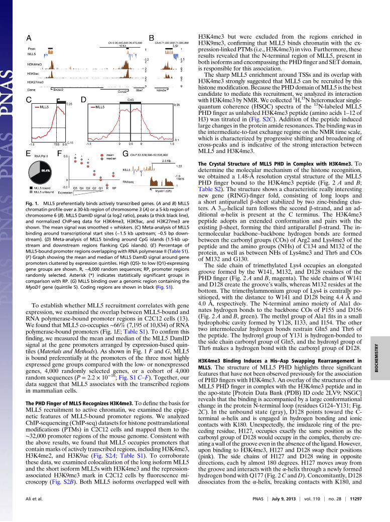
To establish whether MLL5 recruitment correlates with geneexpression, we examined the overlap between MLL5-bound andRNA polymerase-bound promoter regions in C2C12 cells (13).We found that MLL5 co-occupies ∼66% (7,195 of 10,834) of RNApolymerase-bound promoters (Fig. 1E; Table S1). To confirm thisfinding, we measured the mean and median of the MLL5 DamIDsignal at the gene promoters arranged by expression-based quin-tiles (Materials and Methods). As shown in Fig. 1 F and G, MLL5is bound preferentially at the promoters of the three most highlyexpressed gene groups compared with the low- or nonexpressedgenes, 4,000 randomly selected genes, or a cohort of 4,000random sequences (P = 2.2 × 10⁻¹⁶; Fig. S1 C–F). Together, ourdata suggest that MLL5 associates with the transcribed regionsin mammalian cells.
The PHD Finger of MLL5 Recognizes H3K4me3. To define the basis forMLL5 recruitment to active chromatin, we examined the epige-netic features of MLL5-bound promoter regions. We analyzedChIP-sequencing (ChIP-seq) datasets for histone posttranslationalmodifications (PTMs) in C2C12 cells and mapped them to the∼32,000 promoter regions of the mouse genome. Consistent withthe above results, we found that MLL5 occupies promoters thatcontainmarks of actively transcribed regions, includingH3K4me3,H3K4me2, and H3K9ac (Fig. S2A; Table S1). To corroboratethese data, we examined colocalization of the long isoformMLL5and the short isoform MLL5s with H3K4me3 and the repression-associated H3K9me3 mark in C2C12 cells by fluorescence mi-croscopy (Fig. S2B). Both MLL5 isoforms overlapped well with
H3K4me3 but were excluded from the regions enriched inH3K9me3, confirming that MLL5 binds chromatin with the ex-pression-linked PTMs (i.e., H3K4me3) in vivo.Furthermore, theseresults revealed that the N-terminal region of MLL5, present inboth isoforms and encompassing the PHD finger and SET domain,is responsible for this association.The sharp MLL5 enrichment around TSSs and its overlap with
H3K4me3 strongly suggested that MLL5 can be recruited by thishistonemodification. Because thePHDdomain ofMLL5 is the bestcandidate to mediate this recruitment, we analyzed its interactionwith H3K4me3 by NMR.We collected 1H,15N heteronuclear single-quantum coherence (HSQC) spectra of the 15N-labeled MLL5PHD finger as unlabeled H3K4me3 peptide (amino acids 1–12 ofH3) was titrated in (Fig. S2C). Addition of the peptide inducedlarge changes in the protein amide resonances. The binding was inthe intermediate-to-fast exchange regime on the NMR time scale,which is characterized by progressive shifting and broadening ofcross-peaks and is indicative of the strong interaction betweenMLL5 and H3K4me3.
The Crystal Structure of MLL5 PHD in Complex with H3K4me3. Todetermine the molecular mechanism of the histone recognition,we obtained a 1.48-Å resolution crystal structure of the MLL5PHD finger bound to the H3K4me3 peptide (Fig. 2 A and B;Table S2). The structure shows a characteristic really interestingnew gene (RING)-finger fold, consisting of long loops anda short antiparallel β-sheet stabilized by two zinc-binding clus-ters. A 310-helical turn follows the second β-strand, and an ad-ditional α-helix is present at the C terminus. The H3K4me3peptide adopts an extended conformation and pairs with theexisting β-sheet, forming the third antiparallel β-strand. The in-termolecular backbone–backbone hydrogen bonds are formedbetween the carbonyl groups (COs) of Arg2 and Lys4me3 of thepeptide and the amino groups (NHs) of C134 and M132 of theprotein, as well as between NHs of Lys4me3 and Thr6 and COsof M132 and G130.The side chain of trimethylated Lys4 occupies an elongated
groove formed by the W141, M132, and D128 residues of thePHD finger (Fig. 2 A and B, magenta). The side chains of W141and D128 create the groove’s walls, whereas M132 resides at thebottom. The trimethylammonium group of Lys4 is centrally po-sitioned, with the distance to W141 and D128 being 4.4 Å and4.0 Å, respectively. The N-terminal amino moiety of Ala1 do-nates hydrogen bonds to the backbone COs of P155 and D156(Fig. 2 A and B, green). The methyl group of Ala1 fits in a smallhydrophobic cavity formed by Y128, I133, and I154. The othertwo intermolecular hydrogen bonds restrain Gln5 and Thr6 ofthe peptide. The hydroxyl group of Y131 is hydrogen-bonded tothe side chain carbonyl group of Gln5, and the hydroxyl group ofThr6 makes a hydrogen bond with the carboxyl group of D128.
H3K4me3 Binding Induces a His–Asp Swapping Rearrangement inMLL5. The structure of MLL5 PHD highlights three significantfeatures that have not been observed previously for the associationof PHD fingers with H3K4me3. An overlay of the structures of theMLL5 PHD finger in complex with the H3K4me3 peptide and inthe apo-state [Protein Data Bank (PDB) ID code 2LV9; NSGC]reveals that the binding is accompanied by a large conformationalchange in the protein N-terminal loop (residues G124–Y131; Fig.2C). In the unbound state (gray), D128 points toward the C-terminal α-helix and is engaged in hydrogen bonding and ioniccontacts with K180. Unexpectedly, the imidazole ring of the pre-ceding residue, H127, occupies exactly the same position as thecarboxyl group of D128 would occupy in the complex, thereby cre-ating awall of the groove even in the absence of the ligand.However,upon binding to H3K4me3, H127 and D128 swap their positions(pink). The side chains of H127 and D128 swing in oppositedirections, each by almost 180 degrees. H127 moves away fromthe groove and interacts with the α-helix through a newly formedhydrogen bond with Q177 (Fig. 2C andD). Concomitantly, D128dissociates from the α-helix, breaking contacts with K180, and
Fig. 1. MLL5 preferentially binds actively transcribed genes. (A and B) MLL5chromatin profile over a 30-kb region of chromosome 3 (A) or a 5-kb region ofchromosome 6 (B). MLL5 DamID signal (a log2 ratio), peaks (a thick black line),and normalized ChIP-seq data for H3K4me3, H3K9ac, and H3K27me3 areshown. The mean signal was smoothed + whiskers. (C) Meta-analysis of MLL5binding around transcriptional start sites (−1.5 kb upstream; –0.5 bp down-stream). (D) Meta-analysis of MLL5 binding around CpG islands (1.5-kb up-stream and downstream regions flanking CpG islands). (E) Percentage ofMLL5-bound promoter regions overlapping with RNA polymerase II (Table S1).(F) Graph showing the mean and median of MLL5 DamID signal around genepromoters clustered by expression quintiles. High (Q5)- to low (Q1)-expressinggene groups are shown. R, ∼4,000 random sequences; RP, promoter regionsrandomly selected. Asterisk (*) indicates statistically significant groups incomparison with RP. (G) MLL5 binding over a genomic region containing theMyoD1 gene (quintile 5). Coding regions are shown in black (Fig. S1).
Ali et al. PNAS | July 9, 2013 | vol. 110 | no. 28 | 11297
BIOCH
EMISTR
Y
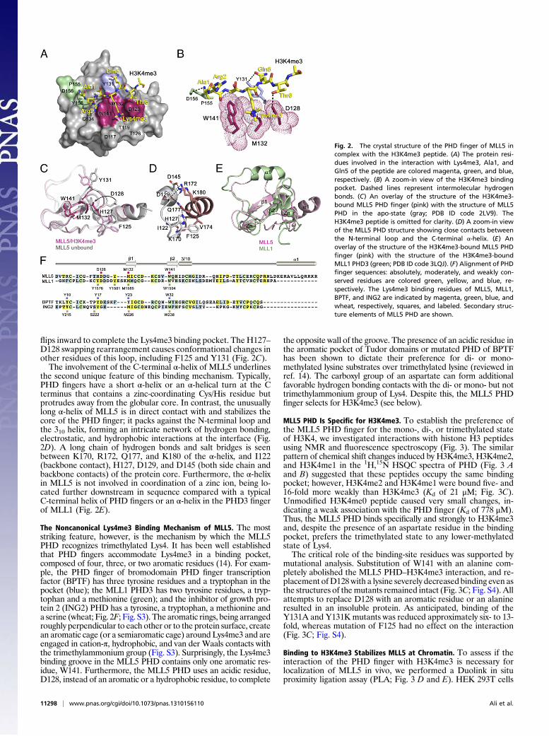
flips inward to complete the Lys4me3 binding pocket. The H127–D128 swapping rearrangement causes conformational changes inother residues of this loop, including F125 and Y131 (Fig. 2C).The involvement of the C-terminal α-helix of MLL5 underlines
the second unique feature of this binding mechanism. Typically,PHD fingers have a short α-helix or an α-helical turn at the Cterminus that contains a zinc-coordinating Cys/His residue butprotrudes away from the globular core. In contrast, the unusuallylong α-helix of MLL5 is in direct contact with and stabilizes thecore of the PHD finger; it packs against the N-terminal loop andthe 310 helix, forming an intricate network of hydrogen bonding,electrostatic, and hydrophobic interactions at the interface (Fig.2D). A long chain of hydrogen bonds and salt bridges is seenbetween K170, R172, Q177, and K180 of the α-helix, and I122(backbone contact), H127, D129, and D145 (both side chain andbackbone contacts) of the protein core. Furthermore, the α-helixin MLL5 is not involved in coordination of a zinc ion, being lo-cated further downstream in sequence compared with a typicalC-terminal helix of PHD fingers or an α-helix in the PHD3 fingerof MLL1 (Fig. 2E).
The Noncanonical Lys4me3 Binding Mechanism of MLL5. The moststriking feature, however, is the mechanism by which the MLL5PHD recognizes trimethylated Lys4. It has been well establishedthat PHD fingers accommodate Lys4me3 in a binding pocket,composed of four, three, or two aromatic residues (14). For exam-ple, the PHD finger of bromodomain PHD finger transcriptionfactor (BPTF) has three tyrosine residues and a tryptophan in thepocket (blue); the MLL1 PHD3 has two tyrosine residues, a tryp-tophan and a methionine (green); and the inhibitor of growth pro-tein 2 (ING2) PHD has a tyrosine, a tryptophan, a methionine anda serine (wheat; Fig. 2F; Fig. S3). The aromatic rings, being arrangedroughly perpendicular to each other or to the protein surface, createan aromatic cage (or a semiaromatic cage) around Lys4me3 and areengaged in cation-π, hydrophobic, and van der Waals contacts withthe trimethylammonium group (Fig. S3). Surprisingly, the Lys4me3binding groove in the MLL5 PHD contains only one aromatic res-idue, W141. Furthermore, the MLL5 PHD uses an acidic residue,D128, instead of an aromatic or a hydrophobic residue, to complete
the opposite wall of the groove. The presence of an acidic residue inthe aromatic pocket of Tudor domains or mutated PHD of BPTFhas been shown to dictate their preference for di- or mono-methylated lysine substrates over trimethylated lysine (reviewed inref. 14). The carboxyl group of an aspartate can form additionalfavorable hydrogen bonding contacts with the di- or mono- but nottrimethylammonium group of Lys4. Despite this, the MLL5 PHDfinger selects for H3K4me3 (see below).
MLL5 PHD Is Specific for H3K4me3. To establish the preference ofthe MLL5 PHD finger for the mono-, di-, or trimethylated stateof H3K4, we investigated interactions with histone H3 peptidesusing NMR and fluorescence spectroscopy (Fig. 3). The similarpattern of chemical shift changes induced by H3K4me3, H3K4me2,and H3K4me1 in the 1H,15N HSQC spectra of PHD (Fig. 3 Aand B) suggested that these peptides occupy the same bindingpocket; however, H3K4me2 and H3K4me1 were bound five- and16-fold more weakly than H3K4me3 (Kd of 21 μM; Fig. 3C).Unmodified H3K4me0 peptide caused very small changes, in-dicating a weak association with the PHD finger (Kd of 778 μM).Thus, the MLL5 PHD binds specifically and strongly to H3K4me3and, despite the presence of an aspartate residue in the bindingpocket, prefers the trimethylated state to any lower-methylatedstate of Lys4.The critical role of the binding-site residues was supported by
mutational analysis. Substitution of W141 with an alanine com-pletely abolished the MLL5 PHD–H3K4me3 interaction, and re-placement ofD128with a lysine severely decreased binding even asthe structures of themutants remained intact (Fig. 3C; Fig. S4). Allattempts to replace D128 with an aromatic residue or an alanineresulted in an insoluble protein. As anticipated, binding of theY131A and Y131K mutants was reduced approximately six- to 13-fold, whereas mutation of F125 had no effect on the interaction(Fig. 3C; Fig. S4).
Binding to H3K4me3 Stabilizes MLL5 at Chromatin. To assess if theinteraction of the PHD finger with H3K4me3 is necessary forlocalization of MLL5 in vivo, we performed a Duolink in situproximity ligation assay (PLA; Fig. 3 D and E). HEK 293T cells
Fig. 2. The crystal structure of the PHD finger of MLL5 incomplex with the H3K4me3 peptide. (A) The protein resi-dues involved in the interaction with Lys4me3, Ala1, andGln5 of the peptide are colored magenta, green, and blue,respectively. (B) A zoom-in view of the H3K4me3 bindingpocket. Dashed lines represent intermolecular hydrogenbonds. (C) An overlay of the structure of the H3K4me3-bound MLL5 PHD finger (pink) with the structure of MLL5PHD in the apo-state (gray; PDB ID code 2LV9). TheH3K4me3 peptide is omitted for clarity. (D) A zoom-in viewof the MLL5 PHD structure showing close contacts betweenthe N-terminal loop and the C-terminal α-helix. (E) Anoverlay of the structure of the H3K4me3-bound MLL5 PHDfinger (pink) with the structure of the H3K4me3-boundMLL1 PHD3 (green; PDB ID code 3LQJ). (F) Alignment of PHDfinger sequences: absolutely, moderately, and weakly con-served residues are colored green, yellow, and blue, re-spectively. The Lys4me3 binding residues of MLL5, MLL1,BPTF, and ING2 are indicated by magenta, green, blue, andwheat, respectively, squares, and labeled. Secondary struc-ture elements of MLL5 PHD are shown.
11298 | www.pnas.org/cgi/doi/10.1073/pnas.1310156110 Ali et al.
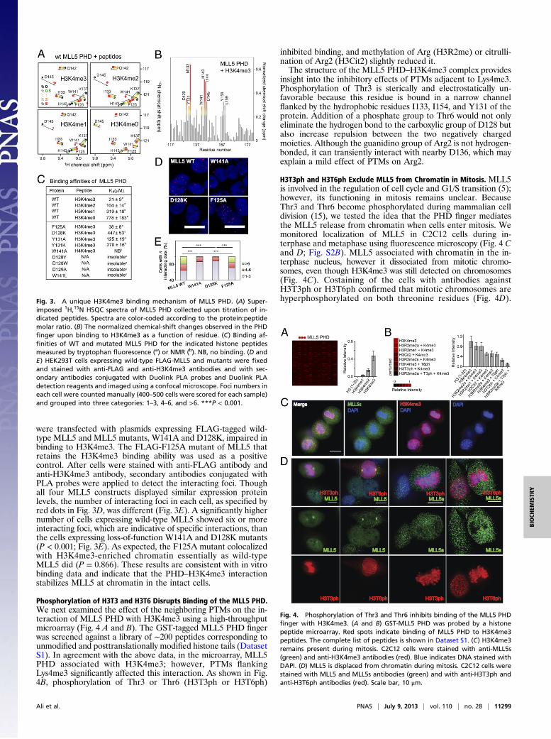
were transfected with plasmids expressing FLAG-tagged wild-type MLL5 and MLL5 mutants, W141A and D128K, impaired inbinding to H3K4me3. The FLAG-F125A mutant of MLL5 thatretains the H3K4me3 binding ability was used as a positivecontrol. After cells were stained with anti-FLAG antibody andanti-H3K4me3 antibody, secondary antibodies conjugated withPLA probes were applied to detect the interacting foci. Thoughall four MLL5 constructs displayed similar expression proteinlevels, the number of interacting foci in each cell, as specified byred dots in Fig. 3D, was different (Fig. 3E). A significantly highernumber of cells expressing wild-type MLL5 showed six or moreinteracting foci, which are indicative of specific interactions, thanthe cells expressing loss-of-function W141A and D128K mutants(P < 0.001; Fig. 3E). As expected, the F125A mutant colocalizedwith H3K4me3-enriched chromatin essentially as wild-typeMLL5 did (P = 0.866). These results are consistent with in vitrobinding data and indicate that the PHD–H3K4me3 interactionstabilizes MLL5 at chromatin in the intact cells.
Phosphorylation of H3T3 and H3T6 Disrupts Binding of the MLL5 PHD.We next examined the effect of the neighboring PTMs on the in-teraction of MLL5 PHD with H3K4me3 using a high-throughputmicroarray (Fig. 4 A and B). The GST-tagged MLL5 PHD fingerwas screened against a library of ∼200 peptides corresponding tounmodified and posttranslationally modified histone tails (DatasetS1). In agreement with the above data, in the microarray, MLL5PHD associated with H3K4me3; however, PTMs flankingLys4me3 significantly affected this interaction. As shown in Fig.4B, phosphorylation of Thr3 or Thr6 (H3T3ph or H3T6ph)
inhibited binding, and methylation of Arg (H3R2me) or citrulli-nation of Arg2 (H3Cit2) slightly reduced it.The structure of the MLL5 PHD–H3K4me3 complex provides
insight into the inhibitory effects of PTMs adjacent to Lys4me3.Phosphorylation of Thr3 is sterically and electrostatically un-favorable because this residue is bound in a narrow channelflanked by the hydrophobic residues I133, I154, and Y131 of theprotein. Addition of a phosphate group to Thr6 would not onlyeliminate the hydrogen bond to the carboxylic group of D128 butalso increase repulsion between the two negatively chargedmoieties. Although the guanidino group of Arg2 is not hydrogen-bonded, it can transiently interact with nearby D136, which mayexplain a mild effect of PTMs on Arg2.
H3T3ph and H3T6ph Exclude MLL5 from Chromatin in Mitosis. MLL5is involved in the regulation of cell cycle and G1/S transition (5);however, its functioning in mitosis remains unclear. BecauseThr3 and Thr6 become phosphorylated during mammalian celldivision (15), we tested the idea that the PHD finger mediatesthe MLL5 release from chromatin when cells enter mitosis. Wemonitored localization of MLL5 in C2C12 cells during in-terphase and metaphase using fluorescence microscopy (Fig. 4 Cand D; Fig. S2B). MLL5 associated with chromatin in the in-terphase nucleus, however it dissociated from mitotic chromo-somes, even though H3K4me3 was still detected on chromosomes(Fig. 4C). Costaining of the cells with antibodies againstH3T3ph or H3T6ph confirmed that mitotic chromosomes arehyperphosphorylated on both threonine residues (Fig. 4D).Fig. 3. A unique H3K4me3 binding mechanism of MLL5 PHD. (A) Super-
imposed 1H,15N HSQC spectra of MLL5 PHD collected upon titration of in-dicated peptides. Spectra are color-coded according to the protein:peptidemolar ratio. (B) The normalized chemical-shift changes observed in the PHDfinger upon binding to H3K4me3 as a function of residue. (C) Binding af-finities of WT and mutated MLL5 PHD for the indicated histone peptidesmeasured by tryptophan fluorescence (a) or NMR (b). NB, no binding. (D andE) HEK293T cells expressing wild-type FLAG-MLL5 and mutants were fixedand stained with anti-FLAG and anti-H3K4me3 antibodies and with sec-ondary antibodies conjugated with Duolink PLA probes and Duolink PLAdetection reagents and imaged using a confocal microscope. Foci numbers ineach cell were counted manually (400–500 cells were scored for each sample)and grouped into three categories: 1–3, 4–6, and >6. ***P < 0.001.
Fig. 4. Phosphorylation of Thr3 and Thr6 inhibits binding of the MLL5 PHDfinger with H3K4me3. (A and B) GST-MLL5 PHD was probed by a histonepeptide microarray. Red spots indicate binding of MLL5 PHD to H3K4me3peptides. The complete list of peptides is shown in Dataset S1. (C) H3K4me3remains present during mitosis. C2C12 cells were stained with anti-MLL5s(green) and anti-H3K4me3 antibodies (red). Blue indicates DNA stained withDAPI. (D) MLL5 is displaced from chromatin during mitosis. C2C12 cells werestained with MLL5 and MLL5s antibodies (green) and with anti-H3T3ph andanti-H3T6ph antibodies (red). Scale bar, 10 μm.
Ali et al. PNAS | July 9, 2013 | vol. 110 | no. 28 | 11299
BIOCH
EMISTR
Y
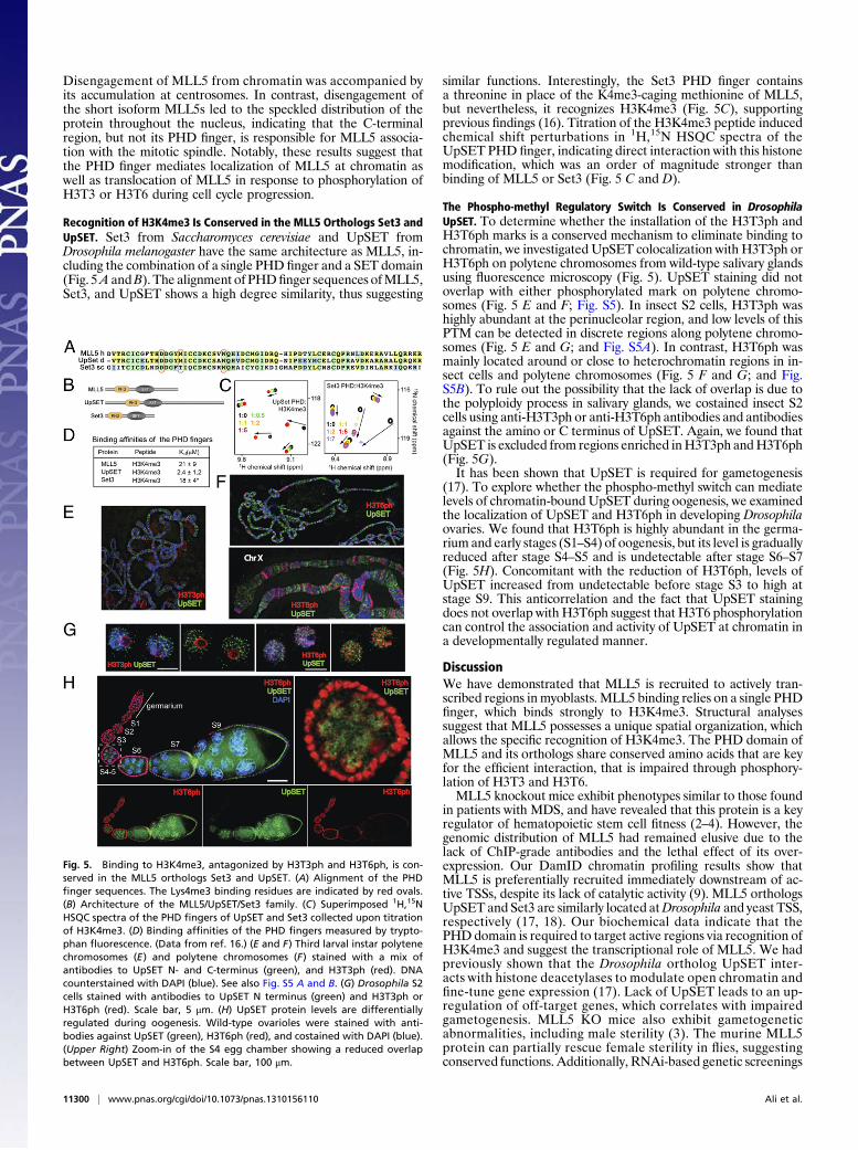
Disengagement of MLL5 from chromatin was accompanied byits accumulation at centrosomes. In contrast, disengagement ofthe short isoform MLL5s led to the speckled distribution of theprotein throughout the nucleus, indicating that the C-terminalregion, but not its PHD finger, is responsible for MLL5 associa-tion with the mitotic spindle. Notably, these results suggest thatthe PHD finger mediates localization of MLL5 at chromatin aswell as translocation of MLL5 in response to phosphorylation ofH3T3 or H3T6 during cell cycle progression.
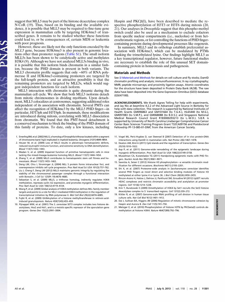
Recognition of H3K4me3 Is Conserved in the MLL5 Orthologs Set3 andUpSET. Set3 from Saccharomyces cerevisiae and UpSET fromDrosophila melanogaster have the same architecture as MLL5, in-cluding the combination of a single PHD finger and a SET domain(Fig. 5A andB). The alignment of PHDfinger sequences ofMLL5,Set3, and UpSET shows a high degree similarity, thus suggesting
similar functions. Interestingly, the Set3 PHD finger containsa threonine in place of the K4me3-caging methionine of MLL5,but nevertheless, it recognizes H3K4me3 (Fig. 5C), supportingprevious findings (16). Titration of the H3K4me3 peptide inducedchemical shift perturbations in 1H,15N HSQC spectra of theUpSET PHD finger, indicating direct interaction with this histonemodification, which was an order of magnitude stronger thanbinding of MLL5 or Set3 (Fig. 5 C and D).
The Phospho-methyl Regulatory Switch Is Conserved in DrosophilaUpSET. To determine whether the installation of the H3T3ph andH3T6ph marks is a conserved mechanism to eliminate binding tochromatin, we investigated UpSET colocalization with H3T3ph orH3T6ph on polytene chromosomes from wild-type salivary glandsusing fluorescence microscopy (Fig. 5). UpSET staining did notoverlap with either phosphorylated mark on polytene chromo-somes (Fig. 5 E and F; Fig. S5). In insect S2 cells, H3T3ph washighly abundant at the perinucleolar region, and low levels of thisPTM can be detected in discrete regions along polytene chromo-somes (Fig. 5 E and G; and Fig. S5A). In contrast, H3T6ph wasmainly located around or close to heterochromatin regions in in-sect cells and polytene chromosomes (Fig. 5 F and G; and Fig.S5B). To rule out the possibility that the lack of overlap is due tothe polyploidy process in salivary glands, we costained insect S2cells using anti-H3T3ph or anti-H3T6ph antibodies and antibodiesagainst the amino or C terminus of UpSET. Again, we found thatUpSET is excluded from regions enriched inH3T3ph andH3T6ph(Fig. 5G).It has been shown that UpSET is required for gametogenesis
(17). To explore whether the phospho-methyl switch can mediatelevels of chromatin-bound UpSET during oogenesis, we examinedthe localization of UpSET and H3T6ph in developing Drosophilaovaries. We found that H3T6ph is highly abundant in the germa-rium and early stages (S1–S4) of oogenesis, but its level is graduallyreduced after stage S4–S5 and is undetectable after stage S6–S7(Fig. 5H). Concomitant with the reduction of H3T6ph, levels ofUpSET increased from undetectable before stage S3 to high atstage S9. This anticorrelation and the fact that UpSET stainingdoes not overlap with H3T6ph suggest that H3T6 phosphorylationcan control the association and activity of UpSET at chromatin ina developmentally regulated manner.
DiscussionWe have demonstrated that MLL5 is recruited to actively tran-scribed regions in myoblasts. MLL5 binding relies on a single PHDfinger, which binds strongly to H3K4me3. Structural analysessuggest that MLL5 possesses a unique spatial organization, whichallows the specific recognition of H3K4me3. The PHD domain ofMLL5 and its orthologs share conserved amino acids that are keyfor the efficient interaction, that is impaired through phosphory-lation of H3T3 and H3T6.MLL5 knockout mice exhibit phenotypes similar to those found
in patients with MDS, and have revealed that this protein is a keyregulator of hematopoietic stem cell fitness (2–4). However, thegenomic distribution of MLL5 had remained elusive due to thelack of ChIP-grade antibodies and the lethal effect of its over-expression. Our DamID chromatin profiling results show thatMLL5 is preferentially recruited immediately downstream of ac-tive TSSs, despite its lack of catalytic activity (9). MLL5 orthologsUpSET and Set3 are similarly located atDrosophila and yeast TSS,respectively (17, 18). Our biochemical data indicate that thePHDdomain is required to target active regions via recognition ofH3K4me3 and suggest the transcriptional role of MLL5. We hadpreviously shown that the Drosophila ortholog UpSET inter-acts with histone deacetylases to modulate open chromatin andfine-tune gene expression (17). Lack of UpSET leads to an up-regulation of off-target genes, which correlates with impairedgametogenesis. MLL5 KO mice also exhibit gametogeneticabnormalities, including male sterility (3). The murine MLL5protein can partially rescue female sterility in flies, suggestingconserved functions. Additionally, RNAi-based genetic screenings
Fig. 5. Binding to H3K4me3, antagonized by H3T3ph and H3T6ph, is con-served in the MLL5 orthologs Set3 and UpSET. (A) Alignment of the PHDfinger sequences. The Lys4me3 binding residues are indicated by red ovals.(B) Architecture of the MLL5/UpSET/Set3 family. (C) Superimposed 1H,15NHSQC spectra of the PHD fingers of UpSET and Set3 collected upon titrationof H3K4me3. (D) Binding affinities of the PHD fingers measured by trypto-phan fluorescence. (Data from ref. 16.) (E and F) Third larval instar polytenechromosomes (E) and polytene chromosomes (F) stained with a mix ofantibodies to UpSET N- and C-terminus (green), and H3T3ph (red). DNAcounterstained with DAPI (blue). See also Fig. S5 A and B. (G) Drosophila S2cells stained with antibodies to UpSET N terminus (green) and H3T3ph orH3T6ph (red). Scale bar, 5 μm. (H) UpSET protein levels are differentiallyregulated during oogenesis. Wild-type ovarioles were stained with anti-bodies against UpSET (green), H3T6ph (red), and costained with DAPI (blue).(Upper Right) Zoom-in of the S4 egg chamber showing a reduced overlapbetween UpSET and H3T6ph. Scale bar, 100 μm.
11300 | www.pnas.org/cgi/doi/10.1073/pnas.1310156110 Ali et al.
suggest thatMLL5may be part of the histone deacetylase complexN-CoR (19). Thus, based on its binding and the available evi-dence, it is possible that MLL5 may be required to fine-tune geneexpression in mammalian cells by targeting H3K4me3 of tran-scribed genes. It remains to be studied whether these functionscould lead to, or be consequences of, certain MDS or leukemiawith poor prognosis.However, these are likely not the only functions encoded by the
MLL5 gene, because H3K4me3 is also present in genomic loca-tions other than promoter regions (Table S1). The small isoformMLL5s has been shown to be catalytically active and methylatesH3K4 (9). Although we have not analyzed MLL5s binding in vivo,it is possible that this isoform binds chromatin in a similar fash-ion, because the PHD domain is present in both variants. TheMLL5 chromatin profile suggests that only ∼60% of RNA poly-merase II and H3K4me3-containing promoters are targeted bythe full-length protein, and an attractive possibility is that theremaining promoters are targeted by MLL5s, which would sug-gest independent functions for each isoform.MLL5 interaction with chromatin is quite dynamic during the
mammalian cell cycle. We show that both MLL5 isoforms detachfrom mitotic chromosomes in dividing myoblasts. Upon detach-ment,MLL5 colocalizes at centrosomes, suggesting additional rolesindependent of its association with chromatin. Several PMTs canaffect the recognition of H3K4me3 by the MLL5 PHD finger—inparticular, H3T3ph and H3T6ph. In mammals, these modificationsare introduced during mitosis, correlating with MLL5 dissociationfrom chromatin. We found that this PMT-based detachment isa conservedmechanism to block the binding of the PHD domain ofthis family of proteins. To date, only a few kinases, including
Haspin and PKCβ(I), have been described to mediate the re-spective phosphorylation of H3T3 or H3T6 during mitosis (20,21). Our analyses in Drosophila suggest that this phospho-methylswitch could also be used as a mechanism to exclude cofactorsfrom specific nuclear compartments (i.e., nucleolus) or from het-erochromatic regions, or for controlling the functions of PHDfinger-containing proteins during developmental processes like oogenesis.In summary, MLL5 and its orthologs establish preferential as-
sociation with H3K4me3, which can be modulated by PTMsflanking the trimethylated lysine. Our findings highlight MLL5 asa key transcriptional regulator, however, future functional studiesare necessary to establish the role of this unusual SET domain-containing protein in homeostasis and pathogenesis.
Materials and MethodsSee SI Materials and Methods for details on cell culture and fly stocks; DamIDchromatin profiling and analysis; immunofluorescence; X-ray crystallography;NMR; peptide microarrays; and proximity ligation assays. Atomic coordinatesfor the structure have been deposited in Protein Data Bank (4L58). The rawdata have been deposited into the Gene Expression Omnibus (GEO) database(GSE47100).
ACKNOWLEDGMENTS. We thank Agnes Telling for help with experiments,and Jay Nix at beamline 4.2.2 of the Advanced Light Source in Berkeley forhelp with data collection. This research is supported by National Institutes ofHealth Grants GM096863 and GM101664 (to T.G.K.), HL65440 (to M.G.),GM097083 (to S.M.P.), and GM068088 (to B.D.S.); and Singapore NationalMedical Research Council Grant R183000293213 (to L.-W.D.). S.B.R. issupported by University of North Carolina Lineberger Comprehensive CancerCenter Basic Sciences Training Program Grant T32CA09156 and PostdoctoralFellowship PF-13-085-01-DMC from the American Cancer Society.
1. EmerlingBM,etal. (2002)MLL5, ahomologofDrosophila trithorax locatedwithina segmentof chromosome band 7q22 implicated in myeloid leukemia.Oncogene 21(31):4849–4854.
2. Heuser M, et al. (2009) Loss of MLL5 results in pleiotropic hematopoietic defects,reduced neutrophil immune function, and extreme sensitivity to DNA demethylation.Blood 113(7):1432–1443.
3. Madan V, et al. (2009) Impaired function of primitive hematopoietic cells in micelacking the mixed-lineage-leukemia homolog MLL5. Blood 113(7):1444–1454.
4. Zhang Y, et al. (2009) MLL5 contributes to hematopoietic stem cell fitness and ho-meostasis. Blood 113(7):1455–1463.
5. Deng LW, Chiu I, Strominger JL (2004) MLL 5 protein forms intranuclear foci, andoverexpression inhibits cell cycle progression. Proc Natl Acad Sci USA 101(3):757–762.
6. Liu J, Cheng F, Deng LW (2012) MLL5 maintains genomic integrity by regulating thestability of the chromosomal passenger complex through a functional interactionwith Borealin. J Cell Sci 125(Pt 19):4676–4685.
7. Sebastian S, et al. (2009) MLL5, a trithorax homolog, indirectly regulates H3K4methylation, represses cyclin A2 expression, and promotes myogenic differentiation.Proc Natl Acad Sci USA 106(12):4719–4724.
8. Wang P, et al. (2009) Global analysis of H3K4 methylation defines MLL family membertargets and points to a role for MLL1-mediated H3K4 methylation in the regulation oftranscriptional initiation by RNA polymerase II. Mol Cell Biol 29(22):6074–6085.
9. Fujiki R, et al. (2009) GlcNAcylation of a histone methyltransferase in retinoic-acid-induced granulopoiesis. Nature 459(7245):455–459.
10. Pijnappel WW, et al. (2001) The S. cerevisiae SET3 complex includes two histone de-acetylases, Hos2 and Hst1, and is a meiotic-specific repressor of the sporulation geneprogram. Genes Dev 15(22):2991–3004.
11. Vogel MJ, Peric-Hupkes D, van Steensel B (2007) Detection of in vivo protein-DNA
interactions using DamID in mammalian cells. Nat Protoc 2(6):1467–1478.12. Deaton AM, Bird A (2011) CpG islands and the regulation of transcription. Genes Dev
25(10):1010–1022.13. Asp P, et al. (2011) Genome-wide remodeling of the epigenetic landscape during
myogenic differentiation. Proc Natl Acad Sci USA 108(22):E149–E158.14. Musselman CA, Kutateladze TG (2011) Handpicking epigenetic marks with PHD fin-
gers. Nucleic Acids Res 39(21):9061–9071.15. Sawicka A, Seiser C (2012) Histone H3 phosphorylation—a versatile chromatin mod-
ification for different occasions. Biochimie 94(11):2193–2201.16. Shi X, et al. (2007) Proteome-wide analysis in Saccharomyces cerevisiae identifies
several PHD fingers as novel direct and selective binding modules of histone H3
methylated at either lysine 4 or lysine 36. J Biol Chem 282(4):2450–2455.17. Rincon-Arano H, Halow J, Delrow JJ, Parkhurst SM, Groudine M (2012) UpSET recruits
HDAC complexes and restricts chromatin accessibility and acetylation at promoter
regions. Cell 151(6):1214–1228.18. Kim T, Buratowski S (2009) Dimethylation of H3K4 by Set1 recruits the Set3 histone
deacetylase complex to 5′ transcribed regions. Cell 137(2):259–272.19. Kittler R, et al. (2007) Genome-scale RNAi profiling of cell division in human tissue
culture cells. Nat Cell Biol 9(12):1401–1412.20. Dai J, Sullivan BA, Higgins JM (2006) Regulation of mitotic chromosome cohesion by
Haspin and Aurora B. Dev Cell 11(5):741–750.21. Metzger E, et al. (2010) Phosphorylation of histone H3T6 by PKCbeta(I) controls de-
methylation at histone H3K4. Nature 464(7289):792–796.
Ali et al. PNAS | July 9, 2013 | vol. 110 | no. 28 | 11301
BIOCH
EMISTR
Y


![Techniques and strategies employing engineered …bleris/papers/2017-TALEs.pdfNCP [31,32]. Chromatin immunoprecipitation and sequencing (ChIP-seq) has revealed dCas9 binding from tens](https://img.pdfslide.us/doc/110x75/60accfbcf2c1682e39595fa9/techniques-and-strategies-employing-engineered-blerispapers2017-talespdf-ncp.jpg)