Embed Size (px)
Citation preview
Molecular and Structural Basisof Cytokine Receptor Pleiotropyin the Interleukin-4/13 SystemSherry L. LaPorte,1 Z. Sean Juo,1 Jana Vaclavikova,1 Leremy A. Colf,1 Xiulan Qi,2 Nicola M. Heller,2 Achsah D. Keegan,2
and K. Christopher Garcia1,*1Howard Hughes Medical Institute, Departments of Molecular and Cellular Physiology, and Structural Biology, Stanford University School
of Medicine, Stanford, CA 94305, USA2Center for Vascular and Inflammatory Diseases and Department of Microbiology and Immunology, University of Maryland Schoolof Medicine, Baltimore, MD, 21201, USA
*Correspondence: [email protected]
DOI 10.1016/j.cell.2007.12.030
SUMMARY
Interleukin-4 and Interleukin-13 are cytokines criticalto the development of T cell-mediated humoral im-mune responses, which are associated with allergyand asthma, and exert their actions through three dif-ferent combinations of shared receptors. Here wepresent the crystal structures of the complete set oftype I (IL-4Ra/gc/IL-4) and type II (IL-4Ra/IL-13Ra1/IL-4, IL-4Ra/IL-13Ra1/IL-13) ternary signaling com-plexes. The type I complex reveals a structural basisfor gc’s ability to recognize six different gc-cytokines.The two type II complexes utilize an unusual top-mounted Ig-like domain on IL-13Ra1 for a novelmode of cytokine engagement that contributes toa reversal in the IL-4 versus IL-13 ternary complex as-sembly sequences, which are mediated through sub-stantially different recognition chemistries. We alsoshow that the type II receptor heterodimer signalswith different potencies in response to IL-4 versusIL-13 and suggest that the extracellular cytokine-receptor interactions are modulating intracellularmembrane-proximal signaling events.
INTRODUCTION
T cell-mediated immunity occurs in successive antigen-driven
and cytokine-driven steps. In the first step, T cell receptors are
activated by specific antigens presented by peptide-MHC com-
plexes on the surface of antigen-presenting cells. In a second
step, these activated T cells are induced to clonally expand by
the cytokine Interleukin-2 (IL-2) and subsequently differentiate
into CD4+ Th1 or Th2 cells as a result of a specific subset of cy-
tokines engaging and activating their respective cytokine recep-
tors (Leonard, 1999). IL-4 is the primary cytokine implicated in
the development of Th2-mediated responses (Seder and Paul,
1994), which are also associated with allergy and asthma
(Barnes, 2002). The cytokines IL-4 and IL-13 are produced by
Th2 cells, recruit and activate IgE-producing B cells, and en-
hance IgE-mediated responses. The strong association of Th2
cytokines with allergic disease has generated interest in target-
ing their receptors for therapeutic intervention (Foster et al.,
2002; Mueller et al., 2002).
IL-4 alpha receptor (IL-4Ra) is a unique member of the com-
mon-gamma chain (gc) family of receptors (Nelms et al., 1999),
with the ability to signal within three different receptor com-
plexes, known as type I and type II receptors (Andrews et al.,
2006; Kelly-Welch et al., 2003; Mueller et al., 2002). On cells of
hematopoietic stem cell origin, the type I receptor comprises
IL-4Ra and the common gamma-chain (gc), which is also shared
by the cytokines IL-2, -7, -9, -15, and -21 (Nelms et al., 1999). On
cells of nonhematopoietic stem cell origin, IL-4 can use the
type II complex, comprising IL-4Ra and IL-13Ra1 (Hilton et al.,
1996; Obiri et al., 1995). This receptor complex is also a func-
tional receptor for IL-13 (Aman et al., 1996), which shares only
�25% homology with IL-4, and this largely explains the overlap
of the biological effects of IL-4 and IL-13 (Wills-Karp, 2004). Type
I receptor complexes can only be formed by IL-4 and are more
active in regulating Th2 development. In contrast, the type II re-
ceptor complex formed by either IL-4 or IL-13 is not found on T
cells and is more active in regulating cells that mediate airway
hypersensitivity and mucus secretion (Andrews et al., 2006). In
fact, IL-13 appears to have its own distinct role in allergic inflam-
mation, acting as a key regulator of allergen-induced airway in-
flammation and goblet cell metaplasia (Wills-Karp, 2004). The
molecular basis for the cytokines’ differing functional responses
is unclear. Both type I and type II receptor complexes signal
through the Jak/STAT cascade, with IL-4Ra associating with
Jak1, gc with Jak3, and IL-13Ra1 with Tyk2. A second mecha-
nism of signal transduction activated by IL-4 and IL-13 leads to
the insulin receptor substrate (IRS) family (Kelly-Welch et al.,
2003).
IL-4 and IL-13 are prototypical four-helix bundle short-chain
cytokines (Moy et al., 2001; Mueller et al., 2002; Walter et al.,
1992; Wlodawer et al., 1992). The IL-4/IL-13 receptors gc,
IL-4Ra, and IL-13Ra1 contain the tandem Fibronectin-III do-
mains and the WSXWS box that form the classical elbow-shaped
‘‘cytokine binding homology region (CHR)’’ that binds to the
Cell 132, 259–272, January 25, 2008 ª2008 Elsevier Inc. 259
helical faces of the cytokine (Bazan, 1990), as originally defined
in the human Growth Hormone (hGH) system (de Vos et al.,
1992). A previous structure of a nonsignaling binary complex be-
tween IL-4 and IL-4Ra indicated a similarity to the hGH-R site I
paradigm (Hage et al., 1999). While the structure of a complete
short-chain cytokine receptor heterodimeric signaling complex
was reported for IL-2 (Wang et al., 2005), the type I and type II
IL-4/13 receptors have several novelties that pose interesting
structural questions (Mueller et al., 2002). First, in the type II com-
plex, the IL-13Ra1 contains an extra N-terminal Ig-like domain
(D1) not found in other receptors of the gc subfamily that is re-
quired for IL-13 signaling but not for IL-4 (Arima et al., 2005). It
is unknown how this top-mounted Ig-like domain participates
in cytokine recognition.
A second novelty pertains to cytokine receptor ‘‘sharing’’ and
the evolutionary relationships between IL-13Ra1, gc, and IL-4Ra
(Boulay et al., 2003; Leonard et al., 1994; Ozaki and Leonard,
2002). While gc is the primary short-chain cytokine shared recep-
tor, IL-4Ra is also shared in three signaling complexes. IL-13Ra1
bears an evolutionary relationship to gc (Boulay et al., 2003), but
its ligand specificity is narrower and it has diverged from a com-
mon ancestral family with the acquisition of the N-terminal Ig-like
domain. It will be interesting to determine if these receptors pos-
sess structural properties conducive to degenerate recognition
and signaling in comparison to their highly specific alpha recep-
tor counterparts. In addition, as only one gc-family complex has
been solved (IL-2) (Wang et al., 2005), the type I complex could
reveal a molecular basis for gc crossreactivity.
Finally, IL-4 and IL-13 can induce distinct functional responses
through the identical type II receptor heterodimers (Kelly-Welch
et al., 2003), raising the question of how ligand specificity is con-
ferred by shared receptors and can influence signaling. Thus, the
IL-4/13 system presents an opportunity to investigate whether
downstream signaling differences through common receptors
have origins in the structural/biophysical properties of the extra-
cellular receptor-cytokine complexes. In order to probe these
questions, we present structural, biochemical, and signaling
studies on the complete set of type I and type II ternary receptor
complexes, IL-4Ra/gc/IL-4, IL-4Ra/IL-13Ra1/IL-4, IL-4Ra/IL-
13Ra1/IL-13.
RESULTS
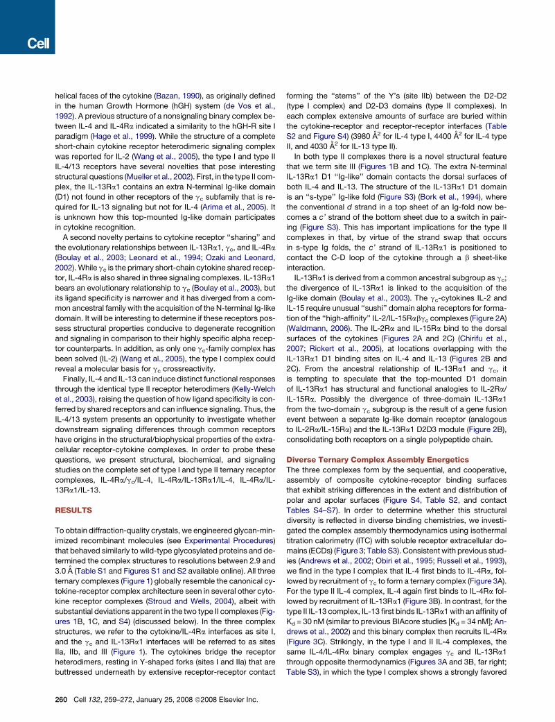
To obtain diffraction-quality crystals, we engineered glycan-min-
imized recombinant molecules (see Experimental Procedures)
that behaved similarly to wild-type glycosylated proteins and de-
termined the complex structures to resolutions between 2.9 and
3.0 A (Table S1 and Figures S1 and S2 available online). All three
ternary complexes (Figure 1) globally resemble the canonical cy-
tokine-receptor complex architecture seen in several other cyto-
kine receptor complexes (Stroud and Wells, 2004), albeit with
substantial deviations apparent in the two type II complexes (Fig-
ures 1B, 1C, and S4) (discussed below). In the three complex
structures, we refer to the cytokine/IL-4Ra interfaces as site I,
and the gc and IL-13Ra1 interfaces will be referred to as sites
IIa, IIb, and III (Figure 1). The cytokines bridge the receptor
heterodimers, resting in Y-shaped forks (sites I and IIa) that are
buttressed underneath by extensive receptor-receptor contact
260 Cell 132, 259–272, January 25, 2008 ª2008 Elsevier Inc.
forming the ‘‘stems’’ of the Y’s (site IIb) between the D2-D2
(type I complex) and D2-D3 domains (type II complexes). In
each complex extensive amounts of surface are buried within
the cytokine-receptor and receptor-receptor interfaces (Table
S2 and Figure S4) (3980 A2 for IL-4 type I, 4400 A2 for IL-4 type
II, and 4030 A2 for IL-13 type II).
In both type II complexes there is a novel structural feature
that we term site III (Figures 1B and 1C). The extra N-terminal
IL-13Ra1 D1 ‘‘Ig-like’’ domain contacts the dorsal surfaces of
both IL-4 and IL-13. The structure of the IL-13Ra1 D1 domain
is an ‘‘s-type’’ Ig-like fold (Figure S3) (Bork et al., 1994), where
the conventional d strand in a top sheet of an Ig-fold now be-
comes a c’ strand of the bottom sheet due to a switch in pair-
ing (Figure S3). This has important implications for the type II
complexes in that, by virtue of the strand swap that occurs
in s-type Ig folds, the c’ strand of IL-13Ra1 is positioned to
contact the C-D loop of the cytokine through a b sheet-like
interaction.
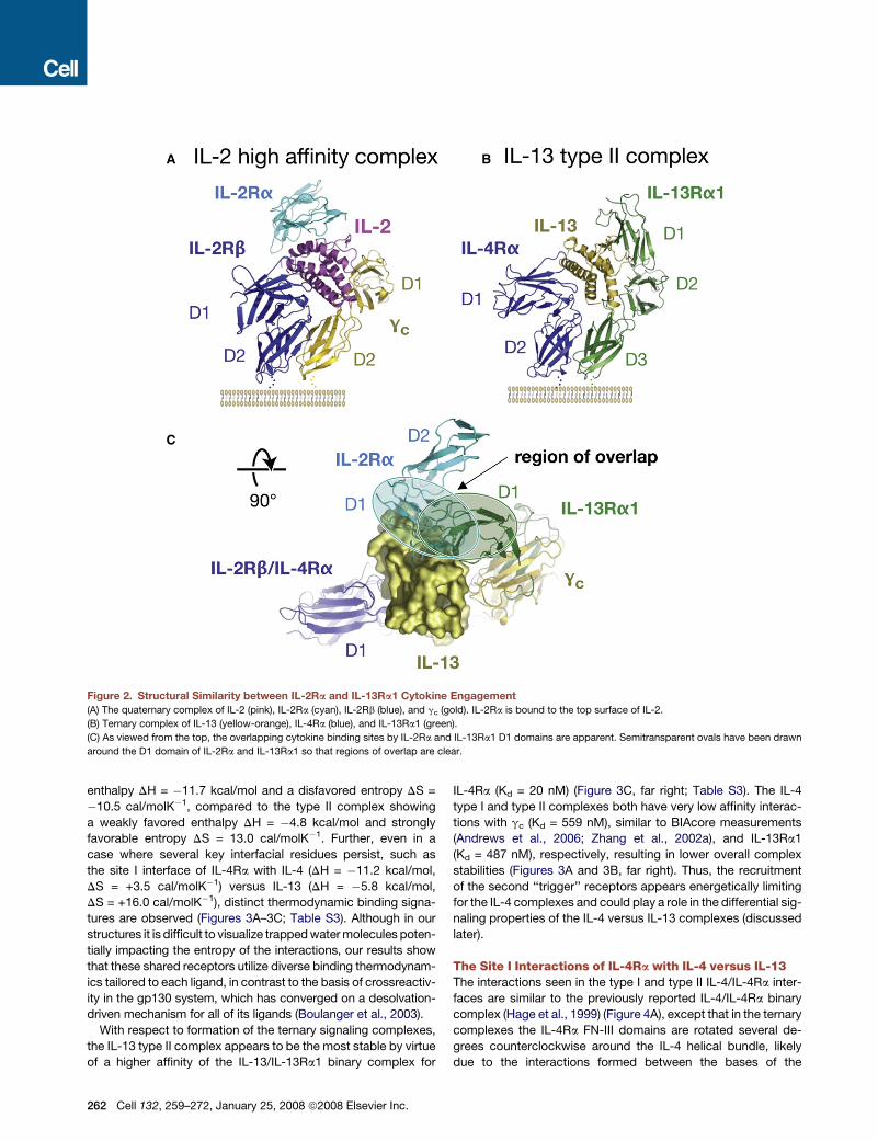
IL-13Ra1 is derived from a common ancestral subgroup as gc;
the divergence of IL-13Ra1 is linked to the acquisition of the
Ig-like domain (Boulay et al., 2003). The gc-cytokines IL-2 and
IL-15 require unusual ‘‘sushi’’ domain alpha receptors for forma-
tion of the ‘‘high-affinity’’ IL-2/IL-15Rabgc complexes (Figure 2A)
(Waldmann, 2006). The IL-2Ra and IL-15Ra bind to the dorsal
surfaces of the cytokines (Figures 2A and 2C) (Chirifu et al.,
2007; Rickert et al., 2005), at locations overlapping with the
IL-13Ra1 D1 binding sites on IL-4 and IL-13 (Figures 2B and
2C). From the ancestral relationship of IL-13Ra1 and gc, it
is tempting to speculate that the top-mounted D1 domain
of IL-13Ra1 has structural and functional analogies to IL-2Ra/
IL-15Ra. Possibly the divergence of three-domain IL-13Ra1
from the two-domain gc subgroup is the result of a gene fusion
event between a separate Ig-like domain receptor (analogous
to IL-2Ra/IL-15Ra) and the IL-13Ra1 D2D3 module (Figure 2B),
consolidating both receptors on a single polypeptide chain.
Diverse Ternary Complex Assembly EnergeticsThe three complexes form by the sequential, and cooperative,
assembly of composite cytokine-receptor binding surfaces
that exhibit striking differences in the extent and distribution of
polar and apolar surfaces (Figure S4, Table S2, and contact
Tables S4–S7). In order to determine whether this structural
diversity is reflected in diverse binding chemistries, we investi-
gated the complex assembly thermodynamics using isothermal
titration calorimetry (ITC) with soluble receptor extracellular do-
mains (ECDs) (Figure 3; Table S3). Consistent with previous stud-
ies (Andrews et al., 2002; Obiri et al., 1995; Russell et al., 1993),
we find in the type I complex that IL-4 first binds to IL-4Ra, fol-
lowed by recruitment of gc to form a ternary complex (Figure 3A).
For the type II IL-4 complex, IL-4 again first binds to IL-4Ra fol-
lowed by recruitment of IL-13Ra1 (Figure 3B). In contrast, for the
type II IL-13 complex, IL-13 first binds IL-13Ra1 with an affinity of
Kd = 30 nM (similar to previous BIAcore studies [Kd = 34 nM]; An-
drews et al., 2002) and this binary complex then recruits IL-4Ra
(Figure 3C). Strikingly, in the type I and II IL-4 complexes, the
same IL-4/IL-4Ra binary complex engages gc and IL-13Ra1
through opposite thermodynamics (Figures 3A and 3B, far right;
Table S3), in which the type I complex shows a strongly favored

Figure 1. Structures of the Type I IL-4, Type II IL-4, and Type II IL-13 Ternary Complexes
(A) The type I complex with IL-4Ra (blue), IL-4 (red), and gc (gold).
(B) The type II IL-4 complex with IL-4Ra (blue), IL-4 (red), and IL-13Ra1 (green).
(C) The type II IL-13 complex with IL-4Ra (blue), IL-13 (yellow-orange), and IL-13Ra1 (green).
The complexes are shown from the side with a cartoon of a membrane underneath (left), and as viewed from the ‘‘top’’ (right). Glycan moieties on Asn residues are
shown in ball-and-stick representation. All figures were generated with PyMol (DeLano, 2002).
Cell 132, 259–272, January 25, 2008 ª2008 Elsevier Inc. 261
enthalpy DH = �11.7 kcal/mol and a disfavored entropy DS =
�10.5 cal/molK�1, compared to the type II complex showing
a weakly favored enthalpy DH = �4.8 kcal/mol and strongly
favorable entropy DS = 13.0 cal/molK�1. Further, even in a
case where several key interfacial residues persist, such as
the site I interface of IL-4Ra with IL-4 (DH = �11.2 kcal/mol,
DS = +3.5 cal/molK�1) versus IL-13 (DH = �5.8 kcal/mol,
DS = +16.0 cal/molK�1), distinct thermodynamic binding signa-
tures are observed (Figures 3A–3C; Table S3). Although in our
structures it is difficult to visualize trapped water molecules poten-
tially impacting the entropy of the interactions, our results show
that these shared receptors utilize diverse binding thermodynam-
ics tailored to each ligand, in contrast to the basis of crossreactiv-
ity in the gp130 system, which has converged on a desolvation-
driven mechanism for all of its ligands (Boulanger et al., 2003).
With respect to formation of the ternary signaling complexes,
the IL-13 type II complex appears to be the most stable by virtue
of a higher affinity of the IL-13/IL-13Ra1 binary complex for
262 Cell 132, 259–272, January 25, 2008 ª2008 Elsevier Inc.
IL-4Ra (Kd = 20 nM) (Figure 3C, far right; Table S3). The IL-4
type I and type II complexes both have very low affinity interac-
tions with gc (Kd = 559 nM), similar to BIAcore measurements
(Andrews et al., 2006; Zhang et al., 2002a), and IL-13Ra1
(Kd = 487 nM), respectively, resulting in lower overall complex
stabilities (Figures 3A and 3B, far right). Thus, the recruitment
of the second ‘‘trigger’’ receptors appears energetically limiting
for the IL-4 complexes and could play a role in the differential sig-
naling properties of the IL-4 versus IL-13 complexes (discussed
later).
The Site I Interactions of IL-4Ra with IL-4 versus IL-13The interactions seen in the type I and type II IL-4/IL-4Ra inter-
faces are similar to the previously reported IL-4/IL-4Ra binary
complex (Hage et al., 1999) (Figure 4A), except that in the ternary
complexes the IL-4Ra FN-III domains are rotated several de-
grees counterclockwise around the IL-4 helical bundle, likely
due to the interactions formed between the bases of the
Figure 2. Structural Similarity between IL-2Ra and IL-13Ra1 Cytokine Engagement
(A) The quaternary complex of IL-2 (pink), IL-2Ra (cyan), IL-2Rb (blue), and gc (gold). IL-2Ra is bound to the top surface of IL-2.
(B) Ternary complex of IL-13 (yellow-orange), IL-4Ra (blue), and IL-13Ra1 (green).
(C) As viewed from the top, the overlapping cytokine binding sites by IL-2Ra and IL-13Ra1 D1 domains are apparent. Semitransparent ovals have been drawn
around the D1 domain of IL-2Ra and IL-13Ra1 so that regions of overlap are clear.

receptors (i.e., the stem of the Y). The principal feature of the
large IL-4/IL-4Ra interface (Figures 4A and S4) remains a charge
complementarity between patches centered on ‘‘hotspot’’ resi-
dues Glu9 and Arg88 on the IL-4 A and C helices (Figure 4A)
(Zhang et al., 2002b), respectively, which form polar contacts
with Tyr13, Tyr183, Ser70, and Asp72 on IL-4Ra.
The site I interface of IL-13 with IL-4Ra (Figure 4B) is presented
on a more compact IL-13 helical scaffold (see Figures 1B and 1C,
top view) (two helical turns shorter in both A and C helices than
IL-4). Although the binding surface and amino acids used
on IL-4Ra to bind IL-13 are nearly identical to that used to con-
tact IL-4 (Figure 4A), their four-helix bundles superimpose poorly
when the complexes are aligned on IL-4Ra (Figure 4C). Yet,
IL-13 exactly recapitulates two critical charged residues by pre-
senting Glu12 and Arg65 to IL-4Ra in the same positions to form
similar receptor contacts as seen for IL-4 residues Glu9 and
Arg88 (Figures 4B–4D), which have been shown by mutagenesis
to be energetically important (Kraich et al., 2006). However, the
composition of remaining IL-13 contact residues with IL-4Ra is
different (Figures 4C and 4D), in accord with the distinct binding
thermodynamics the two cytokines use to bind IL-4Ra (Figures
3B and 3C; Table S3). In the IL-13/IL-4Ra site I, the interface
lacks interactions ringing the charged hotspots observed in the
IL-4/IL-4Ra interface, such as the loss of IL-4 residue Trp91 on
the C helix (Figure 4C). The number of polar bonds per 100 A2
of surface buried in the IL-13/IL-4Ra interface is 1.0, denoting
a substantial loss in specificity relative to the extraordinarily spe-
cific IL-4/IL-4Ra complex (2 polar bonds per 100 A2). Given that
IL-4 binds to IL-4Ra with subnanomolar affinity, and IL-13 has no
measurable affinity for IL-4Ra alone, the energetic value of the
two ‘‘mimicked’’ charged contacts on IL-13 (Kraich et al.,
2006) is apparently manifested within the context of the ‘‘com-
posite’’ interface formed together with the receptor-receptor
contacts (site IIb). In this fashion, the IL-4Ra/IL-13 site I works
in concert with site IIb to engage IL-4Ra through tandem, multi-
point attachment of two weak interfaces.
The Site IIa and Site III InteractionsThe formation of the ternary signaling complex is completed by
the recruitment of a second receptor, either gc or IL-13Ra1,
through structurally separated binding contacts designated sites
IIa (fork of the Y) (Table S5), IIb (stem of the Y) (Table S6), and, in
the cases of the type II complexes, site III (Table S5).
The three site IIa interfaces are uniformly characterized by the
convex elbow of the receptors occupying a canyon on the A and
D helices of the cytokines (Figures 5A–5C), exhibiting excellent
knob-in-holes shape complementarity (Table S2) (Sc = 0.82,
0.73, and 0.65 for the IL-4 type I and IL-4 and IL-13 type II com-
plexes, respectively). Several hydrogen bonds flank the sides of
the apolar canyons in each complex and likely stabilize the
respective docking orientations. Consistent with their ancestral
relationship, IL-13Ra1 and gc present binding sites with conver-
gent structural features (Figures 5A and 5B). The site IIa contacts
in each of the receptor complexes are primarily formed by the
FG2 loop of the membrane-proximal FN-III domains of gc and
IL-13Ra1 (D2 and D3, respectively), but gc also uses the EF1
loop in its D1. The receptor binding surfaces present a somewhat
chemically inert main chain surface that inserts into a cleft in the
bases of cytokines. Both gc and IL-13Ra1 insert an unusual sur-
face-exposed disulfide bond linking the FG2 and BC2 loops, to-
gether with a neighboring side chain (Leu208 for gc, Leu319 for
IL-13Ra1), into the cavity on the cytokine surface (Figures 5A–
5C). One substantial deviation between gc and IL-13Ra1 is that
gc also forms extensive van der Waals contacts with IL-4 through
Tyr103 on the EF1 loop of the D1 domain, resulting in a two-point
site IIa attachment to IL-4 (Figure 5A). This EF-loop interaction is
missing in both type II complexes (Figures 5B and 5C), consis-
tent with mutational data on the IL-13Ra1 EF loop showing minor
effects on binding (Arima et al., 2005).
A stripe of amino acids on the IL-4 and IL-13 A and D helices
demarcates the hydrophobic canyon lined by the alkyl moieties
of both polar and nonpolar amino acids (Figures 5D and 5E).
These side chains part to both sides to form clefts into which
the narrow receptors insert to contact the main chains of the
cytokine helices, facilitating degenerate recognition (Figures 5B
and 5C). The canyon on IL-4 engages nonidentical amino acids
on gc versus IL-13Ra1 in structurally similar fashions (Figures
3A and 3B). For example, IL-4 residues Gln8, Ile11, and Asn15
interact with gc Pro207 and Leu208, which appear to serve as
structural equivalents to Lys318 and Leu319 of IL-13Ra1 (Figures
5A and 5B). Although the binding canyons on IL-4 and IL-13 uti-
lize different amino acids to engage their receptors, they retain
similar surface and shape properties, suggesting a manner of
convergent structural evolution to enable crossreactivity.
The most unique structural feature in the type II complexes is
site III (Figures 5F and 5G). The principal structural characteristic
of site III is an antiparallel beta sheet between the c’ strand of IL-
13Ra1 and the C-D strand of IL-4 and IL-13. Similar sheet-like
contacts form between IL-4 strand residues Asn105 to Thr108
or IL-13 residues Thr88 to Glu91 and the c’ strand residues of
IL-13Ra1 Lys76 to Ile78. However, there is an additional source
of contact in the IL-13 site III that may explain the requirement of
the IL-13Ra1 D1 for signaling in the IL-13 type II complex, in con-
trast to IL-4 (Arima et al., 2005). We also found in mutational
studies that after deletion of IL-13Ra1 D1, the D2D3 CHR module
alone does not detectably bind to IL-13, whereas IL-13Ra1 D2D3
does form a ternary complex with IL-4/IL-4Ra (Figure S5). In the
IL-13 complex site III (Figure 5G), a prominent saucer-shaped,
hydrophobic patch on the IL-13 surface is formed by the alkyl
sides chain of Met 33, Asp87, Lys89, and Trp35, creating a
surface apposing a hydrophobic patch on the tip of IL-13Ra1
D1 composed of Trp65 and Ile78 on the c’ strand (Table S2;
Figure S4). By contrast, the analogous region of IL-4 lacking
the complementary hydrophobic patch (Figures 5F and S4) is ap-
parently relatively devoid of energetic value. However, as for the
site IIa energetics, the energetic basis of the respective IL-13Ra1
D1 dependencies for IL-4 and IL-13 is not explained by the struc-
ture of site III alone but is likely highly energetically coupled to
sites IIa (for IL-13) and IIa/IIb (for IL-4).
Receptor-Receptor Interactions (Site IIb)within the Ternary ComplexesIn all three complexes, extensive contacts (�1200–1300 A2 BSA)
are formed between the membrane-proximal domains of the
receptors (Figure S4 and Table S2). Although the sequences of
gc and IL-13Ra1 are highly divergent (26% identity), the site IIb
Cell 132, 259–272, January 25, 2008 ª2008 Elsevier Inc. 263

264 Cell 132, 259–272, January 25, 2008 ª2008 Elsevier Inc.

interactions appear to utilize a limited degree of molecular mim-
icry to engage the shared IL-4Ra in that several pairwise amino
acid contacts are replicated. For example, both gc and
IL-13Ra1 use structurally equivalent Phe186 and Phe297, and
Pro189 and Pro300, respectively, to interact with a hydrophobic
stretch of IL-4Ra d strand residues Tyr150 through Tyr159 (Table
S6). The receptor-receptor interfaces have very poor shape
complementarity values of 0.49, 0.52, and 0.57 for the three
complexes. By comparison, the excellent shape complementar-
ity of the site IIa interfaces would appear to suggest that the
cytokine-receptor contact is the primary driving force for site II
Figure 4. The Interactions of IL-4Ra with
IL-4 and IL-13
For clarity the overall complexes are shown above
blowups of the site I IL-4 (A) and IL-13 (B) inter-
faces, with color scheme retained from Figure 1.
Zoomed up region of the site I interfaces focuses
on the IL-4Ra EF loop interactions with cytokine
A and C helices (labeled). Key interacting residues
are shown in stick representation, and the ‘‘mim-
icked’’ Arg and Glu hotspot residues found in
both IL-4 and IL-13 are thicker sticks and colored
cyan, with polar contacts drawn as dashed green
lines. (C) ‘‘Open book’’ view of binding epitopes
presented by IL-4 and IL-13 to IL-4Ra. The result-
ing IL-4 and IL-13 cytokine (shown as cylinders)
positions after their respective IL-4Ra were super-
imposed show the relative structural overlap of the
cytokine site I binding sites. (D) Structure-based
sequence alignment between the interacting
portions of helices A and C of IL-4 and IL-13 with
IL-4Ra, where contact positions are in red boxes,
common hydrophobic core residues are high-
lighted in gray, and ‘‘hot spot’’ residues are high-
lighted in cyan to coincide with panels (A), (B),
and (C).
formation. Collectively, the three weak in-
teractions alone (sites IIa, IIb, and III) co-
operatively form a tripartite binding site
where productive affinity is achieved
through multipoint attachment.
How Does gc Crossreact with SixDifferent Cytokines?gc is a signaling receptor for at least six
different short-chain cytokines, including
IL-2, -4, -7, -9, -15, and -21 (Ozaki and
Leonard, 2002). Previously we deter-
mined the structure of the quaternary complex of IL-2 with
IL-2Ra, IL-2Rb, and gc (Wang et al., 2005). With a second gc ter-
nary complex, we analyzed how gc can engage six cytokines with
less than 15% sequence identity (Figure 6). The IL-2 and IL-4 in-
terfaces with gc are strikingly similar, both burying�1000 A2 and
having similar ratios of polar/apolar surface and low specificity
indices (0.7–1.0 polar bonds/100 A2). The gc binding sites on
IL-2 and IL-4 exhibit apolar ‘‘canyons’’ that receive the protrud-
ing elbow of gc through near-ideal shape complementarity
(Figure 6B) (Sc for IL-4 site IIa is 0.82, for IL-2 is 0.84). gc residues
Tyr103 and the disulfide Cys160-Cys209 engage the IL-4 canyon
Figure 3. Contrasting Assembly Sequences, Binding Thermodynamics, and Stabilities of the Three Ternary Complexes
Each row illustrates the reaction in the order in which the binary and ternary complexes assemble (color scheme is maintained from Figure 1). Inset tables list the
thermodynamic parameters calculated based upon the isothermal titration calorimetry (ITC) binding isotherm shown below each reaction diagram. Data are also
summarized in Table S3.
(A) Type I complex: reaction diagram for the interaction of IL-4Ra and IL-4 followed by recruitment of the second receptor, gc. (B) Type II IL-4 complex: the in-
teraction of IL-4Ra and IL-4 followed by the recruitment of the second receptor, IL-13Ra1. In both (A) and (B), the extremely high affinity of IL-4/IL-4Ra (Kd < 1 nM)
is an estimate due to the steepness of the ITC trace. (C) Type II IL-13 complex: the interaction of IL-13Ra1 and IL-13 followed by the recruitment of the second
receptor, IL-4Ra.
Cell 132, 259–272, January 25, 2008 ª2008 Elsevier Inc. 265

in a coupled manner as seen in the IL-2-gc interface (Figure 6B).
Cytokine contact residues on gc loops BC2 and FG2 maintain
similar conformations in both IL-2 and IL-4 complexes, suggest-
ing that crossreactivity is not occurring through structural plas-
ticity of the receptor, but rather a rigid gc surface encounters
extraordinarily complementary shape features on gc-cytokines,
266 Cell 132, 259–272, January 25, 2008 ª2008 Elsevier Inc.
which are further enhanced by specific hydrogen bonds periph-
eral to the canyon.
When IL-2 and IL-4 carbon-a (abbreviation Ca) positions are
compared in the complexes (Figure 6C), five residues each on he-
lices A and D overlap with very close rmsds of 0.2 A and 0.3 A,
respectively, as compared with the overall IL-2/IL-4 rmsd of
Figure 5. The Interactions of IL-4 and IL-13 with gc and IL-13Ra1
For clarity the overall complexes are shown in between blowups of the site IIa (underneath) and III (above) interfaces using the same color scheme as previous
figures. Panels (A), (B), and (C) show zoomed views of the site IIa interfaces of the IL-4 type I, IL-4 type II, and IL-13 type II complexes, respectively, where the
receptor residues are projected on semitransparent cytokine surfaces. IL-4 (red) and IL-13 (yellow-orange) residues underneath the surfaces are labeled in white
and black, respectively, while gc (gold) and IL-13 Ra1(green) residues are labeled in yellow and green, respectively.
(D and E) ‘‘Open book’’ views of the site IIa binding sites. Panel (D) shows the resulting structural positions of IL-4 on IL-4 after superposition of IL-4Ra from the
type I and type II complexes, and panel (E) shows the resulting structural positions of IL-4 on IL-13 after superposition of IL-4Ra from the two different type II
complexes. All interacting residues in the site IIa interfaces are shown as sticks.
Panels (F) and (G) illustrate the site III interfaces in IL-4 and IL-13 type II complexes, respectively, using a similar coloring and labeling scheme as above. The
antiparallel beta sheet between the IL-13Ra1 c’ strand and the cytokine C-D loop is labeled.

1.7 A. The structurally equivalent side chains of IL-2 residues
Glu15, Leu19, Gln126, and Ser130 and IL-4 residues Gln8,
Lys12, Arg121, and Ser125 collectively line the complementary
canyon, making analogous interactions with gc (Figure 6C). Sev-
eral key hydrophobic residues within the helical core, which dic-
tate the resultant positions of ‘‘external’’ interacting side chains
on the helices, are conserved (Figure 6D). We propose these cy-
tokine residue positions constitute a gc ‘‘recognition motif’’ that
likely demarcates residues serving similar structural roles in
other gc-cytokine complexes.
Signaling by the Type I and Type II ReceptorComplexes on CellsGiven the shared use of receptors in the IL-4/13 system, we
asked what the relative potencies of receptor activation were
Figure 6. Basis of Crossreactive Cytokine Recognition by gc
(A) Structural alignment of the IL-2 quaternary (IL-2Ra not shown) and IL-4
type I ternary complexes after superposition on gc (gold). The IL-2 com-
plex is blue, and the IL-4 complex is green.
(B) The conserved apolar ‘‘canyon’’ on the cytokine surfaces (highlighted
in blue) accommodates the protruding gc binding loops (gold and silver
residues).
(C) ‘‘Open book’’ views of the overlap of IL-2 versus IL-4 site IIa binding
sites after superposition of gc.
(D) Structure-based sequence alignment of all known gc cytokines for he-
lices A and D based on gc contact positions by IL-2 and IL-4. Residue
numbers are included and contact residues from IL-2 and IL-4 are marked
with red boxes. Hydrophobic residues are shaded in gray and correspond
predominantly with buried core residues.
for IL-4 versus IL-13 through the type I and II receptors on
cell lines (Figure 7). Since engagement of the type I and
type II receptor complexes leads to the activation of STAT6
(Kelly-Welch et al., 2003), we compared the IL-4-and IL-13-
induced phosphorylation of STAT6 as a measure of receptor
complex activation (Figure 7). In addition, we analyzed the
phosphorylation of STAT3 (Figure S6). We analyzed signaling
by the type I IL-4 receptor in the human B cell line Ramos,
which express gc and IL-4Ra. We analyzed signaling by the
type II receptors in the human epithelial carcinoma cell line
A549, which express IL-4Ra and IL-13Ra1 but do not ex-
press the gc or the decoy receptor IL-13Ra2 (Figure 7A).
Treatment of A549 cells with either IL-4 or IL-13 for 30 min
stimulated the potent tyrosine phosphorylation of STAT6
and the relatively weak phosphorylation of STAT3 (Figures
7B and S6). IL-4 was able to stimulate phosphorylation at sig-
nificantly lower doses (five- to ten-fold) than IL-13. Treatment
of Ramos cells with IL-4 stimulated the tyrosine phosphory-
lation of STAT6 with a dose response similar to that observed
on A549. We also examined the kinetics of STAT6 and STAT3
phosphorylation at several cytokine concentrations (Figures
7C, S6, and S7). At all concentrations tested (1, 5, 50 ng/ml),
IL-4 rapidly induced the tyrosine phosphorylation of STAT6
in A549 reaching plateau levels between 10 and 15 min of
stimulation. However, the response to IL-13 was substan-
tially slower. Treatment of A549 cells with 1 ng/ml, 5 ng/ml,
or 50 ng/ml stimulated the tyrosine phosphorylation of STAT6
to plateau levels at >60, 30, and 15 min, respectively. The relative
delay in STAT6 phosphorylation induced by IL-13 via the type II
receptor complex was most apparent at lower concentrations of
cytokine (1 ng/ml) (Figures S6 and S7). The stimulation of STAT3
phosphorylation by either IL-4 or IL-13 was quite weak in these
cells; however, the pattern of responsiveness was similar to
what we observed for STAT6 (Figure S6). Treatment of Ramos
cells with IL-4 stimulated the tyrosine phosphorylation of
STAT6 with kinetics similar to that observed on A549; IL-4
achieved maximal phosphorylation between 10 and 15 min of
stimulation (Figures 7C and S7). We did not observe the
induction of STAT3 phosphorylation by IL-4 in these cells
(Figure S6). The implications of these signaling data in the con-
text of the structural and thermodynamic data are discussed
further below.
Cell 132, 259–272, January 25, 2008 ª2008 Elsevier Inc. 267

268 Cell 132, 259–272, January 25, 2008 ª2008 Elsevier Inc.
DISCUSSION
The results presented here on the IL-4/13 system bear on several
coupled issues in ligand-receptor interactions, namely (1) the
structural basis of degeneracy versus specificity of shared re-
ceptors, (2) the assembly energetics and sequences of heterodi-
meric receptor complexes, and (3) whether differences in these
extracellular parameters could qualitatively influence the cou-
pling of ligand engagement to intracellular receptor activation.
Surprisingly, we found that the identical type II receptor heter-
odimer, coupled to the same intracellular signaling molecules,
responds to different ligands, IL-4 versus IL-13, with different
signaling potencies and kinetics (Figures 7B, 7C, S6, and S7.
One can consider the initial cytokine-receptor interaction as
the ‘‘driver,’’ and the second receptor recruited for ternary
complex signaling as the ‘‘trigger,’’ akin to terminology previ-
ously proposed for the assembly of gc heterodimeric com-
plexes (Lai et al., 1996). The IL-4/13 system has the unique
property that IL-4 and IL-13 have swapped their trigger and
driver. The evolutionary expansion of receptor usage by switch-
ing driver and trigger modules has also occurred in other re-
ceptors in the family, such as IL-7Ra/gc (Noguchi et al., 1993)
and IL-7Ra/TSLP-R (Pandey et al., 2000). In the IL-4/13 system
this switch is mainly due to the acquisition of the energetically
critical site III in IL-13 and the ‘‘hobbling’’ of its site I. From pre-
vious studies we know that the equilibrium binding affinities of IL-
4 and IL-13 for their heterodimeric receptor complexes on cell
surfaces are similar (Kd ranging from 30–400 pM) (Aman et al.,
1996; Andrews et al., 2002; Hilton et al., 1996; Park et al.,
1987), so the explanation for the potency differences is not sim-
ply receptor occupancy. The facts that IL-4 binds to the IL-4Ra
with very high affinity (Kd�subnanonomolar) (Andrews et al.,
2006; Park et al., 1987) and that the presence of gc or
IL-13Ra1 provides little additional affinity (Murata et al., 1998;
Russell et al., 1993; Zhang et al., 2002a) suggest that driver as-
sociation is dictating IL-4 type II signaling potency. Yet, we find
that the high-affinity driver complex (IL-4/IL-4Ra) recruits the
IL-13Ra1 trigger receptor with very low affinity (Kd �487 nM),
which would be energetically rate limiting for signaling. In con-
trast, IL-13 initially binds to its driver receptor (IL-13Ra1) on
the surface of cells with a relatively slow on-rate but still with
a moderately strong binding constant (Kd �30 nM) (Aman
et al., 1996; Hilton et al., 1996). However, the presence of its
trigger IL-4Ra increases the cell surface affinity for IL-13 from
Figure 7. Comparison of Signaling Activated by IL-4 and IL-13
(A) A549 express IL-4Ra and IL-13Ra1 but not IL-13Ra2 or gc. Expression of
receptor subunits on A549 was analyzed by FACS as described in Experimen-
tal Procedures. The heavy black histograms indicate staining with specific an-
tibody and the dotted histograms indicate staining by the isotype-matched
control.
(B) A549 cells and Ramos cells were washed and treated with various concen-
trations of IL-4 or IL-13 as indicated for 30 min. Cell lysates were prepared
and immunoprecipitated with anti-STAT6. Western blots were probed with
anti-phosphotyrosine antibody. The blots were stripped and reprobed with
anti-STAT6. STAT3 activation is analyzed in Figure S6.
(C) A549 cells and Ramos cells were treated with various concentrations of
IL-4 or IL-13 for various times; STAT6 phosphorylation was analyzed as
described in (B). Data are quantitated in graphical format in Figure S7.

Kd �10 nM to 30 pM (Miloux et al., 1997). Thus, the recruitment
of the trigger receptor (IL-4Ra) by the IL-13/IL-13Ra1 driver
complex is more energetically favorable (Kd �20 nM) than is
the converse trigger receptor (IL-13Ra1) recruitment by the
IL-4/IL-4Ra driver complex (Kd �487 nM). Yet, paradoxically,
we find that IL-4 is more potent at stimulating the tyrosine
phosphorylation of STAT6 in several cell lines.
From the aforementioned energetic results we would predict
that if the trigger receptor becomes limiting, the sensitivity and
kinetics of STAT6 phosphorylation may change. Based on esti-
mates in cell lines, the numbers of IL-4Ra chains (50–5000 mol-
ecules/cell) appear to be limiting as compared to the numbers of
IL-13Ra1 chains (5000–150,000 molecules/cell) (Park et al.,
1987; Obiri et al., 1995). In the two cell lines we analyzed herein,
expression of the IL-4 trigger receptor (gc or IL-13Ra1) was
greater than IL-4Ra (Figure 7A). However, in cells where
IL-13Ra1 is limiting, IL-13 can become more potent than IL-4
(W. Paul, I.S. Juntilla, K. Mizukami, H. Dickenstein, M. Meier-
Schellersheim, R.P. Donnely [NIH], personal communication).
Thus, it appears that when the IL-4 trigger receptor (IL-13Ra1)
is abundant, the high-affinity IL-4/IL-4Ra driver complex deter-
mines the ultimate signaling potency. By comparison, the affinity
of IL-13 for its driver IL-13Ra1 (Kd �30 nM) results in efficient
formation of the IL-13/IL-13Ra1 driver complex even when
IL-13Ra1 is limiting, and the substantially higher affinity of the
IL-13/IL-13Ra1 driver complex for its trigger receptor (IL-4Ra)
(Kd �20 nM) could result in more potent IL-13 signaling versus
IL-4. Thus, the potency of signal transduction can be influenced
by a variety of physical-chemical properties that are communi-
cated through the cytokine receptor ECD interactions, including
the concentration of cytokine and expression levels of receptor,
the order of assembly of the signaling complexes, and the affin-
ities of trigger and driver receptor recruitment. In vivo, the poten-
tial for higher-order assemblies of receptors on the cell surface,
soluble shed receptors in the extracellular millieu, and the ex-
pression of the nonsignaling IL-13Ra2 serving as a decoy could
play unknown roles in IL-4/13 signaling regulation.
Although highly speculative, another possible contributing
factor to the enhanced potency of IL-4 may lie in the relative
orientations of the receptor heterodimers in the two complexes.
It has been shown for gp130 and the Erythropoietin receptor
(EPO-R) that perturbations in the rotational ‘‘pitch’’ of the mem-
brane-proximal intracellular regions, which are proximal to the
Jak binding sites, can modulate signaling potency (Constanti-
nescu et al., 2001; Greiser et al., 2002). Additionally, one study
examined structures of complexes of EPO-R ectodomains with
synthetic peptide agonist and antagonist mimics of EPO (Livnah
et al., 1998) and found that the antagonist complex was rotated
approximately 14 degrees away from the two-fold axis com-
pared to the agonist complex (Figure S8B). We find that, when
the IL-4 and IL-13 type II complexes are superimposed on either
IL-4Ra or IL-13Ra1, the relative orientation of the membrane-
distal domains of the ‘‘partner’’ receptor differs by�7–8 degrees
(Figure S8A). The origin of this difference appears to lie mainly in
the IL-4 versus IL-13 site I interfaces, which have diverged more
in their relative cytokine/receptor positioning than the mem-
brane-proximal regions. This perturbation in orientation could
(Figure S8), in principle, exert a modulatory role on signaling in
a fashion analogous to the EPO peptide mimetics, although
this remains speculative.
Collectively, our structural, biochemical, and signaling results
invite revision of the concept that extracellular ligand engage-
ment of cytokine receptors does not relay ‘‘instructive’’ informa-
tion intracellularly. Experiments in which the ECD of EPO-R was
replaced with the Prolactin ECD, resulting in red blood cell for-
mation, concluded that structurally indiscriminate dimerization
was adequate to initiate cytokine receptor signaling (i.e., ‘‘per-
missive signaling’’) (Socolovsky et al., 1998). While we concur
that downstream signaling specificity is manifested intracellu-
larly, our results appear to suggest that the potency of recep-
tor signaling can be influenced by the biophysical and structural
properties of the extracellular receptor-ligand interactions,
which may, in this case, be utilized as a means for different
ligands to induce divergent signaling responses through shared
receptors in different cellular contexts.
EXPERIMENTAL PROCEDURES
Ternary Complex Protein Expression and Purification
Human IL-4 and IL-13 and the ectodomains of gc, IL-4Ra, and IL-13Ra1 cDNA
were subcloned into pAcGP67-A with C-terminal hexahistidine tags (BD Bio-
sciences, San Diego, CA) and secreted from High-Five insect cells in Insect-
Xpress medium (Lonza, Walkersville, MD). In order to reduce protein heteroge-
neity due to Asn-linked glycosylation, we expressed the molecules in various
glycan-minimized forms. IL-4 was expressed in the presence of tunicamycin
(2.5 mg/l) (Calbiochem, San Diego, CA) to suppress Asn-linked glycosylation.
For IL-4Ra we mutated Asn/Gln in four, out of a possible six, N-linked glyco-
sylation sites (N28Q, N73Q, N109Q, N151Q). IL-13 and IL-13Ra1 were coex-
pressed in the presence of tunicamycin (0.65 mg/l). For the type I complex,
each component was separately purified by gel filtration FPLC Superdex
200 (GE Heathcare, Piscataway, NJ) after which a 1:1:1 molar ratio of IL-4,
IL4Ra, and gc was mixed, digested with carboxypeptidase A (EMD Biosci-
ences, San Diego, CA) to remove C-terminal His-tags, and then repurified by
gel filtration. The IL-4 type II complex was produced in a similar fashion for
crystallization trials, using IL-13Ra1 expressed in the presence of tunicamycin.
For the IL-13 type II complex, IL-13 and IL-13Ra1 coexpressed in the presence
of tunicamycin was digested with carboxypeptidase A and purified as a binary
complex by gel filtration. The IL-4Ra tetra-glycan mutant was separately ex-
pressed and purified and added to the binary complex in a 1:1:1 ratio and pu-
rified by MonoQ FPLC. Ternary complex molecular masses were confirmed by
MALS analysis.
Isothermal Titration Calorimetry
Calorimetric titrations were carried out on a VP-ITC calorimeter (MicroCal,
Northhampton, MA) at 27�C. Gel filtration purified samples were dialyzed
into sterile filtered 10 mM HEPES, pH 7.2, 150 mM NaCl. Data were processed
with OriginLab software. Protein concentrations were determined using the
BCA method. The n, or stoichiometry values determined by ITC, vary by
�25% (e.g., n values between 0.7 and 1.3 for an n value of 1.0) and are con-
sistent with gel filtration molecular weight estimation. In the titrations of binary
complexes, the titrand was used in the cell at a concentration of 5 mM (IL-13Ra)
or 8 mM (IL-4Ra), and the titrant in the syringe at a concentration of 81 mM
(IL-13) or 65 mM (IL-4). In the titration of the ternary complexes two approaches
were used in order to compare similar complexes. Preformed and purified 1:1
IL-4/IL-4Ra binary complex (55–57 mM) was titrated into either gc (8 mM) or
IL-13Ra1 (3 mM). Conversely, preformed and purified 1:1 IL-13/IL-13Ra1
binary complex (3 mM) was the titrand in the cell while IL-4Ra (22 mM) was
the titrant in the syringe.
Ternary Complex Crystallization and X-Ray Data Collection
The IL-4Ra/gc/IL-4 ternary (8–12 mg/ml) was screened for crystallization using
sparse matrix crystallization reagent Wizard III (deCODE Biostructures,
Cell 132, 259–272, January 25, 2008 ª2008 Elsevier Inc. 269

Bainbridge Island, WA). Crystals grew in 8%–12% PEG 8000, 0.1 M HEPES pH
7.5, and 8% ethylene glycol. Data collection was performed at the beamline
11-1, Stanford Synchrotron Radiation Laboratory (SSRL). The crystals dif-
fracted to 2.95 A resolution in space group P212121, with unit cell dimensions
of a = 52.58 A, b = 86.65 A, c = 175.67 A. The data set was indexed, integrated,
and scaled with HKL2000 (Otwinowski and Minor, 1997).
The IL-4Ra/IL-13Ra1/IL-4 ternary complex grew in conditions containing
5%–10% PEG 8000, 0.1 M cacodylate pH 6.5, 0.16 M calcium acetate, and
20% glycerol. Data collection was performed at beamline 8.2.2, Advanced
Light Source (ALS), University of California, Berkeley. The crystals diffracted
to 3.0 A resolution in the space group P21, with unit cell dimensions of
a = 61.64 A, b = 62.84 A, c = 115.13 A, and b = 96.31�. The data set was in-
dexed, integrated, and scaled with HKL2000 (Otwinowski and Minor, 1997).
For the IL-4Ra/IL-13Ra1/IL-13 ternary complex two crystal forms were ob-
tained, using either the fully glycosylated IL-4Ra (form I), or the quadruple gly-
cosylation knockout version as described above (form II). The form I crystals
grew in 10%–15% PEG 8000, 8% ethylene glycol, 0.1 M ammonium citrate
pH 6.0 and were cryopreserved in 23% ethylene glycol. Data collection was
performed at ALS beamline 8.2.1. The crystals diffracted to 3.5 A resolution
with anisotropic diffraction extending to 3.2 A and belong to space group
P212121, with unit cell dimensions of a = 91.98 A, b = 125.10 A, c = 148.27 A,
with two copies of the complex per asymmetric unit. Data were indexed, inte-
grated, and scaled with HKL2000 (Otwinowski and Minor, 1997).
The higher-resolution, glycan-minimized form II crystals were obtained in
12% PEG 6000, 0.2 M ammonium citrate, and 20% glycerol and cryo-pro-
tected with 15% ethylene glycol. These crystals diffracted at SSRL BL 11-1
to 3.0 A with anisotropic diffraction extending to 2.8 A. They belong to the
space group C2, with unit cell dimensions of a = 211.51 A, b = 58.17 A,
c = 64.24 A, with one copy of the complex per asymmetric unit. The data set
was indexed, integrated, and scaled with Mosflm and SCALA as implemented
in CCP4 (CCP4, 1994) .
Structure Determination and Refinement
The IL-4Ra/gc/IL-4 ternary complex structure was determined by molecular
replacement (MR) with the program Phaser (McCoy, 2007) from the CCP4 pro-
gram suite (CCP4, 1994), using the binary complex of IL-4/IL-4Ra (Protein Data
Bank accession code 1IAR) and the gc chain from the IL-2 quaternary complex
(PDB accession code 2B5I) as search models. Rigid-body refinement on the
molecular replacement solution, followed by simulated annealing and individ-
ual B-factor refinement, were performed using CNS. After model rebuilding
with COOT based on composite omit maps and further refinement with CNS
and Refmac5 (Brunger et al., 1998; Emsley and Cowtan, 2004; Murshudov
et al., 1997), the final Rcryst and Rfree were 22.6% and 29.7%, respectively.
For this as well as the two structures described below, Procheck (Laskowksi
et al., 1993) indicates that Ramachandran statistics and the geometry of the
protein models are compatible with structures at similar resolutions (Table S1).
For the IL-4Ra/IL-13Ra1/IL-13 ternary complex, MR solutions were ob-
tained using the diffraction data of form I crystals (with fully glycosylated
IL-4Ra), with IL-4Ra as the search model (PDB accession code 1IAR). These
solutions enabled the establishment of a noncrystallographic symmetry
(NCS) operator relating molecules. Phaser also identified one MR solution
for IL-13, by searching with the averaged IL-13 NMR model (PDB accession
code 1IJZ), and the second copy of IL-13 was properly placed into the crystal
lattice using the NCS operator. Rigid-body refinement was performed on the
MR solutions of the IL-4Ra/IL-13 binary complex. Subsequently, the D2D3
portion of IL-13Ra1 was obtained by Molrep (Vagin and Teplyakov, 2000) using
a ‘‘culled’’ receptor model based on the D2D3 domains of gp130 (PDB acces-
sion code 1BQU). The second copy of the IL-13Ra1 D2D3 domains was then
placed using the NCS operator, and the partial model was rigid-body refined
using CNS. The diffraction data of higher resolution from the form II crystals
of the IL-4Ra/IL-13Ra1/IL-13 ternary complex were used for model rebuilding
and refinement. After molecular replacement with Phaser to place the partially
refined ternary complex model into the current crystal lattice, several rounds of
multicrystal averaging improved the density maps, allowing the rebuilding of
the D2D3 domains of IL-13Ra1. The remaining D1 domain of 13Ra1 was
placed into the complex with Phaser, using a fibronectin type III domain struc-
ture (PDB accession code 1FNA) as search model. Following rigid body refine-
270 Cell 132, 259–272, January 25, 2008 ª2008 Elsevier Inc.
ment, reiterations of model rebuilding with COOT, and structure refinement
using CNS and Refmac5, the final Rcryst and Rfree for this ternary complex
were 25.6% and 31.2%, respectively (Table S1).
For the remaining IL-4Ra/IL-13Ra1/IL-4 ternary complex, the MR search
was performed with Phaser, using models from the other two complexes. Fol-
lowing rigid body refinement, model rebuilding using COOT, and structure re-
finement with CNS and Refmac5, the Rcryst and Rfree were 23.8% and 30.4%,
respectively (Table S1).
Analysis of Receptor Expression and STAT Phosphorylation
Expression of receptor subunits was analyzed by FACS. PE-conjugated anti-
bodies to human IL-4Ra, gC, and IL-13Ra1 were from BD PharMingen (San
Diego, CA); PE-conjugated anti- IL-13Ra2 was from Cell Sciences (Canton,
MA). The signaling experiments were performed as described previously (Za-
morano et al., 1996). Briefly, A549 or Ramos cells were washed three times and
treated with various concentrations of IL-4 or IL-13 (R & D Systems Minneap-
olis, MN) for various times. Cells were harvested and lysed. Equal amounts of
protein (�1 mg) were immunoprecipitated with rabbit anti-STAT6 (S-20, Santa
Cruz Biotechnology, CA). The samples were separated on 7.5% SDS-PAGE
and transferred to polyvinylidene difluoride (PVDF) membrane. The mem-
branes were probed with an anti-phosphotyrosine antibody, RC20-H (Trans-
duction Labs, Lexington, KY). The bound antibody was detected using en-
hanced chemiluminescence. Where indicated the blots were stripped and
probed with anti-STAT6. In some cases, total cell lysate (30 mg/lane) was an-
alyzed using anti-phospho-specific STAT6 (Y641) or STAT3 (Y705) antibodies
(Cell Signaling Technology, Danvers, MA). The films were scanned and ana-
lyzed using NIH Image. The density of signal from the phospho-STAT6 was
normalized to the density of total STAT6 and expressed as phosphorylated
STAT6/total STAT6 ratios. The calculated percent maximum from three inde-
pendent experiments was averaged and graphed ± SEM. The student’s t test
was used to determine the significance of differences between groups.
Supplemental Data
Supplemental Data include eight figures and seven tables and can be found
with this article online at http://www.cell.com/cgi/content/full/132/2/259/
DC1/.
ACKNOWLEDGMENTS
The authors gratefully acknowledge the expert technical assistance of Natalia
Goriatcheva. The authors also gratefully acknowledge Jean-Louis Boulay, Wil-
liam Paul, Ilkka S. Juntilla, Kiyoshi Mizukami, Harold Dickenstein, Martin Me-
ier-Schellersheim, and Raymond P. Donnely (NIH) for discussion and sharing
of unpublished data. This work was supported by the Sandler Program for
Asthma Research (K.C.G., S.L.), the American Cancer Society (S.L.), National
Science Foundation (L.C.), Howard Hughes Medical Institute (K.C.G.), and the
NIH (AI51321) (K.C.G.), AI38985 (A.D.K.) and T32 HL007698 (N.M.H.).
Received: October 3, 2007
Revised: November 20, 2007
Accepted: December 15, 2007
Published: January 24, 2008
REFERENCES
Aman, M.J., Tayebi, N., Obiri, N.I., Puri, R.K., Modi, W.S., and Leonard, W.J.
(1996). cDNA cloning and characterization of the human interleukin 13 recep-
tor alpha chain. J. Biol. Chem. 271, 29265–29270.
Andrews, A.L., Holloway, J.W., Puddicombe, S.M., Holgate, S.T., and Davies,
D.E. (2002). Kinetic analysis of the interleukin-13 receptor complex. J. Biol.
Chem. 277, 46073–46078.
Andrews, A.L., Holloway, J.W., Holgate, S.T., and Davies, D.E. (2006). IL-4
receptor alpha is an important modulator of IL-4 and IL-13 receptor binding:
implications for the development of therapeutic targets. J. Immunol. 176,
7456–7461.

Arima, K., Sato, K., Tanaka, G., Kanaji, S., Terada, T., Honjo, E., Kuroki, R.,
Matsuo, Y., and Izuhara, K. (2005). Characterization of the interaction between
interleukin-13 and interleukin-13 receptors. J. Biol. Chem. 280, 24915–24922.
Barnes, P.J. (2002). Cytokine modulators as novel therapies for asthma. Annu.
Rev. Pharmacol. Toxicol. 42, 81–98.
Bazan, J.F. (1990). Structural design and molecular evolution of a cytokine re-
ceptor superfamily. Proc. Natl. Acad. Sci. USA 87, 6934–6938.
Bork, P., Holm, L., and Sander, C. (1994). The immunoglobulin fold. Struc-
tural classification, sequence patterns and common core. J. Mol. Biol. 242,
309–320.
Boulanger, M.J., Bankovich, A.J., Kortemme, T., Baker, D., and Garcia, K.C.
(2003). Convergent mechanisms for recognition of divergent cytokines by
the shared signaling receptor gp130. Mol. Cell 12, 577–589.
Boulay, J.L., O’Shea, J.J., and Paul, W.E. (2003). Molecular phylogeny within
type I cytokines and their cognate receptors. Immunity 19, 159–163.
Brunger, A.T., Adams, P.D., Clore, G.M., DeLano, W.L., Gros, P., Grosse-
Kunstleve, R.W., Jiang, J.S., Kuszewski, J., Nilges, M., Pannu, N.S., et al.
(1998). Crystallography & NMR system: A new software suite for macromolec-
ular structure determination. Acta Crystallogr. D Biol. Crystallogr. 54, 905–921.
CCP4 (1994). The CCP4 suite: programs for protein crystallography. Acta
Crystallogr. D Biol. Crystallogr. 50, 760–763.
Chirifu, M., Hayashi, C., Nakamura, T., Toma, S., Shuto, T., Kai, H., Yamagata,
Y., Davis, S.J., and Ikemizu, S. (2007). Crystal structure of the IL-15-IL-15Ral-
pha complex, a cytokine-receptor unit presented in trans. Nat. Immunol. 8,
1001–1007.
Constantinescu, S.N., Huang, L.J., Nam, H., and Lodish, H.F. (2001). The
erythropoietin receptor cytosolic juxtamembrane domain contains an essen-
tial, precisely oriented, hydrophobic motif. Mol. Cell 7, 377–385.
de Vos, A.M., Ultsch, M., and Kossiakoff, A.A. (1992). Human growth hormone
and extracellular domain of its receptor: crystal structure of the complex. Sci-
ence 255, 306–312.
DeLano, W.L. (2002). The PyMOL Molecular Graphics System (Palo Alto, CA:
DeLano Scientific).
Emsley, P., and Cowtan, K. (2004). Coot: model-building tools for molecular
graphics. Acta Crystallogr. D Biol. Crystallogr. 60, 2126–2132.
Foster, P.S., Martinez-Moczygemba, M., Huston, D.P., and Corry, D.B. (2002).
Interleukins-4, �5, and �13: emerging therapeutic targets in allergic disease.
Pharmacol. Ther. 94, 253–264.
Greiser, J.S., Stross, C., Heinrich, P.C., Behrmann, I., and Hermanns, H.M.
(2002). Orientational constraints of the gp130 intracellular juxtamembrane do-
main for signaling. J. Biol. Chem. 277, 26959–26965.
Hage, T., Sebald, W., and Reinemer, P. (1999). Crystal structure of the interleu-
kin-4/receptor alpha chain complex reveals a mosaic binding interface. Cell
97, 271–281.
Hilton, D.J., Zhang, J.G., Metcalf, D., Alexander, W.S., Nicola, N.A., and Will-
son, T.A. (1996). Cloning and characterization of a binding subunit of the inter-
leukin 13 receptor that is also a component of the interleukin 4 receptor. Proc.
Natl. Acad. Sci. USA 93, 497–501.
Kelly-Welch, A.E., Hanson, E.M., Boothby, M.R., and Keegan, A.D. (2003). In-
terleukin-4 and interleukin-13 signaling connections maps. Science 300,
1527–1528.
Kraich, M., Klein, M., Patino, E., Harrer, H., Nickel, J., Sebald, W., and Mueller,
T.D. (2006). A modular interface of IL-4 allows for scalable affinity without
affecting specificity for the IL-4 receptor. BMC Biol. 4, 13.
Lai, S.Y., Xu, W., Gaffen, S.L., Liu, K.D., Longmore, G.D., Greene, W.C., and
Goldsmith, M.A. (1996). The molecular role of the common gamma c subunit
in signal transduction reveals functional asymmetry within multimeric cytokine
receptor complexes. Proc. Natl. Acad. Sci. USA 93, 231–235.
Laskowksi, R.A., MacArthur, M.W., Moss, D.S., and Thornton, J.M. (1993).
PROCHECK: a program to check the stereochemical quality of a protein struc-
ture. J. Appl. Cryst. 26, 283.
Leonard, W.J. (1999). Type I cytokines and interferons and their receptors. In
Fundamental Immunology, W. Paul, ed. (Philadelphia: Lippinscott-Raven),
pp. 741–774.
Leonard, W.J., Noguchi, M., and Russell, S.M. (1994). Sharing of a common
gamma chain, gamma c, by the IL-2, IL-4, and IL-7 receptors: implications
for X-linked severe combined immunodeficiency (XSCID). Adv. Exp. Med.
Biol. 365, 225–232.
Livnah, O., Johnson, D.L., Stura, E.A., Farrell, F.X., Barbone, F.P., You, Y., Liu,
K.D., Goldsmith, M.A., He, W., Krause, C.D., et al. (1998). An antagonist pep-
tide-EPO receptor complex suggests that receptor dimerization is not suffi-
cient for activation. Nat. Struct. Biol. 5, 993–1004.
McCoy, A.J. (2007). Solving structures of protein complexes by molecular re-
placement with Phaser. Acta Crystallogr. D Biol. Crystallogr. 63, 32–41.
Miloux, B., Laurent, P., Bonnin, O., Lupker, J., Caput, D., Vita, N., and Ferrara,
P. (1997). Cloning of the human IL-13R alpha1 chain and reconstitution with
the IL4R alpha of a functional IL-4/IL-13 receptor complex. FEBS Lett. 401,
163–166.
Moy, F.J., Diblasio, E., Wilhelm, J., and Powers, R. (2001). Solution structure of
human IL-13 and implication for receptor binding. J. Mol. Biol. 310, 219–230.
Mueller, T.D., Zhang, J.L., Sebald, W., and Duschl, A. (2002). Structure, bind-
ing, and antagonists in the IL-4/IL-13 receptor system. Biochim. Biophys. Acta
1592, 237–250.
Murata, T., Taguchi, J., and Puri, R.K. (1998). Interleukin-13 receptor alpha’ but
not alpha chain: a functional component of interleukin-4 receptors. Blood 91,
3884–3891.
Murshudov, G.N., Vagin, A.A., and Dodson, E.J. (1997). Refinement of macro-
molecular structures by the maximum-likelihood method. Acta Crystallogr.
D Biol. Crystallogr. 53, 240–255.
Nelms, K., Keegan, A.D., Zamorano, J., Ryan, J.J., and Paul, W.E. (1999). The
IL-4 receptor: signaling mechanisms and biologic functions. Annu. Rev. Immu-
nol. 17, 701–738.
Noguchi, M., Yi, H., Rosenblatt, H.M., Filipovich, A.H., Adelstein, S., Modi,
W.S., McBride, O.W., and Leonard, W.J. (1993). Interleukin-2 receptor gamma
chain mutation results in X-linked severe combined immunodeficiency in hu-
mans. Cell 73, 147–157.
Obiri, N.I., Debinski, W., Leonard, W.J., and Puri, R.K. (1995). Receptor for in-
terleukin 13. Interaction with interleukin 4 by a mechanism that does not in-
volve the common gamma chain shared by receptors for interleukins 2, 4, 7,
9, and 15. J. Biol. Chem. 270, 8797–8804.
Otwinowski, Z., and Minor, W. (1997). Processing of X-ray diffraction data col-
lected in oscillation mode. Methods Enzymol. 276, 307–326.
Ozaki, K., and Leonard, W.J. (2002). Cytokine and cytokine receptor pleiotropy
and redundancy. J. Biol. Chem. 277, 29355–29358.
Pandey, A., Ozaki, K., Baumann, H., Levin, S.D., Puel, A., Farr, A.G., Ziegler,
S.F., Leonard, W.J., and Lodish, H.F. (2000). Cloning of a receptor subunit re-
quired for signaling by thymic stromal lymphopoietin. Nat. Immunol. 1, 59–64.
Park, L.S., Friend, D., Sassenfeld, H.M., and Urdal, D.L. (1987). Characteriza-
tion of the human B cell stimulatory factor 1 receptor. J. Exp. Med. 166, 476–
488.
Rickert, M., Wang, X., Boulanger, M.J., Goriatcheva, N., and Garcia, K.C.
(2005). The structure of interleukin-2 complexed with its alpha receptor. Sci-
ence 308, 1477–1480.
Russell, S.M., Keegan, A.D., Harada, N., Nakamura, Y., Noguchi, M., Leland,
P., Friedmann, M.C., Miyajima, A., Puri, R.K., Paul, W.E., et al. (1993). Inter-
leukin-2 receptor gamma chain: a functional component of the interleukin-4
receptor. Science 262, 1880–1883.
Seder, R.A., and Paul, W.E. (1994). Acquisition of lymphokine-producing phe-
notype by CD4+ T cells. Annu. Rev. Immunol. 12, 635–673.
Socolovsky, M., Fallon, A.E., and Lodish, H.F. (1998). The prolactin receptor
rescues EpoR�/� erythroid progenitors and replaces EpoR in a synergistic
interaction with c-kit. Blood 92, 1491–1496.
Cell 132, 259–272, January 25, 2008 ª2008 Elsevier Inc. 271

Stroud, R.M., and Wells, J.A. (2004). Mechanistic diversity of cytokine receptor
signaling across cell membranes. Sci. STKE 2004, re7.
Vagin, A., and Teplyakov, A. (2000). An approach to multi-copy search in mo-
lecular replacement. Acta Crystallogr. D Biol. Crystallogr. 56, 1622–1624.
Waldmann, T.A. (2006). The biology of interleukin-2 and interleukin-15: impli-
cations for cancer therapy and vaccine design. Nat. Rev. Immunol. 6, 595–601.
Walter, M.R., Cook, W.J., Zhao, B.G., Cameron, R.P., Jr., Ealick, S.E., Walter,
R.L., Jr., Reichert, P., Nagabhushan, T.L., Trotta, P.P., and Bugg, C.E. (1992).
Crystal structure of recombinant human interleukin-4. J. Biol. Chem. 267,
20371–20376.
Wang, X., Rickert, M., and Garcia, K.C. (2005). Structure of the quaternary
complex of interleukin-2 with its alpha, beta, and gamma receptors. Science
310, 1159–1163.
Wills-Karp, M. (2004). Interleukin-13 in asthma pathogenesis. Curr. Allergy
Asthma Rep. 4, 123–131.
272 Cell 132, 259–272, January 25, 2008 ª2008 Elsevier Inc.
Wlodawer, A., Pavlovsky, A., and Gustchina, A. (1992). Crystal structure of
human recombinant interleukin-4 at 2.25 A resolution. FEBS Lett. 309, 59–64.
Zamorano, J., Wang, H.Y., Wang, L.M., Pierce, J.H., and Keegan, A.D. (1996).
IL-4 protects cells from apoptosis via the insulin receptor substrate pathway
and a second independent signaling pathway. J. Immunol. 157, 4926–4934.
Zhang, J.L., Buehner, M., and Sebald, W. (2002a). Functional epitope of
common gamma chain for interleukin-4 binding. Eur. J. Biochem. 269,
1490–1499.
Zhang, J.L., Simeonowa, I., Wang, Y., and Sebald, W. (2002b). The high-affin-
ity interaction of human IL-4 and the receptor alpha chain is constituted by two
independent binding clusters. J. Mol. Biol. 315, 399–407.
Accession Numbers
The structure factors and coordinates for the IL-4 type I and IL-4 and IL-13 type
II complexes have been deposited in the Protein Data Bank under accession
codes 3BPL, 3BPN, and 3BPO, respectively.