Embed Size (px)
Citation preview
Virology Reports 6 (2016) 25–31
Contents lists available at ScienceDirect
Virology Reports
j ourna l homepage: www.e lsev ie r .com/ locate /v i rep
Molecular analysis of RNA1 and RNA2 sequences from abetanodavirus isolated from giant grouper (Epinepheluslanceolatus) in Australia
Kalpana Agnihotri a,⁎, Brad Pease a,b, Roger Chong a
a Biosecurity Sciences Laboratory, Biosecurity Queensland, Queensland Department of Agriculture, and Fisheries, Block 12, Health and Food Sciences Precinct, 39 Kessels Road,Coopers Plains Queensland, 4108, PO Box 156, Archerfield BC, Brisbane, QLD, Australiab Department of Agriculture andWater Resources, Building 114, Catalina Crescent, Airport Business Centre, Cairns Airport, Box 96, AAC Cairns International Airport, Cairns, QLD4870, Australia
a r t i c l e i n f o
⁎ Corresponding author.E-mail address: [email protected] (K
http://dx.doi.org/10.1016/j.virep.2016.05.0012214-6695/© 2016 Published by Elsevier B.V. This is an
a b s t r a c t
Article history:Received 11 March 2016Accepted 20 May 2016Available online 26 May 2016
Betanodavirus infections have a significant impact through direct losses and trade restrictionsfor aquaculture sectors in Australia. The giant grouper, Epinephelus lanceolatus, is a high-value, fast-growing species with significant aquaculture potential. With subacute to chronicmortalities reported from a commercial aquaculture facility in northern Queensland, the viralnervous necrosis in the affected fish was confirmed using a RT-qPCR followed by virus isolationusing the SSN-1 cell line. The RNA1 and RNA2 segments were sequenced and nucleotide se-quences were compared with betanodavirus sequences from GenBank. Phylogenetic analysisrevealed that both these sequences clustered with sequences representing red spotted groupernervous necrosis virus genotype and showed high sequence identity to virus sequences affect-ing other grouper species. This is the first report confirming infection by betanodavirus in E.lanceolatus from Australia with successful isolation of the virus in a cell culture system, andanalysis of nearly full length RNA1 and RNA2 sequences.© 2016 Published by Elsevier B.V. This is an open access article under the CC BY-NC-ND license
(http://creativecommons.org/licenses/by-nc-nd/4.0/).
Keywords:BetanodavirusGiant grouperAustralia
1. Introduction
The giant grouper, Epinephelus lanceolatus, is a high-value, fast-growing grouper species with significant aquaculture potential.Over the last two decades, commercial aquaculture of marine groupers has been driven by their high market value in Asia(Harikrishnan et al., 2010). Since then the presence of viral nervous necrosis (VNN) has been recorded in different grouper specieswith disease affecting juvenile and adult fish (Ransangan and Manin, 2012, Tan et al., 2001, Kara et al., 2014, Vendramin et al.,2013). Apart from the groupers, VNN due to betanodavirus infections have been reported in more than 50 fish species in 32fish families worldwide (Munday et al., 2002, Harikrishnan et al., 2010, Oie-World Organisation for Animal Health, 2013). In Aus-tralia, since the presence of betanodaviruses was first reported in 1990 in barramundi (Lates calcarifer) hatchery in north Queens-land (Glazebrook et al., 1990), several Australian native fish species are known to be infected with betanodaviruses (Moody et al.,2009). In the acute infection, a necrotising vacuolation in the retina and the central nervous system tissue is seen histologically.The virus infection is confirmed in these lesions using polyclonal antibodies raised in sheep against barramundi nervous necrosisvirus recombinant coat protein using immunohistochemistry tests (Moody et al., 2009). However, to confirm subclinical infec-tions, isolation of virus using either SSN-1 or E-11 is recommended (Oie-World Organisation for Animal Health, 2013).
. Agnihotri).
open access article under the CC BY-NC-ND license (http://creativecommons.org/licenses/by-nc-nd/4.0/).
26 K. Agnihotri et al. / Virology Reports 6 (2016) 25–31
Betanodaviruses belong to the family Nodaviridae, are small, non-enveloped, spherical viruses with bipartite positive-senseRNA genomes, which are capped but not polyadenylated (Ball and Johnson, 1999, Schneemann et al., 1998). The RNA1 is approx-imately 3100 bases in length and encodes for protein A, which is the viral component of the viral RNA-dependent RNA polymer-ase (RdRp). In infected cells, a sub-genomic RNA3 segment is found, which is approximately 387 bases long transcript of RNA1and codes for two non-structural proteins (Ball and Johnson, 1999).The RNA2, approximately 1400 bases in length, contains a sin-gle open reading frame that encodes the coat protein (Nagai and Nishizawa, 1999). Analysis of the sequences of the variable re-gion of RNA2 from 25 finfish betanodavirus isolate identified four discrete genotypes that were defined as striped jack nervousnecrosis virus (SJNNV), tiger puffer nervous necrosis virus (TPNNV), barfin flounder nervous necrosis virus (BFNNV) and red-spot-ted grouper nervous necrosis virus (RGNNV) (Nishizawa et al., 1997). Turbot necrosis virus (TNV) was reported from Norway; theonly sequence that segregated from four discrete genotypes could possibly be considered as fifth genotype (Johansen et al., 2004).Australian betanodavirus sequences published so far clustered within the RGNNV group, but most of the information used wasfrom partial RNA2 sequences and very limited RNA1 sequences (Hick and Whittington, 2010, Moody et al., 2009). Moreoverthere are no reports till date about betanodavirus infections in giant groupers from Australia. While the grouper industry isexpanding in Australia, the industry is heavily relied on their Asian counterparts about grouper diseases.
2. Materials and methods
Giant grouper (E. lanceolatus) fingerlings were bred from wild caught broodstock in a hatchery in northern Queensland, Aus-tralia. Diseased fingerlings from nursery tanks and grow out ponds exhibiting lethargy, uncoordinated swimming and mortalitieswere submitted to Biosecurity Queensland in 2013 and 2014 for disease investigation.
Histopathology on specimens including eye and brain from these fingerlings were fixed in 10% neutral buffered formalin, andembedded in paraffin. Sections were cut at 4 μm thickness using a microtome (Microm HM325, Thermo), stained withhaematoxylin and eosin (H&E) and examined by light microscopy. Immunohistochemistry test was performed on additional sec-tions cut onto positively charged slides that were dried overnight in a 40 °C incubator using the protocol described earlier (Moodyet al., 2009). Briefly, the slides were dewaxed and rehydrated following 10 min incubation in a 60 °C oven. The sections weretrypsinised and blocked with bovine serum albumin. The sections were stained with sheep polyclonal antibodies raised againstthe recombinant coat protein of Barramundi nervous necrosis virus. The slides were counter stained using rabbit α-sheep IgG(H + L) antibody conjugated to horseradish peroxidase and visualised using diaminobenzidine as the substrate. To confirmthese results, virus isolation and molecular assays were conducted.
Tissues from the eye and brain of each fingerling were aseptically removed and homogenised in pools of no more than fourfish per sample. The tissues were placed in a mortar and pestle with 1200 μL of RLT buffer supplemented with beta-mercapto eth-anol and then ground to a homogenous suspension. RNA was extracted using Qiagen RNeasy Mini Kit according to themanufacturer's instructions (Qiagen Pty Ltd. Australia).
A quantitative reverse transcription PCR (RT-qPCR) for betanodavirus was performed on the extracted samples using 20 μM ofeach primer and 5 μM of the 6-carboxyfluorescein (6fam) probe. Sequences for primers and probes used for the real time RT-qPCRwere NNVqR2T-F: CTT CCT GCC TGA TCC AAC TG; NNVqR2T-R: GTT CTG CTT TCC CAC CAT TTG; NNVqR2T Probe: 6fam-CAA CGACTG CAC CAC GAG TTG-bhq1 adapted from previously published (Hick and Whittington, 2010) for use with the Rotor-Gene 6000(Qiagen Pty Ltd. Australia). Reverse transcription was performed at 50 °C for 20 min, prior to an initial denaturation and hot-startTaq activation at 95 °C for 5 min, followed by amplification using 45 cycles of denaturation at 95 °C for 15 s and annealing/exten-sion at 60 °C for 45 s, fluorescence was acquired after each cycle. The analysis of the RT-qPCR to determine the threshold cycle(CT) for each sample was performed on Rotor-Gene Q series software with a threshold of 0.05, using slope correct and an outlierremoval of 10% to account for background fluorescence.
Virus isolation was attempted using Striped Snakehead (SSN-1) cell line obtained from a commercial supplier (Sigma Aldrich,Australia). The SSN-1 cell line has been shown to be highly permissive to fish betanodaviruses (Iwamoto et al., 1999) and hencewas used. SSN-1 cells were grown and maintained using Leibovitz L-15 medium supplemented with 2 mM glutamine, 15–20%foetal bovine serum, 100 IU penicillin/100 μg streptomycin per ml and fungizone at 25 °C temperature, with subculturing onevery 7–9th day. Approximately 3 × 10⁵ cells per mL were seeded in 2 mL tissue culture tubes and incubated at 25 °C for atleast 24 h. The tissue samples from the eyes and brain of fingerlings were crushed with sterile sand using pestle and mortar.The tissue homogenate was suspended in sterile virus transport medium and centrifuged at 2000 g for 10 min. The cell culturemedium was removed and 100 μL of the clarified supernatant diluted in 900 μL of cell culture medium was used for inoculationof the monolayer of SSN-1 cells. After 2 h the medium was added back to the tubes without removing the clarified supernatant.The tissue culture tubes were incubated at 25 °C for 14 days, labelled as passage 1. At the end of passage 1, cell supernatant wassubcultured on the new monolayer of SSN-1 cells and incubated for another 14 days at 25 °C. Cells were monitored for cytopathiceffect (CPE) every third day using light microscopy. The passage 3 cell culture was used for further amplification and sequencingof RNA1 and RNA2 segments.
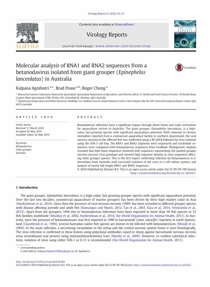
RT-PCR was carried out using the superscript III/platinum Taq high fidelity one-step RT-PCR kit for endpoint detection (Lifetechnologies, Australia). Nearly full length gene sequences for RNA1 and RNA2 were amplified with combination of three setsof primers. The primer sets were chosen from previously published reports (Tan et al., 2001) and were modified to cover theoverlapping regions of both genes. The sequences for the set of primers are: RNA1 RT-PCR primers were NNV1-F1 and NNV1-F6R (approx. 1100 bp); NNV1-F6 and NNV-F10R (approx. 1200 bp); NNV1-F9 and NNV1-R2 (approx. 890 bp) (Fig. 1). The RT-PCR was performed in a 25 μL reaction containing 1x superscript III platinum reaction buffer, 400 nM of each primer selected
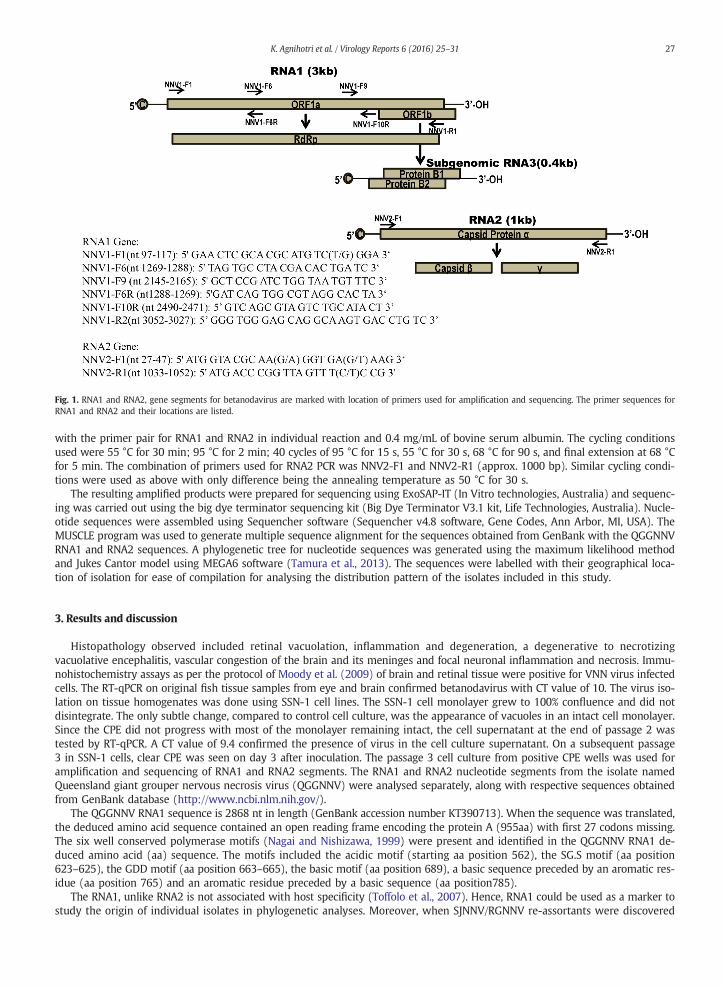
Fig. 1. RNA1 and RNA2, gene segments for betanodavirus are marked with location of primers used for amplification and sequencing. The primer sequences forRNA1 and RNA2 and their locations are listed.
27K. Agnihotri et al. / Virology Reports 6 (2016) 25–31
with the primer pair for RNA1 and RNA2 in individual reaction and 0.4 mg/mL of bovine serum albumin. The cycling conditionsused were 55 °C for 30 min; 95 °C for 2 min; 40 cycles of 95 °C for 15 s, 55 °C for 30 s, 68 °C for 90 s, and final extension at 68 °Cfor 5 min. The combination of primers used for RNA2 PCR was NNV2-F1 and NNV2-R1 (approx. 1000 bp). Similar cycling condi-tions were used as above with only difference being the annealing temperature as 50 °C for 30 s.
The resulting amplified products were prepared for sequencing using ExoSAP-IT (In Vitro technologies, Australia) and sequenc-ing was carried out using the big dye terminator sequencing kit (Big Dye Terminator V3.1 kit, Life Technologies, Australia). Nucle-otide sequences were assembled using Sequencher software (Sequencher v4.8 software, Gene Codes, Ann Arbor, MI, USA). TheMUSCLE program was used to generate multiple sequence alignment for the sequences obtained from GenBank with the QGGNNVRNA1 and RNA2 sequences. A phylogenetic tree for nucleotide sequences was generated using the maximum likelihood methodand Jukes Cantor model using MEGA6 software (Tamura et al., 2013). The sequences were labelled with their geographical loca-tion of isolation for ease of compilation for analysing the distribution pattern of the isolates included in this study.
3. Results and discussion
Histopathology observed included retinal vacuolation, inflammation and degeneration, a degenerative to necrotizingvacuolative encephalitis, vascular congestion of the brain and its meninges and focal neuronal inflammation and necrosis. Immu-nohistochemistry assays as per the protocol of Moody et al. (2009) of brain and retinal tissue were positive for VNN virus infectedcells. The RT-qPCR on original fish tissue samples from eye and brain confirmed betanodavirus with CT value of 10. The virus iso-lation on tissue homogenates was done using SSN-1 cell lines. The SSN-1 cell monolayer grew to 100% confluence and did notdisintegrate. The only subtle change, compared to control cell culture, was the appearance of vacuoles in an intact cell monolayer.Since the CPE did not progress with most of the monolayer remaining intact, the cell supernatant at the end of passage 2 wastested by RT-qPCR. A CT value of 9.4 confirmed the presence of virus in the cell culture supernatant. On a subsequent passage3 in SSN-1 cells, clear CPE was seen on day 3 after inoculation. The passage 3 cell culture from positive CPE wells was used foramplification and sequencing of RNA1 and RNA2 segments. The RNA1 and RNA2 nucleotide segments from the isolate namedQueensland giant grouper nervous necrosis virus (QGGNNV) were analysed separately, along with respective sequences obtainedfrom GenBank database (http://www.ncbi.nlm.nih.gov/).
The QGGNNV RNA1 sequence is 2868 nt in length (GenBank accession number KT390713). When the sequence was translated,the deduced amino acid sequence contained an open reading frame encoding the protein A (955aa) with first 27 codons missing.The six well conserved polymerase motifs (Nagai and Nishizawa, 1999) were present and identified in the QGGNNV RNA1 de-duced amino acid (aa) sequence. The motifs included the acidic motif (starting aa position 562), the SG.S motif (aa position623–625), the GDD motif (aa position 663–665), the basic motif (aa position 689), a basic sequence preceded by an aromatic res-idue (aa position 765) and an aromatic residue preceded by a basic sequence (aa position785).
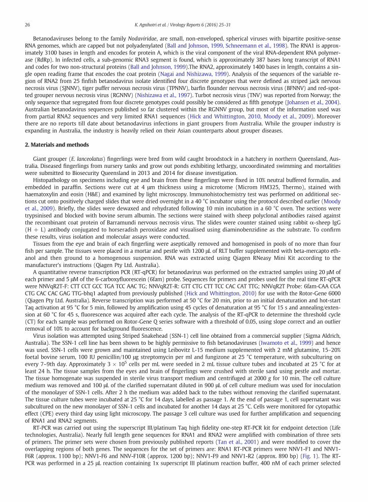
The RNA1, unlike RNA2 is not associated with host specificity (Toffolo et al., 2007). Hence, RNA1 could be used as a marker tostudy the origin of individual isolates in phylogenetic analyses. Moreover, when SJNNV/RGNNV re-assortants were discovered
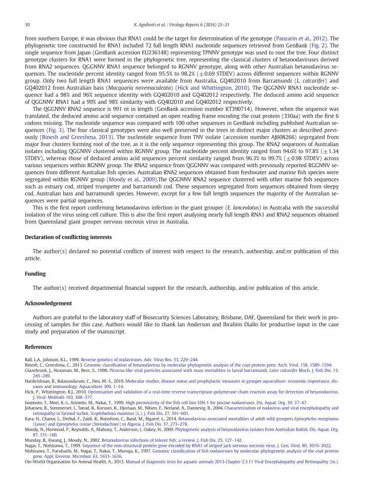
Fig. 2. Phylogenetic analysis for Queensland giant grouper nervous necrosis virus RNA1 nucleotide sequence generated using maximum likelihood method withJukes Cantor model using MEGA 6 software. The single sequence from Japan (GenBank accession EU236148) representing TPNNV genotype was used to rootthe tree. Major genotypes for betanodaviruses are marked indicated by brackets, along with current isolate sequence as highlighted.
28 K. Agnihotri et al. / Virology Reports 6 (2016) 25–31
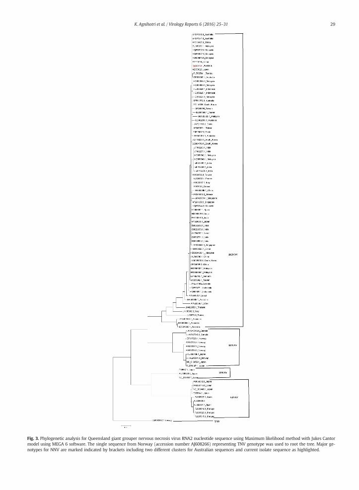
Fig. 3. Phylogenetic analysis for Queensland giant grouper nervous necrosis virus RNA2 nucleotide sequence using Maximum likelihood method with Jukes Cantormodel using MEGA 6 software. The single sequence from Norway (accession number AJ608266) representing TNV genotype was used to root the tree. Major ge-notypes for NNV are marked indicated by brackets including two different clusters for Australian sequences and current isolate sequence as highlighted.
29K. Agnihotri et al. / Virology Reports 6 (2016) 25–31
30 K. Agnihotri et al. / Virology Reports 6 (2016) 25–31
from southern Europe, it was obvious that RNA1 could be the target for determination of the genotype (Panzarin et al., 2012). Thephylogenetic tree constructed for RNA1 included 72 full length RNA1 nucleotide sequences retrieved from GenBank (Fig. 2). Thesingle sequence from Japan (GenBank accession EU236148) representing TPNNV genotype was used to root the tree. Four distinctgenotype clusters for RNA1 were formed in the phylogenetic tree, representing the classical clusters of betanodaviruses derivedfrom RNA2 sequences. QGGNNV RNA1 sequence belonged to RGNNV genotype, along with other Australian betanodavirus se-quences. The nucleotide percent identity ranged from 95.5% to 98.2% (±0.69 STDEV) across different sequences within RGNNVgroup. Only two full length RNA1 sequences were available from Australia, GQ402010 from Barramundi (L. calcarifer) andGQ402012 from Australian bass (Macquaria novemaculeata) (Hick and Whittington, 2010). The QGGNNV RNA1 nucleotide se-quence had a 98% and 96% sequence identity with GQ402010 and GQ402012 respectively. The deduced amino acid sequenceof QGGNNV RNA1 had a 99% and 98% similarity with GQ402010 and GQ402012 respectively.
The QGGNNV RNA2 sequence is 991 nt in length (GenBank accession number KT390714). However, when the sequence wastranslated, the deduced amino acid sequence contained an open reading frame encoding the coat protein (330aa) with the first 6codons missing. The nucleotide sequence was compared with 100 other sequences in GenBank including published Australian se-quences (Fig. 3). The four classical genotypes were also well preserved in the trees in distinct major clusters as described previ-ously (Binesh and Greeshma, 2013). The nucleotide sequence from TNV isolate (accession number AJ608266) segregated frommajor four clusters forming root of the tree, as it is the only sequence representing this group. The RNA2 sequences of Australianisolates including QGGNNV clustered within RGNNV group. The nucleotide percent identity ranged from 94.6% to 97.8% (±1.34STDEV), whereas those of deduced amino acid sequences percent similarity ranged from 96.3% to 99.7% (±0.98 STDEV) acrossvarious sequences within RGNNV group. The RNA2 sequence from QGGNNV was compared with previously reported RGGNNV se-quences from different Australian fish species. Australian RNA2 sequences obtained from freshwater and marine fish species weresegregated within RGNNV group (Moody et al., 2009).The QGGNNV RNA2 sequence clustered with other marine fish sequencessuch as estuary cod, striped trumpeter and barramundi cod. These sequences segregated from sequences obtained from sleepycod, Australian bass and barramundi species. However, except for a few full length sequences the majority of the Australian se-quences were partial sequences.
This is the first report confirming betanodavirus infection in the giant grouper (E. lanceolatus) in Australia with the successfulisolation of the virus using cell culture. This is also the first report analysing nearly full length RNA1 and RNA2 sequences obtainedfrom Queensland giant grouper nervous necrosis virus in Australia.
Declaration of conflicting interests
The author(s) declared no potential conflicts of interest with respect to the research, authorship, and/or publication of thisarticle.
Funding
The author(s) received departmental financial support for the research, authorship, and/or publication of this article.
Acknowledgement
Authors are grateful to the laboratory staff of Biosecurity Sciences Laboratory, Brisbane, DAF, Queensland for their work in pro-cessing of samples for this case. Authors would like to thank Ian Anderson and Ibrahim Diallo for productive input in the casestudy and preparation of the manuscript.
References
Ball, L.A., Johnson, K.L., 1999. Reverse genetics of nodaviruses. Adv. Virus Res. 53, 229–244.Binesh, C., Greeshma, C., 2013. Genomic classification of betanodavirus by molecular phylogenetic analysis of the coat protein gene. Arch. Virol. 158, 1589–1594.Glazebrook, J., Heasman, M., Beer, S., 1990. Picorna-like viral particles associated with mass mortalities in larval barramundi, Lates calcarifer Bloch. J. Fish Dis. 13,
245–249.Harikrishnan, R., Balasundaram, C., Heo, M.-S., 2010. Molecular studies, disease status and prophylactic measures in grouper aquaculture: economic importance, dis-
eases and immunology. Aquaculture 309, 1–14.Hick, P., Whittington, R.J., 2010. Optimisation and validation of a real-time reverse transcriptase-polymerase chain reaction assay for detection of betanodavirus.
J. Virol. Methods 163, 368–377.Iwamoto, T., Mori, K.-I., Arimoto, M., Nakai, T., 1999. High permissivity of the fish cell line SSN-1 for piscine nodaviruses. Dis. Aquat. Org. 39, 37–47.Johansen, R., Sommerset, I., Tørud, B., Korsnes, K., Hjortaas, M., Nilsen, F., Nerland, A., Dannevig, B., 2004. Characterization of nodavirus and viral encephalopathy and
retinopathy in farmed turbot, Scophthalmus maximus (L.). J. Fish Dis. 27, 591–601.Kara, H., Chaoui, L., Derbal, F., Zaidi, R., Boisséson, C., Baud, M., Bigarré, L., 2014. Betanodavirus-associated mortalities of adult wild groupers Epinephelus marginatus
(Lowe) and Epinephelus costae (Steindachner) in Algeria. J. Fish Dis. 37, 273–278.Moody, N., Horwood, P., Reynolds, A., Mahony, T., Anderson, I., Oakey, H., 2009. Phylogenetic analysis of betanodavirus isolates from Australian finfish. Dis. Aquat. Org.
87, 151–160.Munday, B., Kwang, J., Moody, N., 2002. Betanodavirus infections of teleost fish: a review. J. Fish Dis. 25, 127–142.Nagai, T., Nishizawa, T., 1999. Sequence of the non-structural protein gene encoded by RNA1 of striped jack nervous necrosis virus. J. Gen. Virol. 80, 3019–3022.Nishizawa, T., Furuhashi, M., Nagai, T., Nakai, T., Muroga, K., 1997. Genomic classification of fish nodaviruses by molecular phylogenetic analysis of the coat protein
gene. Appl. Environ. Microbiol. 63, 1633–1636.Oie-World Organisation for Animal Health, A., 2013. Manual of diagnostic tests for aquatic animals 2013 Chapter 2.3.11 Viral Encephalopathy and Retinopathy (In.).

31K. Agnihotri et al. / Virology Reports 6 (2016) 25–31
Panzarin, V., Fusaro, A., Monne, I., Cappellozza, E., Patarnello, P., Bovo, G., Capua, I., Holmes, E.C., Cattoli, G., 2012. Molecular epidemiology and evolutionary dynamics ofbetanodavirus in southern Europe. Infect. Genet. Evol. 12, 63–70.
Ransangan, J., Manin, B.O., 2012. Genome analysis of Betanodavirus from cultured marine fish species in Malaysia. Vet. Microbiol. 156, 16–44.Schneemann, A., Reddy, V., Johnson, J.E., 1998. The structure and function of nodavirus particles: a paradigm for understanding chemical biology. Adv. Virus Res. 50,
381–446.Tamura, K., Stecher, G., Peterson, D., Filipski, A., Kumar, S., 2013. MEGA6: molecular evolutionary genetics analysis version 6.0. Mol. Biol. Evol. (mst197).Tan, C., Huang, B., Chang, S.F., Ngoh, G.H., Munday, B., Chen, S.C., Kwang, J., 2001. Determination of the complete nucleotide sequences of RNA1 and RNA2 from greasy
grouper (Epinephelus tauvina) nervous necrosis virus, Singapore strain. J. Gen. Virol. 82, 647–653.Toffolo, V., Negrisolo, E., Maltese, C., Bovo, G., Belvedere, P., Colombo, L., Valle, L.D., 2007. Phylogeny of betanodaviruses and molecular evolution of their RNA polymer-
ase and coat proteins. Mol. Phylogenet. Evol. 43, 298–308.Vendramin, N., Patarnello, P., Toffan, A., Panzarin, V., Cappellozza, E., Tedesco, P., Terlizzi, A., Terregino, C., Cattoli, G., 2013. Viral encephalopathy and retinopathy in
groupers (Epinephelus spp.) in southern Italy: a threat for wild endangered species? BMC Vet. Res. 9, 20.