Embed Size (px)
Citation preview
Dual Role of the Second Extracellular Loop of the CannabinoidReceptor 1: Ligand Binding and Receptor Localization
Kwang H. Ahn, Alexander C. Bertalovitz, Dale F. Mierke, and Debra A. KendallDepartment of Molecular and Cell Biology, University of Connecticut, Storrs, Connecticut (K.H.A., A.C.B, D.A.K.); Departmentof Chemistry, Dartmouth College, Hanover, New Hampshire (D.F.M)
Received April 30, 2009; accepted July 30, 2009
ABSTRACTThe seven transmembrane �-helices of G protein-coupled re-ceptors (GPCRs) are the hallmark of this superfamily. Intrahe-lical interactions are critical to receptor assembly and, for theGPCR subclass that binds small molecules, ligand binding.Most research has focused on identifying the ligand bindingpocket within the helical bundle, whereas the role of the extra-cellular loops remains undefined. Molecular modeling of thecannabinoid receptor 1 (CB1) extracellular loop 2 (EC2), how-ever, suggests that EC2 is poised for key interactions. To testthis possibility, we employed alanine scanning mutagenesis ofCB1 EC2 and identified two distinct regions critical for ligandbinding, G protein coupling activity, and receptor trafficking.Receptors with mutations in the N terminus of EC2 (W255A,N256A) were retained in the endoplasmic reticulum and did notbind the agonist (1R,3R,4R)-3-[2-hydroxy-4-(1,1-dimethylhep-tyl)-phenyl]-4-(3-hydroxypropyl)cyclohexan-1-ol (CP55940) orthe inverse agonist N-(piperidin-1-yl)-5-(4-chlorophenyl)-1-(2,4-dichlorophenyl)-4-methyl-1H-pyrazole-3-carboxamide
(SR141716A). In contrast, the C terminus of EC2 differentiatesagonist and inverse agonist; the P269A, H270A, and I271Areceptors exhibited diminished binding for several agonists butbound inverse agonists SR141716A, N-(piperidin-1-yl)-5-(4-iodophenyl)-1-(2,4-dichlorophenyl)-4-methyl-1H-pyrazole-3-carboxamide (AM251), and 4-[6-methoxy-2-(4-methoxyphenyl)-benzofuran-3-carbonyl]benzonitrile (LY320135) with wild-typereceptor affinity. The F268A receptor involving substitution inthe Cys-X-X-X-Ar motif, displayed both impaired localizationand ligand binding. Other amino acid substitutions at position268 revealed that highly hydrophobic residues are required toaccomplish both functions. It is noteworthy that a F268W re-ceptor was trafficked to the cell surface yet displayed differen-tial binding preference for inverse agonists comparable with theP269A, H270A, and I271A receptors. The findings are consis-tent with a dual role for EC2 in stabilizing receptor assemblyand in ligand binding.
The human cannabinoid receptor 1 (CB1) is a member ofthe rhodopsin-like G protein-coupled receptor (GPCR) family,members of which comprise seven transmembrane helices(TMs) connected by three intracellular loops and three extra-cellular loops. In addition to binding �9-tetrahydrocannabi-nol (�9-THC), the psychoactive constituent of Cannabis sa-tiva, several endogenous cannabinoid receptor agonists havebeen identified, including anandamide (arachidonoylethano-
lamide), 2-arachidonoylglycerol, and 2-arachidonylglycerylether (noladin ether). CB1 also binds a variety of syntheticcannabimimetic compounds such as the nonclassic agonistCP55940, a bicyclic analog of �9-THC, the classic agonistHU-210, and the selective inverse agonists SR141716A (alsoknown as rimonabant), AM251, and LY320135. Upon activa-tion, CB1 modulates synaptic transmission, ultimately play-ing a role in analgesia, anxiety, feeding behavior, and move-ment disorders (Porter and Felder, 2001; Howlett et al.,2002). Thus, understanding the features of CB1 critical forligand binding and receptor localization is important for de-veloping its therapeutic potential.
Molecular modeling analyses of ligand-receptor interac-tions involving CB1 are consistent with the idea that the
This work was supported in part by National Institutes of Health NationalInstitute of General Medical Sciences [Grant GM082054] and by the NationalInstitutes of Health National Institute on Drug Abuse [Grants DA018428,DA020763].
Article, publication date, and citation information can be found athttp://molpharm.aspetjournals.org.
doi:10.1124/mol.109.057356.
ABBREVIATIONS: CB1, cannabinoid receptor 1; GPCR, G protein-coupled receptor; TM, transmembrane helices; EC2, extracellular loop 2; CP55940,(1R,3R,4R)-3-[2-hydroxy-4-(1,1-dimethylheptyl)-phenyl]-4-(3-hydroxypropyl)cyclohexan-1-ol; methanandamide, (R)-N-(2-hydroxy-1-methylethyl)-5Z,8Z,11Z,14Z-eicosatetraenamide; SR141716A, N-(piperidin-1-yl)-5-(4-chlorophenyl)-1-(2,4-dichlorophenyl)-4-methyl-1H-pyrazole-3-carbox-amide; HU-210, (�)-(6aR)-trans-3-(1,1-dimethylheptyl)-6a,7,10,10a-tetrahydro-1-hydroxy-6,6-dimethyl-6H-dibenzo[b,d]pyran-9-methanol; AM251,N-(piperidin-1-yl)-5-(4-iodophenyl)-1-(2,4-dichlorophenyl)-4-methyl-1H-pyrazole-3-carboxamide; LY320135, 4-[6-methoxy-2-(4-methoxyphenyl)benzo-furan-3-carbonyl]benzonitrile; HEK293T, human embryonic kidney cell; PBS, phosphate-buffered saline; TME, Tris/Mg2�/EDTA; GTP�S, guanosine5�-3-O-(thio)triphosphate; ER, endoplasmic reticulum; GFP, green fluorescent protein; PDI, protein disulfide isomerase.
0026-895X/09/7604-833–842$20.00MOLECULAR PHARMACOLOGY Vol. 76, No. 4Copyright © 2009 The American Society for Pharmacology and Experimental Therapeutics 57356/3520649Mol Pharmacol 76:833–842, 2009 Printed in U.S.A.
833
at ASPE
T Journals on July 9, 2018
molpharm
.aspetjournals.orgD
ownloaded from
binding pocket is primarily hydrophobic and within thetransmembrane helix bundle of the receptor, like that ofother GPCRs that bind small molecules (McAllister et al.,2003). In contrast, GPCRs that bind larger ligands, such aspeptide hormones, are thought to do so at the extracellularface of the receptor and typically involve interaction domainsformed by the extracellular N terminus (Holtmann et al.,1995; Lopez de Maturana et al., 2003). However, there is grow-ing evidence that the extracellular loops are also critical forsmall-molecular-weight ligand recognition, selectivity, and/orentrance to the binding pocket (Shi and Javitch, 2004).
Since the crystal structure of bovine rhodopsin was solved(Palczewski et al., 2000), many homology models for therhodopsin-like family A GPCRs, including the cannabinoidreceptors, have been proposed (Shim et al., 2003; Salo et al.,2004; Montero et al., 2005). However, the structure and roleof the extracellular domains are not clearly defined becauseof their mobility, resulting in poorly resolved diffraction pat-terns, and their low sequence homology, relative to the trans-membrane domains. Crystal structures for the �1- and the�2-adrenergic receptors have been solved, providing insightinto the structure of other GPCRs within the family (Cher-ezov et al., 2007; Rasmussen et al., 2007; Rosenbaum et al.,2007; Warne et al., 2008). Although the CB1 receptor belongsto the rhodopsin-like GPCR family, it shares more structuralfeatures with the �-adrenergic receptors than with rhodopsinitself. For instance, both CB1 and the �-adrenergic receptorshave small noncovalently bound ligands, and both receptorsshare an intraloop disulfide bond formed within EC2. CB1shares the Cys-X-X-X-Ar motif [where Ar is an aromaticresidue (i.e., Phe268 in CB1)] that is conserved in EC2 amongGPCRs that bind biogenic amines and peptides. This in-cludes the V1a vasopressin receptor, where the correspond-ing aromatic residue, Phe209, is thought to project into thebinding crevice and mutation at this position results in im-paired agonist binding and intracellular signaling (Conner etal., 2007). Yet CB1 (and its receptor subtype CB2) is highlyunusual in that it has only one extracellular disulfide bondresiding entirely within EC2. Although this EC2 intraloopalso occurs in the �-adrenergic receptors, these receptors inaddition retain the disulfide between TM3 and EC2. Thislatter disulfide bridge is the most common among the rho-dopsin-like family A receptors; most receptors of this sub-class, including rhodopsin, exhibit only this bridge within theextracellular face (Karnik et al., 1988; Strader et al., 1994).
In this study, we have generated a molecular model of CB1,including EC2, that suggests residues of this extracellularloop project into the core of the receptor appropriate for keyinteractions with ligand and other regions of the receptor. Totest this hypothesis, we used alanine-scanning mutagenesisto identify the residues of EC2 within CB1 that are criticalfor ligand binding. Receptors with residues sensitive to mu-tation were further analyzed for binding to a variety of li-gands and for receptor localization. The data indicate thatEC2 is a critical feature of CB1 that plays a role in its ERassembly for subsequent trafficking and in binding someclasses of compounds.
Materials and MethodsCB1 Receptor Model. The model of the human CB1 receptor was
built using the recently published X-ray structure of the human
�2-adrenergic receptor, available as Protein Data Bank ID 2rh1(Rasmussen et al., 2007). The backbone atoms of the transmembranehelices of human CB1 (TM1, Asn112–His143; TM2, Ser152–His181;TM3, Arg186–His219; TM4, Arg230–Trp255; TM5, Asp272–His304;TM6, Met337–Phe368; TM7, Lys376–Lys399) were template-forcedto the TM helices of the �2-adrenergic receptor as defined by theX-ray structure while maintaining proper threading of the interven-ing loops. The �-helix in EC2 of �2-adrenergic receptor was intro-duced into CB1(Ser262–Ile267) based on sequence alignment. Main-taining the topological orientation observed for �2-adrenergicreceptor in the X-ray structure, resulted in the side chain of Cys264projecting toward the extracellular end of TM4 and Cys257, consis-tent with the disulfide bond previously ascribed to CB1 (Shire et al.,1996; Fay et al., 2005). The receptor was extensively energy-mini-mized first using the CVFF force field within Discover (MolecularSimulations, Inc., San Diego, CA) and then CHARMM�� withinNAMD (Phillips et al., 2005). The agonist CP55940 was docked intothe CB1 model guided by previous mutational and modeling studies,clearly indicating a role for Lys192 (Shim et al., 2003; Salo et al.,2004). Twenty orientations of CP55940, with the C3-alkyl tail pro-jecting into the receptor core, as well as extracellularly, were gener-ated and examined by molecular dynamics simulations (100 ps)followed by energy minimization.
Plasmid Construction. All mutants were generated by site-directed mutagenesis (QuikChange; Stratagene, La Jolla, CA) usingthe human CB1 cDNA cloned into pcDNA3.1 as a template, accord-ing to the manufacturer’s instructions. All mutations were confirmedby DNA sequencing.
CB1 Expression and Membrane Preparation. HEK293T cellswere cultured in Dulbecco’s modified Eagle’s medium supplementedwith 10% fetal bovine serum and 3.5 mg/ml glucose at 37°C in 5%CO2. For transient transfection, HEK293T cells were seeded at 8 �105 cells/100-mm dish on the day before transfection. Transfectionwas carried out using the calcium phosphate precipitation method(Chen and Okayama, 1987). Membranes of transfected cells express-ing the wild-type or mutant receptors were prepared as describedpreviously (Abadji et al., 1999). In brief, 24 h after transfection, cellswere harvested, washed twice with phosphate-buffered saline (PBS),and resuspended in PBS containing 4-(2-aminoethyl)benzenesulfo-nyl fluoride, pepstatin A, E-64, bestatin, leupeptin, and aprotinin asprotease inhibitors (Sigma, St. Louis, MO). A membrane fraction wasprepared by nitrogen cavitation at 750 psi for 5 min using a Parr celldisruption bomb, followed by two-step centrifugation, in which thelysate was spun at 500g at 4°C for 10 min and then the supernatantwas spun at 100,000g for 40 min at 4°C. The resulting pellet wasresuspended in TME buffer (25 mM Tris-HCl, 5 mM MgCl2, and 1mM EDTA, pH 7.4) containing 7% sucrose (w/v) at 0.6 �g/�l andstored at �70°C.
Radioligand Binding. Binding assays were performed as de-scribed previously (Murphy and Kendall, 2003). Approximately 30 to40 �g of membranes were incubated at 30°C for 90 min in 200 �l ofTME buffer containing 0.1% fatty acid-free bovine serum albuminusing [3H]CP55940 (139.6 Ci/mmol; PerkinElmer Life and AnalyticalSciences, Waltham, MA) or [3H]SR141716A (42 Ci/mmol; GE Health-care, Piscataway, NJ) for both saturation and competition assays. Insaturation binding assays, at least nine radiolabeled-ligand concen-trations (ranging from 0.24 to 37.60 nM) were used to determine Kd
values of the receptors. In competition binding assays, the cell mem-branes were incubated with fixed tracer concentration typically atthe Kd for the receptor using at least nine concentrations of unla-beled ligands (ranging between 100 pM and 100 �M) as displacingligands. Nonspecific binding was determined in the presence of 1 �Munlabeled ligand in most experiments. Reactions were terminated byadding 250 �l of TME containing 5% bovine serum albumin followedby filtration with a Brandel cell harvester through Whatman GF/Cfilter paper.
GTP�S Binding Assay. For GTP�S binding assay, 15 �g ofmembranes were incubated for 60 min at 30°C in GTP�S binding
834 Ahn et al.
at ASPE
T Journals on July 9, 2018
molpharm
.aspetjournals.orgD
ownloaded from
assay buffer (50 mM Tris-HCl, pH 7.4, 3 mM MgCl2, 0.2 mM EGTA,and 100 mM NaCl) with at least nine concentrations of unlabeledligands (ranging between 100 pM and 100 �M), 0.1 nM [35S]GTP�S(1250 Ci/mmol; PerkinElmer Life Sciences, Waltham, MA), 10 �MGDP, and 0.1% (w/v) bovine serum albumin. Nonspecific binding wasdetermined with 10 �M unlabeled GTP�S (Sigma, St. Louis, MO).The reaction was terminated by rapid filtration through WhatmanGF/C filters. A 4-ml volume of scintillation fluid was added into thevials containing dried filters and the radioactivity trapped in filterswas determined by liquid scintillation counting.
Confocal Microscopy. A day after transfection, HEK293T cellsexpressing CB1 receptors C-terminally fused with the green fluores-cent protein (GFP) were seeded onto 35-mm glass-bottomed dishes(MatTek, Ashland, MA) precoated with poly-D-lysine. Cells werewashed three times with PBS and fixed with 4% paraformaldehydefor 15 min at room temperature. For colocalization studies, the cellswere permeabilized by 0.1% Triton X-100 in DME containing 5%normal goat serum. After incubating with blocking solution (5%normal goat serum in DME) for 30 min at room temperature, thecells were incubated with monoclonal mouse anti-protein disulfideisomerase (anti-PDI; Affinity BioReagents, Golden, CO) diluted1:120 in DME containing 5% normal goat serum. After washing withPBS, cells were incubated with Cy3-labeled donkey anti-mouse sec-ondary antibody (Jackson ImmunoResearch Laboratories, WestGrove, PA) diluted 1:200 for 1 h at room temperature. Cells weremounted in Vectashield mounting medium (Vector Laboratories,Burlingame, CA) and visualized using a Leica TCS SP2 confocalmicroscope (Leica Microsystems, Wetzlar, Germany). Images werecollected from at least three independently transfected cell dishesand processed for presentation in figures using Adobe Photoshop 6.0(Adobe Systems, San Jose, CA).
Data Analysis. All ligand binding assays and GTP�S bindingassays were carried out in duplicate. Data are presented as themean � S.E.M value or the median with the corresponding 95%confidence limits from at least three independent experiments. TheKd and Bmax values were calculated by nonlinear regression (fitted toa one-site binding model) and IC50 values were determined by non-linear regression (fitted to a one-site competition model) using Prismsoftware (GraphPad Software Inc., San Diego, CA). Ki values werethen calculated using the Cheng-Prusoff equation (Cheng and Pru-soff, 1973) based on Kd values obtained from saturation bindinganalyses. EC50 values for the GTP�S binding assays were calculatedusing a sigmoidal dose-response relationship. Ki and EC50 values forthe wild-type and mutant receptors were compared using analysis ofvariance followed by Dunn’s post hoc test for significance. P valuesof � 0.05 were considered to be statistically significant.
ResultsSequence Comparisons and Molecular Modeling of
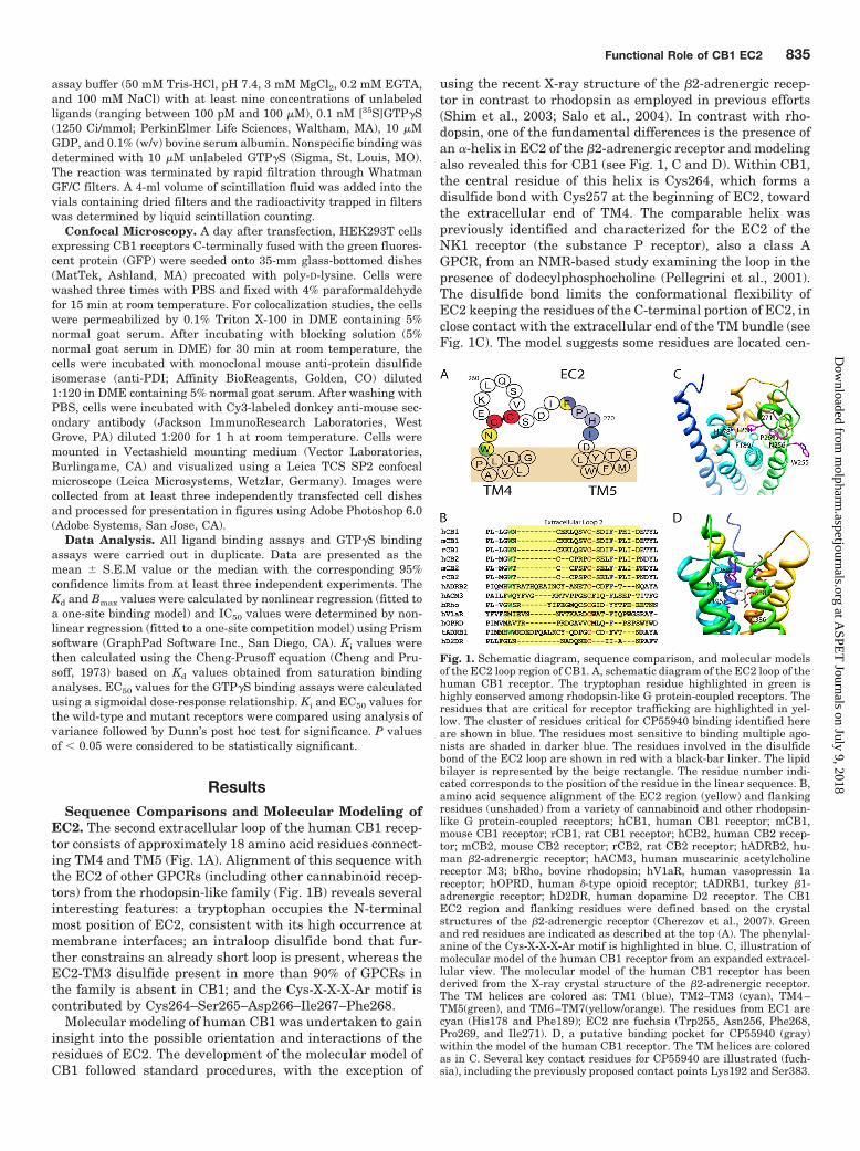
EC2. The second extracellular loop of the human CB1 recep-tor consists of approximately 18 amino acid residues connect-ing TM4 and TM5 (Fig. 1A). Alignment of this sequence withthe EC2 of other GPCRs (including other cannabinoid recep-tors) from the rhodopsin-like family (Fig. 1B) reveals severalinteresting features: a tryptophan occupies the N-terminalmost position of EC2, consistent with its high occurrence atmembrane interfaces; an intraloop disulfide bond that fur-ther constrains an already short loop is present, whereas theEC2-TM3 disulfide present in more than 90% of GPCRs inthe family is absent in CB1; and the Cys-X-X-X-Ar motif iscontributed by Cys264–Ser265–Asp266–Ile267–Phe268.
Molecular modeling of human CB1 was undertaken to gaininsight into the possible orientation and interactions of theresidues of EC2. The development of the molecular model ofCB1 followed standard procedures, with the exception of
using the recent X-ray structure of the �2-adrenergic recep-tor in contrast to rhodopsin as employed in previous efforts(Shim et al., 2003; Salo et al., 2004). In contrast with rho-dopsin, one of the fundamental differences is the presence ofan �-helix in EC2 of the �2-adrenergic receptor and modelingalso revealed this for CB1 (see Fig. 1, C and D). Within CB1,the central residue of this helix is Cys264, which forms adisulfide bond with Cys257 at the beginning of EC2, towardthe extracellular end of TM4. The comparable helix waspreviously identified and characterized for the EC2 of theNK1 receptor (the substance P receptor), also a class AGPCR, from an NMR-based study examining the loop in thepresence of dodecylphosphocholine (Pellegrini et al., 2001).The disulfide bond limits the conformational flexibility ofEC2 keeping the residues of the C-terminal portion of EC2, inclose contact with the extracellular end of the TM bundle (seeFig. 1C). The model suggests some residues are located cen-
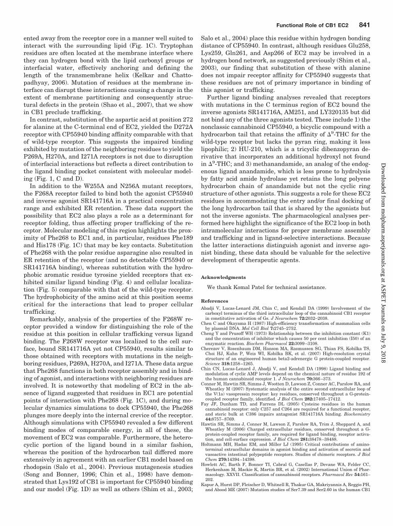
Fig. 1. Schematic diagram, sequence comparison, and molecular modelsof the EC2 loop region of CB1. A, schematic diagram of the EC2 loop of thehuman CB1 receptor. The tryptophan residue highlighted in green ishighly conserved among rhodopsin-like G protein-coupled receptors. Theresidues that are critical for receptor trafficking are highlighted in yel-low. The cluster of residues critical for CP55940 binding identified hereare shown in blue. The residues most sensitive to binding multiple ago-nists are shaded in darker blue. The residues involved in the disulfidebond of the EC2 loop are shown in red with a black-bar linker. The lipidbilayer is represented by the beige rectangle. The residue number indi-cated corresponds to the position of the residue in the linear sequence. B,amino acid sequence alignment of the EC2 region (yellow) and flankingresidues (unshaded) from a variety of cannabinoid and other rhodopsin-like G protein-coupled receptors; hCB1, human CB1 receptor; mCB1,mouse CB1 receptor; rCB1, rat CB1 receptor; hCB2, human CB2 recep-tor; mCB2, mouse CB2 receptor; rCB2, rat CB2 receptor; hADRB2, hu-man �2-adrenergic receptor; hACM3, human muscarinic acetylcholinereceptor M3; bRho, bovine rhodopsin; hV1aR, human vasopressin 1areceptor; hOPRD, human �-type opioid receptor; tADRB1, turkey �1-adrenergic receptor; hD2DR, human dopamine D2 receptor. The CB1EC2 region and flanking residues were defined based on the crystalstructures of the �2-adrenergic receptor (Cherezov et al., 2007). Greenand red residues are indicated as described at the top (A). The phenylal-anine of the Cys-X-X-X-Ar motif is highlighted in blue. C, illustration ofmolecular model of the human CB1 receptor from an expanded extracel-lular view. The molecular model of the human CB1 receptor has beenderived from the X-ray crystal structure of the �2-adrenergic receptor.The TM helices are colored as: TM1 (blue), TM2–TM3 (cyan), TM4–TM5(green), and TM6–TM7(yellow/orange). The residues from EC1 arecyan (His178 and Phe189); EC2 are fuchsia (Trp255, Asn256, Phe268,Pro269, and Ile271). D, a putative binding pocket for CP55940 (gray)within the model of the human CB1 receptor. The TM helices are coloredas in C. Several key contact residues for CP55940 are illustrated (fuch-sia), including the previously proposed contact points Lys192 and Ser383.
Functional Role of CB1 EC2 835
at ASPE
T Journals on July 9, 2018
molpharm
.aspetjournals.orgD
ownloaded from
tral to the receptor interior (e.g., Phe268, Pro269, andIle271). In addition to its projection into the core of thereceptor, the position of Phe268 seems to be coordinated byresidues of EC1 (e.g., His178 and Phe189) (Fig. 1C).
Docking of the nonclassic agonist CP55940 into the recep-tor resulted in a number of different binding modes with suchsimilar energetics that it was difficult to choose a preferredbinding mode. A lack of a clear, energetically defined mode ofligand binding has been described previously for CP55940and CB1 in the literature (Salo et al., 2004). One of thebinding arrangements, with the C3-alkyl chain of CP55940projecting extracellularly, is displayed in Fig. 1D. In thisbinding mode, a number of residues are contributing to theassociation of CP55940, including Lys192, Val196, Leu359,Met363, Ser383, and Cys386. During the molecular dynamicssimulations with the bound CP55940, the EC2 folds over thetransmembrane core of the receptor, particularly the �-heli-cal region, with Phe268 plunging more deeply into the core ofthe receptor and with a “hydrophobic cap” forming on top ofthe ligand.
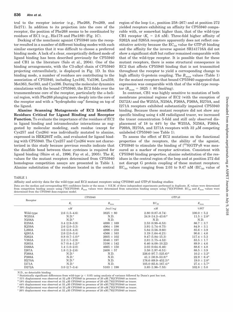
Alanine Scanning Mutagenesis of EC2 IdentifiesResidues Critical for Ligand Binding and ReceptorFunction. To evaluate the importance of the residues of EC2in ligand binding and intramolecular interactions as sug-gested by molecular modeling, each residue (except forCys257 and Cys264) was individually mutated to alanine,expressed in HEK293T cells, and evaluated for ligand bind-ing with CP55940. The Cys257 and Cys264 were not charac-terized in this study because previous results indicate thatthe disulfide bond between these cysteines is required forligand binding (Shire et al., 1996; Fay et al., 2005). The Ki
values for the mutant receptors determined from CP55940homologous competition assays are presented in Table 1.Alanine substitution of the residues located in the central
region of the loop (i.e., position 258–267) and at position 272yielded receptors exhibiting an affinity for CP55940 compa-rable with, or somewhat higher than, that of the wild-typeCB1 receptor (Ki � 2.6 nM). Three-fold higher affinity ofS262A and S265A receptors apparently does not reflect con-stitutive activity because the EC50 value for GTP�S bindingand the affinity for the inverse agonist SR141716A did notshow a significant shift but rather remained comparable withthat of the wild-type receptor. It is possible that for thesemutant receptors, there is some structural consequence inEC2 that affects CP55940 binding that is not transmittedthroughout the receptor to yield a corresponding change tohigh affinity G-protein coupling. The Bmax values (Table 1)for the mutant receptors that bound CP55940 suggested thatexpression was comparable with that of the wild-type recep-tor (Bmax � 3825 � 80 fmol/mg).
In contrast, CB1 was highly sensitive to mutation of bothmembrane proximal regions of EC2 (with the exception ofD272A) and the W255A, N256A, F268A, P269A, H270A, andI271A receptors exhibited substantially impaired CP55940binding. Because these mutant receptors did not show anyspecific binding using 4 nM radioligand tracer, we increasedthe tracer concentration 5-fold and still only observed dis-placement of 18 to 44% by the W255A, N256A, F268A,P269A, H270A, and I271A receptors with 32 �M competingunlabeled CP55940 (see Table 1).
To assess the effect of EC2 mutations on the functionalproperties of the receptors, the ability of the agonist,CP55940 to stimulate the binding of [35S]GTP�S was mea-sured as a marker of receptor activation. Consistent withCP55940 binding properties, alanine substitution of the res-idues in the central region of the loop and at position 272 didnot disrupt G protein coupling of these mutant receptors;EC50 values ranging from 2.03 to 9.47 nM (EC50 value of
TABLE 1Affinity and activity data for the wild-type and EC2 mutant receptors using CP55940 and GTP�S binding studiesData are the median and corresponding 95% confidence limits or the mean � S.E.M. of three independent experiments performed in duplicate. Ki values were determinedfrom competition binding assays using 3HCP55940. Bmax values were determined from saturation binding assays using 3HCP55940. EC50 and Emax values weredetermined from the CP55940 dose-response model.
ReceptorCP55940 GTP�S
Ki Bmax EC50 Emax
nM fmol/mg nM %WT
Wild-type 2.6 (1.5–4.6) 3825 � 80 2.80 (0.87–8.74) 100.0 � 5.2W255A N.D.a N.D. 24.9 (14.2–43.6)* 11.5 � 2.8*N256A N.D.b N.D. N.D. N.D.E258A 3.4 (2.9–3.9) 4898 � 169 2.53 (0.98–6.53) 86.7 � 3.7K259A 2.6 (2.0–3.3) 4964 � 198 3.91 (1.74–8.75) 84.9 � 3.1L260A 3.6 (2.8–4.8) 4996 � 258 5.84 (3.56–9.60) 85.6 � 3.9Q261A 2.6 (2.0–3.4) 4566 � 163 3.19 (1.64–6.21) 90.6 � 4.6S262A 0.8 (0.7–1.0)* 2605 � 102 9.47 (5.84–15.3) 117.4 � 5.3V263A 2.2 (1.7–2.9) 3046 � 197 2.81 (1.75–4.52) 83.5 � 1.7S265A 0.7 (0.4–1.2)* 3106 � 142 6.46 (4.08–10.22) 89.9 � 4.6D266A 1.4 (1.0–2.0) 4665 � 150 2.03 (0.64–6.46) 88.6 � 4.8I267A 1.8 (1.2–2.6) 2408 � 57 3.58 (1.97–6.51) 88.5 � 3.9F268A N.D.a N.D. 226.6 (97.7–525.6)* 10.2 � 3.2*P269A N.D.c N.D. 41.1 (30.9–53.8)* 22.9 � 6.4*H270A N.D.d N.D. 176.6 (69.9–452.3)* 19.0 � 2.8*I271A N.D.e N.D. 105.0 (65.8–167.4)* 27.4 � 5.7*D272A 3.0 (2.7–3.4) 5103 � 198 3.85 (1.96–7.55) 102.8 � 5.0
N.D., no detectable binding.* Statistically significant differences from wild-type (p � 0.05) using analysis of variance followed by Dunn’s post hoc test.a 31% displacement was observed at 32 �M CP55940 in presence of 20 nM 3HCP55940 as tracer.b 18% displacement was observed at 32 �M CP55940 in presence of 20 nM 3HCP55940 as tracer.c 44% displacement was observed at 32 �M CP55940 in presence of 20 nM 3HCP55940 as tracer.d 39% displacement was observed at 32 �M CP55940 in presence of 20 nM 3HCP55940 as tracer.e 26% displacement was observed at 32 �M CP55940 in presence of 20 nM 3HCP55940 as tracer.
836 Ahn et al.
at ASPE
T Journals on July 9, 2018
molpharm
.aspetjournals.orgD
ownloaded from
wild-type � 2.80 nM) were observed (Table 1). However, a10-fold increase or more in EC50 values were observed withthe W255A, N256A, F268A, P269A, H270A, and I271A re-ceptors with considerably reduced Emax values (Emax valuesof 0 to 11.5% by the W255A, N256A, and F268A receptors; 19to 27.4% by P269A, H270A, and I271A receptors) (Table 1).These changes in CP55940 concentration-dependent G pro-tein stimulation paralleled the agonist binding affinity data,suggesting two distinct regions of EC2 are critical forCP55940 sensitivity.
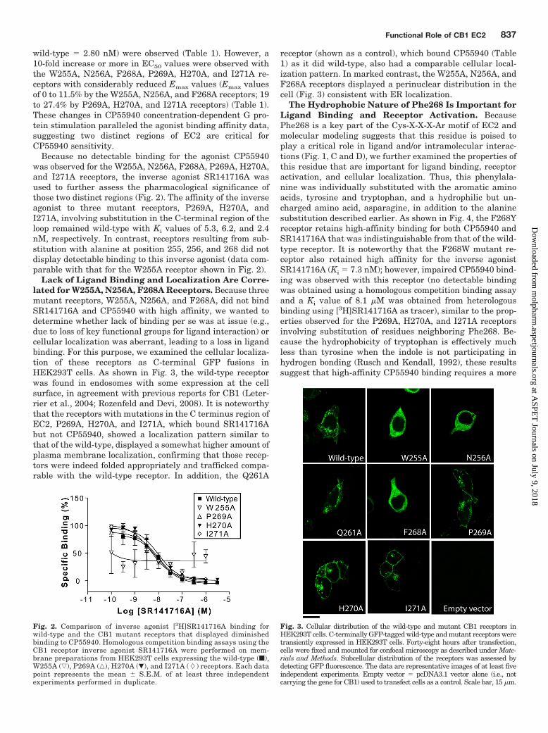
Because no detectable binding for the agonist CP55940was observed for the W255A, N256A, F268A, P269A, H270A,and I271A receptors, the inverse agonist SR141716A wasused to further assess the pharmacological significance ofthose two distinct regions (Fig. 2). The affinity of the inverseagonist to three mutant receptors, P269A, H270A, andI271A, involving substitution in the C-terminal region of theloop remained wild-type with Ki values of 5.3, 6.2, and 2.4nM, respectively. In contrast, receptors resulting from sub-stitution with alanine at position 255, 256, and 268 did notdisplay detectable binding to this inverse agonist (data com-parable with that for the W255A receptor shown in Fig. 2).
Lack of Ligand Binding and Localization Are Corre-lated for W255A, N256A, F268A Receptors. Because threemutant receptors, W255A, N256A, and F268A, did not bindSR141716A and CP55940 with high affinity, we wanted todetermine whether lack of binding per se was at issue (e.g.,due to loss of key functional groups for ligand interaction) orcellular localization was aberrant, leading to a loss in ligandbinding. For this purpose, we examined the cellular localiza-tion of these receptors as C-terminal GFP fusions inHEK293T cells. As shown in Fig. 3, the wild-type receptorwas found in endosomes with some expression at the cellsurface, in agreement with previous reports for CB1 (Leter-rier et al., 2004; Rozenfeld and Devi, 2008). It is noteworthythat the receptors with mutations in the C terminus region ofEC2, P269A, H270A, and I271A, which bound SR141716Abut not CP55940, showed a localization pattern similar tothat of the wild-type, displayed a somewhat higher amount ofplasma membrane localization, confirming that those recep-tors were indeed folded appropriately and trafficked compa-rable with the wild-type receptor. In addition, the Q261A
receptor (shown as a control), which bound CP55940 (Table1) as it did wild-type, also had a comparable cellular local-ization pattern. In marked contrast, the W255A, N256A, andF268A receptors displayed a perinuclear distribution in thecell (Fig. 3) consistent with ER localization.
The Hydrophobic Nature of Phe268 Is Important forLigand Binding and Receptor Activation. BecausePhe268 is a key part of the Cys-X-X-X-Ar motif of EC2 andmolecular modeling suggests that this residue is poised toplay a critical role in ligand and/or intramolecular interac-tions (Fig. 1, C and D), we further examined the properties ofthis residue that are important for ligand binding, receptoractivation, and cellular localization. Thus, this phenylala-nine was individually substituted with the aromatic aminoacids, tyrosine and tryptophan, and a hydrophilic but un-charged amino acid, asparagine, in addition to the alaninesubstitution described earlier. As shown in Fig. 4, the F268Yreceptor retains high-affinity binding for both CP55940 andSR141716A that was indistinguishable from that of the wild-type receptor. It is noteworthy that the F268W mutant re-ceptor also retained high affinity for the inverse agonistSR141716A (Ki � 7.3 nM); however, impaired CP55940 bind-ing was observed with this receptor (no detectable bindingwas obtained using a homologous competition binding assayand a Ki value of 8.1 �M was obtained from heterologousbinding using [3H]SR141716A as tracer), similar to the prop-erties observed for the P269A, H270A, and I271A receptorsinvolving substitution of residues neighboring Phe268. Be-cause the hydrophobicity of tryptophan is effectively muchless than tyrosine when the indole is not participating inhydrogen bonding (Rusch and Kendall, 1992), these resultssuggest that high-affinity CP55940 binding requires a more
Fig. 2. Comparison of inverse agonist [3H]SR141716A binding forwild-type and the CB1 mutant receptors that displayed diminishedbinding to CP55940. Homologous competition binding assays using theCB1 receptor inverse agonist SR141716A were performed on mem-brane preparations from HEK293T cells expressing the wild-type (f),W255A (ƒ), P269A (‚), H270A (�), and I271A (�) receptors. Each datapoint represents the mean � S.E.M. of at least three independentexperiments performed in duplicate.
Fig. 3. Cellular distribution of the wild-type and mutant CB1 receptors inHEK293T cells. C-terminally GFP-tagged wild-type and mutant receptors weretransiently expressed in HEK293T cells. Forty-eight hours after transfection,cells were fixed and mounted for confocal microscopy as described under Mate-rials and Methods. Subcellular distribution of the receptors was assessed bydetecting GFP fluorescence. The data are representative images of at least fiveindependent experiments. Empty vector � pcDNA3.1 vector alone (i.e., notcarrying the gene for CB1) used to transfect cells as a control. Scale bar, 15 �m.
Functional Role of CB1 EC2 837
at ASPE
T Journals on July 9, 2018
molpharm
.aspetjournals.orgD
ownloaded from
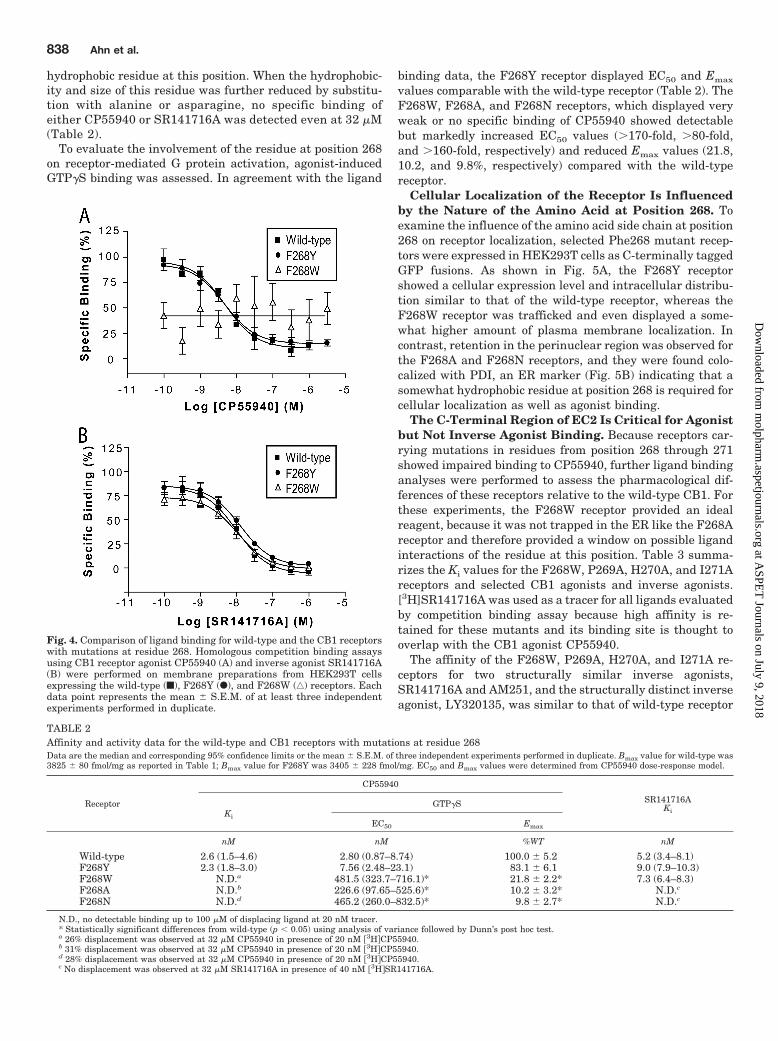
hydrophobic residue at this position. When the hydrophobic-ity and size of this residue was further reduced by substitu-tion with alanine or asparagine, no specific binding ofeither CP55940 or SR141716A was detected even at 32 �M(Table 2).
To evaluate the involvement of the residue at position 268on receptor-mediated G protein activation, agonist-inducedGTP�S binding was assessed. In agreement with the ligand
binding data, the F268Y receptor displayed EC50 and Emax
values comparable with the wild-type receptor (Table 2). TheF268W, F268A, and F268N receptors, which displayed veryweak or no specific binding of CP55940 showed detectablebut markedly increased EC50 values (�170-fold, �80-fold,and �160-fold, respectively) and reduced Emax values (21.8,10.2, and 9.8%, respectively) compared with the wild-typereceptor.
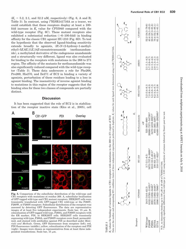
Cellular Localization of the Receptor Is Influencedby the Nature of the Amino Acid at Position 268. Toexamine the influence of the amino acid side chain at position268 on receptor localization, selected Phe268 mutant recep-tors were expressed in HEK293T cells as C-terminally taggedGFP fusions. As shown in Fig. 5A, the F268Y receptorshowed a cellular expression level and intracellular distribu-tion similar to that of the wild-type receptor, whereas theF268W receptor was trafficked and even displayed a some-what higher amount of plasma membrane localization. Incontrast, retention in the perinuclear region was observed forthe F268A and F268N receptors, and they were found colo-calized with PDI, an ER marker (Fig. 5B) indicating that asomewhat hydrophobic residue at position 268 is required forcellular localization as well as agonist binding.
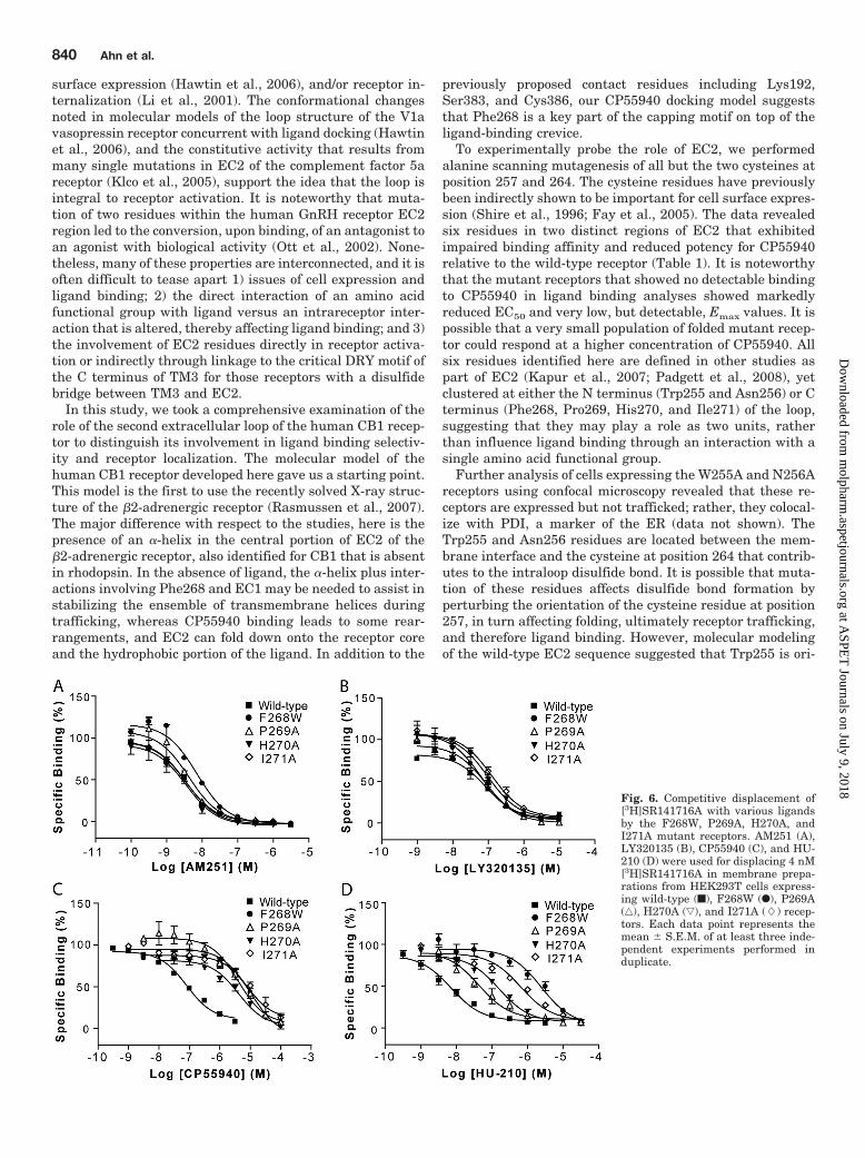
The C-Terminal Region of EC2 Is Critical for Agonistbut Not Inverse Agonist Binding. Because receptors car-rying mutations in residues from position 268 through 271showed impaired binding to CP55940, further ligand bindinganalyses were performed to assess the pharmacological dif-ferences of these receptors relative to the wild-type CB1. Forthese experiments, the F268W receptor provided an idealreagent, because it was not trapped in the ER like the F268Areceptor and therefore provided a window on possible ligandinteractions of the residue at this position. Table 3 summa-rizes the Ki values for the F268W, P269A, H270A, and I271Areceptors and selected CB1 agonists and inverse agonists.[3H]SR141716A was used as a tracer for all ligands evaluatedby competition binding assay because high affinity is re-tained for these mutants and its binding site is thought tooverlap with the CB1 agonist CP55940.
The affinity of the F268W, P269A, H270A, and I271A re-ceptors for two structurally similar inverse agonists,SR141716A and AM251, and the structurally distinct inverseagonist, LY320135, was similar to that of wild-type receptor
Fig. 4. Comparison of ligand binding for wild-type and the CB1 receptorswith mutations at residue 268. Homologous competition binding assaysusing CB1 receptor agonist CP55940 (A) and inverse agonist SR141716A(B) were performed on membrane preparations from HEK293T cellsexpressing the wild-type (f), F268Y (F), and F268W (‚) receptors. Eachdata point represents the mean � S.E.M. of at least three independentexperiments performed in duplicate.
TABLE 2Affinity and activity data for the wild-type and CB1 receptors with mutations at residue 268Data are the median and corresponding 95% confidence limits or the mean � S.E.M. of three independent experiments performed in duplicate. Bmax value for wild-type was3825 � 80 fmol/mg as reported in Table 1; Bmax value for F268Y was 3405 � 228 fmol/mg. EC50 and Bmax values were determined from CP55940 dose-response model.
Receptor
CP55940
SR141716AKiKi
GTP�S
EC50 Emax
nM nM %WT nM
Wild-type 2.6 (1.5–4.6) 2.80 (0.87–8.74) 100.0 � 5.2 5.2 (3.4–8.1)F268Y 2.3 (1.8–3.0) 7.56 (2.48–23.1) 83.1 � 6.1 9.0 (7.9–10.3)F268W N.D.a 481.5 (323.7–716.1)* 21.8 � 2.2* 7.3 (6.4–8.3)F268A N.D.b 226.6 (97.65–525.6)* 10.2 � 3.2* N.D.c
F268N N.D.d 465.2 (260.0–832.5)* 9.8 � 2.7* N.D.c
N.D., no detectable binding up to 100 �M of displacing ligand at 20 nM tracer.* Statistically significant differences from wild-type (p � 0.05) using analysis of variance followed by Dunn’s post hoc test.a 26% displacement was observed at 32 �M CP55940 in presence of 20 nM 3HCP55940.b 31% displacement was observed at 32 �M CP55940 in presence of 20 nM 3HCP55940.d 28% displacement was observed at 32 �M CP55940 in presence of 20 nM 3HCP55940.c No displacement was observed at 32 �M SR141716A in presence of 40 nM 3HSR141716A.
838 Ahn et al.
at ASPE
T Journals on July 9, 2018
molpharm
.aspetjournals.orgD
ownloaded from
(Ki � 5.2, 2.1, and 52.2 nM, respectively) (Fig. 6, A and B;Table 3). In contrast, using [3H]SR141716A as a tracer, wecould establish that these receptors display at least a 100-fold increase in Ki value for CP55940 compared with thewild-type receptor (Fig. 6C). These mutant receptors alsoexhibited a substantial reduction (�6–100-fold) in bindingaffinity for the classic CB1 agonist HU-210 (Fig. 6D). To testthe hypothesis that the observed ligand-binding sensitivityextends broadly to agonists, (R)-N-(2-hydroxy-1-methyl-ethyl)-5Z,8Z,11Z,14Z-eicosatetraenamide (methanandam-ide), a methylated derivative of the endogenous anandamideand a structurally very different, ligand was also evaluatedfor binding to the receptors with mutations in the 268 to 271region. The affinity of the mutants for methanandamide wasalso significantly reduced compared with the wild-type recep-tor (Table 3). These data underscore a role for Phe268,Pro269, His270, and Ile271 of EC2 in binding a variety ofagonists, perturbation of these residues leading to a loss inagonist binding. The insensitivity of inverse agonist bindingto mutations in this region of the receptor suggests that thebinding sites for these two classes of compounds are partiallydistinct.
DiscussionIt has been suggested that the role of EC2 is in stabiliza-
tion of the receptor inactive state (Klco et al., 2005), cell
Fig. 5. Comparison of the subcellular distribution of the wild-type andCB1 receptors with mutations at residue 268. A, subcellular localizationof GFP-tagged wild-type and CB1 mutant receptors. HEK293T cells weretransiently transfected with GFP-tagged CB1 wild-type or the F268Y,F268W, or F268N receptors. Subcellular distribution of the receptors wasassessed by detecting GFP fluorescence. The data are representativeimages of at least five independent experiments. Scale bar, 15 �m. B,colocalization of GFP-tagged wild-type, F268A, and F268N receptors withthe ER marker, PDI, in HEK293T cells. HEK293T cells transientlyexpressing wild-type, F268A, and F268N receptors were fixed, permeabil-ized, and stained with antibodies against PDI as described under Mate-rials and Methods. Green, GFP fluorescence from receptors (left); red,PDI fluorescence (middle); yellow, colocalization of the receptors and PDI(right). Images were chosen as representatives from at least three inde-pendent transfections. Scale bar, 15 �m. T
AB
LE
3K
iva
lues
for
bin
din
gof
vari
ous
liga
nds
toC
B1
rece
ptor
sw
ith
mu
tati
ons
inth
e26
8to
271
regi
onof
EC
2D
ata
are
the
med
ian
and
corr
espo
ndi
ng
95%
con
fide
nce
lim
its
ofth
ree
inde
pen
den
tex
peri
men
tspe
rfor
med
indu
plic
ate.
Ki
valu
esfo
rea
chli
gan
dw
ere
dete
rmin
edu
sin
g4
nM
3H
SR
1417
16A
.
Rec
epto
rK
i
CP
5594
0H
U-2
10M
eth
anan
dam
ide
SR
1417
16A
AM
251
LY
3201
35
nM
Wil
d-ty
pe24
.8(1
4.6–
42.3
)3.
7(2
.1–6
.5)
523
(403
–680
)5.
2(3
.4–8
.1)
2.1
(1.1
–4.2
)52
.2(3
4.3–
79.6
)F
268W
8132
(579
3–11
421)
*16
72(7
57.9
–368
9)*
2009
0(7
371–
5473
0)*
7.3
(6.4
–8.3
)5.
7(3
.9–8
.7)
43.4
(34.
1–55
.2)
P26
9A20
81(1
256–
3446
)*22
.6(1
0.3–
49.5
)*54
70(4
319–
6927
)*5.
3(3
.8–7
.3)
2.4
(1.2
–4.8
)50
.0(2
7.3–
91.6
)H
270A
2576
(149
2–44
46)*
100
(63–
159)
*93
5(2
65–3
297)
6.2
(4.4
–8.7
)2.
0(1
.3–3
.1)
70.4
(52.
1–95
.1)
I271
A33
06(1
422–
7686
)*31
0(1
17–8
22)*
4140
0(5
860–
2925
00)*
2.4
(1.9
–2.9
)1.
7(1
.1–2
.5)
61.2
(46.
1–81
.1)
N.D
.,n
ode
tect
able
bin
din
gu
pto
100
�M
at20
nM
of3
HS
R14
1716
A.
*S
tati
stic
ally
sign
ific
ant
diff
eren
ces
from
wil
d-ty
pe(p
�0.
05)
usi
ng
anal
ysis
ofva
rian
cefo
llow
edby
Du
nn
’spo
sth
octe
st.
Functional Role of CB1 EC2 839
at ASPE
T Journals on July 9, 2018
molpharm
.aspetjournals.orgD
ownloaded from
surface expression (Hawtin et al., 2006), and/or receptor in-ternalization (Li et al., 2001). The conformational changesnoted in molecular models of the loop structure of the V1avasopressin receptor concurrent with ligand docking (Hawtinet al., 2006), and the constitutive activity that results frommany single mutations in EC2 of the complement factor 5areceptor (Klco et al., 2005), support the idea that the loop isintegral to receptor activation. It is noteworthy that muta-tion of two residues within the human GnRH receptor EC2region led to the conversion, upon binding, of an antagonist toan agonist with biological activity (Ott et al., 2002). None-theless, many of these properties are interconnected, and it isoften difficult to tease apart 1) issues of cell expression andligand binding; 2) the direct interaction of an amino acidfunctional group with ligand versus an intrareceptor inter-action that is altered, thereby affecting ligand binding; and 3)the involvement of EC2 residues directly in receptor activa-tion or indirectly through linkage to the critical DRY motif ofthe C terminus of TM3 for those receptors with a disulfidebridge between TM3 and EC2.
In this study, we took a comprehensive examination of therole of the second extracellular loop of the human CB1 recep-tor to distinguish its involvement in ligand binding selectiv-ity and receptor localization. The molecular model of thehuman CB1 receptor developed here gave us a starting point.This model is the first to use the recently solved X-ray struc-ture of the �2-adrenergic receptor (Rasmussen et al., 2007).The major difference with respect to the studies, here is thepresence of an �-helix in the central portion of EC2 of the�2-adrenergic receptor, also identified for CB1 that is absentin rhodopsin. In the absence of ligand, the �-helix plus inter-actions involving Phe268 and EC1 may be needed to assist instabilizing the ensemble of transmembrane helices duringtrafficking, whereas CP55940 binding leads to some rear-rangements, and EC2 can fold down onto the receptor coreand the hydrophobic portion of the ligand. In addition to the
previously proposed contact residues including Lys192,Ser383, and Cys386, our CP55940 docking model suggeststhat Phe268 is a key part of the capping motif on top of theligand-binding crevice.
To experimentally probe the role of EC2, we performedalanine scanning mutagenesis of all but the two cysteines atposition 257 and 264. The cysteine residues have previouslybeen indirectly shown to be important for cell surface expres-sion (Shire et al., 1996; Fay et al., 2005). The data revealedsix residues in two distinct regions of EC2 that exhibitedimpaired binding affinity and reduced potency for CP55940relative to the wild-type receptor (Table 1). It is noteworthythat the mutant receptors that showed no detectable bindingto CP55940 in ligand binding analyses showed markedlyreduced EC50 and very low, but detectable, Emax values. It ispossible that a very small population of folded mutant recep-tor could respond at a higher concentration of CP55940. Allsix residues identified here are defined in other studies aspart of EC2 (Kapur et al., 2007; Padgett et al., 2008), yetclustered at either the N terminus (Trp255 and Asn256) or Cterminus (Phe268, Pro269, His270, and Ile271) of the loop,suggesting that they may play a role as two units, ratherthan influence ligand binding through an interaction with asingle amino acid functional group.
Further analysis of cells expressing the W255A and N256Areceptors using confocal microscopy revealed that these re-ceptors are expressed but not trafficked; rather, they colocal-ize with PDI, a marker of the ER (data not shown). TheTrp255 and Asn256 residues are located between the mem-brane interface and the cysteine at position 264 that contrib-utes to the intraloop disulfide bond. It is possible that muta-tion of these residues affects disulfide bond formation byperturbing the orientation of the cysteine residue at position257, in turn affecting folding, ultimately receptor trafficking,and therefore ligand binding. However, molecular modelingof the wild-type EC2 sequence suggested that Trp255 is ori-
Fig. 6. Competitive displacement of[3H]SR141716A with various ligandsby the F268W, P269A, H270A, andI271A mutant receptors. AM251 (A),LY320135 (B), CP55940 (C), and HU-210 (D) were used for displacing 4 nM[3H]SR141716A in membrane prepa-rations from HEK293T cells express-ing wild-type (f), F268W (F), P269A(‚), H270A (ƒ), and I271A (�) recep-tors. Each data point represents themean � S.E.M. of at least three inde-pendent experiments performed induplicate.
840 Ahn et al.
at ASPE
T Journals on July 9, 2018
molpharm
.aspetjournals.orgD
ownloaded from
ented away from the receptor core in a manner well suited tointeract with the surrounding lipid (Fig. 1C). Tryptophanresidues are often located at the membrane interface wherethey can hydrogen bond with the lipid carbonyl groups orinterfacial water, effectively anchoring and defining thelength of the transmembrane helix (Kelkar and Chatto-padhyay, 2006). Mutation of residues at the membrane in-terface can disrupt these interactions causing a change in theextent of membrane partitioning and consequently struc-tural defects in the protein (Shao et al., 2007), that we showin CB1 preclude trafficking.
In contrast, substitution of the aspartic acid at position 272for alanine at the C-terminal end of EC2, yielded the D272Areceptor with CP55940 binding affinity comparable with thatof wild-type receptor. This suggests the impaired bindingexhibited by mutation of the neighboring residues to yield theP269A, H270A, and I271A receptors is not due to disruptionof interfacial interactions but reflects a direct contribution tothe ligand binding pocket consistent with molecular model-ing (Fig. 1, C and D).
In addition to the W255A and N256A mutant receptors,the F268A receptor failed to bind both the agonist CP55940and inverse agonist SR141716A in a practical concentrationrange and exhibited ER retention. These data support thepossibility that EC2 also plays a role as a determinant forreceptor folding, thus affecting proper trafficking of the re-ceptor. Molecular modeling of this region highlights the prox-imity of Phe268 to EC1 and, in particular, residues Phe189and His178 (Fig. 1C) that may be key contacts. Substitutionof Phe268 with the polar residue asparagine also resulted inER retention of the receptor (and no detectable CP55940 orSR141716A binding), whereas substitution with the hydro-phobic aromatic residue tyrosine yielded receptors that ex-hibited similar ligand binding (Fig. 4) and cellular localiza-tion (Fig. 5) comparable with that of the wild-type receptor.The hydrophobicity of the amino acid at this position seemscritical for the interactions that lead to proper cellulartrafficking.
Remarkably, analysis of the properties of the F268W re-ceptor provided a window for distinguishing the role of theresidue at this position in cellular trafficking versus ligandbinding. The F268W receptor was localized to the cell sur-face, bound SR141716A yet not CP55940, results similar tothose obtained with receptors with mutations in the neigh-boring residues, P269A, H270A, and I271A. These data arguethat Phe268 functions in both receptor assembly and in bind-ing of agonist, and interactions with neighboring residues areinvolved. It is noteworthy that modeling of EC2 in the ab-sence of ligand suggested that residues in EC1 are potentialpoints of interaction with Phe268 (Fig. 1C), and during mo-lecular dynamics simulations to dock CP55940, the Phe268plunges more deeply into the internal crevice of the receptor.Although simulations with CP55940 revealed a few differentbinding modes of comparable energy, in all of these, themovement of EC2 was comparable. Furthermore, the hetero-cyclic portion of the ligand bound in a similar fashion,whereas the position of the hydrocarbon tail differed moreextensively in agreement with an earlier CB1 model based onrhodopsin (Salo et al., 2004). Previous mutagenesis studies(Song and Bonner, 1996; Chin et al., 1998) have demon-strated that Lys192 of CB1 is important for CP55940 bindingand our model (Fig. 1D) as well as others (Shim et al., 2003;
Salo et al., 2004) place this residue within hydrogen bondingdistance of CP55940. In contrast, although residues Glu258,Lys259, Gln261, and Asp266 of EC2 may be involved in ahydrogen bond network, as suggested previously (Shim et al.,2003), our finding that substitution of these with alaninedoes not impair receptor affinity for CP55940 suggests thatthese residues are not of primary importance in binding ofthis agonist or trafficking.
Further ligand binding analyses revealed that receptorswith mutations in the C terminus region of EC2 bound theinverse agonists SR141716A, AM251, and LY320135 but didnot bind any of the three agonists tested. These include 1) thenonclassic cannabinoid CP55940, a bicyclic compound with ahydrocarbon tail that retains the affinity of �9-THC for thewild-type receptor but lacks the pyran ring, making it lesslipophilic; 2) HU-210, which is a tricyclic dibenzopyran de-rivative that incorporates an additional hydroxyl not foundin �9-THC; and 3) methanandamide, an analog of the endog-enous ligand anandamide, which is less prone to hydrolysisby fatty acid amide hydrolase yet retains the long polyenehydrocarbon chain of anandamide but not the cyclic ringstructure of other agonists. This suggests a role for these EC2residues in accommodating the entry and/or final docking ofthe long hydrocarbon tail that is shared by the agonists butnot the inverse agonists. The pharmacological analyses per-formed here highlight the significance of the EC2 loop in bothintramolecular interactions for proper membrane assemblyand trafficking and in ligand-selective interactions. Becausethe latter interactions distinguish agonist and inverse ago-nist binding, these data should be valuable for the selectivedevelopment of therapeutic agents.
Acknowledgments
We thank Komal Patel for technical assistance.
ReferencesAbadji V, Lucas-Lenard JM, Chin C, and Kendall DA (1999) Involvement of the
carboxyl terminus of the third intracellular loop of the cannabinoid CB1 receptorin constitutive activation of Gs. J Neurochem 72:2032–2038.
Chen C and Okayama H (1987) High-efficiency transformation of mammalian cellsby plasmid DNA. Mol Cell Biol 7:2745–2752.
Cheng Y and Prusoff WH (1973) Relationship between the inhibition constant (K1)and the concentration of inhibitor which causes 50 per cent inhibition (I50) of anenzymatic reaction. Biochem Pharmacol 22:3099–3108.
Cherezov V, Rosenbaum DM, Hanson MA, Rasmussen SG, Thian FS, Kobilka TS,Choi HJ, Kuhn P, Weis WI, Kobilka BK, et al. (2007) High-resolution crystalstructure of an engineered human beta2-adrenergic G protein-coupled receptor.Science 318:1258–1265.
Chin CN, Lucas-Lenard J, Abadji V, and Kendall DA (1998) Ligand binding andmodulation of cyclic AMP levels depend on the chemical nature of residue 192 ofthe human cannabinoid receptor 1. J Neurochem 70:366–373.
Conner M, Hawtin SR, Simms J, Wootten D, Lawson Z, Conner AC, Parslow RA, andWheatley M (2007) Systematic analysis of the entire second extracellular loop ofthe V(1a) vasopressin receptor: key residues, conserved throughout a G-protein-coupled receptor family, identified. J Biol Chem 282:17405–17412.
Fay JF, Dunham TD, and Farrens DL (2005) Cysteine residues in the humancannabinoid receptor: only C257 and C264 are required for a functional receptor,and steric bulk at C386 impairs antagonist SR141716A binding. Biochemistry44:8757–8769.
Hawtin SR, Simms J, Conner M, Lawson Z, Parslow RA, Trim J, Sheppard A, andWheatley M (2006) Charged extracellular residues, conserved throughout a G-protein-coupled receptor family, are required for ligand binding, receptor activa-tion, and cell-surface expression. J Biol Chem 281:38478–38488.
Holtmann MH, Hadac EM, and Miller LJ (1995) Critical contributions of amino-terminal extracellular domains in agonist binding and activation of secretin andvasoactive intestinal polypeptide receptors. Studies of chimeric receptors. J BiolChem 270:14394–14398.
Howlett AC, Barth F, Bonner TI, Cabral G, Casellas P, Devane WA, Felder CC,Herkenham M, Mackie K, Martin BR, et al. (2002) International Union of Phar-macology. XXVII. Classification of cannabinoid receptors. Pharmacol Rev 54:161–202.
Kapur A, Hurst DP, Fleischer D, Whitnell R, Thakur GA, Makriyannis A, Reggio PH,and Abood ME (2007) Mutation studies of Ser7.39 and Ser2.60 in the human CB1
Functional Role of CB1 EC2 841
at ASPE
T Journals on July 9, 2018
molpharm
.aspetjournals.orgD
ownloaded from
cannabinoid receptor: evidence for a serine-induced bend in CB1 transmembranehelix 7. Mol Pharmacol 71:1512–1524.
Karnik SS, Sakmar TP, Chen HB, and Khorana HG (1988) Cysteine residues 110and 187 are essential for the formation of correct structure in bovine rhodopsin.Proc Natl Acad Sci U S A 85:8459–8463.
Kelkar DA and Chattopadhyay A (2006) Membrane interfacial localization of aro-matic amino acids and membrane protein function. J Biosci 31:297–302.
Klco JM, Wiegand CB, Narzinski K, and Baranski TJ (2005) Essential role for thesecond extracellular loop in C5a receptor activation. Nat Struct Mol Biol 12:320–326.
Leterrier C, Bonnard D, Carrel D, Rossier J, and Lenkei Z (2004) Constitutiveendocytic cycle of the CB1 cannabinoid receptor. J Biol Chem 279:36013–36021.
Li S, Liu X, Min L, and Ascoli M (2001) Mutations of the second extracellular loop ofthe human lutropin receptor emphasize the importance of receptor activation andde-emphasize the importance of receptor phosphorylation in agonist-induced in-ternalization. J Biol Chem 276:7968–7973.
Lopez de Maturana R, Willshaw A, Kuntzsch A, Rudolph R, and Donnelly D (2003)The isolated N-terminal domain of the glucagon-like peptide-1 (GLP-1) receptorbinds exendin peptides with much higher affinity than GLP-1. J Biol Chem278:10195–10200.
McAllister SD, Rizvi G, Anavi-Goffer S, Hurst DP, Barnett-Norris J, Lynch DL,Reggio PH, and Abood ME (2003) An aromatic microdomain at the cannabinoidCB(1) receptor constitutes an agonist/inverse agonist binding region. J Med Chem46:5139–5152.
Montero C, Campillo NE, Goya P, and Paez JA (2005) Homology models of thecannabinoid CB1 and CB2 receptors. A docking analysis study. Eur J Med Chem40:75–83.
Murphy JW and Kendall DA (2003) Integrity of extracellular loop 1 of the humancannabinoid receptor 1 is critical for high-affinity binding of the ligand CP 55,940but not SR 141716A. Biochem Pharmacol 65:1623–1631.
Ott TR, Troskie BE, Roeske RW, Illing N, Flanagan CA, and Millar RP (2002) Twomutations in extracellular loop 2 of the human GnRH receptor convert an antag-onist to an agonist. Mol Endocrinol 16:1079–1088.
Padgett LW, Howlett AC, and Shim JY (2008) Binding mode prediction of confor-mationally restricted anandamide analogs within the CB1 receptor. J Mol Signal3:5.
Palczewski K, Kumasaka T, Hori T, Behnke CA, Motoshima H, Fox BA, Le Trong I,Teller DC, Okada T, Stenkamp RE, et al. (2000) Crystal structure of rhodopsin: AG protein-coupled receptor. Science 289:739–745.
Pellegrini M, Bremer AA, Ulfers AL, Boyd ND, and Mierke DF (2001) Molecularcharacterization of the substance P*neurokinin-1 receptor complex: developmentof an experimentally based model. J Biol Chem 276:22862–22867.
Phillips JC, Braun R, Wang W, Gumbart J, Tajkhorshid E, Villa E, Chipot C, Skeel
RD, Kale L, and Schulten K (2005) Scalable molecular dynamics with NAMD.J Comput Chem 26:1781–1802.
Porter AC and Felder CC (2001) The endocannabinoid nervous system: uniqueopportunities for therapeutic intervention. Pharmacol Ther 90:45–60.
Rasmussen SG, Choi HJ, Rosenbaum DM, Kobilka TS, Thian FS, Edwards PC,Burghammer M, Ratnala VR, Sanishvili R, Fischetti RF, et al. (2007) Crystalstructure of the human beta2 adrenergic G-protein-coupled receptor. Nature 450:383–387.
Rosenbaum DM, Cherezov V, Hanson MA, Rasmussen SG, Thian FS, Kobilka TS,Choi HJ, Yao XJ, Weis WI, Stevens RC, et al. (2007) GPCR engineering yieldshigh-resolution structural insights into beta2-adrenergic receptor function. Sci-ence 318:1266–1273.
Rozenfeld R and Devi LA (2008) Regulation of CB1 cannabinoid receptor traffickingby the adaptor protein AP-3. FASEB J 22:2311–2322.
Rusch SL and Kendall DA (1992) Signal sequences containing multiple aromaticresidues. J Mol Biol 224:77–85.
Salo OM, Lahtela-Kakkonen M, Gynther J, Jarvinen T, and Poso A (2004) Develop-ment of a 3D model for the human cannabinoid CB1 receptor. J Med Chem47:3048–3057.
Shao C, Shi X, Wehbi H, Zambonelli C, Head JF, Seaton BA, and Roberts MF (2007)Dimer structure of an interfacially impaired phosphatidylinositol-specific phos-pholipase C. J Biol Chem 282:9228–9235.
Shi L and Javitch JA (2004) The second extracellular loop of the dopamine D2receptor lines the binding-site crevice. Proc Natl Acad Sci U S A 101:440–445.
Shim JY, Welsh WJ, and Howlett AC (2003) Homology model of the CB1 cannabinoidreceptor: sites critical for nonclassical cannabinoid agonist interaction. Biopoly-mers 71:169–189.
Shire D, Calandra B, Delpech M, Dumont X, Kaghad M, Le Fur G, Caput D, andFerrara P (1996) Structural features of the central cannabinoid CB1 receptorinvolved in the binding of the specific CB1 antagonist SR 141716A. J Biol Chem271:6941–6946.
Song ZH and Bonner TI (1996) A lysine residue of the cannabinoid receptor is criticalfor receptor recognition by several agonists but not WIN55212-2. Mol Pharmacol49:891–896.
Strader CD, Fong TM, Tota MR, Underwood D, and Dixon RA (1994) Structure andfunction of G protein-coupled receptors. Annu Rev Biochem 63:101–132.
Warne T, Serrano-Vega MJ, Baker JG, Moukhametzianov R, Edwards PC, Hender-son R, Leslie AG, Tate CG, and Schertler GF (2008) Structure of a beta1-adrenergic G-protein-coupled receptor. Nature 454:486–491.
Address correspondence to: Debra A. Kendall, Department of Molecularand Cell Biology, 91 North Eagleville Road, University of Connecticut, Storrs,CT 06269-3125. E-mail: [email protected]
842 Ahn et al.
at ASPE
T Journals on July 9, 2018
molpharm
.aspetjournals.orgD
ownloaded from