Embed Size (px)
Citation preview

MOL #65458
- 1 -
Phenylalanine-544 plays a key role in substrate and inhibitor binding by providing a
hydrophobic packing point at the active site of insulin-regulated aminopeptidase
Anthony L. Albiston1 ,Vi Pham,1,2 , Siying Ye, Leelee Ng, Rebecca A. Lew, Philip E.
Thompson, Jessica K. Holien, Craig J. Morton, Michael W. Parker3 and Siew Yeen Chai4,*
Howard Florey Institute, Florey Neurosciences Institutes (VP, SY, LN, SYC, & ALA) and Centre for
Neuroscience (SYC), University of Melbourne, Parkville, Victoria 3010, Department of Biochemistry &
Molecular Biology, Monash University, Clayton, Victoria 3800, (RAL), Medicinal Chemistry and Drug
Action, Monash Institute of Pharmaceutical Sciences, Parkville, Vic 3052, (PET), St Vincent's Institute
of Medical Research, Fitzroy, Victoria 3065, (JKH, CJM & MWP), Department of Biochemistry and
Molecular Biology, Bio21 Molecular Science and Biotechnology Institute, The University of Melbourne,
30 Flemington Road, Parkville, Victoria 3010, (MWP) AUSTRALIA
Molecular Pharmacology Fast Forward. Published on July 13, 2010 as doi:10.1124/mol.110.065458
Copyright 2010 by the American Society for Pharmacology and Experimental Therapeutics.
This article has not been copyedited and formatted. The final version may differ from this version.Molecular Pharmacology Fast Forward. Published on July 13, 2010 as DOI: 10.1124/mol.110.065458
at ASPE
T Journals on O
ctober 13, 2019m
olpharm.aspetjournals.org
Dow
nloaded from

MOL #65458
- 2 -
Running title: Mutational analysis of IRAP catalytic site
*Corresponding Author:
Siew Yeen Chai
Howard Florey Institute, University of Melbourne,
Parkville, Victoria 3010, Australia,
Tel: 61 3 8344 7782; Fax: 61 3 9348 1707;
E-mail: [email protected]
Number of text pages 22
Number of tables 4
Number of figures 2
Number of references 36
Number of words in the Abstract 200
Number of words in the Introduction 559
Number of words in the Discussion 1314
Abbreviations
aminopeptidase N APN; angiotensin IV Ang IV; arginine8 vasopressin AVP; ethyl 2-amino-7-
hydroxy-4-pyridin-3-yl-4H-chromene-3-carboxylate HFI-142; ethyl 2-acetylamino-7-hydroxy-
4-pyridin-3-yl-4H-chromene-3-carboxylate HFI-419; ethyl 2-amino-7-hydroxy-4-quinolin-3-
yl-4H-chromene-3-carboxylate HFI-435; ethyl 2-acetylamino-7-hydroxy-4-quinolin-3-yl-4H-
chromene-3-carboxylate HFI-437; insulin-regulated aminopeptidase IRAP; L-leucine-4-
methyl-7-coumarinylamide Leu-MCA; 7-amino-4-methylcoumarin MCA; leu-enkephalin
Leu-Enk; leukotriene A4 hydrolase LTA4H; LVV-hemorphin 7 LVV-H7
This article has not been copyedited and formatted. The final version may differ from this version.Molecular Pharmacology Fast Forward. Published on July 13, 2010 as DOI: 10.1124/mol.110.065458
at ASPE
T Journals on O
ctober 13, 2019m
olpharm.aspetjournals.org
Dow
nloaded from

MOL #65458
- 3 -
ABSTRACT
Inhibitors of insulin-regulated aminopeptidase (IRAP) improve memory and are being
developed as a novel treatment for memory loss. In this study, the binding of a class of these
inhibitors to human IRAP was investigated using molecular docking and site-directed
mutagenesis. Four benzopyran-based IRAP inhibitors with different affinities were docked into
a homology model of the catalytic site of IRAP. Two 4-pyridinyl derivatives orientate with the
benzopyran oxygen interacting with the Zn2+ ion and a direct parallel ring-stack interaction
between the benzopyran rings and Phe544. In contrast the two 4-quinolinyl derivatives
orientate in a different manner interacting with the Zn2+ ion via the quinoline nitrogen and
Phe544 contributes an edge-face hydrophobic stacking point with the benzopyran moiety.
Mutagenic replacement of Phe544 with alanine, isoleucine or valine resulted in either complete
loss of catalytic activity or altered hydrolysis velocity that is substrate dependent. Phe544 is
also important for inhibitor binding as these mutations altered the Ki in some cases and docking
of the inhibitors into the corresponding Phe544 mutant models revealed how the interaction
might be disturbed. These findings demonstrate a key role of Phe544 in the binding of the
benzopyran IRAP inhibitors and for optimal positioning of enzyme substrates during catalysis.
This article has not been copyedited and formatted. The final version may differ from this version.Molecular Pharmacology Fast Forward. Published on July 13, 2010 as DOI: 10.1124/mol.110.065458
at ASPE
T Journals on O
ctober 13, 2019m
olpharm.aspetjournals.org
Dow
nloaded from

MOL #65458
- 4 -
INTRODUCTION
Insulin-regulated aminopeptidase (IRAP; EC. 3.4.11.3) is a 165-kDa type II membrane-bound
glycoprotein belonging to the M1 family of zinc metallopeptidases (Albiston et al., 2004b). The
catalytic activity of IRAP has been characterised in vitro with vasopressin, oxytocin, leu-
enkephalin, bradykinin and somatostatin identified as substrates (Herbst et al., 1997; Lew et al.,
2003). Two recent reports have indicated a role for IRAP in the processing of antigenic peptides
for MHC class 1 cross presentation (Saveanu et al., 2009); (Segura et al., 2009). This indicates a
broad substrate specificity, although the main identified substrates for IRAP contain either a
tyrosine or phenylalanine at or near the amino terminus of the peptide. Pharmacological
inhibition of IRAP in the brain improves memory in normal rodents (Albiston et al., 2008; Lee
et al., 2004) and in animals with memory deficits induced by a range of perturbations (Krishnan
et al., 1999; Wisniewski et al., 1993) including chemical disruption of the septo-hippocampal
cholinergic pathway (Albiston et al., 2004a; Olson et al., 2004; Pederson et al., 1998; Pederson
et al., 2001). More recently, inhibition of aminopeptidase activity of IRAP in the brain has been
linked to anti-convulsant properties (Stragier et al., 2006). Currently, there are two classes of
IRAP inhibitors: peptide-based inhibitors (Lew et al., 2003) and the recently identified
benzopyran-based series of inhibitors (Albiston et al., 2008).
Two distinct peptide inhibitors of IRAP have been identified: angiotensin IV (Ang IV) and
LVV-hemorphin-7 (LVV-H7), both competitive inhibitors of the enzyme that bind to the
catalytic site with nanomolar affinities (Lew et al., 2003). Peptidomimetic approaches have
developed further inhibitors of the enzyme (Andersson et al., 2008; Axén et al., 2007; Lukaszuk
et al., 2009; Lukaszuk et al., 2008), predominantly Ang IV analogues with increased stability,
but without significant improvement in affinity for IRAP. A tyrosine near the N-terminus of the
peptide inhibitors is a key amino acid residue for both Ang IV (Tyr2) and LVV-H7 (Tyr4) with
alanine substitution of this amino acid resulting in complete loss of binding to IRAP (Lee et al.,
This article has not been copyedited and formatted. The final version may differ from this version.Molecular Pharmacology Fast Forward. Published on July 13, 2010 as DOI: 10.1124/mol.110.065458
at ASPE
T Journals on O
ctober 13, 2019m
olpharm.aspetjournals.org
Dow
nloaded from

MOL #65458
- 5 -
2003; Sardinia et al., 1993). Specificity is an issue with these peptide inhibitors of IRAP as they
are able to bind to aminopeptidase N (APN) and the G-protein coupled receptors, angiotensin
AT1 and the μ opioid receptors, at micromolar concentration ; Demaegdt et al., 2006).
The second series are small molecule benzopyran-based inhibitors (HFI-series) with nanomolar
affinities for IRAP (Albiston et al., 2008). These were identified using a virtual screening
approach against a homology model of the catalytic domain of IRAP and a subsequent
medicinal chemistry campaign. The most potent inhibitors found to date include either a 4-
(pyridin-3yl) or a 4-(isoquinolin-3-yl) substitutent at the benzopyran and also a 2-amino or 2-
acetamido substitution (Figure 1) (Albiston et al., 2008). While first identified by virtual
screening, no biochemical evidence was available to characterise the molecular basis of
inhibition, which is an important objective for the advancement towards a therapeutic candidate.
The aim of the current study was to investigate the interactions of the inhibitors with the
catalytic site of human IRAP by combining mutagenesis-based biochemical and in silico
approaches. We designed mutagens around Phe544 of IRAP by successively reducing the size
of the side-chain, but maintaining the hydrophobic nature of the site. We have investigated the
effect of these mutations upon the inhibitor affinity, theoretical binding poses and the hydrolysis
of multiple IRAP substrates.
MATERIALS AND METHODS
Materials
Ang IV, Nle1-Ang IV, LVV-H7, (Arg8)-vasopressin (AVP) and leu-enkephalin (Leu-Enk) were
purchased from Auspep, Melbourne, Australia. The fluorescent substrate L-leucine-4-methyl-7-
coumarinylamide (Leu-MCA), its cleavage product 7-amino-4-methylcoumarin (MCA), and all
other reagents were purchased from Sigma (Castle Hill, NSW, Australia). HFI-142 (ethyl 2-
amino-7-hydroxy-4-pyridin-3-yl-4H-chromene-3-carboxylate), HFI-419 (ethyl 2-acetylamino-
This article has not been copyedited and formatted. The final version may differ from this version.Molecular Pharmacology Fast Forward. Published on July 13, 2010 as DOI: 10.1124/mol.110.065458
at ASPE
T Journals on O
ctober 13, 2019m
olpharm.aspetjournals.org
Dow
nloaded from

MOL #65458
- 6 -
7-hydroxy-4-pyridin-3-yl-4H-chromene-3-carboxylate), HFI-435 (ethyl 2-amino-7-hydroxy-4-
quinolin-3-yl-4H-chromene-3-carboxylate) & HFI-437 (ethyl 2-acetylamino-7-hydroxy-4-
quinolin-3-yl-4H-chromene-3-carboxylate) were synthesized by Epichem (Perth, SA, Australia)
according to methods previously described (Albiston et al., 2008).
Molecular modeling of the catalytic site of IRAP
The protease domain of human IRAP (residues Leu140 to Ser533) was modeled on the structure
of the equivalent domain of leukotriene A4 hydrolase, EC 3.3.2.6 (LTA4H), (residues Ser8 to
Leu389, PBD code 1HS6) (Thunnissen et al., 2001). While the overall identity between the
sequences is low, the region immediately surrounding the active site residues, including the
HEXXH and GXMEN motifs, is relatively well conserved with 41% sequence identity. A
sequence alignment of the catalytic domains of several different members of the M1
aminopeptidase family, including IRAP and LTA4H, was used to guide model building (data
not shown). The model was built using COMPOSER in Sybyl6.8 (Tripos Inc, St Louis, MO
USA) and minimized in Sybyl6.8 under the Tripos forcefield, with the final structure having
more than 95% of residues in the allowed region of a Ramachandran plot. Zinc was manually
added to the active site motif after comparison with the zinc-bound LTA4H structure indicated
the conformation of residues in the zinc binding motif was identical in the two proteins. The
quality of the model was confirmed with Verify3D (Eisenberg et al., 1997). Model structures
were examined using Sybyl6.8. Models of mutagens (F544V and F544I) of IRAP were
constructed in PYMOL (Delano, 2002) and minimized under the Tripos forcefield (Clark, 1989)
as described above.
Conformers of the inhibitors HFI-142, HFI-419, HFI-435 and HFI-437 were created using
Omega v2.3.2 and docked into the IRAP models using Fred V.2.2.5 (McGann, 2003). The
IRAP docking box was constructed in Fred Receptor v2.2.5 and contained an inner contour of
This article has not been copyedited and formatted. The final version may differ from this version.Molecular Pharmacology Fast Forward. Published on July 13, 2010 as DOI: 10.1124/mol.110.065458
at ASPE
T Journals on O
ctober 13, 2019m
olpharm.aspetjournals.org
Dow
nloaded from

MOL #65458
- 7 -
68 Å3 and an outer contour of 1690 Å3. A distance of 4 Å was added to the docking box and the
Chemgauss3 scoring function was used which takes into account metal contacts with hydrogen
bond donors and pi stacking interactions with aromatic groups (Supplementary data). The top
twenty poses were retained and visually analyzed in Vida (http://www.eyesopen.com) with
preferred solutions chosen by a number of criteria including the scoring function, preferred
clustering of poses and agreement with mutagenesis data.
Cell culture and transfection
HEK 293T cells were grown in Dulbecco’s Modified Eagle’s Medium (DMEM) (Trace
Biosciences Pty. Ltd, NY, USA) supplemented with 10% heat-inactivated fetal calf serum (HI-
FCS) (Trace Biosciences Pty. Ltd, NY, USA), 100 U/ml penicillin/streptomycin (GIBCO™,
Invitrogen, UK), 250 U/ml Fungizone (GIBCO™, Invitrogen, UK) and 2 mM glutamine (Trace
Biosciences Pty. Ltd) at 37ºC in a modified atmosphere of 95% O2 and 5% CO2. For transient
expression, HEK 293T cells were transfected with either 20 μg pCI-IRAP (wild type IRAP, a
gift from M. Tsujimoto) or the IRAP mutants, or empty vector using Lipofectamine transfection
reagent (Invitrogen, CA, USA) according to the manufacturer’s instructions.
Site-directed mutagenesis
The PCR-based site-directed mutagenesis was carried out with the QuickChange® Site-
Directed Mutagenesis Kit (Stratagene, La Jolla, CA, USA) according to the manufacturer’s
instructions using the full-length cDNA encoding human IRAP construct in pCI-neo vector
(pCI-IRAP) as the template. DNA sequencing with an Applied Biosystems 371 automated
sequencer was used to verify mutations.
This article has not been copyedited and formatted. The final version may differ from this version.Molecular Pharmacology Fast Forward. Published on July 13, 2010 as DOI: 10.1124/mol.110.065458
at ASPE
T Journals on O
ctober 13, 2019m
olpharm.aspetjournals.org
Dow
nloaded from

MOL #65458
- 8 -
Protein determination
Protein concentration was determined with the DC Protein Assay kit (Bio-Rad, CA, USA) using
bovine serum albumin (BSA) as standard.
Enzymatic activity assay
Samples for enzyme analysis were prepared as previously described (Ye et al., 2008). The
enzymatic activities of wild type IRAP and mutants were determined by the hydrolysis of the
synthetic substrate Leu-MCA monitored by the release of a fluorogenic product, MCA, at
excitation and emission wavelengths of 380 and 440 nm, respectively. Assays were performed
in 96-well plates; each well contains between 0.2 - 10 µg solubilised membrane protein, a range
of concentration of substrate in a final volume of 100 µL 50 mM Tris-HCl buffer (pH 7.4).
Non-specific hydrolysis of the substrate was corrected by subtracting the emission from
incubations with membranes transfected with empty vector. Reactions were performed at 37°C
for 30 min within a thermostatted FLEX station fluorescence microplate reader (Molecular
Devices, Sunnyvale, CA). The kinetic parameters (Km and V) were determined by non-linear
fitting of the Michaelis-Menten equation (GraphPad Prism, GraphPad Software Inc., CA, USA),
using final concentrations of Leu-MCA of 15.6 µM – 1 mM. Inhibition constants (Ki) for the
competitive inhibitors were calculated from the relationship IC50 = Ki (1+[S]/Km), where IC50
values were determined over a range of inhibitor concentrations (10-9 to 10-4 M). The Km values
of wild type and mutant IRAP for Leu-MCA were determined from the kinetic studies (Table
1). All data obtained were from at least three separate experiments performed in triplicates.
Determination of substrate degradation
The degradation of substrates by wild type or mutant IRAP was analysed using reverse-phase
HPLC as described previously (Lew et al., 2003). In brief, each peptide substrate (30 µg) was
This article has not been copyedited and formatted. The final version may differ from this version.Molecular Pharmacology Fast Forward. Published on July 13, 2010 as DOI: 10.1124/mol.110.065458
at ASPE
T Journals on O
ctober 13, 2019m
olpharm.aspetjournals.org
Dow
nloaded from

MOL #65458
- 9 -
incubated at 37ºC in Tris-buffered saline (100 mM Tris-HCl, 150 mM NaCl, pH 7.4) with 10 µg
solubilised membrane protein from HEK 293T cells transfected with wild type or mutant IRAP.
At each time point (0, 0.5, 1, 2, 4 and 6h), an aliquot containing 5 µg peptide was removed, and
the reaction stopped by addition of 4 volumes methanol/1% trifluoroacetic acid (TFA). Samples
were dried on a centrifugal vacuum evaporator (Speed-Vac, Savant, Farmingdale, NY, USA)
prior to HPLC analysis using a Agilent 1100 series LC with on-line mass spectrometric detector
(Agilent Technologies, Palo Alto, CA, USA). Samples were loaded onto a Zorbax Eclipse C18
column (maintained at 50ºC) in 1.8% acetonitrile/0.1% TFA/0.02% acetic acid at 0.15 mL/min,
and eluted with a 30 min linear gradient to 60% acetonitrile/0.1% TFA. Fragments were
identified following mass spectral analysis using Agilent ChemStation deconvolution software.
RESULTS
Docking of the HFI-series inhibitors into wild type and mutant IRAP models
Molecular modeling of the catalytic site of IRAP based on the crystal structure of LTA4H
suggested that Phe544 provides a hydrophobic packing point at one side of the active site.
Computational docking of the HFI-series inhibitors (Figure 2) suggested that this may be a key
interaction for each of the inhibitors. For all four compounds, the S-isomer docked better, in
terms of having a lower binding energy/scoring function and also a predominant and consistent
binding mode in the top 20 solutions (Supplementary data). The two pyridinyl analogues (HFI-
142 and HFI-419) display a direct ring-stack interaction between the benzopyran rings and
Phe544 (Figure 2A) and orientate with the Zn2+ ion interacting with the benzopyran oxygen and
in the case of HFI-419, a second interaction with the amide carbonyl. There is also an hydrogen
bond between the hydroxyl moiety and Glu295. Furthermore, numerous van der Waals
interactions between compound and protein are present, particularly with residues Gln293,
Pro296, Glu426, Ala427, Leu483 and Ile540 of the wild-type IRAP. The two quinolinyl
This article has not been copyedited and formatted. The final version may differ from this version.Molecular Pharmacology Fast Forward. Published on July 13, 2010 as DOI: 10.1124/mol.110.065458
at ASPE
T Journals on O
ctober 13, 2019m
olpharm.aspetjournals.org
Dow
nloaded from

MOL #65458
- 10 -
analogues (HFI-435 and HFI-437) orientate in a different manner. Instead of a ring-stack,
Phe544 contributes an edge-face hydrophobic stacking point with the benzopyran (2-amino-4-
aryl-chromene) structure of the inhibitors (Figure 2C). These inhibitors interact with the Zn2+
ion via the quinolinyl nitrogen. The hydroxyl moiety of the quinoline compounds hydrogen
bonds to the side-chains of Lys520 and Ser546. There are also numerous van der Waals
contacts, particularly with Glu295, Met430, Ile461, His464, Glu494, Tyr495 and Phe550.
Docking studies of the HFI inhibitors were similarly carried out on models representing the
F544V and F544I mutants. In comparison to the wild-type structure, HFI-142 was able to dock
in a comparable manner into the F544V mutant, but adopted an alternate binding mode when
docked into the F544I mutant as the modeling predicted that the original pose is not favoured
due to a clash between the side-chain of the isoleucine and the inhibitor. The same is true of
HFI-419, but this inhibitor adopts an alternate “flipped” conformation when docked into the
F544I and F544V mutants; in this pose the 2-acetamido group of HFI-419 is available for
hydrogen bonding with the side-chain of Tyr549 and the Zn2+ interacts with the inhibitor
through the benzopyran oxygen. (Figure 2B).
The two quinolinyl derivatives (HFI-435 and HFI-437) are orientated so that the benzopyran
moiety of the inhibitors form an edge-face stacking interaction with Phe544 (Figure 2C). While
the binding pose is maintained, the side-chains of these mutations do not make contact with the
benzopyran moiety of the inhibitors (Figure 2D) which is expected to result in a loss of binding
affinity.
Kinetic parameters of recombinant wild type and mutant IRAPs
Comparative enzyme kinetic studies were conducted on wild-type and mutant IRAP
(replacement of Phe544 with Ile, Val, Ala) using the synthetic substrate, Leu-MCA. The
turnover rate (Vmax/Km) obtained for the mutants correlated in part with the size and
This article has not been copyedited and formatted. The final version may differ from this version.Molecular Pharmacology Fast Forward. Published on July 13, 2010 as DOI: 10.1124/mol.110.065458
at ASPE
T Journals on O
ctober 13, 2019m
olpharm.aspetjournals.org
Dow
nloaded from

MOL #65458
- 11 -
hydrophobicity of the side-chain (Table 1) with [F544A]IRAP demonstrating no
aminopeptidase activity. The major parameter affected was the Vmax with only modest
alterations in the Michaelis constant (Km) observed for the other mutants (Table 1).
The ability of the mutants to metabolise the peptide substrates AVP and Leu-Enk was also
investigated. The [F544I] mutant metabolised AVP at the same rate as the wild type IRAP, but
was unable to cleave Leu-Enk (Table 2). In keeping with its low Vmax value, the [F544V]
mutant cleaved AVP at < 10% the efficiency of wild-type and was also inactive towards Leu-
Enk (Table 2). The level of perturbation of the catalytic activity of IRAP correlated with the
reduction in the hydrophobic surface area and the size of the side-chain of residue Phe544.
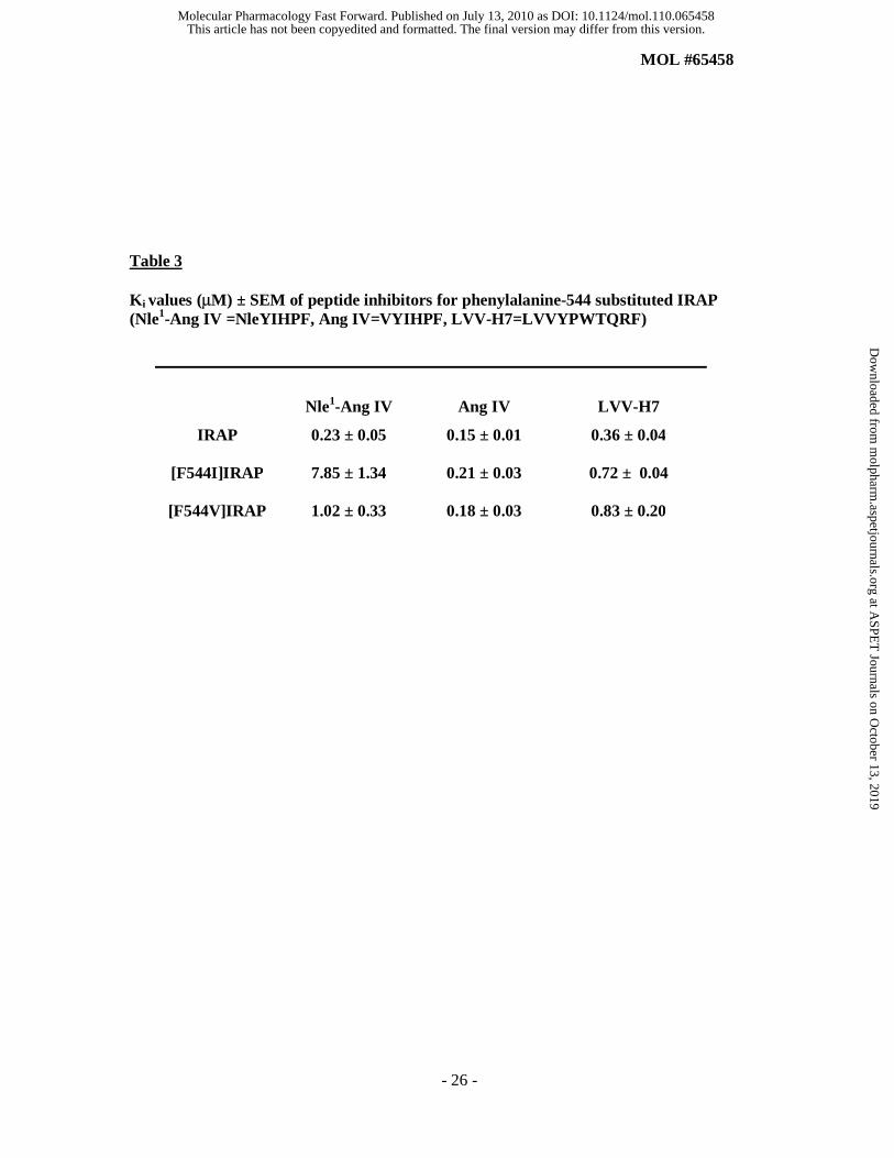
Inhibitory Potencies of Various Classes of IRAP Inhibitors
Peptide Inhibitors
Enzyme inhibition assays were carried out to investigate the effect of the mutations on the
binding affinity of IRAP inhibitors. Substitution of Phe544 with either Ile or Val had no effect
on the affinity of Ang IV for IRAP, whereas these mutations resulted in marked changes in the
affinities for both the Ang IV analogue, Nle1-Ang IV, and LVV-H7. Marked changes were
observed for Nle1-Ang IV, with the affinity being 34-fold lower for the [F544I] mutant and 4.3-
fold lower for the [F544V] mutant compared to wild type (Table 3). In contrast, the affinity of
LVV-H7 for both mutants was approximately 2-fold lower than wild type (Table 3). Originally,
Val1 of Ang IV was substituted for Nle to stabilize the peptide for in vivo studies, presumably to
reduce susceptibility to N-terminal degradation (Sardinia et al., 1994). It is interesting that in
this conserved change to Nle (2-aminohexanoic acid), the unbranched isomer of leucine, the N-
terminal amino acid of Ang IV has such a marked effect on binding to the mutant forms of
IRAP.
This article has not been copyedited and formatted. The final version may differ from this version.Molecular Pharmacology Fast Forward. Published on July 13, 2010 as DOI: 10.1124/mol.110.065458
at ASPE
T Journals on O
ctober 13, 2019m
olpharm.aspetjournals.org
Dow
nloaded from

MOL #65458
- 12 -
HFI-series Inhibitors
Mutation of Phe544 resulted in marked changes in the affinity of some of the inhibitors for
IRAP (Table 4). For the pyridinyl derivatives, only modest changes in affinities of these
inhibitors for the IRAP mutants were observed, except for the affinity of HFI-142 for the
[F544I] mutant where a nearly 10-fold reduction in affinity was observed. In contrast, the
affinities for both HFI compounds containing the quinolinyl group (HFI-435, HFI-437) were
reduced by 10-fold for both the [F544I] and [F544V] mutants. The affinities were decreased to
levels comparable to the affinities of the 4-pyridinyl analogues for wild type IRAP.
DISCUSSION
The benzopyran inhibitors of IRAP are an important new lead towards the development of
therapeutics against dementia, and understanding the molecular basis of the interaction will
assist progress in this field. As yet however, the structure of IRAP has not been solved, such
that models of inhibitor binding are required. We performed computational docking of some of
the most active, IRAP-specific, HFI-series inhibitors (Figure 1) onto a homology model of the
catalytic domain of IRAP (based on the crystal structure of LTA4H). Surprisingly, the docking
results revealed two different binding conformations for these structurally analogous inhibitors,
but indicated in both cases that Phe544 would provide a hydrophobic packing point at one side
of the active site.
In the binding pose adopted by the pyridinyl derivatives, HFI-142 and HFI-419, a ring-stack is
predicted between the benzopyran moieties of the compounds and Phe544 (Figure 2A). This
binding pose contains numerous other interactions including an hydrogen bond from the
hydroxyl moiety of the inhibitors to Glu295 and van der Waals interactions involving Gln293,
Pro296, Glu426, Ala427, Leu483 and Ile540. Although the binding mode of both is comparable,
HFI-419’s higher potency may be due to the added interaction with the Zn through the amide
This article has not been copyedited and formatted. The final version may differ from this version.Molecular Pharmacology Fast Forward. Published on July 13, 2010 as DOI: 10.1124/mol.110.065458
at ASPE
T Journals on O
ctober 13, 2019m
olpharm.aspetjournals.org
Dow
nloaded from

MOL #65458
- 13 -
carbonyl. HFI-142 was able to dock in a comparable manner into the F544V mutant, but
adopted an alternate pose when docked into the F544I mutant due to a clash with the hindered
isoleucine. Consistent with this model, only a modest decrease in affinity of HFI-142 was
observed for the F544V compared to wild type, while a much greater decrease in affinity of
HFI-142 was seen for the F544I mutant IRAP (Table 4). The mutations also impacted
dramatically on the docking of the acetamido derivative, HFI-419, which adopts an alternate
“flipped” conformation when docked into the F544I and F544V mutants, but one that is still
able to make key contacts in the binding site (Figure 2B). That this particular inhibitor retains
significant affinity in the presence of these mutations compared to the other three inhibitors
(Table 4) may be explained by this alternate pose.
The quinolines (HFI-435 and HFI-437) are not able to adopt the aforementioned binding mode
for the pyridinyl compounds as the large quinoline group is too large to enter the small polar
pocket formed by Glu426 and Glu293. Alternatively, they adopt a mode which allows a
stronger interaction with the Zn atom through the quinoline nitrogen. The quinoline compounds
are predicted to be more active than the pyridinyl compounds due to the more favorable co-
ordination with the Zn atom, along with the hydrogen bonding network between the hydroxyl
moiety of the inhibitors and Ser546 and Lys520, and better van der Waals contacts particularly
the quinoline ring with the side-chain of Met430 and the ethyl ester with Ile461. The addition of
an acetyl group to the amino of HFI-435 fills a space and results in HFI-437 having additional
contacts with Phe550 and Tyr495, perhaps accounting for its greater potency.
The significant decline in affinity of HFI-435 and HFI-437 seen with both mutations F544V/I is
likely to be a direct result of a loss of the edge-face hydrophobic interactions between Phe544
and the benzopyran (Figure 2C). In the docking of these inhibitors into the mutant forms, the
binding pose is maintained as the strong interaction with the Zn atom is not hindered by the
This article has not been copyedited and formatted. The final version may differ from this version.Molecular Pharmacology Fast Forward. Published on July 13, 2010 as DOI: 10.1124/mol.110.065458
at ASPE
T Journals on O
ctober 13, 2019m
olpharm.aspetjournals.org
Dow
nloaded from

MOL #65458
- 14 -
mutations as with the pyridinyl compounds. However, the smaller side-chains of these
mutations do not make contact with the benzopyran moiety of the inhibitors (Figure 2D).
From a medicinal chemistry perspective these results are potentially very important, as they
indicate that IRAP is capable of binding these benzopyran inhibitors in multiple alternate
orientations, and this provides for multiple options in the elaboration of the ligands to improve
their pharmaceutical properties. In essence, the enzyme is not seeing one chemical class but two
(and possibly three) and the pyridinyl series and quinolinyl series would be expected to display
different structure-activity relationships. Our preliminary medicinal chemistry campaign to
some extent supports this concept.
As well as the effects on the HFI series, the variable importance of Phe544 in defining the IRAP
catalytic site is reflected in the results obtained for peptide substrate cleavage. Leu-Enk was
unable to be cleaved by either mutant suggesting that a ring stack interaction of the amino-
terminal tyrosine residue of Leu-Enk with Phe544 may be essential for the correct orientation of
the substrate in the catalytic site. In contrast, AVP cleavage by the mutants correlated well with
the Vmax of the mutants, indicating that Phe544 is not playing a pivotal role in the orientation
of AVP in the catalytic site. In keeping with these data is the model for binding of an arginyl
tripeptide substrate to LTA4H. In this model, the corresponding residue to Phe544 in IRAP,
Tyr378, plays a role in defining the S1 and S2’ subsites of the active site (Thunnissen et al.,
2002).
The inhibitory properties of the peptide inhibitors were unaffected by the mutation of Phe544
except for Nle1-Ang IV inhibition of the [F544I] mutant IRAP. These results suggest that the
Phe544 hydrophobic stacking point does not make a significant contribution to the binding
affinities of the peptide inhibitors for IRAP. The 30-fold lower affinity of Nle1-Ang IV for the
[F544I] IRAP compared to wild type is predicted to be due to the side-chain of the isoleucine
This article has not been copyedited and formatted. The final version may differ from this version.Molecular Pharmacology Fast Forward. Published on July 13, 2010 as DOI: 10.1124/mol.110.065458
at ASPE
T Journals on O
ctober 13, 2019m
olpharm.aspetjournals.org
Dow
nloaded from
MOL #65458
- 15 -
clashing with Nle; this is comparable to the interaction of the pyridinyl inhibitors with the
isoleucine side-chain predicted by the in silico modeling (Figure 1).
The aromatic R group residue, represented by Phe544 in IRAP, is conserved throughout the M1
family, with the corresponding residue being either a Phe or a Tyr. The importance of the
Phe544 in the catalytic site of IRAP is consistent with insights obtained from studies on the
crystal structures of a number of M1 aminopeptidases, including human LTA4H (Thunnissen et
al., 2001), bacterial APN (Ito et al., 2006) and plasmodium M1 alanyl aminopeptidase (PfA-
M1) (McGowan et al., 2009). These structures were resolved with the non-specific
aminopeptidase inhibitor bestatin, an analogue of the dipeptide Phe-Leu, present in the active
site. In each case, the phenolic ring of bestatin is demonstrated to be important in stabilising the
binding of bestatin in the catalytic site of these M1 aminopeptidases. In the crystal structure of
bacterial APN, the equivalent amino acid residue, Tyr376, forms a hydrophobic stacking with
the phenyl ring of bestatin in the active site (Ito et al., 2006), (Addlagatta et al., 2008). The
importance of this amino acid residue extends to its interactions with inhibitors of other
aminopeptidases, for example, the LTA4H competitive thioamine inhibitor, (3-(4-
benzyloxyphenoyl)-2-(R)-amino-1-propane thiol) was shown to bind to the zinc and the
hydrophobic pocket with the proximal phenyl ring making stacking interactions with Tyr378
(Phe544 equivalent) and Tyr267 (Thunnissen et al., 2001; Thunnissen et al., 2002). Moreover
Tyr378 in LTA4H also plays a critical role in suicide inactivation of LTA4H epoxide hydrolase
activity (Mueller et al., 1996a; Mueller et al., 1996b).
In conclusion, we demonstrate the involvement of Phe544 in defining the interaction of two
classes of benzopyran derived IRAP inhibitors with its catalytic site, which is reflected in the
altered potencies in binding to wild type and conserved Phe544 mutant IRAP. The docking
studies suggest that for the pyridinyl HFI inhibitors, the benzopyran moiety interacts in a ring
stack with Phe544. In contrast, the docking studies led to the hypothesis that due to the
This article has not been copyedited and formatted. The final version may differ from this version.Molecular Pharmacology Fast Forward. Published on July 13, 2010 as DOI: 10.1124/mol.110.065458
at ASPE
T Journals on O
ctober 13, 2019m
olpharm.aspetjournals.org
Dow
nloaded from

MOL #65458
- 16 -
different orientation of the quinolinyl HFI inhibitors in the catalytic site, Phe544 provides an
edge-face hydrophobic stacking point with the benzopyran. Moreover, we demonstrated that
Phe544 does not play a pivotal role in determining the potencies of the peptide inhibitors. These
new insights into the orientation of the HFI inhibitors in the IRAP catalytic site provide key
information for pharmacophore design for IRAP inhibitors, paving the way for the development
of a new generation of IRAP inhibitors for use as central-acting memory enhancing agents.
This article has not been copyedited and formatted. The final version may differ from this version.Molecular Pharmacology Fast Forward. Published on July 13, 2010 as DOI: 10.1124/mol.110.065458
at ASPE
T Journals on O
ctober 13, 2019m
olpharm.aspetjournals.org
Dow
nloaded from

MOL #65458
- 17 -
REFERENCES
Addlagatta A, Gay L and Matthews BW (2008) Structural basis for the unusual specificity
of Escherichia coli aminopeptidase N. Biochemistry 47:5303-11.
Albiston AL, Morton CJ, Ng HL, Pham V, Yeatman HR, Ye S, Fernando RN, De Bundel
D, Ascher DB, Mendelsohn FA, Parker MW and Chai SY (2008) Identification and
characterization of a new cognitive enhancer based on inhibition of insulin-
regulated aminopeptidase. Faseb J 22:4209-17.
Albiston AL, Pederson ES, Burns P, Purcell B, Wright JW, Harding JW, Mendelsohn FA,
Weisinger RS and Chai SY (2004a) Reversal of scopolamine-induced memory
deficits by LVV-hemorphin 7 in rats in the passive avoidance and Morris water
maze paradigms. Behav Brain Res 154:239-243.
Albiston AL, Ye S and Chai SY (2004b) Membrane bound members of the M1 family:
more than aminopeptidases. Protein Pept Lett 11:491-500.
Andersson H, Demaegdt H, Vauquelin G, Lindeberg G, Karlén A and Hallberg M (2008)
Ligands to the (IRAP)/AT4 receptor encompassing a 4-hydroxydiphenylmethane
scaffold replacing Tyr2. Bioorg Med Chem 16:6924-35.
Axén A, Andersson H, Lindeberg G, Rönnholm H, Kortesmaa J, Demaegdt H, Vauquelin
G, Karlén A and Hallberg M (2007) Small potent ligands to the insulin-regulated
aminopeptidase (IRAP)/AT(4) receptor. J Pept Sci 13:434-44.
Clark M, Cramer, R.D., Van Opdenbosch, N., (1989) Validation of the General Purpose
Tripos 5.2 Force Field. J. Computational Chemistry 10:982-1012.
Delano WL (2002) The PYMOL User's Manual, Delano Scientific, San Carlos, Ca.
Eisenberg D, Luthy R and Bowie JU (1997) VERIFY3D: assessment of protein models
with three-dimensional profiles. Methods Enzymol 277:396-404.
This article has not been copyedited and formatted. The final version may differ from this version.Molecular Pharmacology Fast Forward. Published on July 13, 2010 as DOI: 10.1124/mol.110.065458
at ASPE
T Journals on O
ctober 13, 2019m
olpharm.aspetjournals.org
Dow
nloaded from

MOL #65458
- 18 -
Herbst JJ, Ross SA, Scott HM, Bobin SA, Morris NJ, Lienhard GE and Keller SR (1997)
Insulin stimulates cell surface aminopeptidase activity toward vasopressin in
adipocytes. Am J Physiol 272:E600-6.
Ito K, Nakajima Y, Onohara Y, Takeo M, Nakashima K, Matsubara F, Ito T and
Yoshimoto T (2006) Crystal structure of aminopeptidase N (proteobacteria alanyl
aminopeptidase) from Escherichia coli and conformational change of methionine
260 involved in substrate recognition. J Biol Chem 281:33664-76.
Krishnan R, Hanesworth JM, Wright JW and Harding JW (1999) Structure-binding studies
of the adrenal AT4 receptor: analysis of position two- and three-modified
angiotensin IV analogs. Peptides 20:915-20.
Lee J, Albiston AL, Allen AM, Mendelsohn FAO, Ping SE, Barrett GL, Murphy M,
Morris MJ, McDowall SG and Chai SY (2004) Effect of intracerebroventricular
injection of AT4 receptor ligands, Nle1-angiotensin IV and LVv-hemorphin 7, on
spatial learning in rats. Neuroscience 124:341-349.
Lee JH, Mustafa T, McDowall SG, Mendelsohn FAO, Brennan MB, Lew R, Albiston AL
and Chai SY (2003) Structure-activity study of LVV-hemorphin-7: angiotensin
AT4 receptor ligand and inhibitor of insulin-regulated aminopeptidase (IRAP).
Journal of Pharmacology and Experimental Therapeutics. 305:205-211.
Lew RA, Mustafa T, Ye S, McDowall SG, Chai SY and Albiston AL (2003) Angiotensin
AT4 ligands are potent, competitive inhibitors of insulin regulated aminopeptidase
(IRAP). Journal of Neurochemistry 86:344-350.
Lukaszuk A, Demaegdt H, Feytens D, Vanderheyden P, Vauquelin G and Tourwé D
(2009) The replacement of His(4) in angiotensin IV by conformationally
constrained residues provides highly potent and selective analogues. J Med Chem
52:5612-8.
This article has not been copyedited and formatted. The final version may differ from this version.Molecular Pharmacology Fast Forward. Published on July 13, 2010 as DOI: 10.1124/mol.110.065458
at ASPE
T Journals on O
ctober 13, 2019m
olpharm.aspetjournals.org
Dow
nloaded from

MOL #65458
- 19 -
Lukaszuk A, Demaegdt H, Szemenyei E, Tóth G, Tymecka D, Misicka A, Karoyan P,
Vanderheyden P, Vauquelin G and Tourwé D (2008) Beta-homo-amino acid scan
of angiotensin IV. J Med Chem 51:2291-6.
McGann M, Almond, H., Nicholls, A., Grant, J.A., and Brown, F. (2003) Gaussian
Docking Functions. Biopolymers 68:76-90.
McGowan S, Porter CJ, Lowther J, Stack CM, Golding SJ, Skinner-Adams TS, Trenholme
KR, Teuscher F, Donnelly SM, Grembecka J, Mucha A, Kafarski P, Degori R,
Buckle AM, Gardiner DL, Whisstock JC and Dalton JP (2009) Structural basis for
the inhibition of the essential Plasmodium falciparum M1 neutral aminopeptidase.
Proc Natl Acad Sci U S A 106:2537-42.
Mueller MJ, Andberg MB, Samuelsson B and Haeggstrom JZ (1996a) Leukotriene A4
hydrolase, mutation of tyrosine 378 allows conversion of leukotriene A4 into an
isomer of leukotriene B4. J Biol Chem 271:24345-8.
Mueller MJ, Blomster M, Oppermann UC, Jornvall H, Samuelsson B and Haeggstrom JZ
(1996b) Leukotriene A4 hydrolase: protection from mechanism-based inactivation
by mutation of tyrosine-378. Proc Natl Acad Sci U S A 93:5931-5.
Olson ML, Olson EA, Qualls JH, Stratton JJ, Harding JW and Wright JW (2004)
Norleucine1-angiotensin IV alleviates mecamylamine-induced spatial memory
deficits. Peptides 25:233-241.
Pederson ES, Harding JW and Wright JW (1998) Attenuation of scopolamine-induced
spatial learning impairments by an angiotensin IV analog. Regul Pept 74:97-103.
Pederson ES, Krishnan R, Harding JW and Wright JW (2001) A role for the angiotensin
AT4 receptor subtype in overcoming scopolamine-induced spatial memory deficits.
Regul Pept 102:147-56.
This article has not been copyedited and formatted. The final version may differ from this version.Molecular Pharmacology Fast Forward. Published on July 13, 2010 as DOI: 10.1124/mol.110.065458
at ASPE
T Journals on O
ctober 13, 2019m
olpharm.aspetjournals.org
Dow
nloaded from
MOL #65458
- 20 -
Sardinia MF, Hanesworth JM, Krebs LT and Harding JW (1993) AT4 receptor binding
characteristics: D-amino acid- and glycine- substituted peptides. Peptides 14:949-
54.
Sardinia MF, Hanesworth JM, Krishnan F and Harding JW (1994) AT4 receptor structure-
binding relationship: N-terminal-modified angiotensin IV analogues. Peptides
15:1399-406.
Saveanu L, Carroll O, Weimershaus M, Guermonprez P, Firat E, Lindo V, Greer F,
Davoust J, Kratzer R, Keller SR, Niedermann G and van Endert P (2009) IRAP
identifies an endosomal compartment required for MHC class I cross-presentation.
Science 325:213-7.
Segura E, Albiston AL, Wicks IP, Chai SY and Villadangos JA (2009) Different cross-
presentation pathways in steady-state and inflammatory dendritic cells. Proc Natl
Acad Sci U S A 106:20377-81.
Stragier B, Clinckers R, Meurs A, De Bundel D, Sarre S, Ebinger G, Michotte Y and
Smolders I (2006) Involvement of the somatostatin-2 receptor in the anti-
convulsant effect of angiotensin IV against pilocarpine-induced limbic seizures in
rats. J Neurochem 98:1100-13.
Thunnissen MM, Nordlund P and Haeggstrom JZ (2001) Crystal structure of human
leukotriene A(4) hydrolase, a bifunctional enzyme in inflammation. Nat Struct Biol
8:131-5.
Thunnissen MMGM, Andersson B, Samuelsson B, Wong CH and Haeggstrom JZ (2002)
Crystal structures of leukotriene A4 hydrolase in complex with captopril and two
competitive tight-binding inhibitors. The FASEB Journal 16:1648-1650.
This article has not been copyedited and formatted. The final version may differ from this version.Molecular Pharmacology Fast Forward. Published on July 13, 2010 as DOI: 10.1124/mol.110.065458
at ASPE
T Journals on O
ctober 13, 2019m
olpharm.aspetjournals.org
Dow
nloaded from

MOL #65458
- 21 -
Wisniewski K, Borawska M and Car H (1993) The effect of angiotensin II and its
fragments on post-alcohol impairment of learning and memory. Pol.J.Pharmacol.
45:23-29.
Ye S, Chai SY, Lew RA, Ascher DB, Morton CJ, Parker MW and Albiston AL (2008)
Identification of modulating residues defining the catalytic cleft of insulin-
regulated aminopeptidase. Biochem Cell Biol 86:251-61.
This article has not been copyedited and formatted. The final version may differ from this version.Molecular Pharmacology Fast Forward. Published on July 13, 2010 as DOI: 10.1124/mol.110.065458
at ASPE
T Journals on O
ctober 13, 2019m
olpharm.aspetjournals.org
Dow
nloaded from

MOL #65458
- 22 -
FOOTNOTES
1 These authors contributed equally to this work. 2 is supported by an National Health and
Medical Research Council (NHMRC) Peter Doherty fellowship, 3 is an Australian Research
Council Federation Fellow and an NHMRC Honorary Fellow and 4 was supported by an
NHMRC Senior Research Fellowship.
This research was supported by the Robert J. Kleberg Jr. and Helen C. Kleberg Foundation,
Alzheimer’s Drug Discovery Foundation, a Neurosciences Victoria/Victorian State
Government - Science, Technology and Innovation grant, and National Health and Medical
Research Council Development Grants (#454714 and #520695).
This article has not been copyedited and formatted. The final version may differ from this version.Molecular Pharmacology Fast Forward. Published on July 13, 2010 as DOI: 10.1124/mol.110.065458
at ASPE
T Journals on O
ctober 13, 2019m
olpharm.aspetjournals.org
Dow
nloaded from

MOL #65458
- 23 -
FIGURE LEGENDS
Figure 1
Structures of the benzopyran-based IRAP inhibitors.
(A & C) 4-pyridinyl derivatives (HFI-142 and HFI419), (B&D) 4- quinolinyl derivatives (HFI-
435 and HFI-437). Ki calculated using the synthetic substrate Leu-MCA, Ki = IC50/(1[S]/Km),
where Leu-MCA Km = 38.7 μM; [S] = 25 μM. (Albiston et al., 2008).
Figure 2
Model showing the docking of HFI-419 and HFI-437 to the catalytic site.
(A) Cross-eyed stereo view (above) and Simplified view (below) of HFI-419 (yellow sticks)
docked into the IRAP model. There are interactions with the Zn2+ ion (purple sphere) via the
amide carbonyl and the benzopyran oxygen (shown as dashed lines). Phe544 (white sticks) has
a direct parallel ring stacking interaction with the benzopyran. This picture is rotated 90° along
the x-axis (see arrow) compared to the following panels in this figure. (B) Cross-eyed stereo
(above) and Simplified (below) view of mutation F544I. The mutation results in an unfavorable
clash between the isoleucine side-chain and the benzopyran of HFI-419 (not shown). An
alternative flipped orientation is found to be prominent amongst the top twenty poses which
retains the Zn2+ interaction with the benzopyran oxygen (dashed line) and allows a hydrogen
bond between the acetamide of the inhibitor and Tyr549 (dashed line). There is also an edge-
face interaction between Tyr549 and the pyridinyl ring of the inhibitor. (C) Cross-eyed stereo
view (above) and Simplified view (below) of HFI-437 (yellow sticks) docked into the IRAP
model. This inhibitor has an alternate binding mode to that described for HFI-419 with the
quinoline nitrogen now interacting with the Zn2+ ion. Phe544 is an edge-face stacking point for
this inhibitor. (D) Cross-eyed stereo (above) and Simplified (below) view of the mutation F544I
that would result in a loss of hydrophobic interactions with HFI-437 as the smaller side-chain
does not interact with the benzopyran moiety of the inhibitor. All pictures were constructed
using the molecular modeling software PYMOL (Delano, 2002).
This article has not been copyedited and formatted. The final version may differ from this version.Molecular Pharmacology Fast Forward. Published on July 13, 2010 as DOI: 10.1124/mol.110.065458
at ASPE
T Journals on O
ctober 13, 2019m
olpharm.aspetjournals.org
Dow
nloaded from

MOL #65458
- 24 -
TABLES
Table 1 Kinetic parameters for the hydrolysis of Leu-MCA by wild type IRAP and phenylalanine-544 substituted IRAP
Mutants
Km (µM ± SEM)
Vmax (μM/μg protein.h + SEM)
Vmax/Km
(µg protein-1.h-1)
IRAP 47.1 ± 6.2 144.4 ± 5.1 3.1
[F544I]IRAP 63.7 ± 12.6 56.5 ± 3.0 0.9
[F544V]IRAP 101 ± 10.6 9.3±1.1 0.1
This article has not been copyedited and formatted. The final version may differ from this version.Molecular Pharmacology Fast Forward. Published on July 13, 2010 as DOI: 10.1124/mol.110.065458
at ASPE
T Journals on O
ctober 13, 2019m
olpharm.aspetjournals.org
Dow
nloaded from

MOL #65458
- 25 -
Table 2 Degradation of vasopressin (AVP) and Leu-enkephalin (Leu-Enk) by wild-type and phenylalanine-544 substituted IRAP. The catalytic activity for the mutants is expressed relative to wild type (1.0).
Enzyme
Vasopressin (AVP)
CYFQNCPRG
Leu-Enk
YGGFL
IRAP 1.0 1.0
[F544I]IRAP 1.0 No activity
[F544V]IRAP <0.1 No activity
This article has not been copyedited and formatted. The final version may differ from this version.Molecular Pharmacology Fast Forward. Published on July 13, 2010 as DOI: 10.1124/mol.110.065458
at ASPE
T Journals on O
ctober 13, 2019m
olpharm.aspetjournals.org
Dow
nloaded from
MOL #65458
- 26 -
Table 3 Ki values (μM) ± SEM of peptide inhibitors for phenylalanine-544 substituted IRAP (Nle1-Ang IV =NleYIHPF, Ang IV=VYIHPF, LVV-H7=LVVYPWTQRF)
Nle1-Ang IV
Ang IV
LVV-H7
IRAP 0.23 ± 0.05 0.15 ± 0.01 0.36 ± 0.04
[F544I]IRAP 7.85 ± 1.34 0.21 ± 0.03 0.72 ± 0.04
[F544V]IRAP 1.02 ± 0.33 0.18 ± 0.03 0.83 ± 0.20
This article has not been copyedited and formatted. The final version may differ from this version.Molecular Pharmacology Fast Forward. Published on July 13, 2010 as DOI: 10.1124/mol.110.065458
at ASPE
T Journals on O
ctober 13, 2019m
olpharm.aspetjournals.org
Dow
nloaded from

MOL #65458
- 27 -
Table 4 Ki values (μM) ± SEM of HFI inhibitors for phenylalanine-544 substituted IRAP
HFI-142
HFI-435
HFI-419
HFI-437
IRAP 2.01 ± 0.51 0.33± 0.14 0.75 ± 0.30 0.031± 0.004
[F544I]IRAP 16.42 ± 5.22 3.72 ± 0.62 1.83 ± 0.34 0.20 ± 0.03
[F544V]IRAP 4.10 ± 0.08 2.51 ± 0.52 0.67 ± 0.13 0.27 ± 0.08
This article has not been copyedited and formatted. The final version may differ from this version.Molecular Pharmacology Fast Forward. Published on July 13, 2010 as DOI: 10.1124/mol.110.065458
at ASPE
T Journals on O
ctober 13, 2019m
olpharm.aspetjournals.org
Dow
nloaded from

C D
A B
O
N
NH2OH
O CH3
O
HFI-142
O
N
NH2OH
O CH3
O
O
N
NHOH
O CH3
O
O CH3
O
N
NHOH
O CH3
O
O CH3
HFI-142 Ki 2.01 μM HFI-435 Ki 0.33 μM
HFI-419 Ki 0.75 μM HFI-437 Ki 0.03 μM
Figure 1
This article has not been copyedited and formatted. The final version may differ from this version.Molecular Pharmacology Fast Forward. Published on July 13, 2010 as DOI: 10.1124/mol.110.065458
at ASPE
T Journals on O
ctober 13, 2019m
olpharm.aspetjournals.org
Dow
nloaded from

This article has not been copyedited and formatted. The final version may differ from this version.Molecular Pharmacology Fast Forward. Published on July 13, 2010 as DOI: 10.1124/mol.110.065458
at ASPE
T Journals on O
ctober 13, 2019m
olpharm.aspetjournals.org
Dow
nloaded from

This article has not been copyedited and formatted. The final version may differ from this version.Molecular Pharmacology Fast Forward. Published on July 13, 2010 as DOI: 10.1124/mol.110.065458
at ASPE
T Journals on O
ctober 13, 2019m
olpharm.aspetjournals.org
Dow
nloaded from


![MOL #89482 Title Page The antiallergic mast cell ...molpharm.aspetjournals.org/content/molpharm/early/2013/10/10/mol... · benzopyrano[2,3-b]pyridine-3-carboxylic acid; BRL10833,](https://img.pdfslide.us/doc/110x75/5a8dd0697f8b9adb648ce60d/mol-89482-title-page-the-antiallergic-mast-cell-23-bpyridine-3-carboxylic.jpg)