Embed Size (px)
Citation preview

Module 6: Immunology
AnS 536Spring 2015
Innate and Adaptive Immunity in the Newborn
Fetus exists in a sterile environment prior to birth Neonates require an efficient host
defense
Types of Immunity
Innate Adaptive Humoral Cell-Mediated Active Passive
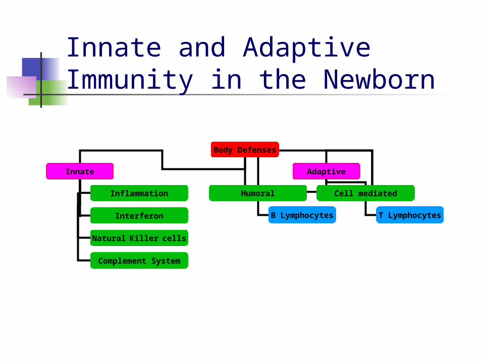
Body Defenses
Innate Adaptive
Inflammation
Interferon
Natural Killer cells
Complement System
Humoral Cell mediated
B Lymphocytes T Lymphocytes
Innate and Adaptive Immunity in the Newborn
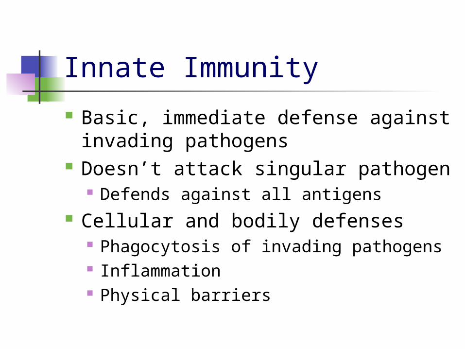
Innate Immunity
Basic, immediate defense against invading pathogens
Doesn’t attack singular pathogen Defends against all antigens
Cellular and bodily defenses Phagocytosis of invading pathogens Inflammation Physical barriers

Innate Immunity in the Newborn Innate
Non-specific defense mechanisms that respond immediately to antigens present in the body
Include: Physical barriers (skin, mucous membranes) Inflammation (reaction of the body to tissue
damage) Phagocytosis (neutrophils, monocytes, and tissue
macrophages) NK cells Interferon Complement
Generalized response not specific to a particular antigen

Innate Immune System
Without adaptive immunity, neonate relies on complement and effector cells of innate immune system
Phagocytic ability of cells is normal/increased, but less responsive to activation by lymphokines
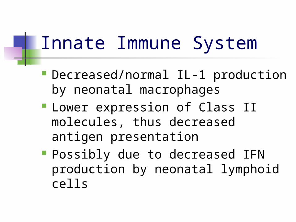
Innate Immune System
Decreased/normal IL-1 production by neonatal macrophages
Lower expression of Class II molecules, thus decreased antigen presentation
Possibly due to decreased IFN production by neonatal lymphoid cells
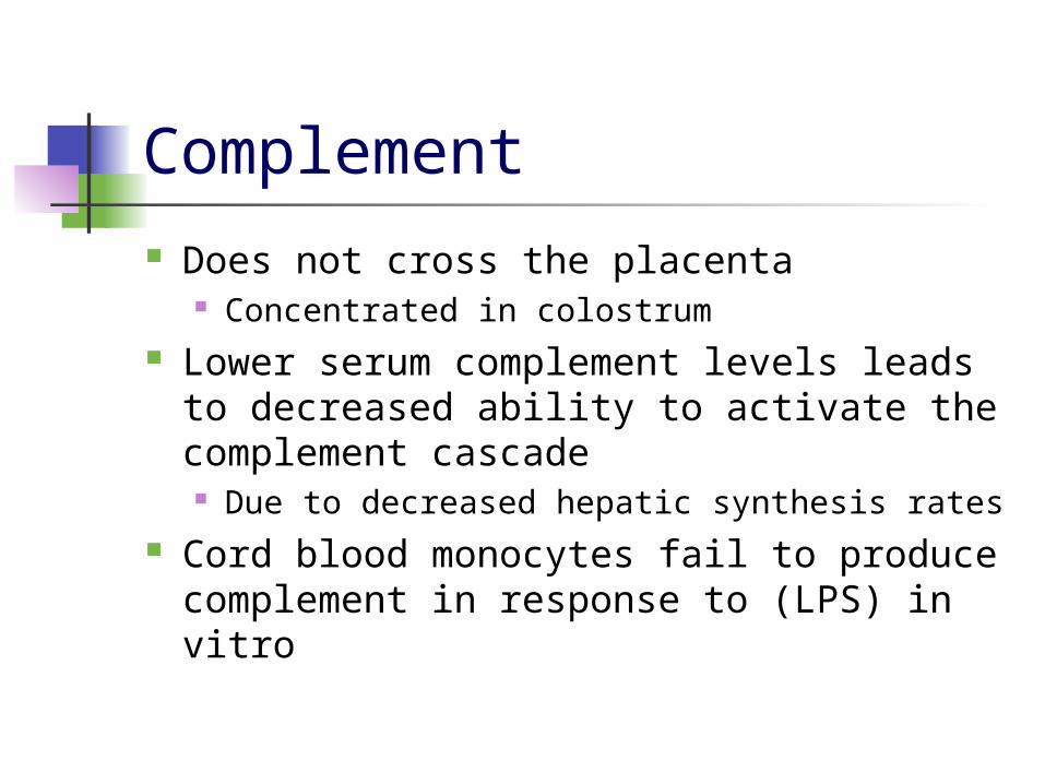
Complement Does not cross the placenta
Concentrated in colostrum Lower serum complement levels leads
to decreased ability to activate the complement cascade Due to decreased hepatic synthesis rates
Cord blood monocytes fail to produce complement in response to (LPS) in vitro
Innate and Adaptive Immunity in the Newborn
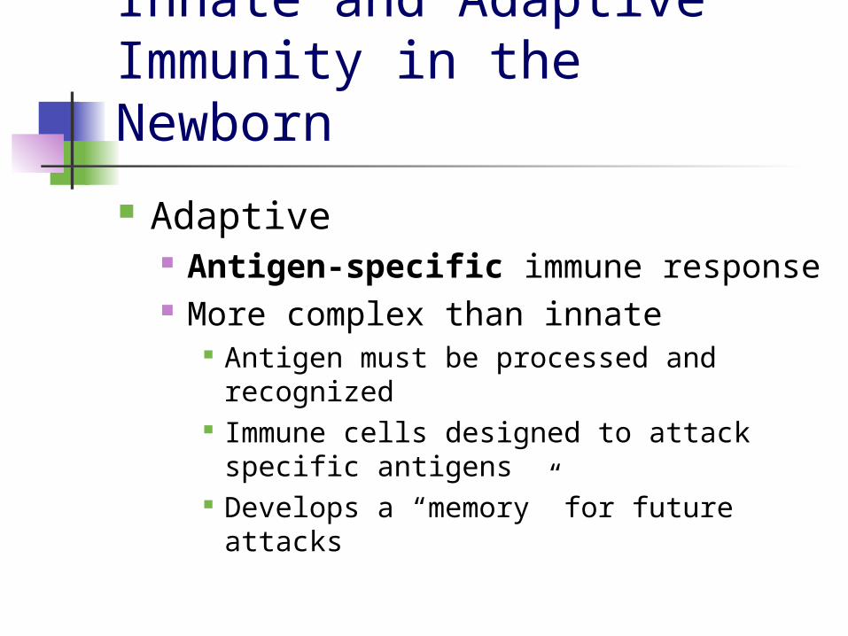
Adaptive Antigen-specific immune response More complex than innate
Antigen must be processed and recognized
Immune cells designed to attack specific antigens
Develops a “memory” for future attacks

Adaptive Immunity
Increased level of defense Attacks specific pathogen
Memorizes that pathogen in case of future need
Mostly cellular responses Divided into humoral and cell
mediated immunity

Adaptive Immunity
B cell development Pluripotent stem cells within blood
islands of yolk sac give rise to progenitor cells
Progenitor cells migrate to the fetal liver where B cell development begins
Later in fetal development, bone marrow assumes this function

Humoral Immunity Immunity using macromolecules B cells
Type of lymphocyte that is formed in bone marrow
Possesses a protein on outer surface called B cell receptor
Antibodies/Immunoglobulins Produced by B cells Takes out bacteria and viruses
Complement System Helps phagocytic cells clear pathogens Causes cytolysis of target cell

Passive Immunity
Antibodies passed from one individual to another
Natural Transfer of antibodies through placenta or colostrum
Artificial Taking antibodies for a specific pathogen from
immune individual to non-immune Short duration

Active Immunity
Antibodies are produced when immune system is exposed to an antigen Artificially acquired
Vaccines Naturally acquired
Memory T cells
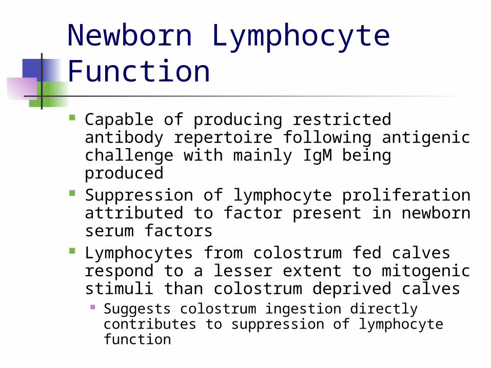
Newborn Lymphocyte Function Capable of producing restricted antibody
repertoire following antigenic challenge with mainly IgM being produced
Suppression of lymphocyte proliferation attributed to factor present in newborn serum factors
Lymphocytes from colostrum fed calves respond to a lesser extent to mitogenic stimuli than colostrum deprived calves Suggests colostrum ingestion directly
contributes to suppression of lymphocyte function
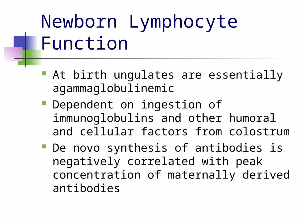
Newborn Lymphocyte Function At birth ungulates are essentially
agammaglobulinemic Dependent on ingestion of
immunoglobulins and other humoral and cellular factors from colostrum
De novo synthesis of antibodies is negatively correlated with peak concentration of maternally derived antibodies

Immunoglobulins
Mark cells for attack or destroy cells themselves
Five types IgM IgE IgD IgG IgA
Immunoglobulins
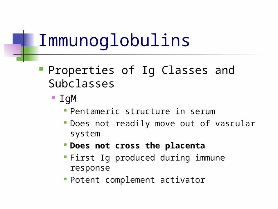
Properties of Ig Classes and Subclasses IgM
Pentameric structure in serum Does not readily move out of vascular
system Does not cross the placenta First Ig produced during immune response Potent complement activator
IgM
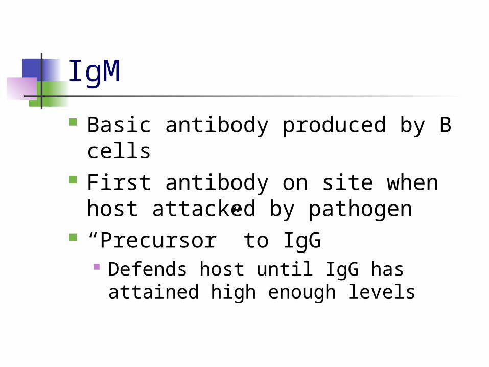
Basic antibody produced by B cells First antibody on site when host
attacked by pathogen “Precursor” to IgG
Defends host until IgG has attained high enough levels
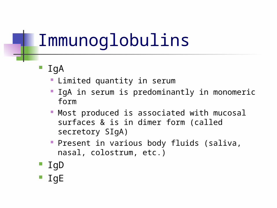
Immunoglobulins IgA
Limited quantity in serum IgA in serum is predominantly in monomeric form Most produced is associated with mucosal
surfaces & is in dimer form (called secretory SIgA)
Present in various body fluids (saliva, nasal, colostrum, etc.)
IgD IgE
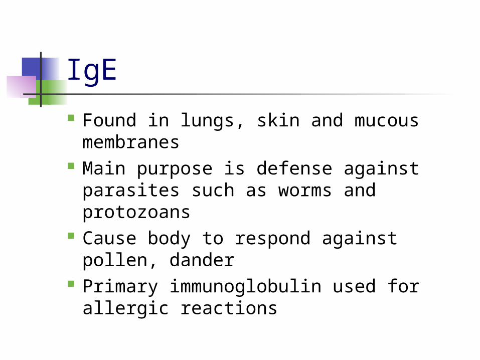
IgE Found in lungs, skin and mucous
membranes Main purpose is defense against
parasites such as worms and protozoans
Cause body to respond against pollen, dander
Primary immunoglobulin used for allergic reactions
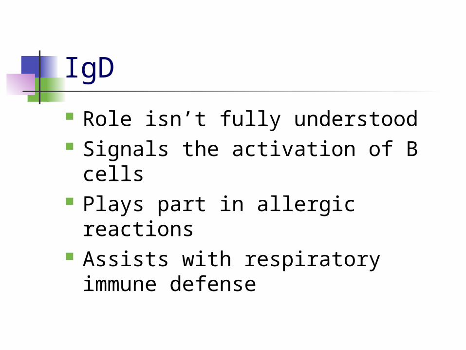
IgD
Role isn’t fully understood Signals the activation of B cells Plays part in allergic reactions Assists with respiratory immune
defense

Immunoglobulins IgG
Monomeric structure in serum Predominant Ig in serum 4 subclasses
IgG1 have Fc region that effectively bind to macrophage Fc receptors
IgG2 does not cross the human placenta IgG3 most effective activator of complement and
also has Fc regions like IgG1
IgG4

IgG
Major immunoglobulin of defense Part of secondary immune response This is the only antibody that can
cross through the placenta Causes agglutination of pathogens Coats surface of foreign bodies for
ingestion by phagocytes Activates complement system

IgG in Human Fetus Level of IgG in fetus are correlated to the level in
mother IgG must be bound to neonatal Fc receptor (FcRn)
or face degradation by lysosomal enzymes Certain regions of Africa showed limited transfer of
IgG because FcRn was saturated with IgG (Palmeira, et al. 2012)
Fetus starts to acquire maternal antibodies beginning at the 13th week of gestation
Largest amount of transfer happens in third trimester At full term, fetal IgG concentrations surpass the
mothers by 20-30% Maternal age, parity, weight and delivery type
have no effect on IgG concentration

Adaptive Immunity Antibody Response
Following antigenic stimulation in the adult, initial antibody response consists mainly of the production of IgM
Maturation of the humoral immune response involves “class switching”
Rearrangement of genes within DNA to produce IgG
Rearrangements are delayed during fetal life

Adaptive Immunity
Normal numbers of B & T cells are present at birth
Humoral immune responses are functionally immature Due to regulatory imbalance between
T cell mediated help & suppression Also due to B cell immaturity

Adaptive Immunity
Newborns are better able to respond to protein antigens then capsular polysaccharide antigens
Demonstrate delayed ability to switch from IgM to IgG Results in developmental lag prior to
attainment of adult levels of serum IgM, IgG, and IgA
Adaptive Immunity
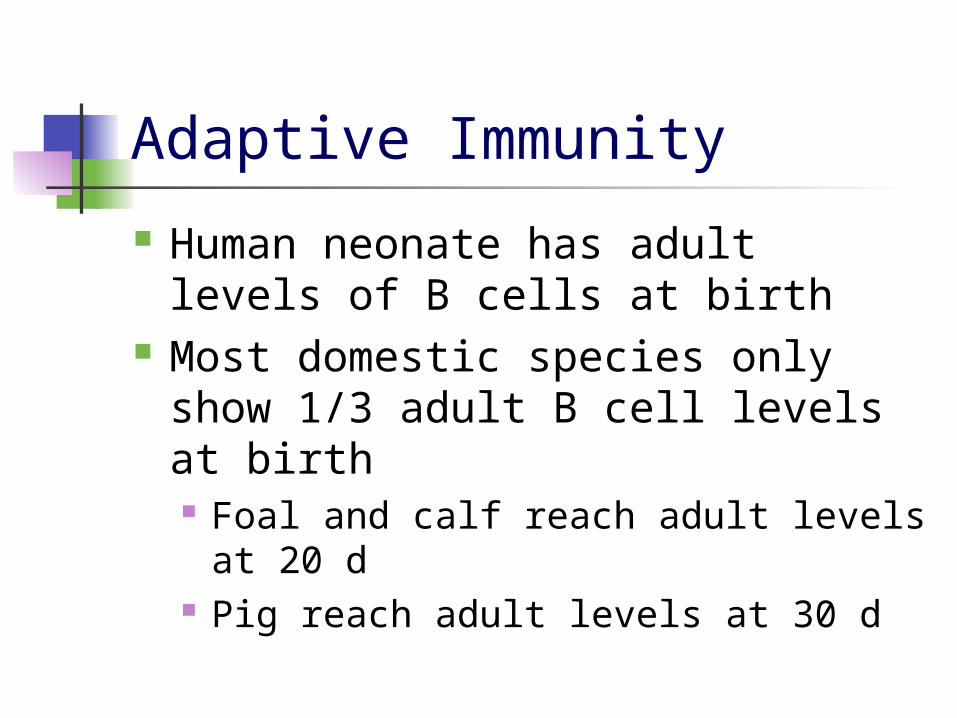
Human neonate has adult levels of B cells at birth
Most domestic species only show 1/3 adult B cell levels at birth Foal and calf reach adult levels at 20
d Pig reach adult levels at 30 d
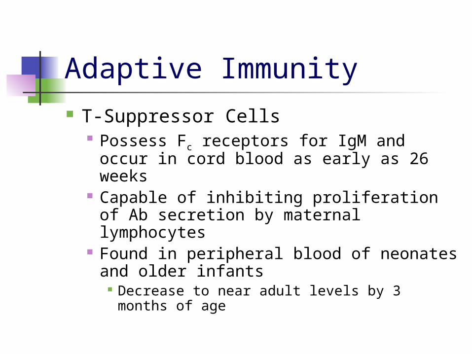
Adaptive Immunity T-Suppressor Cells
Possess Fc receptors for IgM and occur in cord blood as early as 26 weeks
Capable of inhibiting proliferation of Ab secretion by maternal lymphocytes
Found in peripheral blood of neonates and older infants
Decrease to near adult levels by 3 months of age

Adaptive Immunity
Summary Regulatory imbalance between T cell
mediated help and suppression, as well as B cell immaturity
Gap between development of lymphocytes and their ability to effectively generate a normal antibody response

Antimicrobial Sources in Neonatal Immunity
Antimicrobial peptides classification: Defensins
α-defensins Human neutrophil peptide 1 to 4 (HNP1-4) Human defensins 5 to 6 (HD5-6)
β-defensins HBD1-4
Expressed primarily by epithelial cells
Cathelicidins Have been thought to only be expressed in
mammals LL-37 only one found in humans Highly variant antimicrobial peptides

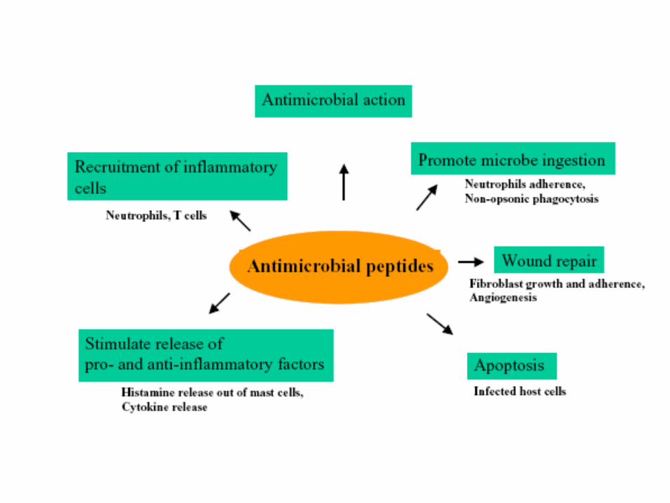
Antimicrobial Sources in Neonatal Immunity
Antimicrobial peptides are cationic Have an affinity to negatively charged
microbial membranes Have been shown to kill Gram+ and Gram-
bacteria, fungi, parasites, certain enveloped viruses, and cancer cells in vitro
Expressed in many organs in the body Neutrophils, paneth cells, epithelial cells of
skin, respiratory & gastrointestinal tracts, urogenital system, kidney pancreas, and placenta
Expression is continual (α-defensins and HBD1) or induced (HBD2-4)
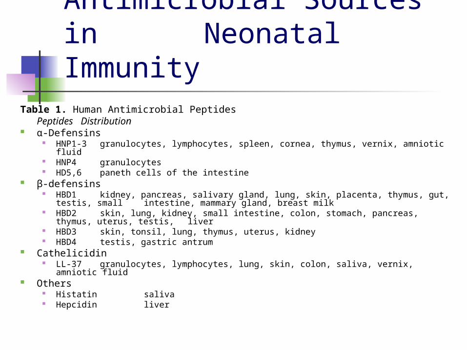
Antimicrobial Sources in Neonatal Immunity
Table 1. Human Antimicrobial PeptidesPeptides Distribution
α-Defensins HNP1-3 granulocytes, lymphocytes, spleen, cornea, thymus, vernix, amniotic fluid HNP4 granulocytes HD5,6 paneth cells of the intestine
β-defensins HBD1 kidney, pancreas, salivary gland, lung, skin, placenta, thymus, gut, testis,
small intestine, mammary gland, breast milk HBD2 skin, lung, kidney, small intestine, colon, stomach, pancreas, thymus,
uterus, testis, liver HBD3 skin, tonsil, lung, thymus, uterus, kidney HBD4 testis, gastric antrum
Cathelicidin LL-37 granulocytes, lymphocytes, lung, skin, colon, saliva, vernix, amniotic fluid
Others Histatin saliva Hepcidin liver

Maternal Recognition of Fetus Some embryonic proteins are recognized
as foreign due to paternal origin Maternal immune response is impaired
during pregnancy Fetal trophoblast
Maternal blood circulation contacts Anatomic barrier between dam and fetus Syncytiotrophoblastic layer of cells has been
found to be lacking in transplacental antigens Most likely reason why maternal immune system
doesn’t reject the fetus
Maternal Recognition of Fetus
Fetus creates its own protection Idoleamine 2,3-dioxygenase (IDO)
Destroys tryptophan (TRP) TRP fuels I-cells, that then attack foreign
tissue

Active vs. Passive Immunity
Active The production of antibodies as a
result of exposure to an antigen Natural exposure Artificially acquired
Vaccines contain modified antigens that initiate an immune response without causing the disease
Initial response produces memory T lymphocytes or B lymphocytes
Active vs. Passive Immunity Passive
Direct transfer of antibodies actively formed by another person or animal
“Borrowed” immunity Transfer of IgG from the mother to fetus across
the placenta during gestation Ingestion of colostrum transfers IgA
Antibodies are usually broken down before one month of age
Antibody-synthesizing ability does not develop before one month of age

Colostrum and its Role First time neonate will receive antibodies
from mother Stomach is porous at birth to allow absorption Absorption at max for first six hours post birth Can acquire antibodies for up to 24 hours, but
transfer hindered Without adequate intake of colostrum,
newborn will have less productive life Higher risk of morbidity, mortality, decreased
growth rates and first lactation milk production in dairy calves (Fidler, et al. 2007)
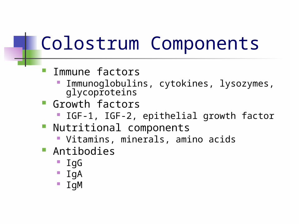
Colostrum Components Immune factors
Immunoglobulins, cytokines, lysozymes, glycoproteins
Growth factors IGF-1, IGF-2, epithelial growth factor
Nutritional components Vitamins, minerals, amino acids
Antibodies IgG IgA IgM

Changing Absorption of IgG
Difructose anhydride III Indigestible disaccharide which promotes
absorption of calcium and magnesium in intestines
Improves absorption of IgG in newborns Feed colostrum in one feeding Heat treated colostrum
Decreases microbial count while maintaining IgG levels

Changing Absorption of IgG Use of colostrum replacers
Colostrum replacers (CR) had less transfer of passive immunity when compared to colostrum (Fidler, et al. 2011)
However, the more CR the calves received, the better the transfer
Feeding sodium bicarbonate NaHCO3 can increase IgG concentration up to a
point (Cabral, et al. 2011) Feed colostrum in two feedings

Ig Deficiencies Hypogammaglobulinemia
Lack or decrease of one or more types of antibodies
Fetuses that don’t receive antibodies through the placenta fall into this category
IgG deficiencies More susceptible to infections such as pneumonia,
bronchitis and others Often occurs when there’s also a deficiency in IgA
or IgM Cause unknown but has possible genetic ties