Embed Size (px)
Citation preview
Modulation of protein aggregation by polyethyleneglycol conjugation: GCSF as a case study
RAHUL S. RAJAN,1 TIANSHENG LI,1 MOHINI ARAS,4 CHRISTOPHER SLOEY,1
WESTON SUTHERLAND,2 HIROMI ARAI,5 ROBERT BRIDDELL,2 OLAF KINSTLER,3
ALEXIS M.K. LUERAS,1 YU ZHANG,1 HEATHER YEGHNAZAR,1 MICHAEL TREUHEIT,1
AND DAVID N. BREMS1
Departments of 1Pharmaceutics, 2Hematology, and 3Protein Science, Amgen Inc., Thousand Oaks, California 91320, USA4Department of Chemical Engineering, University of California, Berkeley, California 94720, USA5Cal State Channel Islands, Department of Biology, Camarillo, California 93012, USA
(RECEIVED November 30, 2005; FINAL REVISION February 3, 2006; ACCEPTED February 9, 2006)
Abstract
Polyethylene glycol (PEG) conjugation to proteins has emerged as an important technology to producedrug molecules with sustained duration in the body. However, the implications of PEG conjugation toprotein aggregation have not been well understood. In this study, conducted under physiological pH andtemperature, N-terminal attachment of a 20 kDa PEG moiety to GCSF had the ability to (1) preventprotein precipitation by rendering the aggregates soluble, and (2) slow the rate of aggregation relative toGCSF. Our data suggest that PEG-GCSF solubility was mediated by favorable solvation of watermolecules around the PEG group. PEG-GCSF appeared to aggregate on the same pathway as that ofGCSF, as evidenced by (a) almost identical secondary structural transitions accompanying aggregation,(b) almost identical covalent character in the aggregates, and (c) the ability of PEG-GCSF to rescueGCSF precipitation. To understand the role of PEG length, the aggregation properties of free GCSF werecompared to 5kPEG-GCSF and 20kPEG-GCSF. It was observed that even 5kPEG-GCSF avoidedprecipitation by forming soluble aggregates, and the stability toward aggregation was vastly improvedcompared to GCSF, but only marginally less stable than the 20kPEG-GCSF. Biological activitymeasurements demonstrated that both 5kPEG-GCSF and 20kPEG-GCSF retained greater activity afterincubation at physiological conditions than free GCSF, consistent with the stability measurements. Thedata is most compatible with a model where PEG conjugation preserves the mechanism underlyingprotein aggregation in GCSF, steric hindrance by PEG influences aggregation rate, while aqueoussolubility is mediated by polar PEG groups on the aggregate surface.
Keywords: GCSF; protein aggregation; protein stability; polyethylene glycol; solubility
Protein aggregation represents a dominant degradationpathway that is often encountered during the developmentof therapeutic proteins in biotechnology (Mitraki and King1989; Mitraki et al. 1991; Clark 2001). Protein aggregates
have also been shown to be intimately associated with thepathogenesis of many neurodegenerative disorders, includ-ing Alzheimer’s disease, Huntington’s disease, Parkinson’sdisease, and Retinitis Pigmentosa (Chen et al. 2002; Stefaniand Dobson 2003; Li et al. 2004; Rajan and Kopito 2005).There is a growing body of evidence to suggest that proteinaggregates are inherently toxic to cells, even those notspecifically associated with an amyloid disease (Bence et al.2001; Stefani and Dobson 2003). In parenterally deliveredformulations, protein aggregates have the potential to causeadverse reactions in patients, such as aggregation at the site
ps0520040 Rajan et al. ARTICLE RA
Reprint requests to: Rahul S. Rajan, Department of Pharmaceutics,Amgen Inc., Thousand Oaks, CA 91320, USA; e-mail: [email protected]; fax: 805-375-5794; or David N. Brems, Department of Pharmaceu-tics, Amgen Inc., Thousand Oaks, CA 91320, USA; e-mail: [email protected]; fax: 805-375-5794.Article published online ahead of print. Article and publication date
are at http://www.proteinscience.org/cgi/doi/10.1110/ps.052004006.
Protein Science (2006), 15:1063–1075. Published by Cold Spring Harbor Laboratory Press. Copyright � 2006 The Protein Society 1063
JOBNAME: PROSCI 15#5 2006 PAGE: 1 OUTPUT: Thursday April 6 16:36:56 2006
csh/PROSCI/111777/PS0520040

of administration and the production of neutralizing anti-bodies (Hermeling et al. 2004). The formation of aggre-gates is also a major concern during manufacturing steps,storage, and shipping of the protein drug. Due to the insolu-ble nature of many protein aggregates, and the hurdles inisolating intermediates, it has been inherently difficult tostudy the mechanisms involved in the aggregation of thera-peutically important proteins until recently (Plakoutsi et al.2005; Seefeldt et al. 2005; Williams et al. 2005). Thus,developing rational approaches to understanding and con-trolling protein aggregation represents a major challengefor pharmaceutical drug development.
PEG is a water-soluble, biocompatible polymer that iscommonly utilized as an additive in protein formulations,and to facilitate crystallization of proteins (Cleland andJones 1996; Kerwin et al. 2002). Historically, conjugationof protein therapeutics with PEG has been performed toimprove the half-life of the protein in the blood serumbecause the large size of the PEG-conjugated moleculeslows down renal clearance (Molineux 2002). PEG con-jugation has also been used in protein encapsulationsystems, where it improved the encapsulation efficiencyand lowered the initial rate of release (Hora et al. 1990;Al-Azzam et al. 2005). However, there was no systematiccorrelation between the extent of protein aggregation andimprovements in the above processes. It has been pre-viously documented that PEG conjugation retards proteinprecipitation from the liquid state (Katre 1990; Kim andPark 2001); but there is an absence of systematic studiesinvestigating the mechanism underlying this phenomenonin the literature.
In the present study, the effects of PEG conjugation tothe aggregation properties of a therapeutic protein, granu-locyte colony stimulating factor (GCSF) have been inves-tigated. GCSF is a ;19 kDa four-helix bundle protein thatbelongs to the family of hematopoietic cytokines (Hillet al. 1993; Chaiken and Williams 1996; Kolvenbach et al.1997; Brems 2002; Raso et al. 2005). It regulates thegrowth and differentiation of hematopoietic progenitor cellsto functionally activate the formation of mature neutrophils.GCSF, marketed by Amgen under the trade name ofNEUPOGEN, has been widely used to treat neutropeniathat is often induced by myelosuppressive chemotherapy(Metcalf et al. 1996; Buchsel et al. 2002). Neulasta is thenext generation of GCSF, and has a 20-kDa PEG groupattached to the N terminus of the protein. It exhibitscomparable clinical benefits as GCSF, but due to its longhalf-life in the serum, only needs to be dosed once perchemotherapy cycle, as opposed to repeated daily adminis-tration for GCSF (Molineux 2002).
The shelf life of GCSF, commercially formulated inacidic conditions, is >2 yr at 2°C to 8°C (Herman et al.1996). However, at higher pH, such as neutral pH, GCSFhas a tendency to aggregate at moderate to high protein
concentrations and elevated temperatures (Krishnan et al.2002; Raso et al. 2005). Since the GCSF concentrationsrequired in the blood stream for effective action are in thepicomolar range (Brems 2002), it does not affect the useof GCSF as a drug, as GCSF aggregation is highly concen-tration dependent (Raso et al. 2005). However, aggrega-tion can pose severe restrictions on the handling of theprotein during the manufacturing process as well as forhigh concentration dosage forms. It is shown here that20kPEG-GCSF has the added advantage that even atrelatively high concentrations it does not precipitate outof solution. Unlike GCSF, which had a tendency to precipi-tate at neutral pH and 37°C (Krishnan et al. 2002; Rasoet al. 2005), 20kPEG-GCSF formed soluble aggregates underthe same conditions. The presence of the PEG moiety didnot perturb the secondary structural transitions or covalentinteractions involved in the aggregation process. Our datasuggests that the pathway of PEG-GCSF soluble aggregateformation was similar to that of GCSF precipitation.Strikingly, conjugation of the PEG group to GCSF greatlyretarded the rate as well as the extent of aggregate forma-tion, for both short- and long-length PEG-GCSF. In additionto greater bioavailability, PEG conjugation thus representsa powerful approach to enhancing the inherent stability ofGCSF toward aggregation while preserving the propertiesof the molecule.
Results
Conjugation of 20kPEG to GCSF preventedprotein precipitation
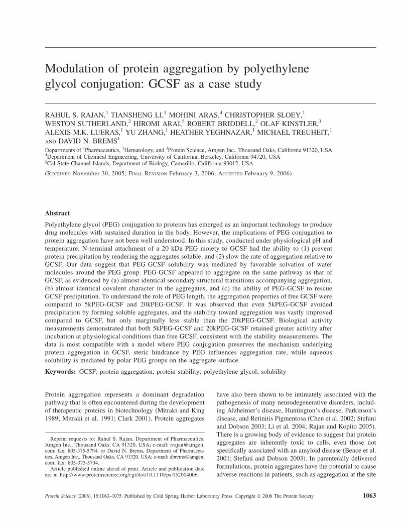
To compare the solubility properties of 20kPEG-GCSFand GCSF under physiological conditions, both proteinswere incubated at 37°C in 100 mM sodium phosphate atpH 6.9, for the times indicated in Figure 1A. A concen-tration of 5 mg/mL was chosen as the kinetics of GCSFaggregation were optimal for this study (>50% after 48 h),and lower concentrations precipitated very slowly (Raso et al.2005). At each time point, the sample was centrifuged andthe protein concentration measured using absorption at280 nm. The percentage of insoluble protein was calculatedfrom the difference in the protein concentration betweentime zero and the subsequent time points. It was observedthat while GCSF rapidly precipitated, 20kPEG-GCSF stayedcompletely soluble. To test the effect of free PEG on thesolubility of GCSF, solutions containing equimolar con-centrations of GCSF and free 20kPEG were incubated atpH 6.9, 37°C for 48 h, and compared to solutions of GCSFand 20kPEG-GCSF (Fig. 1B,C). Free PEG was not able toprevent GCSF precipitation, as observed by the formation ofvisible precipitates in the presence of free PEG (Fig. 1C)and turbidity at 600 nm (Fig. 1B). Further, from size-exclusion chromatography, the percent of GCSF that was
Rajan et al.
1064 Protein Science, vol. 15
JOBNAME: PROSCI 15#5 2006 PAGE: 2 OUTPUT: Thursday April 6 16:36:57 2006
csh/PROSCI/111777/PS0520040
insoluble after a 48-h incubation (52.83 6 1.36%) wasfound to be essentially the same as that obtained for GCSFin the presence of 1:1 free 20kPEG (52.1 6 1.8%),supporting this conclusion.
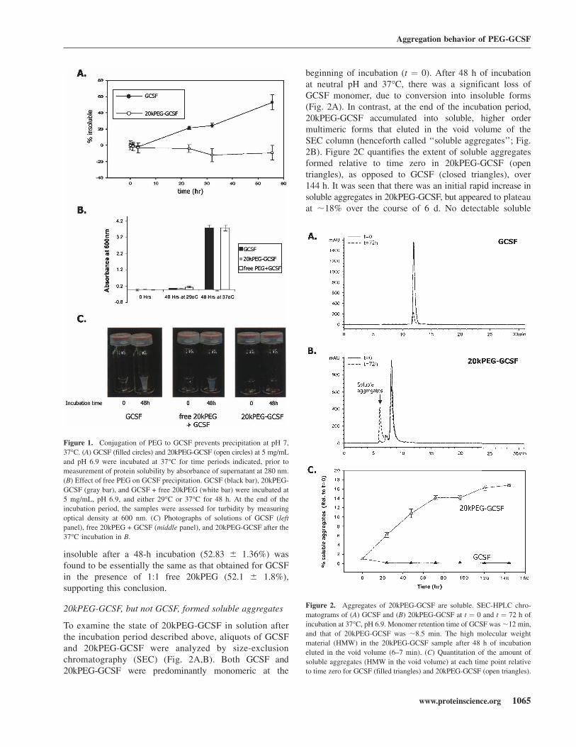
20kPEG-GCSF, but not GCSF, formed soluble aggregates
To examine the state of 20kPEG-GCSF in solution afterthe incubation period described above, aliquots of GCSFand 20kPEG-GCSF were analyzed by size-exclusionchromatography (SEC) (Fig. 2A,B). Both GCSF and20kPEG-GCSF were predominantly monomeric at the
beginning of incubation (t ¼ 0). After 48 h of incubationat neutral pH and 37°C, there was a significant loss ofGCSF monomer, due to conversion into insoluble forms(Fig. 2A). In contrast, at the end of the incubation period,20kPEG-GCSF accumulated into soluble, higher ordermultimeric forms that eluted in the void volume of theSEC column (henceforth called ‘‘soluble aggregates’’; Fig.2B). Figure 2C quantifies the extent of soluble aggregatesformed relative to time zero in 20kPEG-GCSF (opentriangles), as opposed to GCSF (closed triangles), over144 h. It was seen that there was an initial rapid increase insoluble aggregates in 20kPEG-GCSF, but appeared to plateauat ;18% over the course of 6 d. No detectable soluble
Figure 1. Conjugation of PEG to GCSF prevents precipitation at pH 7,
37°C. (A) GCSF (filled circles) and 20kPEG-GCSF (open circles) at 5 mg/mL
and pH 6.9 were incubated at 37°C for time periods indicated, prior to
measurement of protein solubility by absorbance of supernatant at 280 nm.
(B) Effect of free PEG on GCSF precipitation. GCSF (black bar), 20kPEG-
GCSF (gray bar), and GCSF + free 20kPEG (white bar) were incubated at
5 mg/mL, pH 6.9, and either 29°C or 37°C for 48 h. At the end of the
incubation period, the samples were assessed for turbidity by measuring
optical density at 600 nm. (C) Photographs of solutions of GCSF (left
panel), free 20kPEG + GCSF (middle panel), and 20kPEG-GCSF after the
37°C incubation in B.
Figure 2. Aggregates of 20kPEG-GCSF are soluble. SEC-HPLC chro-
matograms of (A) GCSF and (B) 20kPEG-GCSF at t ¼ 0 and t ¼ 72 h of
incubation at 37°C, pH 6.9. Monomer retention time of GCSF was;12 min,
and that of 20kPEG-GCSF was ;8.5 min. The high molecular weight
material (HMW) in the 20kPEG-GCSF sample after 48 h of incubation
eluted in the void volume (6–7 min). (C) Quantitation of the amount of
soluble aggregates (HMW in the void volume) at each time point relative
to time zero for GCSF (filled triangles) and 20kPEG-GCSF (open triangles).
Aggregation behavior of PEG-GCSF
www.proteinscience.org 1065
JOBNAME: PROSCI 15#5 2006 PAGE: 3 OUTPUT: Thursday April 6 16:36:57 2006
csh/PROSCI/111777/PS0520040
Fig. 1 live 4/C
aggregates were observed in GCSF in the same time period.Further, incubation with free PEG did not cause GCSF toform soluble aggregates (data not shown). Thus, conjugationof a 20-kDa PEG group to GCSF prevented protein pre-cipitation by rendering the aggregates soluble.
20kPEG-GCSF soluble aggregates and GCSFprecipitate shared a similar extent of oligomericand covalent character
A number of experiments were carried out to investigatethe mechanism underlying the formation of soluble aggre-gates in the case of PEG-GCSF. First, the extent ofmultimer formation between GCSF and 20kPEG-GCSFwas investigated. One possible explanation for the ap-pearance of a single peak near the void volume in SEC isthat the ‘‘soluble aggregate’’ of 20kPEG-GCSF may bea single, homogenous species. On the other hand, the voidvolume peak may, in fact, contain several oligomericspecies, which elute as one peak due to the limitations ofSEC resolution. To distinguish between these possibili-ties, and to compare the size distribution of the solubleaggregate with respect to the insoluble precipitate of GCSF,isolated soluble aggregate species and the solubilizedGCSF pellet were analyzed by SDS-PAGE (Fig. 3A, leftpanel). The solubilized pellet from the free PEG + GCSFcombination was analyzed as a control. The monomericform of GCSF (with or without free PEG) ran close to thepredicted mass of ;18 kDa (‘‘GCSF,’’ closed arrow). Onthe other hand, the monomeric form of 20kPEG-GCSF ranat an apparent size of ;55 kDa, much larger than thepredicted mass of 38 kDa (‘‘20kPEG-GCSF,’’ open blockarrow). GCSF from solubilized pellets of either GCSF aloneor GCSF + free PEG, as well as the 20kPEG-GCSF solubleaggregate migrated as an ensemble of monomer, dimer,trimer, and higher order multimers. Thus, the void volume(soluble aggregate) peak in the SEC of 20kPEG-GCSF didnot represent one species, but was composed of a distribu-tion of multimeric forms that were similar to that observedin the GCSF pellet, as assessed by SDS-PAGE.
To evaluate whether these multimers were covalentlylinked, the above samples were also treated with DTT to re-duce disulfide bonds, prior to loading (Fig. 3A, right panel).It was seen that the reduced samples contained mainly themonomer, with small amounts of the dimer (‘‘GCSF2,’’closed arrow for the GCSF and GCSF + free PEG samplesand ‘‘20kPEG-GCSF2,’’ open blocked arrow for the 20kPEG-GCSF sample). The presence of higher order multimers inthe nonreduced gel suggested that they were covalent. Theelimination of these higher order multimer bands indicatedthat they were disulfide linked, for both the GCSF pellet aswell as the 20kPEG-GCSF soluble aggregate.
To quantify and compare the extent of higher ordercovalent oligomers present in the GCSF pellet and in the
20kPEG-GCSF soluble aggregate, these samples (thesolubilized pellet of GCSF and the isolated solubleaggregate of 20kPEG-GCSF, respectively) were analyzedby denatured size-exclusion chromatography (D-SEC)(Fig. 3B), under nonreducing as well as reducing con-ditions. The nonreduced solubilized pellet from GCSFexhibited a monomeric species at ;34 min, a dimer at
Figure 3. Similar extent of oligomeric and covalent character in GCSF
and 20kPEG-GCSF aggregates. (A) Silver-stained SDS-PAGE gels of
solubilized pellets of GCSF and GCSF + free 20kPEG, and isolated soluble
aggregate of 20kPEG-GCSF. The gels were run both nonreduced (left
panel) and reduced (right panel). The monomer and dimer positions for
GCSF (closed arrow) and 20kPEG-GCSF (open block arrow) are in-
dicated. (B) Denatured SEC chromatograms are shown for solubilized
GCSF pellet (top panel) and isolated 20kPEG-GCSF soluble aggregates
(bottom panel). Samples were either reduced and alkylated (‘‘Red’’), or run
as alkylated but nonreduced (‘‘NR’’). The percent covalent character for
each sample from this analysis has been indicated.
Rajan et al.
1066 Protein Science, vol. 15
JOBNAME: PROSCI 15#5 2006 PAGE: 4 OUTPUT: Thursday April 6 16:37:14 2006
csh/PROSCI/111777/PS0520040

;30 min, and higher order multimers at ;22 min, nearthe void volume (Fig. 3B, upper panel). The nonreducedsoluble aggregate from 20kPEG-GCSF eluted differently;only the monomer peak at ;26 min and the void volumepeak at ;23 min were apparent (Fig. 3B, lower panel).Presumably, this was due to the much larger size of the20kPEG-GCSF molecule compared to GCSF, coupledwith a larger hydrodynamic radius due to solvated SDSmolecules, the same forces that caused it to run largerthan expected on SDS-PAGE. Treatment with DTT, as inthe case of SDS-PAGE, resulted in predominantly mono-meric and some dimeric forms in both samples, accom-panied by complete loss of higher oligomeric forms.From this analysis, the percent of covalent forms in theGCSF pellet sample was found to be 62.4 6 0.23%, whilethat for the soluble aggregate sample from 20kPEG-GCSF was 64.1 6 0.18%. These observations revealedthat the distribution of covalent, higher order multimersfound in the GCSF pellet was preserved in the 20kPEG-GCSF soluble aggregate, and therefore suggested thatconjugation of PEG to GCSF did not alter the forces thatheld the aggregates together.
Secondary structural transitions that accompanyaggregation are conserved between GCSFand 20kPEG-GCSF
One hypothesis to explain why 20kPEG-GCSF did notprecipitate under conditions that favored precipitation ofGCSF would be that conjugation of the PEG moietyretarded the secondary structural transitions in the proteinthat were responsible for aggregation. As seen in thecrystal structure (Hill et al. 1993), native GCSF containedmostly helical structures. This was further verified bya signal at 1656 cm�1 in second-derivative FTIR, as shownpreviously (Arakawa et al. 1995, Krishnan et al. 2002). Onthe other hand, aggregation of GCSF involved a conforma-tional transition, as evidenced by strong intensity bands at1620 cm�1 and 1695 cm�1, corresponding to a large extentof intermolecular b-sheet (Krishnan et al. 2002). To testwhether presence of the 20-kDa PEG group influences thetemperature-dependent shift in GCSF conformation fromhelix to b-sheet at neutral pH, FTIR spectra were recordedfor GCSF and 20kPEG-GCSF with single-degree temper-ature increments, from 35–45°C (Fig. 4A). The protein wasincubated for 1 min at each temperature prior to the spectralscan. Figure 4A (left panel) shows that GCSF at 35°Cwas predominantly a-helical as evidenced by the band at;1655 cm�1. With increasing temperature, the b-sheetcontent, reflected in the 1622 cm�1 and 1695 cm�1 bands,significantly increased. At 45°C, most of the a-helical struc-ture was converted to b-sheet. Secondary structural tran-sitions for 20kPEG-GCSF accompanied by the same increasesin temperature are shown in Figure 4A (right panel). It was
seen that both the starting spectral profile, as well as thechanges in the spectra with temperature, were almostidentical between GCSF and 20kPEG-GCSF.
Figure 4B shows a comparison of the secondary struc-tures of the GCSF precipitate and the isolated 20kPEG-GCSF soluble aggregate as revealed by second-derivativeFTIR spectroscopy. FTIR was optimal for this purpose asit could analyze secondary structure in both solid as wellas liquid forms. The GCSF pellet as well as the 20kPEG-GCSF soluble aggregate possessed very similar extent ofintermolecular b-sheet content, as evidenced by theoverlapping peak positions and areas (for both the 1622and 1695 cm�1 peaks). The stretching frequency and areaunder the curve for the 1655 cm�1 peak, corresponding toa-helical structures, was also conserved between thepellet and the soluble aggregate.
These observations suggested that conjugation of thePEG moiety to GCSF did not alter the helix to sheettransition that accompanied aggregation.
Figure 4. Conformational transitions and aggregate structure are pre-
served in PEG-GCSF. (A) PEG conjugation does not perturb the protein
structure and transitions responsible for GCSF aggregation. GCSF (left
panel) or PEG-GCSF (right panel) was incubated in a temperature ramp
experiment from 35°–45°C. The secondary structure at each temperature
was assessed by second-derivative FTIR. Helical structure was detected as
a band at 1655 cm�1, while the intermolecular b-sheet was detected as
a pair of bands at 1622 cm�1 and 1695 cm�1. (B) Comparison of the
secondary structure of the GCSF precipitate and 20kPEG-GCSF soluble
aggregate by FTIR.
Aggregation behavior of PEG-GCSF
www.proteinscience.org 1067
JOBNAME: PROSCI 15#5 2006 PAGE: 5 OUTPUT: Thursday April 6 16:37:18 2006
csh/PROSCI/111777/PS0520040

Rescue of GCSF precipitation by 20kPEG-GCSF
For proteins, it is generally regarded that the mechanism ofaggregation involves the self-association of intermediatesalong the folding pathway (Fink 1998; Havel et al. 1998) ormisfolded states (Canet et al. 1999; Stefani and Dobson2003). The similarity in secondary structural transitionsaccompanying aggregation and the extent of covalentcharacter in the aggregates of GCSF and 20kPEG-GCSFsuggested a similarity between the pathway of GCSFprecipitation, and the intermediates involved, to that of20kPEG-GCSF soluble aggregate formation. To further testthis, GCSF was incubated with either 1:1 20kPEG-GCSF(2.5 mg/mL each) or 2:1 20kPEG-GCSF (5 mg/mL GCSF,2.5 mg/mL 20kPEG-GCSF), and the extent of precipitationof these samples was compared to GCSF alone or GCSFincubated with free 20kPEG (latter two at 5 mg/mL) (Fig.5A). After 48 h of incubation at pH 6.9 and 37°C, asexpected, there was significant increase in sample turbiditydue to precipitation, measured as absorbance at 700 nm, inthe GCSF (black bar) and GCSF + free 20kPEG (white bar)samples. However, for both GCSF + 20kPEG-GCSFsamples, there was a dramatic decrease in the turbidity.Thus, co-incubation of GCSF with 20kPEG-GCSF effec-tively suppressed or ‘‘rescued’’ protein precipitation.
To investigate the fate of GCSF and PEG-GCSF in thesesamples, the supernatants were analyzed by SEC, as thistechnique separates the monomeric forms of the twoproteins. Figure 5B shows SEC chromatograms of GCSFincubated with 1:1 20kPEG-GCSF at the t ¼ 0 h, t ¼ 60 h,and t ¼ 120 h time points. At t ¼ 0, the sample containedtwo major peaks at ;11 min and 8.7 min corresponding toGCSF monomer and 20kPEG-GCSF monomer, respec-tively. At t ¼ 60 h and t ¼ 120 h, there was a large decreasein the GCSF monomer peak, a shift in the peak correspond-ing to PEG-GCSF by ;0.3 min, and the appearance ofa soluble aggregate peak. The shift in the PEG-GCSFmonomer peak suggested the formation of a GCSF:PEG-GCSF heterodimer. In the absence of precipitation, thegrowth of the heterodimer and soluble aggregate peaksconcomitant with loss of GCSF monomer peak suggestedan association between GCSF and PEG-GCSF.
Given the specificity of the aggregation process (Speedet al. 1996; Rajan et al. 2001; Krebs et al. 2004), theseobservations, taken together with results from Figures 3and 4, strongly suggest that the pathway of GCSF precipi-tation was the same as that for 20kPEG-GCSF solubleaggregate formation.
Difference in aqueous solvation properties ofGCSF and PEG-GCSF
The previous experiments indicated that the mechanismunderlying solubility of the aggregates was not due toa perturbation of the nature of the GCSF protein moiety.
The PEG group, however, does possess hydrophilic oxygenspecies in each repeat unit of the polymer. We thereforesought to determine whether the improved solubility ofPEG-GCSF aggregates were due to the favorable orienta-tion of solvent (water) molecules around PEG in spite ofthe increase in hydrophobicity due to aggregation. To verifythis hypothesis, the ability of GCSF or PEG-GCSF topartition to an organic phase from an aqueous phase wastested. In this regard, water–octanol systems have beenused extensively to investigate solvation properties of waterfor the molecule under consideration (Leo 1976; Wimley et al.1996). Figure 6A shows SEC chromatograms of 0.5 mg/mLGCSF with 0.5 mg/mL free 20kPEG in the aqueous phaseprior to and after overnight (16 h) incubation in a mixtureof 90% aqueous phase (phosphate buffer at pH 6.9) and10% octanol, with constant rotating to ensure that equilib-rium was attained. Very little GCSF remained in theaqueous phase (2.4 6 0.76%), suggesting that most of theGCSF had partitioned away from the aqueous phase. Incontrast, the vast majority of 20kPEG-GCSF under the sameconditions remained in the aqueous phase (93.2 6 4.9%),
Figure 5. 20kPEG-GCSF can rescue GCSF precipitation. (A) GCSF was
incubated for 48 h at pH 6.9, 37°C either alone (black bar), or with 1:1
20kPEG-GCSF (gray bar), 2:1 20kPEG-GCSF (hatched bar), or free 20k
PEG (white bar). The starting GCSF concentration was 5 mg/mL, and
protein turbidity was monitored by absorbance at 700 nm. (B) GCSF was
co-incubated with 1:1 20kPEG-GCSF as described above, and supernatants
analyzed by SEC are shown at t ¼ 0, t ¼ 60 h and t ¼ 120 h. The peaks
corresponding to GCSF monomer, PEG-GCSF monomer, and soluble
oligomers are shown.
Rajan et al.
1068 Protein Science, vol. 15
JOBNAME: PROSCI 15#5 2006 PAGE: 6 OUTPUT: Thursday April 6 16:37:21 2006
csh/PROSCI/111777/PS0520040
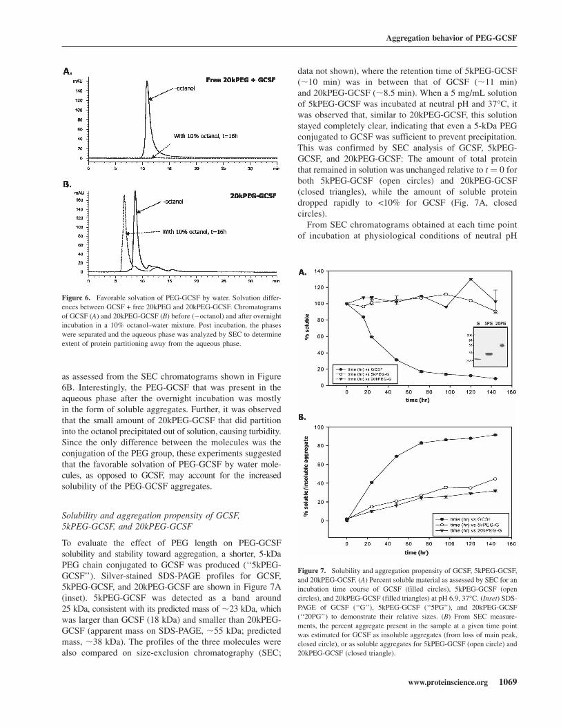
as assessed from the SEC chromatograms shown in Figure6B. Interestingly, the PEG-GCSF that was present in theaqueous phase after the overnight incubation was mostlyin the form of soluble aggregates. Further, it was observedthat the small amount of 20kPEG-GCSF that did partitioninto the octanol precipitated out of solution, causing turbidity.Since the only difference between the molecules was theconjugation of the PEG group, these experiments suggestedthat the favorable solvation of PEG-GCSF by water mole-cules, as opposed to GCSF, may account for the increasedsolubility of the PEG-GCSF aggregates.
Solubility and aggregation propensity of GCSF,5kPEG-GCSF, and 20kPEG-GCSF
To evaluate the effect of PEG length on PEG-GCSFsolubility and stability toward aggregation, a shorter, 5-kDaPEG chain conjugated to GCSF was produced (‘‘5kPEG-GCSF’’). Silver-stained SDS-PAGE profiles for GCSF,5kPEG-GCSF, and 20kPEG-GCSF are shown in Figure 7A(inset). 5kPEG-GCSF was detected as a band around25 kDa, consistent with its predicted mass of ;23 kDa, whichwas larger than GCSF (18 kDa) and smaller than 20kPEG-GCSF (apparent mass on SDS-PAGE, ;55 kDa; predictedmass, ;38 kDa). The profiles of the three molecules werealso compared on size-exclusion chromatography (SEC;
data not shown), where the retention time of 5kPEG-GCSF(;10 min) was in between that of GCSF (;11 min)and 20kPEG-GCSF (;8.5 min). When a 5 mg/mL solutionof 5kPEG-GCSF was incubated at neutral pH and 37°C, itwas observed that, similar to 20kPEG-GCSF, this solutionstayed completely clear, indicating that even a 5-kDa PEGconjugated to GCSF was sufficient to prevent precipitation.This was confirmed by SEC analysis of GCSF, 5kPEG-GCSF, and 20kPEG-GCSF: The amount of total proteinthat remained in solution was unchanged relative to t ¼ 0 forboth 5kPEG-GCSF (open circles) and 20kPEG-GCSF(closed triangles), while the amount of soluble proteindropped rapidly to <10% for GCSF (Fig. 7A, closedcircles).
From SEC chromatograms obtained at each time pointof incubation at physiological conditions of neutral pH
Figure 6. Favorable solvation of PEG-GCSF by water. Solvation differ-
ences between GCSF + free 20kPEG and 20kPEG-GCSF. Chromatograms
of GCSF (A) and 20kPEG-GCSF (B) before (�octanol) and after overnight
incubation in a 10% octanol–water mixture. Post incubation, the phases
were separated and the aqueous phase was analyzed by SEC to determine
extent of protein partitioning away from the aqueous phase.
Figure 7. Solubility and aggregation propensity of GCSF, 5kPEG-GCSF,
and 20kPEG-GCSF. (A) Percent soluble material as assessed by SEC for an
incubation time course of GCSF (filled circles), 5kPEG-GCSF (open
circles), and 20kPEG-GCSF (filled triangles) at pH 6.9, 37°C. (Inset) SDS-PAGE of GCSF (‘‘G’’), 5kPEG-GCSF (‘‘5PG’’), and 20kPEG-GCSF
(‘‘20PG’’) to demonstrate their relative sizes. (B) From SEC measure-
ments, the percent aggregate present in the sample at a given time point
was estimated for GCSF as insoluble aggregates (from loss of main peak,
closed circle), or as soluble aggregates for 5kPEG-GCSF (open circle) and
20kPEG-GCSF (closed triangle).
Aggregation behavior of PEG-GCSF
www.proteinscience.org 1069
JOBNAME: PROSCI 15#5 2006 PAGE: 7 OUTPUT: Thursday April 6 16:37:24 2006
csh/PROSCI/111777/PS0520040
and 37°C, the fraction of total material that existed eitheras a soluble aggregate (5kPEG-GCSF and 20kPEG-GCSF)or insoluble aggregate (GCSF) was calculated (Fig. 7B). Itwas seen that aggregate formation in 5kPEG-GCSF (opencircles) was vastly reduced compared to GCSF (closedcircles), and was only slightly increased compared to20kPEG-GCSF (closed triangles). Thus, attachment of evena 5kPEG to GCSF greatly stabilized the molecule againstintermolecular association leading to aggregation.
Conjugation of PEG stabilized against loss of in vitrobioactivity due to aggregation
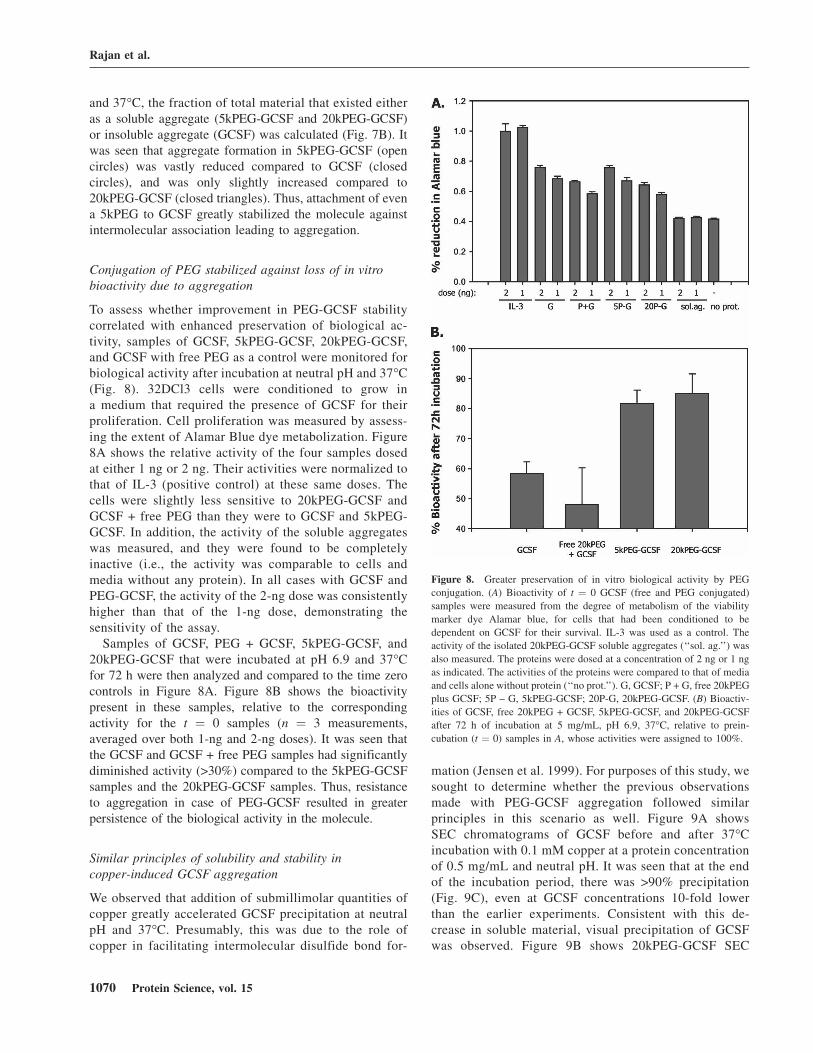
To assess whether improvement in PEG-GCSF stabilitycorrelated with enhanced preservation of biological ac-tivity, samples of GCSF, 5kPEG-GCSF, 20kPEG-GCSF,and GCSF with free PEG as a control were monitored forbiological activity after incubation at neutral pH and 37°C(Fig. 8). 32DCl3 cells were conditioned to grow ina medium that required the presence of GCSF for theirproliferation. Cell proliferation was measured by assess-ing the extent of Alamar Blue dye metabolization. Figure8A shows the relative activity of the four samples dosedat either 1 ng or 2 ng. Their activities were normalized tothat of IL-3 (positive control) at these same doses. Thecells were slightly less sensitive to 20kPEG-GCSF andGCSF + free PEG than they were to GCSF and 5kPEG-GCSF. In addition, the activity of the soluble aggregateswas measured, and they were found to be completelyinactive (i.e., the activity was comparable to cells andmedia without any protein). In all cases with GCSF andPEG-GCSF, the activity of the 2-ng dose was consistentlyhigher than that of the 1-ng dose, demonstrating thesensitivity of the assay.
Samples of GCSF, PEG + GCSF, 5kPEG-GCSF, and20kPEG-GCSF that were incubated at pH 6.9 and 37°Cfor 72 h were then analyzed and compared to the time zerocontrols in Figure 8A. Figure 8B shows the bioactivitypresent in these samples, relative to the correspondingactivity for the t ¼ 0 samples (n ¼ 3 measurements,averaged over both 1-ng and 2-ng doses). It was seen thatthe GCSF and GCSF + free PEG samples had significantlydiminished activity (>30%) compared to the 5kPEG-GCSFsamples and the 20kPEG-GCSF samples. Thus, resistanceto aggregation in case of PEG-GCSF resulted in greaterpersistence of the biological activity in the molecule.
Similar principles of solubility and stability incopper-induced GCSF aggregation
We observed that addition of submillimolar quantities ofcopper greatly accelerated GCSF precipitation at neutralpH and 37°C. Presumably, this was due to the role ofcopper in facilitating intermolecular disulfide bond for-
mation (Jensen et al. 1999). For purposes of this study, wesought to determine whether the previous observationsmade with PEG-GCSF aggregation followed similarprinciples in this scenario as well. Figure 9A showsSEC chromatograms of GCSF before and after 37°Cincubation with 0.1 mM copper at a protein concentrationof 0.5 mg/mL and neutral pH. It was seen that at the endof the incubation period, there was >90% precipitation(Fig. 9C), even at GCSF concentrations 10-fold lowerthan the earlier experiments. Consistent with this de-crease in soluble material, visual precipitation of GCSFwas observed. Figure 9B shows 20kPEG-GCSF SEC
Figure 8. Greater preservation of in vitro biological activity by PEG
conjugation. (A) Bioactivity of t ¼ 0 GCSF (free and PEG conjugated)
samples were measured from the degree of metabolism of the viability
marker dye Alamar blue, for cells that had been conditioned to be
dependent on GCSF for their survival. IL-3 was used as a control. The
activity of the isolated 20kPEG-GCSF soluble aggregates (‘‘sol. ag.’’) was
also measured. The proteins were dosed at a concentration of 2 ng or 1 ng
as indicated. The activities of the proteins were compared to that of media
and cells alone without protein (‘‘no prot.’’). G, GCSF; P + G, free 20kPEG
plus GCSF; 5P – G, 5kPEG-GCSF; 20P-G, 20kPEG-GCSF. (B) Bioactiv-
ities of GCSF, free 20kPEG + GCSF, 5kPEG-GCSF, and 20kPEG-GCSF
after 72 h of incubation at 5 mg/mL, pH 6.9, 37°C, relative to prein-
cubation (t ¼ 0) samples in A, whose activities were assigned to 100%.
Rajan et al.
1070 Protein Science, vol. 15
JOBNAME: PROSCI 15#5 2006 PAGE: 8 OUTPUT: Thursday April 6 16:37:27 2006
csh/PROSCI/111777/PS0520040
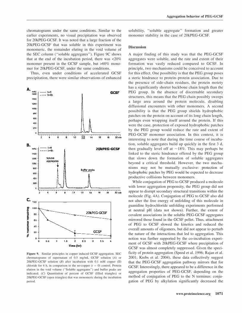
chromatograms under the same conditions. Similar to theearlier experiments, no visual precipitation was observedfor 20kPEG-GCSF. It was noted that a large fraction of the20kPEG-GCSF that was soluble in this experiment wasmonomeric, the remainder eluting in the void volume ofthe SEC column (‘‘soluble aggregates’’). Figure 9C showsthat at the end of the incubation period, there was <20%monomer present in the GCSF sample, but >60% mono-mer for 20kPEG-GCSF, under the same conditions.Thus, even under conditions of accelerated GCSF
precipitation, there were similar observations of enhanced
solubility, ‘‘soluble aggregate’’ formation and greatermonomer stability in the case of 20kPEG-GCSF.
Discussion
A major finding of this study was that the PEG-GCSFaggregates were soluble, and the rate and extent of theirformation was vastly reduced compared to GCSF. Inprinciple, two mechanisms could be conceived to accountfor this effect. One possibility is that the PEG group posesa steric hindrance to protein–protein association. Due tothe presence of side-chain residues, the protein moietyhas a significantly shorter backbone chain length than thePEG group. In the absence of discernable secondarystructures, this means that the PEG chain possibly sweepsa large area around the protein molecule, disablingdiffusional encounters with other monomers. A secondpossibility is that the PEG group shields hydrophobicpatches on the protein on account of its long chain length,perhaps even wrapping itself around the protein. If thiswere the case, protection of exposed hydrophobic patchesby the PEG group would reduce the rate and extent ofPEG-GCSF monomer association. In this context, it isinteresting to note that during the time course of incuba-tion, soluble aggregates build up quickly in the first 3 d,then gradually level off at ;18%. This may perhaps belinked to the steric hindrance offered by the PEG groupthat slows down the formation of soluble aggregatesbeyond a critical threshold. However, the two mecha-nisms may not be mutually exclusive: protection ofhydrophobic patches by PEG would be expected to decreaseproductive collisions between monomers.
While conjugation of PEG to GCSF produced a moleculewith lower aggregation propensity, the PEG group did notappear to disrupt secondary structural transitions within themolecule (Fig. 4A). Conjugation of PEG to GCSF also didnot alter the free energy of unfolding of this molecule inguanidine hydrochloride unfolding experiments performedat neutral pH (data not shown). Further, the extent ofcovalent associations in the soluble PEG-GCSF aggregatesmirrored those found in the GCSF pellet. Thus, attachmentof PEG to GCSF slowed the kinetics and reduced theoverall amounts of oligomers, but did not appear to perturbthe nature of the interactions that led to aggregation. Thisnotion was further supported by the co-incubation experi-ment of GCSF with 20kPEG-GCSF where precipitation ofGCSF was almost completely suppressed. Given the speci-ficity of protein aggregation (Speed et al. 1996; Rajan et al.2001; Krebs et al. 2004), these data collectively suggestthat the PEG-GCSF aggregation pathway mirrors that forGCSF. Interestingly, there appeared to be a difference in theaggregation properties of PEG-GCSF, depending on themethod of conjugation of PEG to the N terminus: conju-gation of PEG by alkylation significantly decreased the
Figure 9. Similar principles in copper induced GCSF aggregation. SEC
chromatograms of supernatant of 0.5 mg/mL GCSF solution (A) or
20kPEG-GCSF solution (B) after incubation with 0.1 mM copper (II)
chloride for 4 h, in comparison to the no-copper (t ¼ 0) control. Protein
elution in the void volume (‘‘Soluble aggregates’’) and buffer peaks are
indicated. (C) Quantitation of percent of GCSF (filled triangles) or
20kPEG-GCSF (open triangles) that was monomeric during the incubation
period.
Aggregation behavior of PEG-GCSF
www.proteinscience.org 1071
JOBNAME: PROSCI 15#5 2006 PAGE: 9 OUTPUT: Thursday April 6 16:37:30 2006
csh/PROSCI/111777/PS0520040
aggregation propensity compared to that obtained byacylation (Kinstler et al. 1996). This suggested a furtherinvolvement of the charge of the N-terminal amino groupon the aggregation properties of PEG-GCSF.
In this study, it was observed that both 5kPEG-GCSF and20kPEG-GCSF retained significantly higher bioactivitiescompared to free GCSF after 72 h of incubation at neutralpH and 37°C. However, while there was expected to be;20% monomer from the stability studies at the 72-h timepoint for free GCSF, there was still;50% to 60% bioactivityremaining. This phenomenon could perhaps be accountedfor by the higher variability of the bioassay compared to thestability assay. If the sample size were sufficiently large, it islikely the average bioactivity values would begin to con-verge with the stability numbers. However, the overall trendin bioactivity measurements was completely consistentwith the predictions made by the stability assay, and gavegreater confidence to the use of PEG-GCSF for highersustained bioactivity in vitro as well in vivo.
During design of the protein drug in question, thelength of PEG that needs to be attached to the protein isa key consideration. Thus far, this consideration has beenprimarily driven by the serum half-life afforded by thesize of the resulting PEG-conjugated drug molecule. Thisstudy showed that in addition to serum half-life, theaggregation propensity of the PEG-drug molecule inconsideration should also be evaluated. The data with5kPEG-GCSF indicated that a small PEG moiety attachedto the drug could result in a large benefit for proteinstability toward aggregation. In other words, even if alarge increase in serum half-life is not desired, conjuga-tion of PEG to a drug molecule may be considered solelyfrom the point of view of a stability advantage towardaggregation. The improvements in monomer stability canbe expected to correlate with improvements not only instorage and handling, but also in greater retention ofbiological activity, as shown in this study.
This study revealed that polyethylene glycol conjugatedGCSF remained soluble under conditions where nativeGCSF precipitated. This phenomenon could possibly beexplained by the very hydrophilic nature of the PEG groupcompared to protein moiety. The oxygen atoms in PEGgroup may form favorable hydrogen bond interactions withwater, and make the process of solvation by water aroundPEG-protein more favorable than around the protein alone.In this event, in spite of protein–protein association, therewould be sufficient water molecules around the PEG-protein multimers to keep the entire assembly soluble. Thismodel would predict the existence of PEG-protein‘‘micelles’’ with PEG-protein assemblies containing a hy-drophobic protein core and a hydrophilic PEG exterior thatis completely solvated by water. Indeed, the water/organicsolvent partition experiments revealed a greater preferencefor PEG-GCSF to remain in the aqueous phase, supporting
this model. It is possible that protein unfolding mighthave occurred during the transfer process, and from thisexperiment it would be difficult to assess the solubilityof the folded versus unfolded forms of GCSF. However, theformation of soluble aggregates in this experiment by20kPEG-GCSF is intriguing: it suggests a highly polarnature of the exterior of PEG-GCSF aggregates, perhapsby formation of PEG-protein micelles, thereby preventingtransfer into the octanol phase. The presence of the oxygengroups in each repeat unit of the PEG group wouldpotentially be a good site for solvation by water, by virtueof hydrogen bonding between the water hydrogens and thelone pair of electrons on the PEG oxygen atoms.
Controlling protein aggregation represents a majorchallenge during the development of protein therapeutics.Several approaches have been employed at improvingprotein solubility and stability. The most common oneinvolves reengineering the protein via site-directed mu-tagenesis. In the case of GCSF, three groups have un-dertaken this challenge (Lu et al. 1999; Bishop et al.2001; Luo et al. 2002), using the knowledge obtained byX-ray structure determination of this molecule (Hill et al.1993; Lovejoy et al. 1993). While these approachesproduced GCSF mutants with greater thermodynamicstability and biological activity than the wild-type mol-ecule, it was difficult to predict the aggregation propertiesof the resulting mutant proteins. A second approachinvolves the addition of excipients to reduce aggregation,such as sucrose, which has been shown to inhibit theaggregation propensity of GCSF (Krishnan et al. 2002).Protein aggregation has also been suppressed by novelapproaches such as application of high pressure (Foguelet al. 1999) and protein glycosylation (Sinclair and Elliott2005). While these approaches are useful, they do notsubstantially alter the pharmokinetic profile of the mol-ecule in blood serum. Protein PEG conjugation, at least inthe case of GCSF, thus represents a unique solution, as itprevents protein precipitation and severely retards oligo-merization, while simultaneously conferring greatly im-proved pharmacokinetic properties to the molecule. If thiswere found to be true for other molecules, this combina-tion would greatly serve the cause of producing stable yetefficacious therapeutic protein drugs.
Materials and methods
Reagents and protein
All chemicals were of the highest purity grade available fromcommercial sources. Linear 5k or 20k PEG products werepurchased from Shearwater polymers. Pharmaceutical qualityrhGCSF was produced and purified at Amgen, Inc. usingheterologous expression in Escherichia coli. PEGylation andpurification of GCSF with either 5 kDa or 20 kDa PEG-aldehydewas achieved using reductive alkylation as previously described
Rajan et al.
1072 Protein Science, vol. 15
JOBNAME: PROSCI 15#5 2006 PAGE: 10 OUTPUT: Thursday April 6 16:37:32 2006
csh/PROSCI/111777/PS0520040
(Kinstler et al. 2002). This method has been shown to selectivelyconjugate a single linear PEG moiety to the N terminus ofGCSF, as evidenced by endoproteinase mapping, MALDI TOF-MS, and size-exclusion HPLC with on-line light scattering(Kinstler et al. 1996, 2002). The purity of the unmodified andmodified versions of GCSF were comparable and typically above95%, as assessed by size-exclusion chromatography (SEC).
SEC
SEC was performed using a Phenomenex Biosep SEC-S 30004.6 mm 3 3000 mm column on an Agilent 1100 system withdiode array detection. The mobile phase was 50 mM sodiumphosphate, 100 mM sodium chloride, 5% ethanol (pH 7.5), andthe flow rate was 0.3 mL/min. It has been shown earlier that thepresence of ethanol does not alter the aggregation profile asquantified by SEC, but its presence improves column perfor-mance and peak shape (Ratto et al. 1997). The total time for themethod was 35 min, and the column eluate was monitored at215 nm. Peak areas in the chromatograms were used to quantifyamounts of monomer and soluble aggregate. Chromatograms foreach peak were analyzed in two ways: either as percent remainingrelative to the amount at time zero, or as a percent of the totalprotein present at that particular time point.
Aggregation conditions
For aggregation experiments involving incubation of a singleprotein, the final protein concentration was always 5 mg/mL,and the buffer was 100 mM sodium phosphate, pH 6.9. GCSF orPEG-GCSF was diluted into this solution from a stock solutionof 50 mg/mL, kept in 20 mM sodium acetate, pH 4.0. Theprotein concentrations were estimated at 280 nm, where thePEG moiety has negligible absorbance. For GCSF and PEG-GCSF (5k or 20k), equal concentrations in the mg/mL unitscorrespond to equimolar concentrations. This procedure wasfound to give identical results compared to a process of dilutingit first in pH 4 buffer followed by exchange into pH 6.9 bufferby dialysis at 4°C. No microfiltration was carried out. Proteins wereincubated at 37°C for times indicated. Prior to SEC analysis,samples were centrifuged at 14,000g for 5 min to remove anyprecipitate. The extent of insoluble protein was calculated as thedifference between the total protein present before the incubationand the soluble protein present after the incubation, based on peakareas in the respective SEC chromatograms. For coincubationexperiments, a 1:1 ratio represented a concentration of 2.5 mg/mLfor each protein. Turbidity measurements were performed byreading the optical density at either 600 nm or 700 nm. For SDS-PAGE and denatured size-exclusion (D-SEC) analysis, GCSFinsoluble aggregates were washed three times with 100 mMsodium phosphate buffer to remove any soluble material. Thematerial each time was centrifuged at 14,000g for 5 min and thesupernatent was removed. Pellets were solubilized using 10 mMacetate, 5% sorbitol (pH 5.2), containing 2% sodium dodecylsulphate (SDS), and 10 mM iodoacetamide (IAA). The resultingsolution was heated at 80°C for 20 min to achieve a clear solution.To prepare soluble aggregates of 20kPEG-GCSF quantitativelyand rapidly, a solution of the protein at 5 mg/mL and pH 6.9 in100 mM sodium phosphate was incubated at 50°C for 1 h. Thiscondition was found to produce >95% soluble aggregates. Forcopper-induced aggregation experiments, 1 mM copper (II) chloridewas diluted from a stock solution of 1 M in water. The finalprotein concentration was 0.5 mg/mL, diluted from an eight timesGCSF stock and a 203 PEG-GCSF stock, into 100 mM sodium
phosphate, pH 6.9. The pH of the final solutions was checkedto ensure that the pH did not deviate from 6.9. Proteinswere incubated for t ¼ 4 h at 37°C, centrifuged, and anal-yzed by SEC similar to the temperature induced aggregationexperiments.
SDS-PAGE
A 4% to 20% gradient gel was used (Novex/Invitrogen). Proteinwas visualized by silver staining. Prior to loading, samples wereheated at 80°C for 20 min. For sample reduction, 10 mM DTTwas added to the sample buffer prior to heating.
Denatured size-exclusion chromatography (D-SEC)
For analysis by nonreduced D-SEC, SDS and IAAwere added toa final concentration of 2% and 15 mM, respectively, to thesamples and the resulting solution was then incubated at 80°Cfor 20 min. For reduced D-SEC, SDS and DTT were added toa final concentration of 2% and 10 mM, respectively. Sampleswere analyzed on an Agilent 1050 HPLC with diode arraydetection. Two 7.8 mm 3 300 mm G3000 Tosoh TSKSWxLcolumns were employed in series. The mobile phase was 100 mMsodium chloride, 150 mM sodium phosphate, 0.1% SDS,pH 6.9. The sample chamber was kept at room temperatureand the buffer was filtered just prior to starting the HPLC method.
FTIR
Samples were analyzed by Bomem MB-series FTIR followingdrying using single-bounce attenuated total reflectance. Usingan Omega CN76000, sample temperature was adjusted in 1°Cincrements with 10-min equilibration times.
Water–octanol partition experiments
One hundred micrograms of GCSF or 20kPEG-GCSF was addedfrom a 12 mg/mL stock to obtain 180 mL total of a solution of100 mM phosphate buffer (pH 6.9). To this solution, either20 mL of octanol or control buffer were added to make a totalof 200 mL (10% octanol). Samples were incubated overnight ina rotating container to ensure adequate mixing and equilibrium.The next morning, the phases were allowed to settle and theprotein was sampled from the aqueous phase or the controlsample. After centrifugation (13,000 rpm for 5 min), samples(10 mL) were analyzed by size-exclusion column as describedabove and the amount of protein was quantified as the areaunder the curve of the HPLC chromatogram.
Biological activity measurements
The bioassay was conducted in flat-bottomed 96-well plates. Foreach sample, 32DCl3 cells that had been previously selected forproliferative response to rh-GCSF were plated in triplicate ata concentration of 10,000 cells per well. Cells were plated in100 mL of growth medium (Iscove’s Modified Dubelcco’sMedium) supplemented with 2.5% fetal bovine serum per well.Samples were diluted in the same growth medium and added tothe appropriate wells in 100 mL to give a final concentration ofeither 1 ng/mL or 2 ng/mL. Recombinant murine IL-3 was usedas a positive growth control at the same concentrations. Plateswere incubated for 4 d at 37°C, 5% CO2, ambient O2, and fullhumidity. After 4 d of incubation, 20 mL of Alamar blue dye was
Aggregation behavior of PEG-GCSF
www.proteinscience.org 1073
JOBNAME: PROSCI 15#5 2006 PAGE: 11 OUTPUT: Thursday April 6 16:37:32 2006
csh/PROSCI/111777/PS0520040

added to each well. Plates were then incubated for an additional2 d before reading absorbance of each well at wavelengths of570 nm and 600 nm. The percent reduction of Alamar blue wascalculated based on the formula provided by the manufacturer(Biosource). A higher reduction of the dye correlates witha larger number of cells per well and thus acts as an indirectmeasure of proliferation.
Acknowledgments
We thank Susan Hershenson for stimulating discussions anduseful comments; Ravi Ali and Grace Chu for technical support;and Krishnan Sampath, Yatin Gokarn, Arnold McAuley, VasuDharmavaram, Dean Liu, Bruce Mason, Ramil Latypov, TimOsslund, Carl Kolvenbach, and Bruce Kerwin for helpful feedback.We thank Amgen manufacturing and process development teamsfor material availability.
References
Al-Azzam, W., Pastrana, E.A., King, B., Mendez, J., and Griebenow, K. 2005.Effect of the covalent modification of horseradish peroxidase with poly(ethylene glycol) on the activity and stability upon encapsulation inpolyester microspheres. J. Pharm. Sci. 94: 1808–1819.
Arakawa, T., Horan, T.P., Leong, K., Prestrelski, S.J., Narhi, L.O., and Hu, S.1995. Structure and activity of granulocyte colony-stimulating factorderived from CHO cells containing cDNA coding for alternatively splicedsequences. Arch. Biochem. Biophys. 316: 285–289.
Bence, N.F., Sampath, R., and Kopito, R.R. 2001. Impairment of the ubiquitinproteasome system by protein aggregation. Science 292: 1552–1555.
Bishop, B., Koay, D.C., Sartorelli, A.C., and Regan, L. 2001. Reengineeringgranulocyte colony-stimulating factor for enhanced stability. J. Biol. Chem.276: 33465–33470.
Brems, D.N. 2002. The kinetics of G-CSF folding. Protein Sci. 11: 2504–2511.Buchsel, P.C., Forgey, A., Grape, F.B., and Hamann, S.S. 2002. Granulocyte
macrophage colony-stimulating factor: Current practice and novelapproaches. Clin. J. Oncol. Nurs. 6: 198–205.
Canet, D., Sunde, M., Last, A.M., Miranker, A., Spencer, A., Robinson, C.V.,and Dobson, C.M. 1999. Mechanistic studies of the folding of humanlysozyme and the origin of amyloidogenic behavior in its disease-relatedvariants. Biochemistry 38: 6419–6427.
Chaiken, I.M. and Williams, W.V. 1996. Identifying structure–function relation-ships in four-helix bundle cytokines: Towards de novo mimetics design.Trends Biotechnol. 14: 369–375.
Chen, S., Ferrone, F.A., and Wetzel, R. 2002. Huntington’s disease age-of-onsetlinked to polyglutamine aggregation nucleation. Proc. Natl. Acad. Sci. 99:11884–11889.
Clark, E.D. 2001. Protein refolding for industrial processes. Curr. Opin.Biotechnol. 12: 202–207.
Cleland, J.L. and Jones, A.J. 1996. Stable formulations of recombinant humangrowth hormone and interferon-gamma for microencapsulation in bio-degradable microspheres. Pharm. Res. 13: 1464–1475.
Fink, A.L. 1998. Protein aggregation: Folding aggregates, inclusion bodies andamyloid. Fold. Des. 3: R9–R23.
Foguel, D., Robinson, C.R., Caetano de Sousa Jr., P., Silva, J.L., andRobinson, A.S. 1999. Hydrostatic pressure rescues native protein fromaggregates. Biotechnol. Bioeng. 63: 552–558.
Havel, H.A., Kauffman, E.W., Plaisted, S.M., and Brems, D.N. 1998. Re-versible self-association of bovine growth hormone during equilibriumunfolding. Biochemistry 25: 6533–6538.
Herman, A.C., Boone, T.C., and Lu, H.S. 1996. Characterization, formulation,and stability of Neupogen (Filgrastim), a recombinant human granulocyte-colony stimulating factor. In Formulation, characterization and stability ofprotein drugs (ed. R. Pearlman and Y.J. Wang), pp. 303–328. PlenumPress, New York.
Hermeling, S., Crommelin, D.J., Schellekens, H., and Jiskoot, W. 2004.Structure–immunogenicity relationships of therapeutic proteins. Pharm.Res. 21: 897–903.
Hill, C.P., Osslund, T.D., and Eisenberg, D. 1993. The structure of granulocyte-colony-stimulating factor and its relationship to other growth factors. Proc.Natl. Acad. Sci. 90: 5167–5171.
Hora, M.S., Rana, R.K., Nunberg, J.H., Tice, T.R., Gilley, R.M., andHudson, M.E. 1990. Controlled release of interleukin-2 from biodegradablemicrospheres. Biotechnology (N.Y.) 8: 755–758.
Jensen, P.Y., Bonander, N., Horn, N., Turner, Z., and Farver, O. 1999.Expression, purification and copper-binding studies for the first metal-binding domain of Menkes protein. Eur. J. Biochem. 264: 890–896.
Katre, N.V. 1990. Immunogenicity of recombinant IL-2 modified by covalentattachment of polyethylene glycol. J. Immunol. 144: 209–213.
Kerwin, B.A., Chang, B.S., Gregg, C.V., Gonneli, M., Li, T., andStambini, G.B. 2002. Interactions between PEG and type I soluble tumornecrosis factor receptor: Modulation by pH and by PEGylation at the Nterminus. Protein Sci. 11: 1825–1833.
Kim, J.J. and Park, K. 2001. Glucose-binding property of pegylated concanav-alin A. Pharm. Res. 18: 794–799.
Kinstler, O.B., Brems, D.N., Lauren, S.L., Paige, A.G., Hamburger, J.B.,and Treuheit, M.J. 1996. Characterization and stability of N-terminallyPEGylated rhG-CSF. Pharm. Res. 13: 996–1002.
Kinstler, O.B., Molineux, G., Treuheit, M., Ladd, D., and Gegg, C. 2002.Characterization and stability of N-terminally PEGylated rhG-CSF. Adv.Drug Deliv. Rev. 54: 477–485.
Kolvenbach, C.G., Narhi, L.O., Philo, J.S., Li, T., Zhang, M., and Arakawa, T.1997. Granulocyte-colony stimulating factor maintains a thermally stable,compact, partially folded structure at pH 2. J. Pept. Res. 50: 310–318.
Krebs, M.R., Morozova-Roche, L.A., Daniel, K., Robinson, C.V., andDobson, C.M. 2004. Observation of sequence specificity in the seedingof protein amyloid fibrils. Protein Sci. 13: 1933–1938.
Krishnan, S., Chi, E.Y., Webb, J.N., Chang, B.S., Shan, D., Goldenberg, M.,Manning, M.C., Randolph, T.W., and Carpenter, J.F. 2002. Aggregation ofgranulocyte colony stimulating factor under physiological conditions:Characterization and thermodynamic inhibition. Biochemistry 41: 6422–6431.
Leo, A.J. 1976. Dependence of hydrophobicity of apolar molecules on theirmolecular volume. J. Med. Chem. 19: 611–615.
Li, J., Zhu, M., Manning-Bog, A.B., Di Monte, D.A., and Fink, A.L. 2004.Dopamine and L-dopa disaggregate amyloid fibrils: Implications forParkinson’s and Alzheimer’s disease. FASEB J. 18: 962–964.
Lovejoy, B., Cascio, D., and Eisenberg, D. 1993. Crystal structure of canineand bovine granulocyte-colony stimulating factor (G-CSF). J. Mol. Biol.234: 640–653.
Lu, H.S., Fausset, P.R., Narhi, L.O., Horan, T., Shingawa, T., Shimamoto, G.,and Boone, T.C. 1999. Chemical modification and site-directed mutagen-esis of methionine residues in recombinant human granulocyte colony-stimulating factor: Effect on stability and biological activity. Arch.Biochem. Biophys. 362: 1–11.
Luo, P., Hayes, R.J., Chan, C., Stark, D.M., Hwang, M.Y., Jacinto, J.M.,Juvvadi, P., Chung, H.S., Kundu, A., Ary, M.L., et al. 2002. Development ofa cytokine analog with enhanced stability using computational ultrahighthroughput screening. Protein Sci. 11: 1218–1226.
Metcalf, D., Robb, L., Dunn, A.R., Mifsud, S., and DiRago, L. 1996. Role ofgranulocyte-macrophage colony-stimulating factor and granulocyte colony-stimulating factor in the development of an acute neutrophil inflammatoryresponse in mice. Blood 88: 3755–3764.
Mitraki, A. and King, J. 1989. Amino acid substitutions influencingintracellular protein folding pathways. Biotechnology (N.Y.) 7: 690–697.
Mitraki, A., Fane, B., Haase-Pettingell, C., Sturtevant, J., and King, J. 1991.Global suppression of protein folding defects and inclusion body formation.Science 253: 54–58.
Molineux, G. 2002. Pegylation: Engineering improved pharmaceuticals forenhanced therapy. Cancer Treat. Rev. Suppl A: 13–16.
Plakoutsi, G., Bemporad, F., Calamai, M., Taddei, N., Dobson, C.M., andChiti, F. 2005. Evidence for a mechanism of amyloid formation involvingmolecular reorganization within native-like precursor aggregates. J. Mol.Biol. 351: 910–922.
Rajan, R.S. and Kopito, R.R. 2005. Suppression of wild-type rhodopsinmaturation by mutants linked to autosomal dominant retinitis pigmentosa.J. Biol. Chem. 14: 1284–1291.
Rajan, R.S., Illing, M.E., Bence, N.F., and Kopito, R.R. 2001. Specificity inintracellular protein aggregation and inclusion body formation. Proc. Natl.Acad. Sci. 98: 13060–13065.
Raso, S.W., Abel, J., Barnes, J.M., Maloney, K.M., Pipes, G., Treuheit, M.J.,King, J., and Brems, D.N. 2005. Aggregation of granulocyte-colonystimulating factor in vitro involves a conformationally altered monomericstate. Protein Sci. 14: 2246–2257.
Ratto, J.J., O’Conner, S.R., Distler, A.R., Wu, G.-M., Hummel, D.,Treuheit, M.J., Herman, A.C., and Davis, J.M. 1997. Ethanol-sodiumchloride-phosphate mobile phase for size-exclusion chromatography ofpoly(ethylene glycol) modified proteins. J. Chromatogr. A 763: 337–344.
Rajan et al.
1074 Protein Science, vol. 15
JOBNAME: PROSCI 15#5 2006 PAGE: 12 OUTPUT: Thursday April 6 16:37:33 2006
csh/PROSCI/111777/PS0520040
Seefeldt, M.B., Kim, Y.S., Tolley, K.P., Seely, J., Carpenter, J.F., andRandolph, T.W. 2005. High pressure studies of aggregation of recombinanthuman interleukin-1 receptor antagonist: Thermodynamics, kinetics andapplication of accelerated formulation studies. Protein Sci. 14: 2258–2266.
Sinclair, A.M. and Elliott, S. 2005. Glycoengineering: The effect of glyco-sylation on the properties of therapeutic proteins. J. Pharm. Sci. 94: 1626–1635.
Speed, M.A., Wang, D.I., and King, J. 1996. Specific aggregation of partiallyfolded polypeptide chains: The molecular basis of inclusion body compo-sition. Nat. Biotechnol. 14: 1283–1287.
Stefani, M. and Dobson, C.M. 2003. Protein aggregation and aggregatetoxicity: New insights into protein folding, misfolding diseases andbiological evolution. J. Mol. Med. 81: 678–699.
Williams, A.D., Sega, M., Chen, M., Kheterpal, I., Geva, M., Berthelier, V.,Kaleta, D.T., Cook, K.D., and Wetzel, R. 2005. Structural properties ofAb protofibrils stabilized by a small molecule. Proc. Natl. Acad. Sci. 102:7115–7120.
Wimley, W.C., Creamer, T.P., and White, S.H. 1996. Solvation energies ofamino acid side chains and backbone in a family of host–guest pentapep-tides. Biochemistry 35: 5109–5124.
Aggregation behavior of PEG-GCSF
www.proteinscience.org 1075
JOBNAME: PROSCI 15#5 2006 PAGE: 13 OUTPUT: Thursday April 6 16:37:34 2006
csh/PROSCI/111777/PS0520040