Embed Size (px)
Citation preview
lable at ScienceDirect
Biomaterials 30 (2009) 1113–1120
Contents lists avai
Biomaterials
journal homepage: www.elsevier .com/locate/biomateria ls
Modulation of hepatocyte phenotype in vitro via chemomechanical tuningof polyelectrolyte multilayers
Alice A. Chen a,e,1, Salman R. Khetani a,1, Sunyoung Lee b, Sangeeta N. Bhatia a,c,d, Krystyn J. Van Vliet b,*
a Division of Health Sciences and Technology, Massachusetts Institute of Technology, 77 Massachusetts Avenue, Cambridge, MA 02139, USAb Department of Materials Science and Engineering, Massachusetts Institute of Technology, 77 Massachusetts Avenue, Cambridge, MA 02139, USAc Department of Electrical Engineering and Computer Science, Massachusetts Institute of Technology, 77 Massachusetts Avenue, Cambridge, MA 02139, USAd Division of Medicine, Brigham and Women’s Hospital, 75 Francis Street, Boston, MA 02115, USAe School of Engineering and Applied Sciences, Harvard University, 29 Oxford Street, Cambridge, MA 02138, USA
a r t i c l e i n f o
Article history:Received 9 September 2008Accepted 31 October 2008Available online 28 November 2008
Keywords:HepatocytePolyelectrolyte multilayersComplianceSurface modificationChemomechanics
* Corresponding author. Department of MaterialMassachusetts Institute of Technology, 77 MassachCambridge, MA 02139, USA. Tel.: þ1 617 253 3315; fa
E-mail address: [email protected] (K.J. Van Vliet).1 These authors contributed equally to this work.
0142-9612/$ – see front matter � 2008 Elsevier Ltd.doi:10.1016/j.biomaterials.2008.10.055
a b s t r a c t
It is increasingly appreciated that since cell and tissue functions are regulated by chemomechanicalstimuli, precise control over such stimuli will improve the functionality of tissue models. However, due tothe inherent difficulty in decoupling these cues as presented by extracellular materials, few studies haveexplored the independent modulation of biochemical and mechanical stimuli towards the manipulationof sustained cellular processes. Here, we demonstrate that both mechanical compliance and ligandpresentation of synthetic, weak polyelectrolyte multilayers (PEMs) can be tuned independently toinfluence the adhesion and liver-specific functions of primary rat hepatocytes over extended in vitroculture (two weeks). These synthetic PEMs exhibited elastic moduli E ranging over 200 kPa -< E< 142 MPa, as much as one thousand-fold more compliant than tissue-culture polystyrene(E w 2.5 GPa). The most compliant of these PEM substrata promoted hepatocyte adhesion and spheroidalmorphology. Subsequent modification of PEMs with type I collagen and the proteoglycan decorin did notalter substrata compliance, but enhanced the retention of spheroids on surfaces and stabilized hepaticfunctions (albumin and urea secretion, CYP450 detoxification activity). Decorin exhibited uniquecompliance-mediated effects on hepatic functions, down-regulating the hepatocyte phenotype whenpresented on highly compliant substrata while up-regulating hepatocyte functions when presented onincreasingly stiffer substrata. These results show that phenotypic functions of liver models can bemodulated by leveraging synthetic polymers to study and optimize the interplay of biochemical andmechanical cues at the cell–material interface. More broadly, these results suggest an enabling approachfor the systematic design of functional tissue models applied to drug screening, cell-based therapies andfundamental studies in development, physiology and disease.
� 2008 Elsevier Ltd. All rights reserved.
1. Introduction
Tissue functions depend on the reciprocal and dynamic inter-actions of cells with their surrounding microenvironment or niche,which includes biochemical and mechanical stimuli defined byneighboring cells and extracellular matrices. Accordingly, it isbecoming increasingly clear that the development of functional invitro models of tissue patho/physiology depends on the ability tounderstand, predict, and harness the chemical and mechanical
s Science and Engineering,usetts Avenue, Room 8-237,x: þ1 617 253 8745.
All rights reserved.
properties of extracellular substrata [1,2]. Several descriptivestudies have highlighted the cooperative effects of ligand presen-tation and substrata stiffness on cellular functions ranging fromadhesion and motility to morphogenesis and remodeling [3,4];furthermore, these findings have led to the development of newsynthetic substrata offering improved control over independentbiochemical and mechanical cues [5–12]. In particular, poly(acrylamide) (PA) hydrogels of approximate elastic modulus (E) of101–105 Pa have been surface-functionalized with adhesionproteins or ligands, and used extensively to study chemo-mechanical effects on a variety of cell fate processes includingfibroblast migration and contractility [5], endothelial cell adhesion[6,7], myotube formation [8], stem cell differentiation [9], andhepatocyte spreading [10]. Polyethylene glycol (PEG)-basedsubstrata exhibiting similar E comparable to those of PA hydrogelshave also been used to evaluate the effects of mechanical
A.A. Chen et al. / Biomaterials 30 (2009) 1113–11201114
compliance on cellular morphology and phenotype [11]. However,because changes in composition or extent of crosslinking in naturaland aforementioned synthetic systems may also affect surfaceligand density, configurations, and distensibility [12], the interplaybetween biochemical and mechanical cues on cellular fates has notyet been fully decoupled. A system amenable to independentmodulation of chemical composition, stiffness, and ligand presen-tation has the potential to help elucidate the mechanisms ofcooperative chemomechanical feedback, as well as aid in thedevelopment of highly functional in vitro models of tissues.
Weak polyelectrolyte multilayers (PEMs) comprising poly(acrylic acid)/poly(allylamine hydrochloride) are ionically cross-linked hydrogels that serve as a robust model system with uniqueadvantages for decoupling the effects of chemical and mechanicalstimuli on cellular processes. Distinct from PA or PEG syntheticsystems, the elastic modulus of these weak PEM substrata is variedover several orders of magnitude ranging 105–108 Pa throughcontrol of layer-by-layer (LbL) assembly pH [7]. LbL assembly isperformed by alternately dipping substrates (e.g., glass or tissue-culture polystyrene, TCPS) in solutions of polyanion and polycationchains with defined pH; for these weak PEMs, the degree of ioniccrosslinking between the polyanion and polycation chains, andthus the mechanical stiffness defined by E, increases as pHincreases from pH 2.0 to pH 6.5. Extensive characterization of thesePEM films, hydrated at near-neutral pH in water or buffered cellmedia, has confirmed that assembly pH reliably modulatesmechanical stiffness over this range, without concurrent or statis-tically significant differences in surface roughness, surface charge,or hydrophobicity/philicity as determined by total interactionenergy [13]. We have previously employed these weak PEMs toshow that increasing the stiffness of a given PEM composition viaincreased crosslinking can enhance the adherence of vascularendothelial cells [7] and fibroblasts [14], and that ligand function-alization of such PEMs can be achieved to avoid inadvertent alter-ation of the substrata [14]. Therefore, these PEMs are uniquelysuited for the independent modulation of substrata mechanicalcompliance and ligand presentation for cell types less amenable tosustained in vitro manipulation and function.
Here, we leverage PEMs to systematically study the effects ofmechanical and biochemical cues on primary rat hepatocytestowards the development of a functional in vitro model of livertissue. Freshly isolated primary hepatocytes are widely consideredto be ideal for construction of liver tissue models useful in funda-mental biological studies [15,16], bio-artificial liver devices [17],and drug screening [18]; yet these cells rapidly (hours to a fewdays) lose viability and phenotypic functions upon isolation fromthe native in vivo microenvironment of the liver [15,19]. Severalstudies have enhanced the phenotypic functions of hepatocytes invitro by modifying the chemomechanical microenvironment viagels formed from natural proteins (e.g., Matrigel� [20–22] andcollagen gel sandwiches [23]). However, natural gel- and sandwich-culture systems are not ideal for systematic chemomechanicalmanipulation and testing due to variability among protein batchesand challenges in decoupling or systematically varying themechanical and biochemical properties. Synthetic polymericsystems do not generally suffer from such disadvantages but, todate, have not sustained hepatocytes for longer than one weekwithout additional stromal cells or ligands requiring chemicalconjugation to the polymer backbone [24–26]. Furthermore,compliance of PEMs used in previous reports was not quantifiedindependently of added cellular or extracellular matrix cues; somesurface chemistry modifications can inadvertently increase themechanical stiffness of PEMs [14], such that the actual complianceof substrata after chemical modification should be confirmed. Thus,the systematic and reproducible investigation of biochemical andmechanical stimuli on pure hepatic phenotype has not been fully
explored towards facile engineering of defined PEM microenvi-ronments which influence broad classes of liver-specific functionsover extended in vitro culture.
In this study, weak PEM substrata with elastic moduli rangingfrom 105 to 108 Pa were used to evaluate the independent andsynergistic effects of multiple biochemical cues (type I collagen,proteoglycan decorin) and mechanical compliance on the adhesion,morphology and phenotypic functions of primary rat hepatocytes.Hepatocyte functions were evaluated on substrata that were che-momechanically optimized to promote the attachment and reten-tion of hepatic spheroids, which have previously been shown tostabilize several liver-specific functions [27–29]. More specifically,we demonstrated retention of broad classes of hepatic functions(albumin secretion, urea synthesis and CYP450 1A activity) for twoweeks on optimized PEMs, as compared to the well-known loss ofphenotype of primary hepatocytes on collagen-coated TCPS and toshorter retention durations achieved via other synthetic hydrogels.We also observed that substrata stiffness modulated the functionaleffects of substrata-bound decorin ligand on the duration and levelsof hepatic functions. Decorin, previously shown to induce functionsin primary rat hepatocytes when presented on collagen-coatedTCPS [15], retained such behavior on stiff PEMs; however, thisproteoglycan down-regulated hepatic functions when presented onhighly compliant PEMs, a previously unreported finding. We thusconclude that liver-specific functions are modulated strongly by thecoupling between ligand presentation and mechanical complianceof the synthetic substrata, over a wide range of elastic moduliachievable in a scalable synthetic substrata platform.
2. Materials and methods
2.1. Preparation of PEM substrata
Poly(acrylic acid) (PAA, Polysciences; Mw w 70,000 g/mol) and poly(allylaminehydrochloride) (PAH, Sigma–Aldrich; Mw w 90,000 g/mol) were prepared as dilutesolutions of polyelectrolytes (0.01 M by repeating unit molecular weight) in deion-ized water, and pH adjusted to 2.0, 4.0, or 6.5 using HCl or NaOH. Layer-by-layer(LbL) assembly was employed, using a programmable slide stainer (Zeiss) to coatmulti-well tissue culture-treated polystyrene plates (TCPS, Becton Dickinson) andglass coverslips (VWR International) with alternating layers of PAA and PAHadjusted to the same pH, resulting in ionically crosslinked PEMs [7]. Substrata aredenoted by assembly pH, e.g., ‘‘PEM 2.0’’ indicates that the substrata were assembledfor PAA and PAH solutions both adjusted to pH¼ 2.0, with PAA as the last dippingsolution. The number of layers was varied to obtain a uniform hydrated thickness ofw100 nm: PEM 2.0, 4.0, and 6.5 samples contained 11, 15, and 42 bilayers, respec-tively [7]. Prior to cell seeding, all surfaces were sterilized in 70% EtOH for 1 h, fol-lowed by 3� rinses with sterile ddH20. Substrates were coated with 100 mg/mLcollagen I or 100 mg/mL collagen I pre-mixed with 25 mg/mL decorin (Sigma) for2–3 h at 37 �C.
2.2. Rat hepatocyte isolation and culture
Hepatocytes were isolated from 2–3 month old adult female Lewis rats (CharlesRiver Laboratories) using a modified procedure of Seglen [30] and seeded at0.3�106 cells per well (12-well plates modified with PEMs) in serum-free culturemedium comprising high glucose DMEM, 0.5 U/mL insulin, 7 ng/mL glucagons,7.5 mg/mL hydrocortisone, 10 U/mL penicillin, and 10 mg/mL streptomycin. Cells werecultured in serum-free medium at 37 �C, 5% CO2 for 6–8 h to allow for attachment,followed by removal of unattached cells and replacement with serum-supple-mented (10% FBS) medium. Culture medium was sampled and replaced daily.
2.3. Quantification of hepatocyte adhesion and functions
Quantification of cell adhesion was performed by counting cells in phasecontrast micrographs (Nikon Eclipse TE200 and CoolSnap-HQ Digital CCD camera)taken 6–8 h after cell seeding. Six 10� magnification fields of cells per conditionwere averaged for each condition and normalized to the average number adhered topositive control substratum (collagen-modified TCPS). Albumin content in condi-tioned media was measured using an enzyme linked immunosorbent assay (ELISA)with horseradish peroxidase detection and peroxidase substrate 3,30 ,5,50-tetrame-thylbenzidine as described previously [15]. Urea concentration was quantified usinga colorimetric endpoint assay based on acid- and heat-catalyzed condensation ofurea with diacetylmonoxime (Stanbio Labs). Cytochrome P450 (CYP1A1) enzymaticactivity was measured by quantifying the amount of resorufin produced from the
A.A. Chen et al. / Biomaterials 30 (2009) 1113–1120 1115
CYP-mediated cleavage of ethoxyresorufin O-deethylase (EROD). EROD (5 mM) wasincubated with cell cultures for 30 min, medium was collected, and resorufin fluo-rescence was quantified at 571/585 nm (excitation/emission wavelengths).
2.4. Statistical analysis
Statistical significance was determined using two-tailed t-test or one-wayANOVA (analysis of variance) with Tukey’s post-hoc test. Unless otherwise noted,error bars represent SEM (standard error of mean), with sample size (n) and p-valuefor each data set indicated in the corresponding figure captions.
3. Results
3.1. Effects of substrata compliance on hepatocyte adhesionand morphology
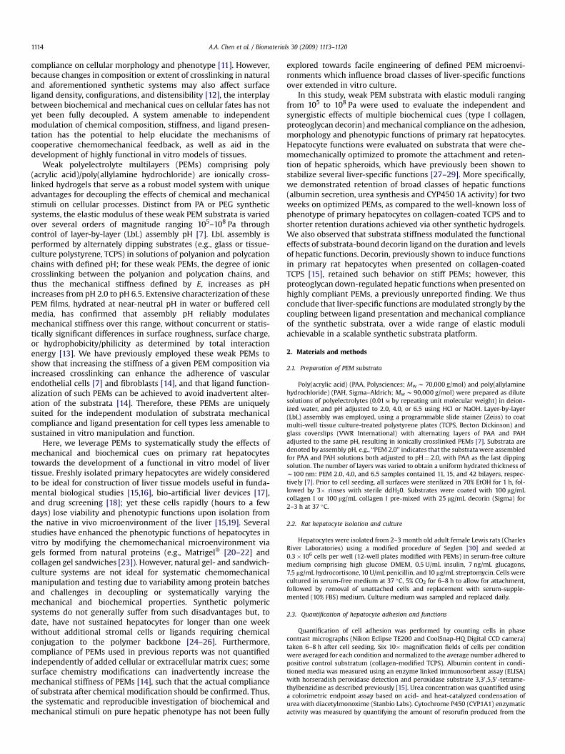
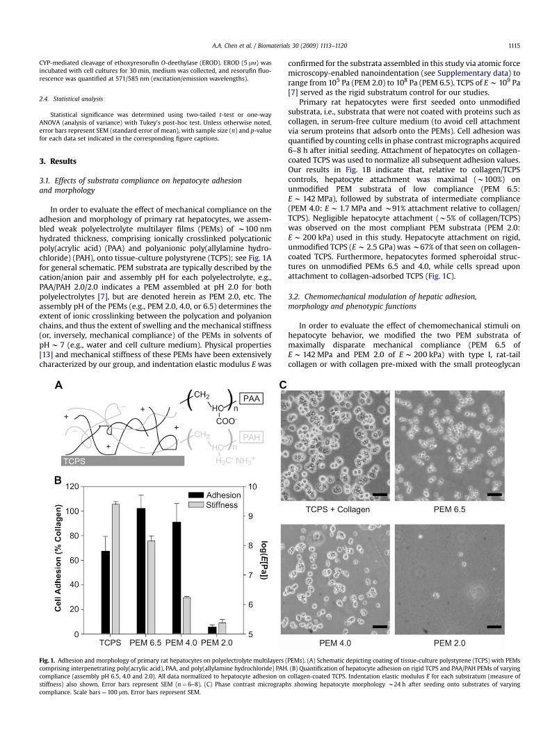
In order to evaluate the effect of mechanical compliance on theadhesion and morphology of primary rat hepatocytes, we assem-bled weak polyelectrolyte multilayer films (PEMs) of w100 nmhydrated thickness, comprising ionically crosslinked polycationicpoly(acrylic acid) (PAA) and polyanionic poly(allylamine hydro-chloride) (PAH), onto tissue-culture polystyrene (TCPS); see Fig. 1Afor general schematic. PEM substrata are typically described by thecation/anion pair and assembly pH for each polyelectrolyte, e.g.,PAA/PAH 2.0/2.0 indicates a PEM assembled at pH 2.0 for bothpolyelectrolytes [7], but are denoted herein as PEM 2.0, etc. Theassembly pH of the PEMs (e.g., PEM 2.0, 4.0, or 6.5) determines theextent of ionic crosslinking between the polycation and polyanionchains, and thus the extent of swelling and the mechanical stiffness(or, inversely, mechanical compliance) of the PEMs in solvents ofpH w 7 (e.g., water and cell culture medium). Physical properties[13] and mechanical stiffness of these PEMs have been extensivelycharacterized by our group, and indentation elastic modulus E was
Fig. 1. Adhesion and morphology of primary rat hepatocytes on polyelectrolyte multilayers (comprising interpenetrating poly(acrylic acid), PAA, and poly(allylamine hydrochloride) PAHcompliance (assembly pH 6.5, 4.0 and 2.0). All data normalized to hepatocyte adhesion onstiffness) also shown. Error bars represent SEM (n¼ 6–8). (C) Phase contrast micrographcompliance. Scale bars¼ 100 mm. Error bars represent SEM.
confirmed for the substrata assembled in this study via atomic forcemicroscopy-enabled nanoindentation (see Supplementary data) torange from 105 Pa (PEM 2.0) to 108 Pa (PEM 6.5). TCPS of E w 109 Pa[7] served as the rigid substratum control for our studies.
Primary rat hepatocytes were first seeded onto unmodifiedsubstrata, i.e., substrata that were not coated with proteins such ascollagen, in serum-free culture medium (to avoid cell attachmentvia serum proteins that adsorb onto the PEMs). Cell adhesion wasquantified by counting cells in phase contrast micrographs acquired6–8 h after initial seeding. Attachment of hepatocytes on collagen-coated TCPS was used to normalize all subsequent adhesion values.Our results in Fig. 1B indicate that, relative to collagen/TCPScontrols, hepatocyte attachment was maximal (w100%) onunmodified PEM substrata of low compliance (PEM 6.5:E w 142 MPa), followed by substrata of intermediate compliance(PEM 4.0: E w 1.7 MPa and w91% attachment relative to collagen/TCPS). Negligible hepatocyte attachment (w5% of collagen/TCPS)was observed on the most compliant PEM substrata (PEM 2.0:E w 200 kPa) used in this study. Hepatocyte attachment on rigid,unmodified TCPS (E w 2.5 GPa) was w67% of that seen on collagen-coated TCPS. Furthermore, hepatocytes formed spheroidal struc-tures on unmodified PEMs 6.5 and 4.0, while cells spread uponattachment to collagen-adsorbed TCPS (Fig. 1C).
3.2. Chemomechanical modulation of hepatic adhesion,morphology and phenotypic functions
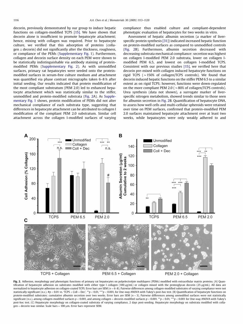
In order to evaluate the effect of chemomechanical stimuli onhepatocyte behavior, we modified the two PEM substrata ofmaximally disparate mechanical compliance (PEM 6.5 ofE w 142 MPa and PEM 2.0 of E w 200 kPa) with type I, rat-tailcollagen or with collagen pre-mixed with the small proteoglycan
PEMs). (A) Schematic depicting coating of tissue-culture polystyrene (TCPS) with PEMs. (B) Quantification of hepatocyte adhesion on rigid TCPS and PAA/PAH PEMs of varyingcollagen-coated TCPS. Indentation elastic modulus E for each substratum (measure ofs showing hepatocyte morphology w24 h after seeding onto substrates of varying
A.A. Chen et al. / Biomaterials 30 (2009) 1113–11201116
decorin, previously demonstrated by our group to induce hepaticfunctions on collagen-modified TCPS [15]. We have shown thatdecorin alone is insufficient to promote hepatocyte attachment;hence, mixing with collagen was required. Prior to hepatocyteculture, we verified that this adsorption of proteins (colla-gen� decorin) did not significantly alter the thickness, roughness,or compliance of the PEMs (Supplementary Fig. 1). Furthermore,collagen and decorin surface density on each PEM were shown tobe statistically indistinguishable via antibody staining of protein-modified PEMs (Supplementary Fig. 2). As with unmodifiedsurfaces, primary rat hepatocytes were seeded onto the protein-modified surfaces in serum-free culture medium and attachmentwas quantified via phase contrast micrographs taken 6–8 h afterinitial seeding. Our results indicated that protein modification ofthe most compliant substratum (PEM 2.0) led to enhanced hepa-tocyte attachment which was statistically similar to the stiffer,unmodified and protein-modified substrata (Fig. 2A). As Supple-mentary Fig. 1 shows, protein modification of PEMs did not altermechanical compliance of each substrata type, suggesting thatdifferences in hepatocyte attachment can be attributed to collagen Imodification of the compliant PEM 2.0 substratum. Similar cellattachment across the collagen I-modified surfaces of varying
Fig. 2. Adhesion, morphology and phenotypic functions of primary rat hepatocytes on polytification of hepatocyte adhesion on substrates modified with either type I collagen (10normalized to hepatocyte adhesion on collagen-coated TCPS. Error bars are SEM (n¼ 6–8). Pastatistically significant (n.s.). #p< 0.01 vs. ‘TCPSþ CollþDec’, **p< 0.01, ***p< 0.001, for Onprotein-modified substrates: cumulative albumin secretion over two weeks. Error bars arsignificant (n.s.), among collagen-modified surfaces p< 0.001, and among collagenþ decorinpost-hoc test. (C) Hepatocyte morphology on collagen-coated substrata of varying compligenþ decorin was similar. Scale bars¼ 100 mm. Error bars represent SEM.
compliance thus enabled culture and compliant-dependentphenotypic evaluation of hepatocytes for two weeks in vitro.
Assessment of hepatic albumin secretion (a marker of liver-specific protein synthesis [31]) indicated increased hepatic functionon protein-modified surfaces as compared to unmodified controls(Fig. 2B). Furthermore, albumin secretion decreased withdecreasing substrata mechanical compliance: secretion was higheston collagen I-modified PEM 2.0 substrata, lower on collagen I-modified PEM 6.5, and lowest on collagen I-modified TCPS.Consistent with our previous studies [15], we verified here thatdecorin pre-mixed with collagen induced hepatocyte functions onrigid TCPS (w150% of collagen/TCPS controls). We found thatdecorin induced hepatic functions on the stiffer PEM 6.5 to a similarextent as on rigid TCPS; however, functions were down-regulatedon the more compliant PEM 2.0 (w80% of collagen/TCPS controls).Urea synthesis (data not shown), a surrogate marker of liver-specific nitrogen metabolism, showed trends similar to those seenfor albumin secretion in Fig. 2B. Quantification of hepatocyte DNA,to assess how well cells and multi-cellular spheroids were retainedover time on PEM surfaces, confirmed that protein-modified PEM2.0 surfaces maintained hepatocyte attachment over at least twoweeks, while hepatocytes were only weakly adhered to and
electrolyte multilayers (PEMs) modified with extracellular matrix proteins. (A) Quan-0 mg/mL) or collagen mixed with the proteoglycan decorin (25 mg/mL). All data areirwise differences among collagen-modified substrates of varying compliance were not
e-way ANOVA with Tukey’s post-hoc test. (B) Quantification of hepatocyte functions one SEM (n¼ 3). Pairwise differences among unmodified surfaces were not statistically-modified surfaces p< 0.001. **p< 0.01, ***p< 0.001 for One-way ANOVA with Tukey’s
ance, 2 days post-seeding. Hepatocyte morphology on substrata modified with colla-
A.A. Chen et al. / Biomaterials 30 (2009) 1113–1120 1117
released within one week from PEM 6.5 surfaces (SupplementaryFig. 3). Lastly, hepatocytes formed stable, spheroidal aggregates ofapproximately 50–100 mm diameter on protein-modified PEM 2.0surfaces, while the extent of cell spreading increased with reduc-tion in substrata compliance (Fig. 2C).
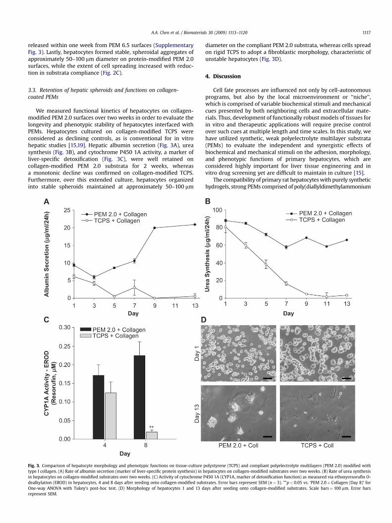
3.3. Retention of hepatic spheroids and functions on collagen-coated PEMs
We measured functional kinetics of hepatocytes on collagen-modified PEM 2.0 surfaces over two weeks in order to evaluate thelongevity and phenotypic stability of hepatocytes interfaced withPEMs. Hepatocytes cultured on collagen-modified TCPS wereconsidered as declining controls, as is conventional for in vitrohepatic studies [15,19]. Hepatic albumin secretion (Fig. 3A), ureasynthesis (Fig. 3B), and cytochrome P450 1A activity, a marker ofliver-specific detoxification (Fig. 3C), were well retained oncollagen-modified PEM 2.0 substrata for 2 weeks, whereasa monotonic decline was confirmed on collagen-modified TCPS.Furthermore, over this extended culture, hepatocytes organizedinto stable spheroids maintained at approximately 50–100 mm
Fig. 3. Comparison of hepatocyte morphology and phenotypic functions on tissue-culture ptype I collagen. (A) Rate of albumin secretion (marker of liver-specific protein synthesis) in hin hepatocytes on collagen-modified substrates over two weeks. (C) Activity of cytochrome Pdealkylation (EROD) in hepatocytes, 4 and 8 days after seeding onto collagen-modified subsOne-way ANOVA with Tukey’s post-hoc test. (D) Morphology of hepatocytes 1 and 13 drepresent SEM.
diameter on the compliant PEM 2.0 substrata, whereas cells spreadon rigid TCPS to adopt a fibroblastic morphology, characteristic ofunstable hepatocytes (Fig. 3D).
4. Discussion
Cell fate processes are influenced not only by cell-autonomousprograms, but also by the local microenvironment or ‘‘niche’’,which is comprised of variable biochemical stimuli and mechanicalcues presented by both neighboring cells and extracellular mate-rials. Thus, development of functionally robust models of tissues forin vitro and therapeutic applications will require precise controlover such cues at multiple length and time scales. In this study, wehave utilized synthetic, weak polyelectrolyte multilayer substrata(PEMs) to evaluate the independent and synergistic effects ofbiochemical and mechanical stimuli on the adhesion, morphology,and phenotypic functions of primary hepatocytes, which areconsidered highly important for liver tissue engineering and invitro drug screening yet are difficult to maintain in culture [15].
The compatibility of primary rat hepatocytes with purely synthetichydrogels, strong PEMs comprised of poly(diallyldimethylammonium
olystyrene (TCPS) and compliant polyelectrolyte multilayers (PEM 2.0) modified withepatocytes on collagen-modified substrates over two weeks. (B) Rate of urea synthesis450 1A (CYP1A, marker of detoxification function) as measured via ethoxyresorufin O-trates. Error bars represent SEM (n¼ 3). **p< 0.05 vs. ‘PEM 2.0þ Collagen (Day 8)’ forays after seeding onto collagen-modified substrates. Scale bars¼ 100 mm. Error bars
A.A. Chen et al. / Biomaterials 30 (2009) 1113–11201118
chloride) (PDAC) and poly(sulfonated styrene) (PSS), was firstdemonstrated by Kidambi et al. [25]. The authors showed that PSS-terminated substrata promoted hepatocyte adhesion andspreading; yet liver-specific functions (albumin and urea produc-tion) on these synthetic substrates declined over one week inculture and were comparable to those seen on unmodified tissue-culture polystyrene [25]. Furthermore, the authors did not evaluatethe dependence of hepatocyte morphology and functions onvarying substrata mechanical compliances. In a follow-up study,the authors created co-cultures of hepatocytes and fibroblasts onprotein-free surfaces by utilizing micropatterned domains of PEMsadhesive to either hepatocytes or fibroblasts [26]. The authorsverified the previously reported and well-known ‘‘co-cultureeffect’’ on their surfaces by showing that 3T3 murine embryonicfibroblasts were able to induce functions in primary rat hepatocytesvia heterotypic signaling [16,32,33]. It was unclear from this study,however, whether there were any synergistic effects of substratemechanical compliance and heterotypic cell–cell interactions onliver-specific functions of hepatocytes. More recently, Janorkar et al.functionalized polyacrylic acid (PAA)/polyethyleneimine (PEI)PEMs with extracellular matrix-like polypeptides to enhance liver-specific functions [24]. Despite these experimental developmentsusing PEMs, substrata compliance in the aforementioned reportscould not be tuned via assembly pH and was not quantified inde-pendently of adjunct cells or ligands. Using PA hydrogels, Semler etal. polymerized, cut, and adhered to tissue-culture plate surfacesa 9-condition array of substrates with varying elastic moduli anddensities of immobilized fibronectin, identifying high complianceregimes (E¼ 1.9 kPa) in which hepatocyte cell–cell interactionsdominated over hepatocyte–fibronectin interactions [10]. Whilehighlighting the impact of mechanical and biochemical cooperativesignaling on liver-specific functions and gene expression, theconditions of this study were limited to relatively small variationsin mechanical stiffness (5.6–19 kPa) and a single protein (fibro-nectin). Furthermore, this study required multiple tedious steps forsystem assembly, which limits the facile, reproducible investigationof multiple stimuli towards engineering defined microenviron-ments for hepatocytes or other cells.
We probed hepatocyte attachment on fully synthetic PEMsubstrata that varied in compliance over several orders of magni-tude (elastic moduli E ranging from 0.2 to 2500 MPa), using serum-free culture medium to avoid cell attachment due to serum proteinspre-adsorbing onto the substrate. We found that hepatocytes dis-played maximal attachment (w100% of adhesion to TCPS/collagencontrol, E w 2500 MPa) on substrata of lower mechanical compli-ance (PEM 6.5, E w 142 MPa; and PEM 4.0, E w 1.7 MPa), whilenegligible attachment was observed on the most compliantsubstrata (PEM 2.0, E w 0.2 MPa). We note that it is possible thesesubstrata are sufficiently thin such that traction exerted by hepato-cytes at adhesion sites could increase the effective or perceivedstiffness of these substrata. Although calculation of such effectivestiffness requires several assumptions and estimates of cell-generated forces and adhesion site radii that have not yet been wellquantified for hepatocytes, our recent model of reduced adhesionsite displacement on coatings of finite thickness [34] indicates thatthis effective stiffness would increase by the same factor (much lessthan 10-fold) for each of these three PEM substrata of identicalthickness w100 nm. The inverse correlation between unfunction-alized substrata compliance and percentage of cell adhesion/spreading is consistent with previous observations for endothelialcells [7] and fibroblasts cultured on these PEMs [35], as well as forother adherent tissue cell types on polymer hydrogels [36]. Whilethe mechanism of this protein-free hepatocyte adhesion tounfunctionalized, weak PEM substrata is currently unknown,differential adhesion of hepatocytes on varying compliance cannotbe attributed to surface charge, energy, or roughness of these PEMs,
as we have shown that these physical properties are statisticallyindistinguishable for these substrata [13]. Lack of hepatocyteattachment to PEM 2.0 was therefore most likely due to reducedcell–substratum adhesion via unstable focal contact or adhesioncomplexes, as observed with other adherent tissue cell types suchas fibroblasts [1]. On the stiffer PEM substrata (PEM 6.5 and 4.0),however, the balance of cell–cell and cell–substrata interactionsstabilized formation of hepatic aggregates for several days inculture. In comparison, as expected from our previous work [15],hepatocytes on collagen-modified, rigid TCPS rapidly spread toadopt a fibroblastic morphology. Thus, the results of our primaryhepatocyte adhesion studies are consistent with others showingthat a variety of adhesive tissue cell types, including fibroblasts,cardiomyocytes and endothelial cells, adhere poorly to highlycompliant hydrogels. The magnitude of substrata compliance thatis sufficient to significantly reduce (or promote) cell adhesiondepends on the cell type, substrata composition, and protein/ligandcoating density, such that this trend is typically reported forinternal comparisons for a given substrata system and cell type[1,7,10].
Unmodified PEM substrata of stiffness greater than E w 0.2 MPapromoted attachment and aggregation of hepatocytes; however,we found that cells detached from these stiffer substrata after onlya few days of culture, due ostensibly to the dominance of cell–cellover cell–substrata interactions. We thus modified substrata withtype I collagen, an extracellular matrix protein that has been shownto promote hepatocyte attachment for several weeks in culture[15]. We found that protein modification affected neither thethickness nor the mechanical compliance of the stiffest and mostcompliant PEM substrata. The PEM system therefore enabledindependent comparison of hepatocyte functions over severalweeks in well-defined chemomechanical microenvironments.Following collagen modification, hepatic spheroid formation wasobserved on PEMs exhibiting both low (PEM 6.5) and high (PEM2.0) compliance. Spheroidal aggregates displayed higher levels ofbroad classes of liver-specific functions (albumin secretion, ureasynthesis and CYP450 1A activity) as compared to well-spreadhepatocytes on collagen-modified TCPS controls, a finding that isconsistent with previously published reports [27–29]. However,previous methods to create hepatic spheroids (e.g., tumor-derivedMatrigel�, roller bottles, non-adhesive dishes) are eitherconfounded by coupled changes in ligand density (i.e., Matrigel�),or limited by challenges in handling and processing spheroids asthey form and coalesce to become large cellular masses withnecrotic cores. In this study, the compliant, collagen-modified PEM2.0 substrata promoted attachment and long-term (two weeks)retention of hepatic spheroids (approximately 50–100 mm indiameter) over stiffer PEM 6.5 substrata, as evaluated by thequantification of adherent hepatocyte DNA over time. Tetheredspheroids eliminated the need for sedimentation steps duringculture medium changes for suspended spheroid cultures, andfacilitated evaluation of hepatic morphology and functions withvarying chemomechanical stimuli on a reproducible and syntheticPEM platform.
Type I collagen typically does not induce liver-specific functionsin hepatocytes and is instead used with hepatocytes primarily as anadhesive cue on solid substrates. Although we used type I collagento promote long-term retention of highly functional hepaticspheroids on PEM substrata, a primary objective of this study wasto utilize the tunable PEM system to investigate the incorporationof hepatocyte-stabilizing biochemical cues on mechanically distinctsubstrata. Several such molecular cues have been previouslyimplicated in induction of hepatic functions when presented aloneon solid substrates or in combination with adhesive factors such ascollagen [15,16,37]. Using a functional genomic screen on hepato-cyte–fibroblast co-cultures, we have previously shown that
A.A. Chen et al. / Biomaterials 30 (2009) 1113–1120 1119
decorin, a chondroitin sulfate–dermatan sulfate proteoglycan thatbinds collagen and is present in the native liver [38,39], can inducephenotypic functions in primary rat hepatocytes adhered tocollagen-coated TCPS [15]. In this study, we sought to evaluate theinterplay of decorin and substrata mechanical compliance on thehepatic phenotype over extended in vitro culture. We observed thatthe presence of decorin caused hepatic functions of culturedhepatocytes to be up-regulated similarly on rigid TCPS and PEM 6.5substrates. However, on highly compliant PEM 2.0 substrata, thepresence of decorin led to down-regulation of liver-specific func-tions in hepatic spheroids, and such down-regulation was notattributable to statistically significant differences in initial attach-ment of cells or to differential retention of hepatic spheroids oncollagen-only controls (evaluated by DNA quantification over time).Furthermore, the inability of decorin to enhance hepatic functionson PEM 2.0 was not due to saturation of albumin secretion, ashepatocytes secrete much higher albumin levels (w2 to 3-fold)upon co-cultivation with supporting fibroblasts at the cell seedingdensities used in this study [15,39]. Such multifaceted effects ofmechanical compliance and ligand type/density are not unexpected[8,20], given the common components of mechanotransductiveand other functional signaling pathways. Semler et al. have alsoreported a monotonic coupling for fibronectin-functionalizedpolyacrylamide hydrogels [10], noting increased albumin secretionfor gels of lower compliance and constant fibronectin concentra-tion; however, the authors considered shorter durations (oneweek) and a much narrower range of substrata stiffness (shearelastic storage modulus G’ w 2–9 kPa) than those considered in thisstudy.
Although we demonstrated via antibody staining that the extentof collagen and decorin adsorption to PEMs was statisticallyindistinguishable, it remains possible that ligand orientation and/oraltered collagen fibril structure [38,40] may differ among thesePEMs (or between the PEMs and the TCPS) in a manner correlativewith mechanical compliance. It is further possible that PEMmechanical compliance may not only define the initial microenvi-ronment that modulates cell adhesion, but also the subsequentcapacity of cell-generated proteins and ligands to be produced and/or absorbed to these substrata. These correlative factors are verychallenging to quantify on hydrogel surfaces and are beyond thescope of the present study, but remain important considerations inthe distinction between causal and correlative effects of substratastiffness on tissue cell function. We also note that the mechanicalstiffness of the most compliant hydrogels considered in this study(E w 200 kPa) is within the range of normal liver tissue measuredvia various methods (E w 1–750 kPa) [41–43]; however, it is diffi-cult to draw a direct analogy between the tunable, synthetic PEMplatform used in vitro here and microenvironments present inpatho/physiological states in vivo. Future studies in our laboratorieswill further tune and improve hepatic functions in vitro via the useof combinatorial mixtures of polymer-tethered ligands and solublefactors (e.g., growth factors), on two- and three-dimensionalmaterials that display spatial variations in compliance and ligand-tether flexibility.
5. Conclusion
We demonstrate that hepatocyte morphology and broad classesof phenotypic functions can be modulated via independent andsynergistic tuning of biochemical and mechanical stimuli as pre-sented on synthetic polymeric substrata. Facile assembly andsynergistic effects of high substrata compliance and collagenpresentation onto a standard 2D culture surface enabled creation ofa robust, pure-hepatocyte tissue model which displayed enhancedliver-specific functions over collagen-modified TCPS controls fortwo weeks. We also discovered compliance-mediated effects of the
proteoglycan decorin on hepatic functions, with hepatocyte func-tions down-regulated on highly compliant surfaces as compared tocollagen-only controls but up-regulated on increasing PEM stiff-ness. Potential applications of our multi-well platform includemedium- to high-throughput evaluation of interactions betweenexogenous compounds (e.g., drugs, environmental toxicants) andthe various microenvironmental cues used to modulate fateprocesses of primary hepatocytes. Lastly, our approach of modu-lating chemomechanical cues towards improvement of cellularfunctions in vitro is amenable to multiple cell types (e.g., stem andprecursor cells) for applications such as drug screening, cell-basedtherapies or the fundamental study of chemomechanical processesunderlying tissue function and disease.
Acknowledgements
We gratefully acknowledge K. Hudson, M. Akiyama and S. Katzfor rat hepatocyte isolations and media preparation, Y. Li forgeneration of recombinant protein and assistance with cell culture,S. Kambhampati and E. Liu for assistance with biochemical assays,and J.M. Maloney and J.M. Milwid for assistance with PEM substrataassembly. Funding was provided by the NSF Graduate ResearchFellowship (A.A.C.), NSF CAREER Awards (K.J.V.V. and S.N.B.),Arnold & Mabel Beckman Foundation Young Investigator Award(K.J.V.V.), NIH NIDDK (S.N.B.), and the David and Lucile PackardFoundation (S.N.B.). K.J.V.V. and S.L. acknowledge use of sharedexperimental facilities in the NSF MRSEC Center for MaterialsScience and Engineering (award number DMR 02-13282).
Appendix. Supplementary data
Supplementary data associated with this article can be found inthe online version at doi:10.1016/j.biomaterials.2008.10.055.
References
[1] Discher DE, Janmey P, Wang YL. Tissue cells feel and respond to the stiffness oftheir substrate. Science 2005;310(5751):1139–43.
[2] Lee S, Mandic J, Van Vliet KJ. Chemomechanical mapping of ligand–receptorbinding kinetics on cells. Proc Natl Acad Sci U S A 2007;104(23):9609–14.
[3] Deroanne CF, Lapiere CM, Nusgens BV. In vitro tubulogenesis of endothelialcells by relaxation of the coupling extracellular matrix–cytoskeleton. Car-diovasc Res 2001;49(3):647–58.
[4] Vailhe B, Lecomte M, Wiernsperger N, Tranqui L. The formation of tubularstructures by endothelial cells is under the control of fibrinolysis andmechanical factors. Angiogenesis 1998;2(4):331–44.
[5] Pelham RJ, Wang YL. Cell locomotion and focal adhesions are regulated bysubstrate flexibility. Proc Natl Acad Sci U S A 1997;94(25):13661–5.
[6] Yeung T, Georges PC, Flanagan LA, Marg B, Ortiz M, Funaki M, et al. Effects ofsubstrate stiffness on cell morphology, cytoskeletal structure, and adhesion.Cell Motil Cytoskeleton 2005;60(1):24–34.
[7] Thompson MT, Berg MC, Tobias IS, Rubner MF, Van Vliet KJ. Tuning complianceof nanoscale polyelectrolyte multilayers to modulate cell adhesion. Biomate-rials 2005;26(34):6836–45.
[8] Engler AJ, Griffin MA, Sen S, Bonnemann CG, Sweeney HL, Discher DE. Myo-tubes differentiate optimally on substrates with tissue-like stiffness: patho-logical implications for soft or stiff microenvironments. J Cell Biol 2004;166(6):877–87.
[9] Engler AJ, Sen S, Sweeney HL, Discher DE. Matrix elasticity directs stem celllineage specification. Cell 2006;126(4):677–89.
[10] Semler EJ, Lancin PA, Dasgupta A, Moghe PV. Engineering hepatocellularmorphogenesis and function via ligand-presenting hydrogels with gradedmechanical compliance. Biotechnol Bioeng 2005;89(3):296–307.
[11] Peyton SR, Raub CB, Keschrumrus VP, Putnam AJ. The use of poly(ethyleneglycol) hydrogels to investigate the impact of ECM chemistry and mechanicson smooth muscle cells. Biomaterials 2006;27(28):4881–93.
[12] Walton EB, Oommen B, Van Vliet KJ. How stiff and thin can an engineeredextracellular matrix be? Modeling molecular forces at the cell–matrix inter-face. Conf Proc IEEE Eng Med Biol Soc 2007;2007:6419–21.
[13] Lichter JA, Thompson MT, Delgadillo M, Nishikawa T, Rubner MF, Van Vliet KJ.Substrata mechanical stiffness can regulate adhesion of viable bacteria. Bio-macromolecules 2008;9(6):1571–8.
[14] Thompson MT, Berg MC, Tobias IS, Lichter JA, Rubner MF, Van Vliet KJ.Biochemical functionalization of polymeric cell substrata can alter mechanicalcompliance. Biomacromolecules 2006;7(6):1990–5.
A.A. Chen et al. / Biomaterials 30 (2009) 1113–11201120
[15] Khetani SR, Szulgit G, Del Rio JA, Barlow C, Bhatia SN. Exploring interactionsbetween rat hepatocytes and nonparenchymal cells using gene expressionprofiling. Hepatology 2004;40(3):545–54.
[16] Bhatia SN, Balis UJ, Yarmush ML, Toner M. Effect of cell–cell interactions inpreservation of cellular phenotype: cocultivation of hepatocytes and non-parenchymal cells. FASEB J 1999;13(14):1883–900.
[17] Allen JW, Hassanein T, Bhatia SN. Advances in bioartificial liver devices. Hepa-tology 2001;34(3):447–55.
[18] Hewitt NJ, Lechon MJ, Houston JB, Hallifax D, Brown HS, Maurel P, et al.Primary hepatocytes: current understanding of the regulation of metabolicenzymes and transporter proteins, and pharmaceutical practice for the use ofhepatocytes in metabolism, enzyme induction, transporter, clearance, andhepatotoxicity studies. Drug Metab Rev 2007;39(1):159–234.
[19] Guguen-Guillouzo C, Guillouzo A. Modulation of functional activities incultured rat hepatocytes. Mol Cell Biochem 1983;53–54(1–2):35–56.
[20] Semler EJ, Ranucci CS, Moghe PV. Mechanochemical manipulation of hepa-tocyte aggregation can selectively induce or repress liver-specific function.Biotechnol Bioeng 2000;69(4):359–69.
[21] Gross-Steinmeyer K, Stapleton PL, Tracy JH, Bammler TK, Lehman T, Strom SC,et al. Influence of Matrigel-overlay on constitutive and inducible expression ofnine genes encoding drug-metabolizing enzymes in primary human hepato-cytes. Xenobiotica 2005;35(5):419–38.
[22] Semler EJ, Moghe PV. Engineering hepatocyte functional fate through growthfactor dynamics: the role of cell morphologic priming. Biotechnol Bioeng2001;75(5):510–20.
[23] Dunn JC, Tompkins RG, Yarmush ML. Hepatocytes in collagen sandwich:evidence for transcriptional and translational regulation. J Cell Biol 1992;116(4):1043–53.
[24] Janorkar AV, Rajagopalan P, Yarmush ML, Megeed Z. The use of elastin-likepolypeptide–polyelectrolyte complexes to control hepatocyte morphologyand function in vitro. Biomaterials 2008;29(6):625–32.
[25] Kidambi S, Lee I, Chan C. Controlling primary hepatocyte adhesion andspreading on protein-free polyelectrolyte multilayer films. J Am Chem Soc2004;126(50):16286–7.
[26] Kidambi S, Sheng L, Yarmush ML, Toner M, Lee I, Chan C. Patterned co-cultureof primary hepatocytes and fibroblasts using polyelectrolyte multilayertemplates. Macromol Biosci 2007;7(3):344–53.
[27] Tong JZ, De Lagausie P, Furlan V, Cresteil T, Bernard O, Alvarez F. Long-term culture of adult rat hepatocyte spheroids. Exp Cell Res 1992;200(2):326–32.
[28] Hamilton GA, Westmorel C, George AE. Effects of medium composition on themorphology and function of rat hepatocytes cultured as spheroids andmonolayers. In Vitro Cell Dev Biol Anim 2001;37(10):656–67.
[29] Dvir-Ginzberg M, Elkayam T, Aflalo ED, Agbaria R, Cohen S. Ultrastructural andfunctional investigations of adult hepatocyte spheroids during in vitro culti-vation. Tissue Eng 2004;10(11–12):1806–17.
[30] Seglen PO. Preparation of isolated rat liver cells. Methods Cell Biol 1976;13:29–83.[31] Bhatia SN, Toner M, Tompkins RG, Yarmush ML. Selective adhesion of hepa-
tocytes on patterned surfaces. Ann N Y Acad Sci 1994;745:187–209.[32] Corlu A, Ilyin G, Cariou S, Lamy I, Loyer P, Guguen-Guillouzo C. The coculture:
a system for studying the regulation of liver differentiation/proliferationactivity and its control. Cell Biol Toxicol 1997;13(4–5):235–42.
[33] Khetani SR, Bhatia SN. Microscale culture of human liver cells for drugdevelopment. Nat Biotechnol 2008;26(1):120–6.
[34] Maloney JM, Walton EB, Bruce CM, Van Vliet KJ. Influence of finite thicknessand stiffness on cellular adhesion-induced deformation of compliantsubstrata. Phys Rev 2008;78(4 Pt 1):041923.
[35] Berg MC, Yang SY, Hammond PT, Rubner MF. Controlling mammalian cellinteractions on patterned polyelectrolyte multilayer surfaces. Langmuir 2004;20(4):1362–8.
[36] Janmey PA, McCulloch CA. Cell mechanics: integrating cell responses tomechanical stimuli. Annu Rev Biomed Eng 2007;9:1–34.
[37] Khetani SR, Chen AA, Ranscht B, Bhatia SN. T-cadherin modulates hepatocytefunctions in vitro. FASEB J 2008;22(11):3768–75.
[38] Pogany G, Vogel KG. The interaction of decorin core protein fragments withtype I collagen. Biochem Biophys Res Commun 1992;189(1):165–72.
[39] Hogemann B, Edel G, Schwarz K, Krech R, Kresse H. Expression of biglycan,decorin and proteoglycan-100/CSF-1 in normal and fibrotic human liver.Pathol Res Pract 1997;193(11–12):747–51.
[40] Pogany G, Hernandez DJ, Vogel KG. The in vitro interaction of proteoglycanswith type I collagen is modulated by phosphate. Arch Biochem Biophys 1994;313(1):102–11.
[41] Wells RG. The role of matrix stiffness in regulating cell behavior. Hepatology2008;47(4):1394–400.
[42] Li Z, Dranoff JA, Chan EP, Uemura M, Sevigny J, Wells RG. Transforming growthfactor-beta and substrate stiffness regulate portal fibroblast activation inculture. Hepatology 2007;46(4):1246–56.
[43] Constantinides G, Kalcioglu ZI, McFarland M, Smith JF, Van Vliet KJ. Probingmechanical properties of fully hydrated gels and biological tissues. J Biomech2008.
Chen et. al., Submitted to Biomaterials,
1
SUPPLEMENTARY DATA
Supplementary Methods
PEM surface characterization
Hydrated thicknesses of PEMs in 150 mM phosphate buffered saline at pH = 7.2 were
measured through atomic force microscopy (AFM; PicoPlus, Agilent Technologies). Scratches were
made with razor blades on PEMs assembled on glass coverslips, and thicknesses of PEMs were
measured near scratch regions from AFM height images acquired in tapping mode. Tapping mode
imaging at randomly selected positions across the sample confirmed the uniform coverage of PEM
across the sample surface area analyzed in cell culture, as has been established for this assembly
protocol and is supported by uniform immunofluorescence staining of the protein-functionalized PEMs
described below.
AFM cantilever spring constants were measured as discussed elsewhere [7] with the thermal
noise method. The deflection sensitivity of each AFM cantilever (nm/V), and cantilever spring
constants, nominally k = 0.1 N/m, were measured for each experiment and used for analyses of elastic
moduli of PEMs. Elastic moduli were calculated by applying a modified Hertzian model of spherical
contact via AFM force spectroscopy as discussed in detail elsewhere [7, 14]. Force-displacement
responses collected from AFM force spectroscopy were converted offline to force-separation curves
through the Scanning Probe Imaging Processor (Image Metrology), followed by customized analyses
to calculate elastic moduli through the scientific computing software Igor (Wavemetrics). Note that,
although hepatocyte experiments were conducted on PEMs assembled on TCPS and these
thickness/stiffness experiments were conducted on PEMs assembled on glass to enable scratching, we
have found the elastic moduli E of these PEMs to be independent of these substrate differences for the
hydrogel thicknesses considered here, within the standard error of measurement among replicate
samples (e.g., see Ref. 3 on TCPS and Ref. 11 on glass).
Chen et. al., Submitted to Biomaterials,
2
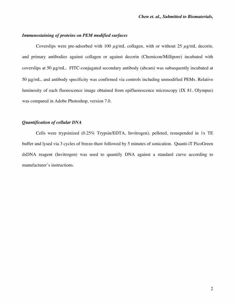
Immunostaining of proteins on PEM modified surfaces
Coverslips were pre-adsorbed with 100 µg/mL collagen, with or without 25 µg/mL decorin,
and primary antibodies against collagen or against decorin (Chemicon/Millipore) incubated with
coverslips at 50 µg/mL. FITC-conjugated secondary antibody (abcam) was subsequently incubated at
50 µg/mL, and antibody specificity was confirmed via controls including unmodified PEMs. Relative
luminosity of each fluorescence image obtained from epifluorescence microscopy (IX 81, Olympus)
was compared in Adobe Photoshop, version 7.0.
Quantification of cellular DNA
Cells were trypsinized (0.25% Trypsin/EDTA, Invitrogen), pelleted, resuspended in 1x TE
buffer and lysed via 3 cycles of freeze-thaw followed by 5 minutes of sonication. Quanti-iT PicoGreen
dsDNA reagent (Invitrogen) was used to quantify DNA against a standard curve according to
manufacturer’s instructions.
Chen et. al., Submitted to Biomaterials,
3
Supplementary Figures
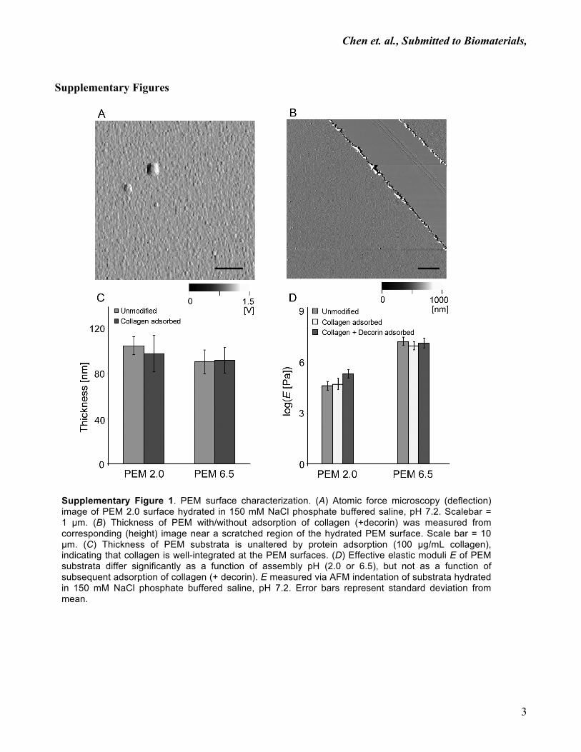
Supplementary Figure 1. PEM surface characterization. (A) Atomic force microscopy (deflection) image of PEM 2.0 surface hydrated in 150 mM NaCl phosphate buffered saline, pH 7.2. Scalebar = 1 µm. (B) Thickness of PEM with/without adsorption of collagen (+decorin) was measured from corresponding (height) image near a scratched region of the hydrated PEM surface. Scale bar = 10 µm. (C) Thickness of PEM substrata is unaltered by protein adsorption (100 µg/mL collagen), indicating that collagen is well-integrated at the PEM surfaces. (D) Effective elastic moduli E of PEM substrata differ significantly as a function of assembly pH (2.0 or 6.5), but not as a function of subsequent adsorption of collagen (+ decorin). E measured via AFM indentation of substrata hydrated in 150 mM NaCl phosphate buffered saline, pH 7.2. Error bars represent standard deviation from mean.
Chen et. al., Submitted to Biomaterials,
4
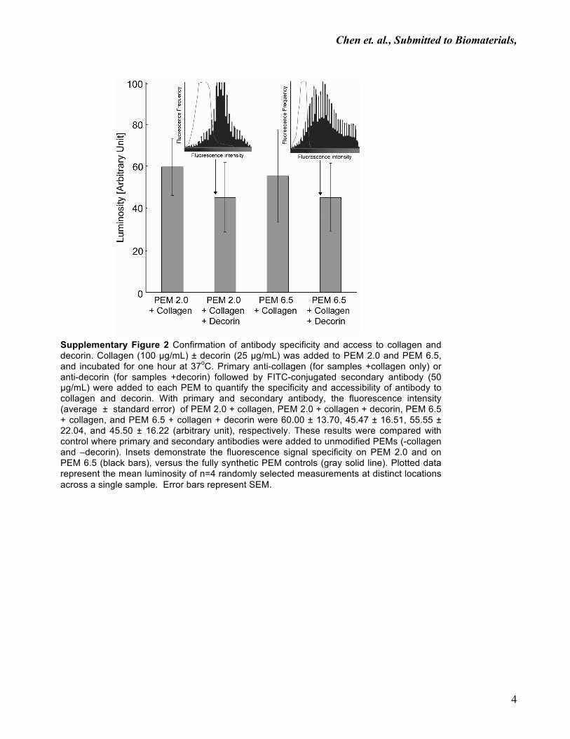
Supplementary Figure 2 Confirmation of antibody specificity and access to collagen and decorin. Collagen (100 µg/mL) ± decorin (25 µg/mL) was added to PEM 2.0 and PEM 6.5, and incubated for one hour at 37oC. Primary anti-collagen (for samples +collagen only) or anti-decorin (for samples +decorin) followed by FITC-conjugated secondary antibody (50 µg/mL) were added to each PEM to quantify the specificity and accessibility of antibody to collagen and decorin. With primary and secondary antibody, the fluorescence intensity (average ± standard error) of PEM 2.0 + collagen, PEM 2.0 + collagen + decorin, PEM 6.5 + collagen, and PEM 6.5 + collagen + decorin were 60.00 ± 13.70, 45.47 ± 16.51, 55.55 ± 22.04, and 45.50 ± 16.22 (arbitrary unit), respectively. These results were compared with control where primary and secondary antibodies were added to unmodified PEMs (-collagen and –decorin). Insets demonstrate the fluorescence signal specificity on PEM 2.0 and on PEM 6.5 (black bars), versus the fully synthetic PEM controls (gray solid line). Plotted data represent the mean luminosity of n=4 randomly selected measurements at distinct locations across a single sample. Error bars represent SEM.
Chen et. al., Submitted to Biomaterials,
5
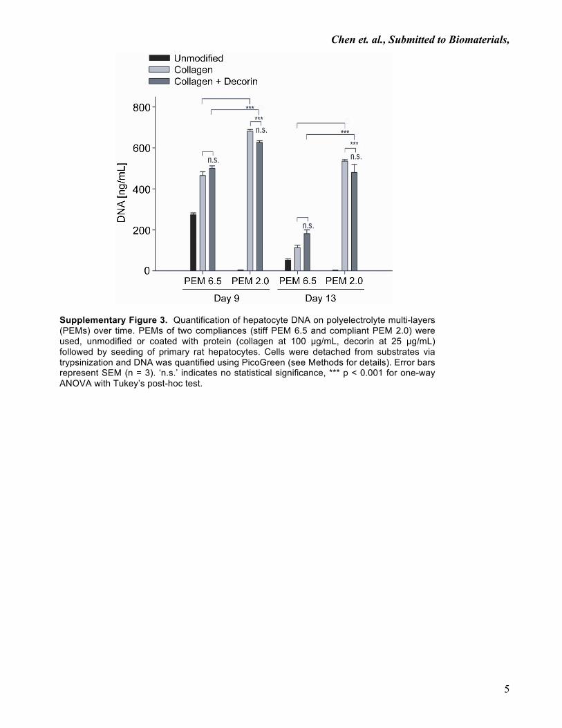
Supplementary Figure 3. Quantification of hepatocyte DNA on polyelectrolyte multi-layers (PEMs) over time. PEMs of two compliances (stiff PEM 6.5 and compliant PEM 2.0) were used, unmodified or coated with protein (collagen at 100 µg/mL, decorin at 25 µg/mL) followed by seeding of primary rat hepatocytes. Cells were detached from substrates via trypsinization and DNA was quantified using PicoGreen (see Methods for details). Error bars represent SEM (n = 3). ‘n.s.’ indicates no statistical significance, *** p < 0.001 for one-way ANOVA with Tukey’s post-hoc test.