Embed Size (px)
Citation preview
N
EUROPSYCHOPHARMACOLOGY
2001
–
VOL
.
25
,
NO
.
S5
© 2001 American College of NeuropsychopharmacologyPublished by Elsevier Science Inc. 0893-133X/01/$–see front matter655 Avenue of the Americas, New York, NY 10010 PII S0893-133X(01)00322-0
The characterization of the molecular correlates of sleep and wakefulness is essential to understand the restorative processes occurring during sleep and the cellular mechanisms underlying sleep regulation. In order to determine what molecular changes occur during the sleep-waking cycle, we have recently performed a systematic screening of gene expression in the brain of sleeping, sleep deprived, and spontaneously awake rats. Out of the
�
10,000 genes screened so far, a small minority (
�
0.5%) was differentially expressed in the cerebral cortex across behavioral states. Most genes were upregulated in wakefulness and sleep deprivation relative to sleep, while only a few were upregulated in sleep relative to wakefulness and sleep deprivation. Almost all the genes upregulated in sleep, and several genes upregulated in wakefulness and sleep deprivation, did not match any known sequence.
Known genes expressed at higher levels in wakefulness and sleep deprivation could be grouped into functional categories: immediate early genes/transcription factors, genes related to energy metabolism, growth factors/adhesion molecules, chaperones/heat shock proteins, vesicle and synapse-related genes, neurotransmitter/hormone receptors, neurotransmitter transporters, enzymes, and others. Although the characterization of the molecular correlates of sleep, wakefulness, and sleep deprivation is still in progress, it is already apparent that the transition from sleep to waking can affect basic cellular functions such as RNA and protein synthesis, neural plasticity, neurotransmission, and metabolism.
(Neuropsychopharmacology 25:S28-S35, 2001).
© 2001 American College of Neuropsychopharmacology.Published by Elsevier Science Inc.
Modulation of Brain Gene Expression during Sleep and Wakefulness:A Review of Recent Findings
Giulio Tononi, M.D., Ph.D. and Chiara Cirelli, M.D., Ph.D.
KEY
WORDS
:
Sleep deprivation; Microarray; Differential display
GENE EXPRESSION AND THE HOMEOSTATICREGULATION OF SLEEP
Considering the typical duration of sleep-wake statesand the time constants of their regulation (minutes tohours rather than seconds), it is plausible that gene ex-pression in the brain is subject to significant modula-tions across behavioral states. For this reason, over the
past several years, we have investigated the expressionof thousand of transcripts in the brain of sleeping, wak-ing, or sleep deprived animals (Cirelli and Tononi 1998,2000a,b). The goal of these studies is to characterize spe-cific patterns of gene expression that distinguish sleepfrom wakefulness. The identification of such patternsmay help us to understand the homeostatic regulationof sleep and its functional consequences.
IMMEDIATE EARLY GENES EXPRESSION INSLEEP AND WAKEFULNESS
Our initial studies focused on the expression of the so-called immediate early genes (IEGs), which are rapidly
From the The Neurosciences Institute, 10640 John J. Hopkins Drive,San Diego, CA 92121
Address correspondence to: Chiara Cirelli, Department of Psychi-atry, U of Wisconsin at Madison, 6001 Research Park Boulevard,Madison, WI 53719, E-mail: [email protected]
N
EUROPSYCHOPHARMACOLOGY
2001
–
VOL
.
25
,
NO
.
S5
Gene Expression in Sleep and Wakefulness
S29
induced by a large number of extracellular stimuli. Thebest known of such genes,
c-fos
, shows increased mRNAlevels after a few minutes of stimulation, and its proteinproduct, Fos, is synthesized shortly thereafter and canbe detected for several hours. The expression of
c-fos
can thus serve as a marker of neuronal activity. In addi-tion,
c-fos
and other IEGs function as transcriptionalregulators (Sheng and Greenberg 1990) and may there-fore regulate the transcription of a number of targetgenes that could be directly relevant to the homeostasisand functions of sleep. The results of our work (Pompe-iano et al. 1994; Cirelli et al. 1995) and that of severalother laboratories (reviewed in Cirelli and Tononi
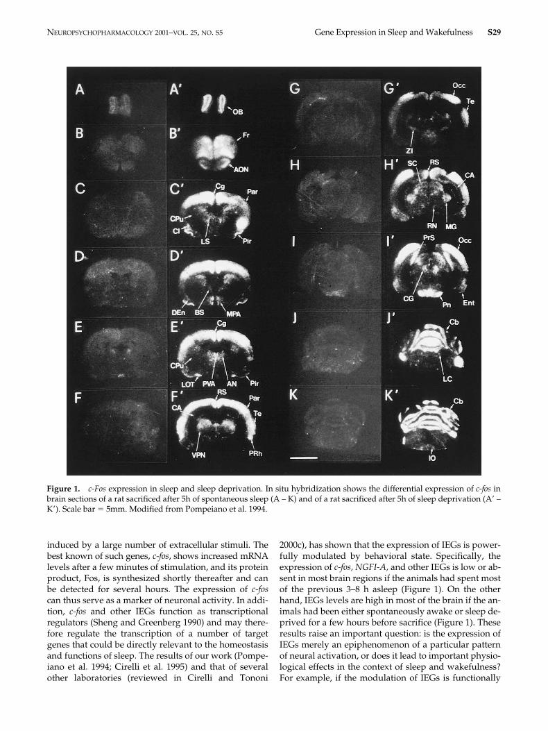
2000c), has shown that the expression of IEGs is power-fully modulated by behavioral state. Specifically, theexpression of
c-fos,
NGFI-A,
and other IEGs is low or ab-sent in most brain regions if the animals had spent mostof the previous 3–8 h asleep (Figure 1). On the otherhand, IEGs levels are high in most of the brain if the an-imals had been either spontaneously awake or sleep de-prived for a few hours before sacrifice (Figure 1). Theseresults raise an important question: is the expression ofIEGs merely an epiphenomenon of a particular patternof neural activation, or does it lead to important physio-logical effects in the context of sleep and wakefulness?For example, if the modulation of IEGs is functionally
Figure 1. c-Fos expression in sleep and sleep deprivation. In situ hybridization shows the differential expression of c-fos inbrain sections of a rat sacrificed after 5h of spontaneous sleep (A – K) and of a rat sacrificed after 5h of sleep deprivation (A� –K�). Scale bar � 5mm. Modified from Pompeiano et al. 1994.
S30
G. Tononi and C. Cirelli N
EUROPSYCHOPHARMACOLOGY
2001
–
VOL
.
25
,
NO
.
S5
significant, other, late genes should in turn be activatedor deactivated in an orchestrated way during the transi-tion from sleep to wakefulness.
SYSTEMATIC ANALYSIS OF CHANGES INGENE EXPRESSION BETWEEN SLEEP
AND WAKEFULNESS
We employed mRNA differential display and cDNA mi-croarray technology to systematically establish the dif-ferences in gene expression that occur between sleep andwakefulness. We have compared brain gene expressionafter short (three hours) and sustained (eight hours) peri-ods of sleep, spontaneous wakefulness, and sleep depri-vation. We have also examined gene expression in thebrain of rats chronically deprived of either total sleep orREM sleep for long periods of time (4 to 14 days). Ourstudy focused on the cerebral cortex because it appearsto be the brain region most significantly affected by sleepdeprivation in humans (Horne 1988; Drummond et al.2000). Moreover, several influential hypotheses aboutthe functions of sleep (Maquet 1995) suggest that the ce-rebral cortex is the main target of the restorative effectsof sleep. We estimated that, so far, we have examined
�
10,000 transcripts. Because the number of genes ex-pressed in the rat cerebral cortex is likely to range be-tween 15,000 and 30,000 (Milner and Sutcliffe 1983), ourscreening is probably extensive, but not yet exhaustive.
While these studies are still in progress, several gen-eral conclusions can already be drawn, as is summarizedbelow. We found that only a small minority of thegenes expressed in the cerebral cortex (
�
0.5%) are up ordown-regulated between sleep and wakefulness or af-ter different periods of sleep deprivation. Most of thedifferentially expressed genes show higher mRNA lev-els after spontaneous wakefulness and/or after sleep dep-rivation than after sleep, while only a small minority ofgenes is upregulated during sleep. Most genes upregu-lated by 3–8 h of sleep deprivation are also upregulatedby spontaneous wakefulness, although the increase inmRNA levels is generally more significant in the formercondition. Moreover, more genes are upregulated aftersustained (eight hours) or very long (days) periods ofsleep deprivation than after short (three hours) periodsof sleep loss. Most of the transcripts upregulated dur-ing spontaneous wakefulness and/or sleep deprivationcorrespond to known genes and can be grouped in fewfunctional categories. Interestingly, different categoriescorrespond to different periods of sleep deprivation.On the other hand, almost all the genes whose mRNAlevels are higher in sleep relative to wakefulness andsleep deprivation do not match any known sequence.The only exception so far is the “sleep” gene that codesfor the membrane protein E25. Even in this case, how-ever, the function of this protein is unknown.
GENES WHOSE EXPRESSION INCREASES AFTER SHORT PERIODS (3 H) OFSPONTANEOUS WAKEFULNESS
AND SLEEP DEPRIVATION
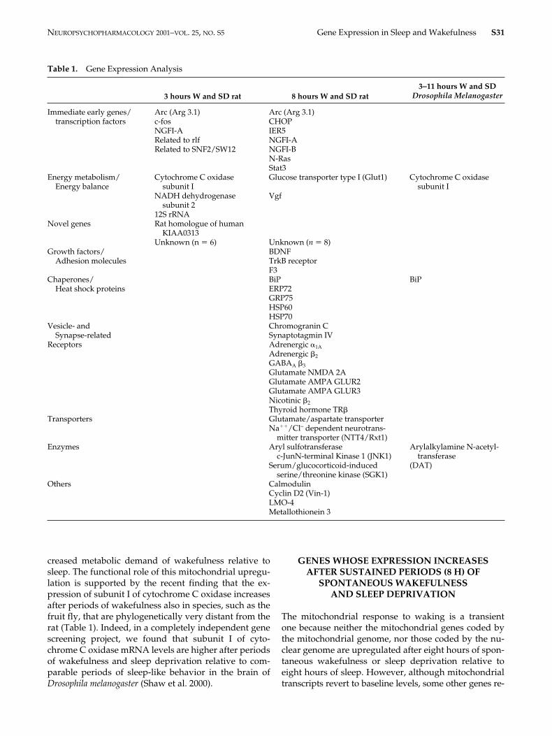
Two classes of genes show prominent changes after threehours of wakefulness or sleep deprivation: IEGs/tran-scription factors and mitochondrial genes encoded bythe mitochondrial genome (Cirelli and Tononi 1998,2000b; Table 1). The IEGs group includes
Arc, c-fos,NGFI-A,
the rat homolog of the human Zn-15 relatedzinc finger (
rlf
) gene, which has been implicated in tran-scriptional regulation, and AA117313, probably similarto the human global transcription activator SNF2/SWI2.In addition to
Arc
and
NGFI-A
(which are upregulatedafter both 3 h and 8 h of wakefulness and sleep depriva-tion), other IEGs/transcription factors are induced aftersustained (8 h) periods of sleep loss (Cirelli and Tononi2000a). They include
CHOP, IER5, NGFI-B, N-Ras
, and
Stat3.
Many of these genes may play a role in promotinggene transcription during wakefulness and may be in-volved in triggering the expression of other “late” genessuch those identified after long periods of sleep loss.
Three genes coded by the mitochondrial genomewere found to be induced by short periods of wakeful-ness and sleep deprivation: subunit I of cytochrome Coxidase, subunit 2 of NADH dehydrogenase, and 12SrRNA. The rapid regulation in the expression of mito-chondrial genes in response to being awake for threehours was unexpected. Cytochrome C oxidase is theterminal enzyme of the respiratory chain, and plays acrucial role in the regulation of oxidative metabolism(Wong-Riley et al. 1997). This enzyme is made up ofseveral subunits, some of which (e.g. subunit I) are codedby the mitochondrial genome, while others (e.g. sub-unit IV) are coded by the nuclear genome. Interestingly,changes in mRNA levels between sleep and wakeful-ness involve only the mitochondrial genes coded by themitochondrial genome and not those coded by the nu-clear genome. Mitochondria seem to contain excessamounts of nuclear-encoded cytochrome C oxidase sub-units. Changes in neuronal activity and energy demandaffect the transcription of mitochondrially encoded sub-units of cytochrome C oxidase more quickly and moresignificantly than that of the nuclear subunits (Wong-Riley et al. 1997). Thus, it is the synthesis of mitochon-drially encoded subunits, followed by the holoenzymeassembly, that is governed by dynamic local energyneeds. Cerebral glucose is almost exclusively metabo-lized through mitochondrial oxidative phosphorylationand glucose metabolism is 20–30% higher in wakeful-ness than in nonREM sleep in several species, includingthe rat (Ramm and Frost 1983). The increased expres-sion of mitochondrial genes after three hours of wake-fulness suggests a previously unsuspected mechanismby which neurons and/or glia can adapt to the in-
N
EUROPSYCHOPHARMACOLOGY
2001
–
VOL
.
25
,
NO
.
S5
Gene Expression in Sleep and Wakefulness
S31
Table 1.
Gene Expression Analysis
3 hours W and SD rat 8 hours W and SD rat3–11 hours W and SD
Drosophila Melanogaster
Immediate early genes/ Arc (Arg 3.1) Arc (Arg 3.1)transcription factors c-fos CHOP
NGFI-A IER5Related to rlf NGFI-ARelated to SNF2/SW12 NGFI-B
N-RasStat3
Energy metabolism/Energy balance
Cytochrome C oxidasesubunit I
Glucose transporter type I (Glut1) Cytochrome C oxidasesubunit I
NADH dehydrogenasesubunit 2
Vgf
12S rRNANovel genes Rat homologue of human
KIAA0313Unknown (n
�
6) Unknown (
n
�
8)Growth factors/ BDNF
Adhesion molecules TrkB receptorF3
Chaperones/ BiP BiPHeat shock proteins ERP72
GRP75HSP60HSP70
Vesicle- and Chromogranin CSynapse-related Synaptotagmin IV
Receptors Adrenergic
�
1A
Adrenergic
�
2
GABA
A
�
3
Glutamate NMDA 2AGlutamate AMPA GLUR2Glutamate AMPA GLUR3Nicotinic
�
2
Thyroid hormone TR
�
Transporters Glutamate/aspartate transporterNa
��
/Cl
–
dependent neurotrans-mitter transporter (NTT4/Rxt1)
Enzymes Aryl sulfotransferasec-JunN-terminal Kinase 1 (JNK1)
Arylalkylamine N-acetyl-transferase
Serum/glucocorticoid-inducedserine/threonine kinase (SGK1)
(DAT)
Others CalmodulinCyclin D2 (Vin-1)LMO-4Metallothionein 3
creased metabolic demand of wakefulness relative tosleep. The functional role of this mitochondrial upregu-lation is supported by the recent finding that the ex-pression of subunit I of cytochrome C oxidase increasesafter periods of wakefulness also in species, such as thefruit fly, that are phylogenetically very distant from therat (Table 1). Indeed, in a completely independent genescreening project, we found that subunit I of cyto-chrome C oxidase mRNA levels are higher after periodsof wakefulness and sleep deprivation relative to com-parable periods of sleep-like behavior in the brain of
Drosophila melanogaster
(Shaw et al. 2000).
GENES WHOSE EXPRESSION INCREASES AFTER SUSTAINED PERIODS (8 H) OF
SPONTANEOUS WAKEFULNESSAND SLEEP DEPRIVATION
The mitochondrial response to waking is a transientone because neither the mitochondrial genes coded bythe mitochondrial genome, nor those coded by the nu-clear genome are upregulated after eight hours of spon-taneous wakefulness or sleep deprivation relative toeight hours of sleep. However, although mitochondrialtranscripts revert to baseline levels, some other genes re-
S32
G. Tononi and C. Cirelli N
EUROPSYCHOPHARMACOLOGY
2001
–
VOL
.
25
,
NO
.
S5
lated to energy metabolism are markedly upregulatedafter eight hours of wakefulness (Table 1). One of thesegenes is
Glut1,
one of the major glucose transporters re-sponsible for the transfer of glucose from blood to neu-rons and glia. Thus,
Glut1
induction may represent an-other mechanism employed by the brain to respond tothe increased energy requirements of the waking state.
Several heat shock proteins and molecular chaper-ones such as HSP60, HSP70, and BiP show highermRNA levels after sustained periods of wakefulness.BiP, the major chaperone of the endoplasmic reticulum(ER), associates with nascent glycoproteins and secre-tory polypeptides during assembly in the ER and retainthem in an assembly-competent shape. Increases inmRNA and/or protein levels of
BiP
and heat shock pro-teins occur during stress conditions (e.g. ischemia, se-vere glucose deprivation, Ca
��
depletion) that maycause the accumulation of unfolded or malfoded pro-teins in cells (see Cirelli and Tononi 2000a for refs). Inthese abnormal conditions Bip may target unfoldedproteins for degradation or participate in their refold-ing. The finding that molecular chaperones and heatshock proteins are induced in a completely physiologi-cal condition, i.e. during spontaneous wakefulness, wasalso unexpected. The events responsible for
BiP
induc-tion are still unclear, but an increase in protein synthe-sis, notably of proteins that require assembly in the ER,could play a role. The
Aplysia
homolog of
BiP
, for in-stance, is induced after long-term sensitization training,and has been hypothesized to play a role in the foldingof newly synthesized proteins involved in synapticplasticity (Kuhl et al. 1992). Despite the fact that the roleof
BiP
induction during wakefulness is still unknown,its functional importance is once again supported bythe finding that BiP mRNA levels are induced after pe-riods of wakefulness and sleep deprivation in the brainof
Drosophila melanogaster
(Shaw et al. 2000; Table 1).Another major group of genes upregulated after
eight hours of wakefulness includes components of thepresynaptic and postsynaptic neurotransmission ma-chinery, such as vesicle and synaptic-related genes, sub-units of several neurotransmitter receptors, both excita-tory and inhibitory, and neurotransmitter transporters.The finding that AMPA receptor subunits GluR2 andGluR3 mRNA levels are higher after spontaneouswakefulness and sleep deprivation relative to sleep isparticularly intriguing. The number and density ofAMPA receptors at the postsynaptic membrane hasbeen shown to be regulated by synaptic activity, andendocytosis and exocytosis of AMPA receptors regulatesynaptic plasticity. Moreover, in the adult brain in vivo,long-term potentiation and long-term depression areassociated with increases and decreases, respectively, ofprotein levels of GluR1 and GluR2 (Heynen et al. 2000).Thus, the upregulation of components of the presynap-tic and postsynaptic neurotransmission machinery may
represent a general compensatory response of the brainto the increased synaptic neurotransmission duringwakefulness relative to sleep. In addition, the inductionof at least some of these genes, such as
GluR1
and
GluR2
, may more specifically mediate the occurrence ofplastic phenomena during wakefulness.
In this regard, it is important to emphasize that, irre-spective of the category in which they are listed, severalof the genes upregulated in wakefulness and sleep dep-rivation relative to sleep have been implicated in themolecular mechanisms of neural plasticity. This is thecase not only for the genes coding for GluR1 and GluR2,but also for those coding for several IEGs (Arc, c-fos,CHOP, NGFI-A), as well as for the growth factor BDNFand its receptor TrkB, the adhesion molecule F3, BiP,synaptotagmin IV, calmodulin, and a few others. Over-all, our data suggest that the transcription (and possiblytranslation) of plasticity-related genes is favored duringwakefulness relative to sleep.
GENE EXPRESSION IN SLEEP ANDWAKEFULNESS AND THE
NORADRENERGIC SYSTEM
Many transcripts upregulated during wakefulness areinduced diffusely in the cerebral cortex and in many otherbrain regions. We hypothesized that a key factor responsi-ble for their induction might be the level of activity ofneuromodulatory systems such as the noradrenergic andthe serotoninergic systems. These systems project dif-fusely to most of the brain and regulate gene expres-sion. Moreover, their activity is strictly state-dependent.
During sleep, locus coeruleus neurons fire regularlyat very low rates, whereas during wakefulness they fireat higher rates and emit phasic, short bursts of action po-tentials in response to salient events (Aston-Jones andBloom 1981a). Norepinephrine released diffusely bythese neurons over large portions of the brain enhancesinformation transmission and promotes attentive pro-cesses by increasing
�
activity in the EEG (Cape andJones 2000 and refs therein). Norepinephrine can enablevarious forms of activity-dependent synaptic plasticityand can stimulate gene transcription. To assess the roleof the noradrenergic system in the induction of gene ex-pression during wakefulness, we used rats whose behav-ior and brain electrical activity were continually moni-tored and in which the left locus coeruleus was lesioned(Cirelli et al. 1996; Cirelli and Tononi 2000b). Thus,when these animals were awake, norepinephrinewould be released only on the right side. The rats withunilateral locus coeruleus lesions behaved normallyand showed normal amounts of sleep and wakefulness.Furthermore, as demonstrated by EEG analysis, brainelectrical activity seemed normal and essentially indis-tinguishable between the right and the left side of their
N
EUROPSYCHOPHARMACOLOGY
2001
–
VOL
.
25
,
NO
.
S5
Gene Expression in Sleep and Wakefulness
S33
brain. The expression of
c-fos
,
NGFI-A
,
P-CREB,
Arc,
and
BDNF
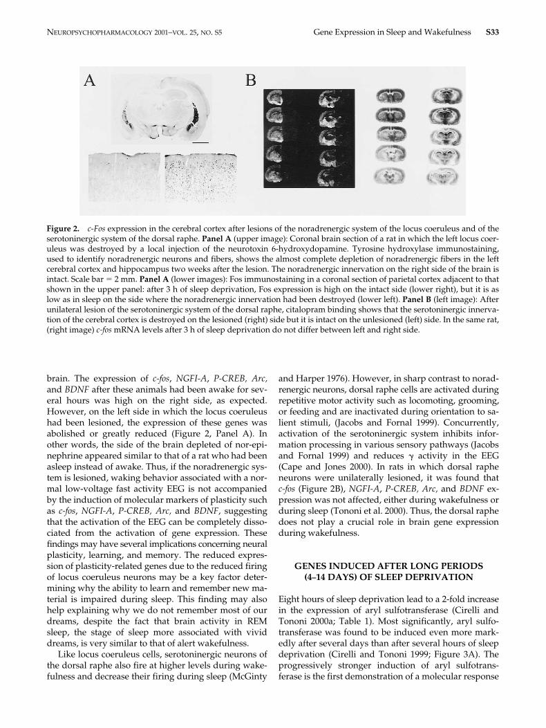
after these animals had been awake for sev-eral hours was high on the right side, as expected.However, on the left side in which the locus coeruleushad been lesioned, the expression of these genes wasabolished or greatly reduced (Figure 2, Panel A). Inother words, the side of the brain depleted of nor-epi-nephrine appeared similar to that of a rat who had beenasleep instead of awake. Thus, if the noradrenergic sys-tem is lesioned, waking behavior associated with a nor-mal low-voltage fast activity EEG is not accompaniedby the induction of molecular markers of plasticity suchas
c-fos
,
NGFI-A
,
P-CREB,
Arc,
and
BDNF
, suggestingthat the activation of the EEG can be completely disso-ciated from the activation of gene expression. Thesefindings may have several implications concerning neuralplasticity, learning, and memory. The reduced expres-sion of plasticity-related genes due to the reduced firingof locus coeruleus neurons may be a key factor deter-mining why the ability to learn and remember new ma-terial is impaired during sleep. This finding may alsohelp explaining why we do not remember most of ourdreams, despite the fact that brain activity in REMsleep, the stage of sleep more associated with vividdreams, is very similar to that of alert wakefulness.
Like locus coeruleus cells, serotoninergic neurons ofthe dorsal raphe also fire at higher levels during wake-fulness and decrease their firing during sleep (McGinty
and Harper 1976). However, in sharp contrast to norad-renergic neurons, dorsal raphe cells are activated duringrepetitive motor activity such as locomoting, grooming,or feeding and are inactivated during orientation to sa-lient stimuli, (Jacobs and Fornal 1999). Concurrently,activation of the serotoninergic system inhibits infor-mation processing in various sensory pathways (Jacobsand Fornal 1999) and reduces
�
activity in the EEG(Cape and Jones 2000). In rats in which dorsal rapheneurons were unilaterally lesioned, it was found that
c-fos
(Figure 2B),
NGFI-A
,
P-CREB,
Arc,
and
BDNF
ex-pression was not affected, either during wakefulness orduring sleep (Tononi et al. 2000). Thus, the dorsal raphedoes not play a crucial role in brain gene expressionduring wakefulness.
GENES INDUCED AFTER LONG PERIODS(4
–
14 DAYS) OF SLEEP DEPRIVATION
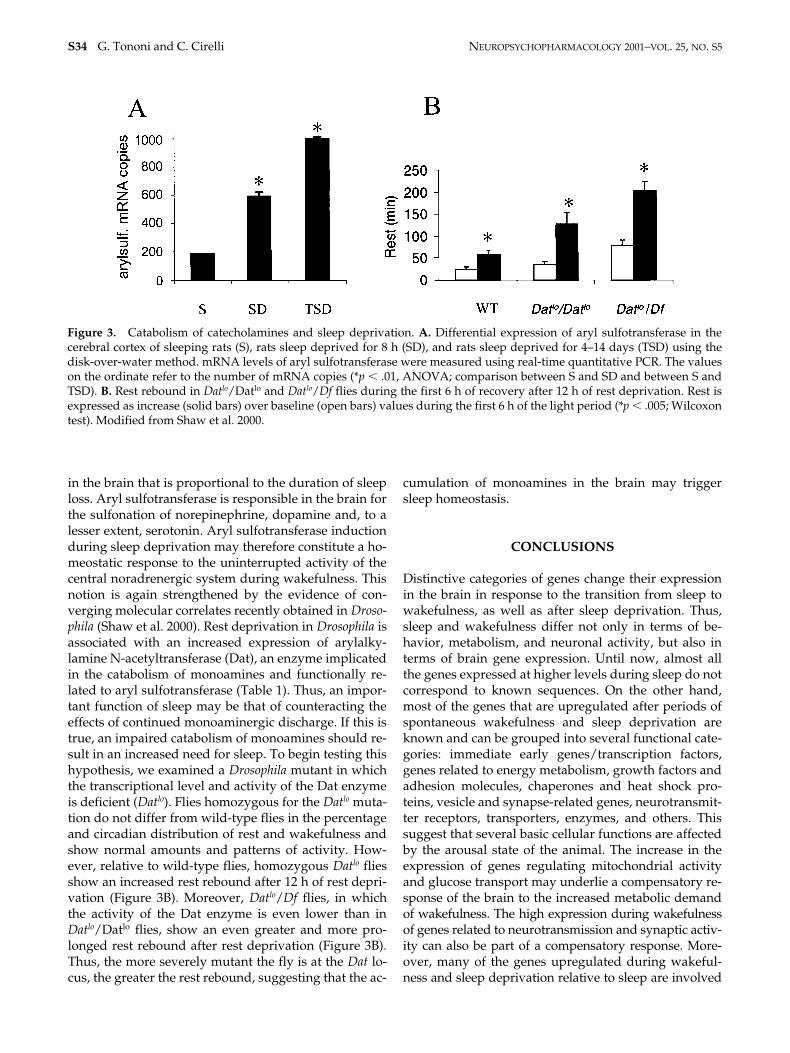
Eight hours of sleep deprivation lead to a 2-fold increasein the expression of aryl sulfotransferase (Cirelli andTononi 2000a; Table 1). Most significantly, aryl sulfo-transferase was found to be induced even more mark-edly after several days than after several hours of sleepdeprivation (Cirelli and Tononi 1999; Figure 3A). Theprogressively stronger induction of aryl sulfotrans-ferase is the first demonstration of a molecular response
Figure 2. c-Fos expression in the cerebral cortex after lesions of the noradrenergic system of the locus coeruleus and of theserotoninergic system of the dorsal raphe. Panel A (upper image): Coronal brain section of a rat in which the left locus coer-uleus was destroyed by a local injection of the neurotoxin 6-hydroxydopamine. Tyrosine hydroxylase immunostaining,used to identify noradrenergic neurons and fibers, shows the almost complete depletion of noradrenergic fibers in the leftcerebral cortex and hippocampus two weeks after the lesion. The noradrenergic innervation on the right side of the brain isintact. Scale bar � 2 mm. Panel A (lower images): Fos immunostaining in a coronal section of parietal cortex adjacent to thatshown in the upper panel: after 3 h of sleep deprivation, Fos expression is high on the intact side (lower right), but it is aslow as in sleep on the side where the noradrenergic innervation had been destroyed (lower left). Panel B (left image): Afterunilateral lesion of the serotoninergic system of the dorsal raphe, citalopram binding shows that the serotoninergic innerva-tion of the cerebral cortex is destroyed on the lesioned (right) side but it is intact on the unlesioned (left) side. In the same rat,(right image) c-fos mRNA levels after 3 h of sleep deprivation do not differ between left and right side.
S34
G. Tononi and C. Cirelli N
EUROPSYCHOPHARMACOLOGY
2001
–VOL. 25, NO. S5
in the brain that is proportional to the duration of sleeploss. Aryl sulfotransferase is responsible in the brain forthe sulfonation of norepinephrine, dopamine and, to alesser extent, serotonin. Aryl sulfotransferase inductionduring sleep deprivation may therefore constitute a ho-meostatic response to the uninterrupted activity of thecentral noradrenergic system during wakefulness. Thisnotion is again strengthened by the evidence of con-verging molecular correlates recently obtained in Droso-phila (Shaw et al. 2000). Rest deprivation in Drosophila isassociated with an increased expression of arylalky-lamine N-acetyltransferase (Dat), an enzyme implicatedin the catabolism of monoamines and functionally re-lated to aryl sulfotransferase (Table 1). Thus, an impor-tant function of sleep may be that of counteracting theeffects of continued monoaminergic discharge. If this istrue, an impaired catabolism of monoamines should re-sult in an increased need for sleep. To begin testing thishypothesis, we examined a Drosophila mutant in whichthe transcriptional level and activity of the Dat enzymeis deficient (Datlo). Flies homozygous for the Datlo muta-tion do not differ from wild-type flies in the percentageand circadian distribution of rest and wakefulness andshow normal amounts and patterns of activity. How-ever, relative to wild-type flies, homozygous Datlo fliesshow an increased rest rebound after 12 h of rest depri-vation (Figure 3B). Moreover, Datlo/Df flies, in whichthe activity of the Dat enzyme is even lower than inDatlo/Datlo flies, show an even greater and more pro-longed rest rebound after rest deprivation (Figure 3B).Thus, the more severely mutant the fly is at the Dat lo-cus, the greater the rest rebound, suggesting that the ac-
cumulation of monoamines in the brain may triggersleep homeostasis.
CONCLUSIONS
Distinctive categories of genes change their expressionin the brain in response to the transition from sleep towakefulness, as well as after sleep deprivation. Thus,sleep and wakefulness differ not only in terms of be-havior, metabolism, and neuronal activity, but also interms of brain gene expression. Until now, almost allthe genes expressed at higher levels during sleep do notcorrespond to known sequences. On the other hand,most of the genes that are upregulated after periods ofspontaneous wakefulness and sleep deprivation areknown and can be grouped into several functional cate-gories: immediate early genes/transcription factors,genes related to energy metabolism, growth factors andadhesion molecules, chaperones and heat shock pro-teins, vesicle and synapse-related genes, neurotransmit-ter receptors, transporters, enzymes, and others. Thissuggest that several basic cellular functions are affectedby the arousal state of the animal. The increase in theexpression of genes regulating mitochondrial activityand glucose transport may underlie a compensatory re-sponse of the brain to the increased metabolic demandof wakefulness. The high expression during wakefulnessof genes related to neurotransmission and synaptic activ-ity can also be part of a compensatory response. More-over, many of the genes upregulated during wakeful-ness and sleep deprivation relative to sleep are involved
Figure 3. Catabolism of catecholamines and sleep deprivation. A. Differential expression of aryl sulfotransferase in thecerebral cortex of sleeping rats (S), rats sleep deprived for 8 h (SD), and rats sleep deprived for 4–14 days (TSD) using thedisk-over-water method. mRNA levels of aryl sulfotransferase were measured using real-time quantitative PCR. The valueson the ordinate refer to the number of mRNA copies (*p � .01, ANOVA; comparison between S and SD and between S andTSD). B. Rest rebound in Datlo/Datlo and Datlo/Df flies during the first 6 h of recovery after 12 h of rest deprivation. Rest isexpressed as increase (solid bars) over baseline (open bars) values during the first 6 h of the light period (*p � .005; Wilcoxontest). Modified from Shaw et al. 2000.
NEUROPSYCHOPHARMACOLOGY 2001–VOL. 25, NO. S5 Gene Expression in Sleep and Wakefulness S35
in neural plasticity, suggesting that plastic changes, in asmuch as they require the induction of genes, occur dur-ing wakefulness rather than during sleep. Several heatshock proteins and chaperones involved in protein fold-ing and endoplasmic reticulum functions also showhigher expression in wakefulness than in sleep. Theanalysis of the genes induced by long-term sleep depri-vation is still in progress, but one transcript whose ex-pression increases in proportion to the duration ofwakefulness has already been identified. This transcriptcodes for the enzyme aryl sulfotransferase involved inthe catabolism of catecholamines, and its induction maysignify that an important function of sleep is to preventthe uninterrupted activity of the noradrenergic system.
ACKNOWLEDGMENTS
This work was carried out as part of the experimental neurobiol-ogy program at The Neurosciences Institute, which is supportedby Neurosciences Research Foundation. The Foundation re-ceives major support for this program from Novartis Pharma-ceutical Corporation. We thank Glen A. Davis, Marijo C. Gall-ina, and Donald F. Robinson for their expert contribution.
REFERENCES
Aston-Jones G, Bloom FE (1981a): Activity of norepineph-rine-containing locus coeruleus neurons in behaving ratsanticipates fluctuations in the sleep-waking cycle. J Neu-rosci 1:876–886
Cape EG, Jones BE (2000): Differential modulation of high-fre-quency �-electroencephalogram activity and sleep-wakestate by noradrenaline and serotonin microinjectionsinto the region of cholinergic basalis neurons. J Neurosci18:2653–2666
Cirelli C, Pompeiano M, Tononi G (1995): c-Fos expressionin the rat brain after sleep deprivation. J Sleep Res 4:92–106
Cirelli C, Pompeiano M, Tononi G (1996): Neuronal geneexpression in the waking state: a role for the locus coer-uleus. Science 274:1211–1215
Cirelli C, Tononi G (1998): Differences in gene expressionbetween sleep and waking as revealed by mRNA differ-ential display. Mol Brain Res 56:293–305
Cirelli C, Tononi G (1999): Changes in gene expression in thecerebral cortex of rats after short-term and long-termsleep deprivation. Sleep 22 (Suppl 1):113
Cirelli C, Tononi G (2000a): Gene expression in the brainacross the sleep-waking cycle. Brain Res 885:303–321
Cirelli C, Tononi G (2000b): Differential expression of plas-
ticity-related genes in waking and sleep and their regu-lation by the noradrenergic system. J Neurosci 20:9187–9194
Cirelli C, Tononi G (2000c): On the functional significance ofc-fos induction during the sleep/waking cycle. Sleep23:453–469
Drummond SP, Brown GG, Gillin JC, Stricker JL, Wong EC,Buxton RB (2000): Altered brain response to verbal learn-ing following sleep deprivation. Nature 403:655–657
Heynen AJ, Quinian EM, Bae DC, Bear MF (2000): Bidirec-tional, activity-dependent regulation of glutamate recep-tors in the adult hippocampus in vivo. Neuron 28:527–536
Horne JA (1988): Why we sleep. The functions of sleep inhumans and other mammals. Oxford, Oxford Univer-sity Press.
Jacobs BL, Fornal CA (1999): Activity of serotonergic neu-rons in behaving animals. Neuropsychopharmacol 21:9S–15S
Kuhl D, Kennedy TE, Barzilai A, Kandel ER (1992): Long-term sensitization training in Aplysia leads to anincrease in the expression of BiP, the major proteinchaperon of the ER. J Cell Biol 19:1069–1076
Maquet P (1995): Sleep function(s) and cerebral metabolism.Behav Brain Res 69:75–83
McGinty DJ, Harper RM (1976): Dorsal raphe neurons:depression of firing during sleep in cats. Brain Res 101:569–575
Milner FD, Sutcliffe JG (1983): Gene expression in rat brain.Nucleic Acid Res 11:5497–5520
Pompeiano M, Cirelli C, Tononi G (1994): Immediate-earlygenes in spontaneous wakefulness and sleep: Expres-sion of c-fos and NGFI-A mRNA and protein. J SleepRes 3:80–96
Ramm P, Frost BJ (1983): Regional metabolic activity in therat brain during sleep-wake activity. Sleep 6:196–216
Shaw PJ, Cirelli C, Greenspan RJ, Tononi G (2000): Corre-lates of sleep and waking in Drosophila Melanogaster.Science 287:1834–1837
Sheng M, Greenberg ME (1990): The regulation and functionof c-fos and other immediate early genes in the nervoussystem. Neuron 4:477–485
Tononi G, Cirelli C, Shaw PJ (2000): The Molecular correlatesof sleep, waking, and sleep deprivation. In Borbély A,Hayaishi O, Sejnowski TJ, Altman JS (eds), HumanFrontier Workshop VIII, The Regulation of Sleep. Stras-bourg, HFSP, pp. 155–167.
Wong-Riley MTT, Mullen MA, Huang Z, Guyer C (1997):Brain cytochrome oxidase subunit complementaryDNAs: isolation, subcloning, sequencing, light and elec-tron microscopic in situ hybridization of transcripts,and regulation by neuronal activity. Neuroscience 76:1035–1055