Embed Size (px)
Citation preview

Modified Oxygen Defect Chemistry at Transition Metal OxideHeterostructures Probed by Hard X‑ray Photoelectron Spectroscopyand X‑ray DiffractionYan Chen,†,‡,$ Dillon D. Fong,∥ F. William Herbert,†,§ Julien Rault,⊥ Jean-Pascal Rueff,⊥,#
Nikolai Tsvetkov,†,‡ and Bilge Yildiz*,†,‡,§
†Laboratory of Electrochemical Interfaces, ‡Department of Nuclear Science and Engineering, §Department of Materials Science andEngineering, Massachusetts Institute of Technology, Cambridge, Massachusetts 02139, United States∥Materials Science Division, Argonne National Laboratory, Argonne, Illinois 60439, United States⊥Synchrotron SOLEIL, l’Orme des Merisiers, Saint-Aubin, France#Sorbonne Universites, UPMC Univ Paris 06, CNRS, Laboratoire de Chimie Physique − Matiere et Rayonnement, 11 rue Pierre etMarie Curie, 75005 Paris, France
*S Supporting Information
ABSTRACT: Transition metal oxide heterostructures areinteresting due to the distinctly different properties that canarise from their interfaces, such as superconductivity, highcatalytic activity, and magnetism. Oxygen point defects canplay an important role at these interfaces in inducingpotentially novel properties. The design of oxide hetero-structures in which the oxygen defects are manipulated toattain specific functionalities requires the ability to resolve thestate and concentration of local oxygen defects across buriedinterfaces. In this work, we utilized a novel combination ofhard X-ray photoelectron spectroscopy (HAXPES) and highresolution X-ray diffraction (HRXRD) to probe the localoxygen defect distribution across the buried interfaces of oxideheterolayers. This approach provides a nondestructive way to qualitatively probe locally the oxygen defects in transition metaloxide heterostructures. We studied two trilayer structures as model systems: the La0.8Sr0.2CoO3−δ/(La0.5Sr0.5)2CoO4−δ/La0.8Sr0.2CoO3−δ (LSC113/LSC214) and the La0.8Sr0.2CoO3−δ/La2NiO4+δ/La0.8Sr0.2CoO3−δ (LSC113/LNO214) on SrTiO3 (001)single crystal substrates. We found that the oxygen defect chemistry of these transition metal oxides was strongly impacted by thepresence of interfaces and the properties of the adjacent phases. Under reducing conditions, the LSC113 in the LSC113/LNO214trilayer had less oxygen vacancies than the LSC113 in the LSC113/LSC214 trilayer and the LSC113 single phase film. On the otherhand, LSC214 and LNO214 were more reduced in the two trilayer structures when in contact with the LSC113 layer compared totheir single phase counterparts. The results point out a potential way to modify the local oxygen defect states at oxideheterointerfaces.
1. INTRODUCTION
Ionic defects, particularly oxygen defects, are known to affectthe properties of transition metal oxides.1−3 Controlling oxygendefects electrochemically4−6 may pave the way to modifying themagnetic, electronic, and transport properties of transitionmetal oxides. The importance of oxygen defects has motivatedthe development of techniques to quantify their totalconcentration in bulk materials, such as thermogravimetricanalysis (TGA), dilatometry,2 and Raman spectroscopy.7−9 Inaddition to their total concentration, it is important to knowtheir spatial distribution near extended defects, such as surfaces,grain boundaries, and heterophase boundaries. Knowing theirconcentration and structure near the interfaces can allow tuningof the local functionality of the material. Resolving oxygen
defect states near heterointerfaces buried beneath the surface isimportant for advancing our understanding of the uniqueproperties that arise at such interfaces, including magnetism,10
electronic conductivity,11,12 ionic conductivity,13,14 and catalyticactivity.15−23 Fascinating recent examples include the reversiblecontrol of the oxygen defect content by external bias near theCo/GdOx heterointerface to tailor the magnetic anisotropy ofCo,10 and near the interface of Ti/Pr1−xCaxMnO3 metal-oxidelayers to reversibly change the charge-carrier transportcharacteristics for resistive switching.24
Received: February 23, 2018Revised: April 17, 2018Published: April 17, 2018
Article
pubs.acs.org/cmCite This: Chem. Mater. 2018, 30, 3359−3371
© 2018 American Chemical Society 3359 DOI: 10.1021/acs.chemmater.8b00808Chem. Mater. 2018, 30, 3359−3371

Obtaining information about local oxygen defect states nearthe buried heterointerfaces of oxides, however, remains achallenging task. Previous successful attempts include the worksof Kim et al.,25,26 who used scanning transmission electronmicroscopy (STEM) to map the oxygen vacancies in oxide thinfilms based on the changes in local lattice parameters. Theyreported different oxygen vacancy distributions in (La0.5Sr0.5)-CoO3 films epitaxially grown on NdGaO3 and (LaA-lO3)0.3(Sr2AlTaO6)0.7 substrates,
25 and this difference in oxygenvacancy concentration was associated with distinct magneticproperties in the two systems studied. The same authors alsoobserved the local accumulation of oxygen vacancies near theBiFeO3/LaxSr1−xMnO3 interface,26 and they proposed thatthese vacancies impact the polarization switching behavior nearthe interface. The oxidation state of the cation as related to theoxygen defect concentration near buried interfaces can also beobtained by X-ray photoelectron spectroscopy and sputterdepth-profiling. For example, Tsvetkov et al.27 performed suchanalysis across the interface of La0.8Sr0 .2CoO3−δ/(La0.5Sr0.5)2CoO4−δ, for which the results were linked to fasteroxygen reduction kinetics important for fuel cells. Despite thesemotivating and successful examples, the destructive nature ofthe experimental methods may introduce uncertainties in thequantification of the point defects near interfaces. In this work,we implemented a nondestructive approach combining hard X-ray photoelectron spectroscopy (HAXPES)28−30 and highresolution X-ray diffraction (HRXRD) to qualitatively probethe spatial distribution of oxygen defects in and across theburied interfaces of thin trilayer model structures. The highenergy of photons used in HAXPES enabled probing depthsgreater than 10 nm, which was comparable to the totalthickness of the trilayer samples. By varying the photon energyand the photoelectron emission angle, the oxygen defectcontent from different depths across the buried interface was
estimated by analyzing the transition metal valence state andthe cation composition. Complementary to HAXPES, thelattice parameters measured by HRXRD allowed quantificationof the changes in the oxygen defect concentration of each layer.Our approach is nondestructive and can be adapted broadly toother oxide heterolayers to understand the effect of interfaceson oxygen defects, and there by on the unique interface-induced properties.11−23
In this study, we assessed trilayer heterostructures as modelsystems made of perovskite (ABO3) and Ruddlesden−Popper(RP) (A′2B′O4) oxides. Perovskite and RP oxides have beenstudied widely for oxygen reduction and oxygen transport infuel cells and separation membranes.31,32 In particular, cobaltiteperovskite-RP heterostructures (with B = Co) have attractedgreat interest recently because they can present significantlyenhanced oxygen reduction kinetics compared to their singlephase counterparts.16−21 The mechanisms behind such anenhancement were studied with both experiments andtheoretical calculations33−36 for heterostructures ofLa0.8Sr0.2CoO3−δ (LSC113) and (La0.5Sr0.5)2CoO4−δ (LSC214),where δ is the oxygen nonstoichiometry. Until now, however,the potential role of oxygen defects at or near these interfaceshas been missing from the picture. In this work, wecharacterized the oxygen defect chemistry near the buriedinterfaces between LSC113 and two RP-type oxides that havevery different oxygen defect chemistries: LSC214 and La2NiO4+δ(LNO214). At the experimental conditions in this work, thedominant oxygen defects in single phase LSC214 are oxygenvacancies, and in LNO214 oxygen interstitials. These twoheterostructures in the form of trilayers on single crystal SrTiO3(STO) (001) are denoted as LSC113/LSC214 and LSC113/LNO214, respectively. We found that the LSC113 with LNO214 isless reducible than the LSC113 alone or the LSC113 with LSC214.On the other hand, the LSC214 and LNO214 lose oxygen more
Figure 1. Structure of LSC113/LSC214 and LSC113/LNO214 trilayer model systems. (a) Transmission electron microscopy (TEM) images of thetrilayer structure. Pt serves as the capping layer for TEM imaging. (b) X-ray reflectivity (XRR) of LSC113/LSC214 and LSC113/LNO214 trilayers intheir as-prepared state. The inset schematics represent the LSC113/LSC214 and LSC113/LNO214 trilayer model structures, in which the black squaresand white squares symbolize the oxygen interstitials and oxygen vacancies, respectively.
Chemistry of Materials Article
DOI: 10.1021/acs.chemmater.8b00808Chem. Mater. 2018, 30, 3359−3371
3360

easily when they are adjacent to LSC113 in the trilayers thantheir single phase counterparts. Our findings indicate that onecan engineer the overall defect chemistry of a material byconstructing superlattices with complementary defect behav-iors, and thereby engineer the functionality.
2. METHODSThe LSC113/LSC214, LSC113/LNO214 trilayer and reference singlephase films were grown by PLD using a KrF excimer laser with thewavelength 248 nm and a laser fluence of 1.9 J/cm2. The number ofpulses for the bottom LSC113, LSC214 or LNO214, and top LSC113layers were 3450, 3000, and 3000, respectively. The deposition wascarried out at 700 °C under 10 mTorr oxygen pressures. After thegrowth process, the films were cooled down to room temperature in 2Torr oxygen pressure with a cooling rate of 5 °C/min.The HAXPES experiment was carried out using the endstation
located at the GALAXIES beamline of the SOLEIL synchrotron in
France. The photon energy covers the range 2.3 to 12 keV, andelectrons with kinetic energies up to 12 keV can be analyzed.37 Theangle between the direction of the incident X-ray beam and thephotoelectron detection is fixed to be 90°. The sample can be tilted tochange the emission angle, as shown in Supporting Information (SI)Figure S1. Due to the large penetration depth of HAXPES, the C 1speak is too weak to serve as the external peak calibration for spectra atsmall emission angles. Considering that the chemical environment ofLa is quite similar for all the layers and expected not to show anychanges upon annealing, all the La 3d spectra in this work were alignedto be at 833.3 eV. The C 1s peak is at 284.8 eV when it is detectablefor spectra with large emission angles (more surface sensitive). TheHAXPES spectra were analyzed using the CasaXPS software. The (La+ Sr)/Co ratios and Sr/La ratio were quantified based La 3d, Co 2p,and Sr 3d peaks. The Co valence states were identified from Co 2ppeaks.
The lattice parameters of the trilayers and single phase films in theiras-prepared condition and after annealing were measured by HRXRD
Figure 2. Depth profile of Co valence state in the trilayer thin films. (a) Representative Co 2p spectra collected with photon energy of 3000 eV as afunction of emission angle from 85° to 15°, shown for the as-prepared LSC113/LSC214 trilayer. Each spectra contain cumulative informationextending from the surface to the probing depth, as marked by the gray arrow (the length of arrow is not to scale). (b−d) Comparison of the shapeof the Co 2p3/2 satellite peak in LSC113/LSC214 among the (b) as-prepared state, and after annealing at (c) 300 °C and at (d) 500 °C for 1 h. (e−g)Coss
2+/(Coss2+ + Coss
3+) ratio quantified for different probing depths in the LSC113/LSC214 trilayer film obtained by varying the detection angle, f(θ), atthe photon energy of 3000 eV, and by varying the photon energy, f(Ephoton), at the emission angle of 45°, shown for (e) the as-prepared state, andafter annealing at (f) 300 °C and (g) 500 °C for 1 h. Coss
2+/(Coss2+ + Coss
3+) qualitatively represents the relative presence of Co2+ in the films, whichwas calculated by fitting the satellite peaks as demonstrated in (a−d) for spectra collected at 15°. The probing depth is estimated as three times theinelastic mean free path of Co 2p3/2 photoelectrons (SI section S1). The colored rectangles mark the approximate position of the different layers,with blue for LSC113, red for LSC214, and gray for the STO substrate.
Chemistry of Materials Article
DOI: 10.1021/acs.chemmater.8b00808Chem. Mater. 2018, 30, 3359−3371
3361

using a Rigaku Smartlab diffractometer equipped with a 2-bounce Ge
(220) channel-cut monochromator and Cu Kα1 radiation.Transmission electron microscopy (TEM) samples were fabricated
in the Helios Nanolab 600 dual beam focused ion beam milling
system. The Ga ion beam was operated at a voltage and current that
was varied in a range of 30 keV−5 keV and 9.5 nA−28 pA,
respectively. TEM measurements were performed using a JEOL 2010
FEG microscope. All the crystal structures in this manuscript are
visualized using the Vesta software.38
3. RESULTS AND DISCUSSION
3.1. LSC113/LSC214 and LSC113/LNO214 Trilayer ModelSystems. LSC113/LSC214 and LSC113/LNO214 trilayer filmsgrown by PLD were epitaxial with the substrate and had (001)out-of-plane orientation, as shown in the TEM images in Figure1a. Based on the TEM images and X-ray reflectivitymeasurements, the thicknesses of each layer were 3−4 nm,with a total thickness of ∼10 nm, as shown in Figure 1b. Thesamples were examined in their as-prepared state as well as afterbeing reduced by annealing at 300 and 500 °C for 1 h at 10−10
Figure 3. Comparison of the reduction of Co in LSC113 in the LSC113/LSC214 and LSC113/LNO214 trilayers: (a−d): Co 2p3/2 satellite peak intensity(a), Co 2p3/2 main peak position (b), valence band (V.B.) spectra (c), and La 3d5/2 (d). The data shown in (a−d) were collected at 80° emissionangle, corresponding to a probing depth within the top LSC113 layer in the trilayers. The bottom two spectra in each panel are from the as-preparedsamples, while the top spectra represent the state after annealing at 500 °C. (e−f) Comparison of Coss
2+/(Coss2+ + Coss
3+) in the LSC113 single phasereference film, and in the LSC113/LSC214 and LSC113/LNO214 trilayers, (e) in the as-prepared state and (f) after annealing at 500 °C. The probingdepth is defined in the caption of Figure 2. The colored rectangles mark the approximate position of different layers, with blue for LSC113, red forLSC214, green for LNO214, and gray for the STO substrate.
Chemistry of Materials Article
DOI: 10.1021/acs.chemmater.8b00808Chem. Mater. 2018, 30, 3359−3371
3362

mbar in an ultrahigh vacuum chamber. A LSC113 film alwaysserved as the top layer in the trilayer structure to ensure thatthe oxygen surface exchange kinetics during the annealing forboth types of trilayers were the same. The results werecompared among these trilayers and single-phase ∼10 nm-thickfilms of LSC113, LSC214, and LNO214 on STO (001) substrates,prepared under the same conditions.3.2. Oxygen Defects across the Buried Interfaces
Measured by HAXPES. We determined the transition metalvalence states and cation composition by HAXPES in the thinfilm trilayers and across their interfaces. This informationallowed us to compare the oxygen vacancy and oxygeninterstitial content in the trilayer structures with respect tothe single-phase thin film reference structures. Here, a decreasein the emission angle from 85° to 15° with respect to thesurface normal implies an increase in probing depth. Theprobing depth at each emission angle and photon energy isdefined as three times the inelastic mean free path, λ, of thephotoelectron as described in SI section S1.The chemicalinformation obtained by HAXPES at a given angle and photonenergy is cumulative information from the top surface throughdifferent depths of the sample up to the maximum probingdepth. For instance, the information obtained at 80°−85° ismainly from the topmost LSC113 layers in the LSC113/LSC214and in the LSC113/LNO214 trilayers; at an emission angle of 15°the spectra contain cumulative information extending from thesurface into the STO substrate (Figure 2a).First, we quantified the distribution of the Co valence state
across the depth and buried interfaces of LSC113/LSC214trilayer structures from the Co 2p photoelectron spectrameasured as a function of photon energy and emission angle.The Co oxidation state is an important factor for determiningthe electronic and ionic conductivity of the LSC113 and LSC214,as well as for the electron transfer process during oxygenreduction. Representative Co 2p spectra collected at differentemission angles are shown in Figure 2a for the as-preparedLSC113/LSC214 trilayer. The Co 2p spectrum contains two mainpeaks, Co 2p1/2 and Co 2p3/2, corresponding to different spin−orbit couplings. For each main peak, there is also a satellite peakat higher binding energy because of the extra electronstransferred from the O 2p orbitals to the Co 3d orbital.These electrons screen the electric field of the holes in the Co2p orbital created during the photoelectron emission process.The shape and position of the satellite peaks, particularly theCo 2p3/2 satellite peak, are widely used to identify the valencestate of Co, as summarized in SI section S2. Under thepreparation conditions during pulsed laser deposition of thesefilms, the valence states of Co in LSC113 are predominantly 3+with a small amount of 4+,39,40 while in LSC214 Co is mainly3+.41 After annealing in reducing conditions as described above,the 2+ valence state of Co is expected to be part of theequilibrium in LSC113
39 and in LSC214.41 The binding energies
of the Co 2p3/2 satellite peak for Co2+and for Co3+ are at ∼786
eV42−46 and ∼789 eV,42,47−49 respectively (Figure 2a, SI TableS1). To compare the Co valence state as a function of probingdepth among different samples, we fitted the Co 2p spectra asshown in Figure 2. To qualitatively represent and compare theamount of Co2+ in the different films, we assessed the fractionalpresence of Co2+, quantified as the ratio of the Co2+ 2p3/2satellite peak area to the sum of the areas of the Co2+ and Co3+
2p3/2 satellite peaks. This ratio is denoted as Coss2+/(Coss
2+ +Coss
3+). There is no evidence that the ratio of satellite peaks(Coss
2+/Coss3+) increases linearly with the Co2+/Co3+ ratio.
However, it is known that higher peak intensity for the Co2+
satellite arises from a higher Co2+ content, and similarly for theCo3+ case (see references listed in SI Table S1), even if the ratioof satellite peak intensities does not have to be linear.Therefore, we did not make any conclusion about the absolutechange of the Co2+/Co3+ ratio based on the satellite peak ratiosin this manuscript. To reproduce the asymmetric shapes of themain peaks as accurately as possible and minimize their impacton the satellite peak quantification (Figure 2a), we fitted the Co2p3/2 and Co 2p1/2 regions consistently with three peaks asperformed in ref 50. It should be noted these three fitted peaksdo not correspond to different valence states. More details onthe fitting parameters can be found in SI section S3. The errorbars in Figure 2 and in Figure 3 for Co2+ss/(Co
3+ss + Co2+ss)
represent the uncertainty introduced by the fitting procedure.The impact of different fitting parameters, including fittingrange, peak shape, and position, on the final result wereevaluated, and the standard deviation of the fitting results wasused to show the uncertainty introduced by the fittingprocedure. Considering the fact that we did all the measure-ments during the same beam time at the same end-station atSOLEIL synchrotron, we believe that the uncertainty arisingfrom measurement conditions, such as the fluctuation of beamintensity or the measurement geometry, is negligible incomparing the samples to each other.The Coss
2+/(Coss2+ + Coss
3+) ratio quantified either as a functionof emission angle, f(θ), or as a function of photon energy,f(EPhoton), is plotted versus probing depth in Figure 2e−g. Asshown in SI section S1, the probing depth is estimated as threetimes the inelastic mean free path of Co 2p 3/2 photoelectrons.Each point contains cumulative information from the topsurface to the probing depth. A general increase in the Co2+
content at each depth can be seen after annealing the specimenat 300 and 500 °C at a low oxygen pressure. This is consistentwith the annealing induced reduction and creation of oxygenvacancies in the LSC113 and LSC214 layers. Concurrent with theincreased intensity of the Co2+ 2p3/2 satellite peak, the Co mainpeak position also shifted to lower binding energies (SI FigureS4b). The increase of the Coss
2+/(Coss2+ + Coss
3+) ratio is moreevident in the LSC113 layers after annealing at 500 °C; hereCoss
2+/(Coss2+ + Coss
3+) decreases slightly when the main samplingregion enters into the LSC214 layer and increases again whenentering the bottom LSC113 layer (Figure 2g). This effect isobserved because LSC214 is more difficult to reduce by formingoxygen vacancies (i.e., it has a higher oxygen vacancy formationenergy) than LSC113.
27,36 Finally, it is worth reminding that,while the plots in Figure 2e−g show a spatially varying profile,the HAXPES intrinsically measures accumulated data from thesurface into varying depths in the samples, and so the finalnonzero values for Coss
2+/(Coss2+ + Coss
3+) should not beinterpreted as the presence of Co2+ in the STO substrate.Having demonstrated that HAXPES enables profiling of the
Co valence state across the buried heterointerfaces of theLSC113/LSC214 trilayer, we now examine the valence state ofCo in LSC113 when it is adjacent to two different oxides, i.e.either LSC214 or LNO214 within the two trilayer structures. Adirect examination of all the Co 2p3/2 peaks and the valenceband edge (in Figure 3a, b, c) reveals the dependence of the Covalence state in LSC113 on the neighboring layer. First, the Co
2+
2p3/2 satellite peak intensity is higher for LSC113 with LSC214 asthe neighboring oxide than the one with LNO214, both at theas-prepared state and after annealing at 500 °C (Figure 3a).Second, the main Co 2p3/2 peak of LSC113neighboring with
Chemistry of Materials Article
DOI: 10.1021/acs.chemmater.8b00808Chem. Mater. 2018, 30, 3359−3371
3363
LSC214 is located at a lower binding energy than that of LSC113neighboring with LNO214 (Figure 3b). Third, the valence bandedge in LSC113 was also impacted by the neighboring oxidelayer. When the LSC113 film is reduced at elevated temperaturesand low oxygen pressures, formation of oxygen vacanciesinduce defect states and shift the valence band edge up.36,51−53
The valence band edge of LSC113 with LSC214 and of LSC113with LNO214 both up-shifted (i.e., to lower binding energy)after annealing at 500 °C (Figure 3c). This upshift is larger forLSC113 with LSC214, indicating a more significant reduction ofCo in the LSC113 layer when it neighbors LSC214. All three ofthese results are consistent among each other and indicate amore reduced Co in LSC113 when adjacent to LSC214 thanwhen it is adjacent to LNO214. It is worth also noting that theLa 3d core level emission from LSC113 does not depend on thecomposition of the neighboring oxide (Figure 3d).More quantitatively, the Coss
2+/(Coss2+ + Coss
3+) ratio in LSC113neighboring with LSC214 is higher than that in LSC113neighboring with LNO214, for both the as-prepared state andafter annealing at 500 °C (Figure 3e, f). The amounts of Co2+
in both the top and the bottom LSC113 layers in the LSC113/LSC214 trilayer significantly increase after annealing at 500 °C(Figure 3e, f). However, the amount of Co2+ in the LSC113/LNO214 trilayer only slightly increases after annealing at 500 °C(Figure 3f). The Coss
2+/(Coss2+ + Coss
3+) ratios in the LSC113single phase reference film and in the LSC113 layer of theLSC113/LSC214 trilayer are very similar to each other. Anydifference in the reduced states of these two specimens was notpossible to resolve by HAXPES, but was more clearly resolvedby the HRXRD measurements that are presented later.
Due to the strong overlap between the Ni 2p and La 3dpeaks, and between the Ni 3p and Co 3p peaks (SI Figure S8),the Ni valence state in the LNO214 layers could not bedetermined from HAXPES. However, the results obtained fromHRXRD measurements (discussed later in the paper) can beused to examine the relative reducibility of Ni in the referenceLNO214 film and in the LSC113/LNO214 trilayer structure.It is interesting that the Coss
2+/(Coss2+ + Coss
3+) ratio was highnear the first interface (Figure 2e), indicating possibly a quitehigh Co2+ concentration in that region in the as-prepared state.This situation may arise because of Sr enrichment near theLSC113/LSC214 interface after high temperature synthesis, aspreviously reported by another group.34 However, it is only onedata point that showed very high Co2+ content for the as-prepared state, and we will show in the later section we ruledout the possibility of Sr migration as the possible mechanism ofdifferent defect chemistry in the trilayer structure.Having shown that the Co valence state in LSC113 depends
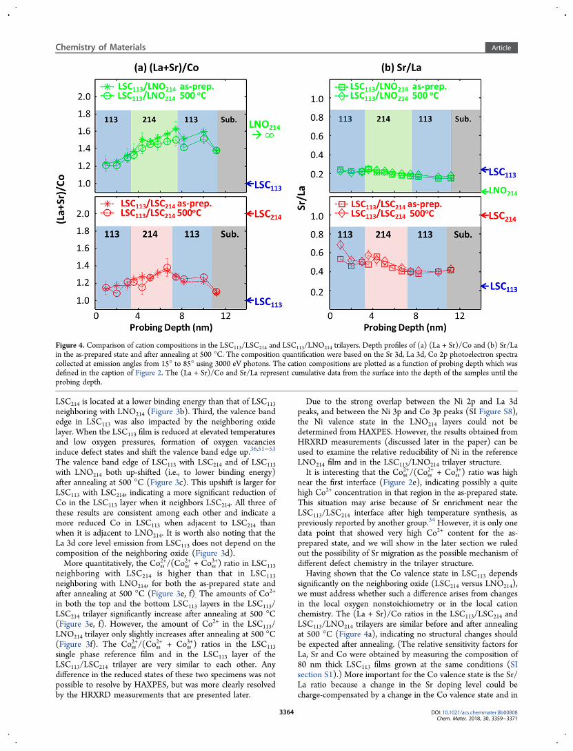
significantly on the neighboring oxide (LSC214 versus LNO214),we must address whether such a difference arises from changesin the local oxygen nonstoichiometry or in the local cationchemistry. The (La + Sr)/Co ratios in the LSC113/LSC214 andLSC113/LNO214 trilayers are similar before and after annealingat 500 °C (Figure 4a), indicating no structural changes shouldbe expected after annealing. (The relative sensitivity factors forLa, Sr and Co were obtained by measuring the composition of80 nm thick LSC113 films grown at the same conditions (SIsection S1).) More important for the Co valence state is the Sr/La ratio because a change in the Sr doping level could becharge-compensated by a change in the Co valence state and in
Figure 4. Comparison of cation compositions in the LSC113/LSC214 and LSC113/LNO214 trilayers. Depth profiles of (a) (La + Sr)/Co and (b) Sr/Lain the as-prepared state and after annealing at 500 °C. The composition quantification were based on the Sr 3d, La 3d, Co 2p photoelectron spectracollected at emission angles from 15° to 85° using 3000 eV photons. The cation compositions are plotted as a function of probing depth which wasdefined in the caption of Figure 2. The (La + Sr)/Co and Sr/La represent cumulative data from the surface into the depth of the samples until theprobing depth.
Chemistry of Materials Article
DOI: 10.1021/acs.chemmater.8b00808Chem. Mater. 2018, 30, 3359−3371
3364

the oxygen vacancy concentration.40,54 As shown in Figure 4b,the Sr/La ratio in the as-prepared LSC113/LSC214 film decreasesas the probing depth goes from the top surface into the bulk.This is due to the Sr enrichment at/near the surface, aphenomenon that is generally observed for doped perovskiteoxides.51,55−57 After 500 °C, the Sr/La distribution remainsunchanged for the LSC113/LNO214 trilayer, and it increasesmainly at the very top surface of the LSC113/LSC214 trilayer. Onthe other hand, the Co2+ fraction clearly increased within theLSC113/LSC214 trilayer after annealing at 500 °C (Figure 3e, f).If the enrichment of Sr2+ on the La-site were the only change inchemistry at 500 °C, then we should have found a more
oxidized Co in LSC113, but we found a more reduced Co.Therefore, the more reduced Co is consistent with an overallincrease of oxygen vacancy concentration within the LSC113/LSC214 trilayers to an extent significantly greater than that inthe LSC113/LNO214 trilayer (Figure 3e,f). Lastly, we note thatone reason why the (La + Sr)/Co ratio in LSC113/LNO214 andLSC113/LSC214 are larger than 1 is that the chemicalinformation obtained by HAXPES is a cumulative informationacross different depths of the sample. Even when the probingdepth, here referring to three times the inelastic mean free pathλ, is mainly within the top LSC113 layer, the data carry alsosome information from the underlying LSC214 or LNO214. This
Figure 5. Lattice expansion and contraction due to oxygen nonstoichiometry change, quantified by HRXRD in LSC113/LSC214, LSC113/LNO214trilayer structures and their single phase counterparts. (a) 2θ−ω scans of LSC113/LSC214 (top) and LSC113/LNO214 (bottom), as-prepared and afterannealing at 300 and 500 °C for 1 h. (b) The schematics show the expansion of the c lattice parameter in LSC113 and LSC214 films due to the increaseof oxygen vacancies, and the contraction of c in LNO214 due to the loss of oxygen interstitials after annealing. (c−e) Comparison of the HRXRDpatterns to show the relative changes in the out-of-plane lattice parameter, c, in LSC113, LSC214, and LNO214: (c) LSC113 (002) peak in 2θ−ω scansfor LSC113, LSC113/LSC214, and LSC113/LNO214, as-prepared and after annealing at 500 °C. (d) LSC214 (008) and (e) LNO214 (008) peaks in 2θ−ωscans for the single phase and trilayer structures in the as-prepared state and after annealing at 500 °C. The dashed lines mark the position of theLSC113 (002) peak, LSC214 (008) peak, and LNO214 (008) peak.
Chemistry of Materials Article
DOI: 10.1021/acs.chemmater.8b00808Chem. Mater. 2018, 30, 3359−3371
3365

can also explain the seemly larger (La + Sr)/Co ratio (andsmaller Sr/La ratio) in LSC1113/LNO214 compared to that inLSC113/LSC214. Considering that HAXPES is always moresensitive to the surface, Sr enrichment on the surface of LSC113mentioned above can also lead to an A to B ratio larger than 1for both LSC113/LSC214 and LSC113/LNO214.In summary, combining the information on the Co valence
state (Figures 2 and 3) and the cation composition (Figure 4)deduced from HAXPES, we found that the Co in LSC113neighboring LSC214 (in LSC113/LSC214) is more reducible thanthe Co in LSC113 neighboring LNO214 (in LSC113/LNO214).This is because of a larger concentration of oxygen vacanciesthat form in LSC113 of the LSC113/LSC214 than in LSC113 of theLSC113/LNO214, after annealing at elevated temperatures in areducing atmosphere. From this finding, we can conclude thatthe oxygen nonstoichiometry in thin LSC113 layers can besignificantly influenced by the nature of the adjacent oxide.3.3. Oxygen Defect States Probed by HRXRD. The
oxygen nonstoichiometry in transition metal oxides impacts thelattice parameter, commonly referred to as chemical strain.58,59
In this section, we present results from HRXRD analysis. Wequantify the relative changes in the lattice parameters inLSC113/LSC214 and LSC113/LNO214 trilayers and relate thelattice expansion or contraction to the relative changes in theoxygen nonstoichiometry. This approach for observing theoxygen defect chemistry in thin films and buried layers iscomplementary to the HAXPES analysis presented in theprevious section.The LSC113, LSC214, LNO214 layers within the LSC113/
LSC214 and LSC113/LNO214 trilayers remained epitaxial withthe STO (001) substrate before and after annealing (Figure5a). After annealing at 500 °C for 1 h, the out-of-plane latticeparameter, c, of LSC113 in LSC113/LSC214 expanded by 0.6%(Figure 5a, Table 1), while that of LSC113 in LSC113/LNO214did not show any detectable change. The c for LSC113 in thesingle phase film is larger than that for either of the trilayerstructures, both in the as-prepared state and after 500 °Cannealing (Figure 5c). The lattice parameter of LSC113 isknown60,61 to increase with oxygen nonstoichiometry (δ), i.e.,with the oxygen vacancy concentration, as depicted schemati-cally in Figure 5b. The lattice parameter and oxygennonstoichiometry of the as-prepared samples and after heatingat temperature T were defined to be cas‑prep,, δas‑prep, cT, and δT.
To relate the change in the lattice parameter (Δc = cT − cas‑prep.)to the relative changes in the oxygen nonstoichiometry (Δδ =δT − δas‑prep.), we used the chemical expansion coefficient,
=εχ
∂∂ 0.053
vand =ε
χ∂∂
1.543v
2
2 , where ε = Δcc
and χ = δv 3
,
quantified by Chen et al.60 for La0.8Sr0.2CoO3−δ (SI section S3).The c lattice parameter and the annealing-induced changes inthe c (Δc) and in the oxygen nonstoichiometric (Δδ) forLSC113 single phase, LSC113 in LSC113/LSC214, and LSC113 inLSC113/LNO214 are shown in Table 1. We note that, in relatingthe Δc to Δδ above, the chemical expansion coefficientdeduced for bulk powder specimens was used.60 Furthermore,we also included the impact of the in-plane clamping by thesubstrate, by calculating how the chemical expansion shouldpropagate into changes in c for clamped films, using anapproximate Young’s modulus and Poisson ratios as shown inSI section S3. For the LSC113 in LSC113/LNO214, the Δδ after500 °C was too small to be detected by our HRXRDmeasurement. The Δδ for single phase LSC113 after 500 °C isestimated to be 0.188, which is larger than that of LSC113 in theLSC113/LSC214 and the LSC113/LNO214 trilayers.For RP phases, it is known that the c lattice parameter
decreases when the oxide loses oxygen interstitials, but itincreases when the oxide loses lattice oxygen and forms oxygenvacancies62−65 (SI Figure S10). We found that the c parameterof LSC214 increased after annealing in reducing conditions,while the c parameter of LNO214 decreased (Figure 5a, b, Table1). For LSC214 with 50% Sr used in this study, the oxygenvacancy is the dominant defect for the conditions involved inthe preparation and annealing of these films.41 The dominantoxygen defect in LNO214, however, is oxygen interstitials, evenat temperatures as high as 900 °C and under reducingconditions of 10−11 bar oxygen partial pressure.63 Therefore, wecan conclude that, in this work after annealing at 500 °C, theLSC214 has a higher concentration of oxygen vacancies, whilethe LNO214 has a lower concentration of oxygen interstitials.Compared with the one in the LSC113/LSC214 trilayer structure,single phase LSC214 has a smaller c lattice parameter (Figure 5d,Table 1). On the other hand, the LNO214 single phase shows alarger c than that in the LSC113/LNO214 trilayer (Figure 5e).These results indicate more oxygen vacancies are present in theLSC214 and less oxygen interstitials are present in LNO214 filmswhen they are in contact with LSC113 in the trilayers. To relate
Table 1. Comparison of the c Lattice Parameter, the Change in the c Lattice Parameter (Δc), in the Out-of-Plane Strain State, ε(Δc/c), and in the Oxygen Nonstoichiometry (Δδ) in La0.8Sr0.2CoO3−δ (LSC113), (La0.5Sr0.5)2CoO4−δ (LSC214), and La2NiO4+δ(LNO214) in Trilayer Structures and in Single Phase Films after Annealing at 500 °C
Sample Condition c (Å) Δc (Å) ε Δδ Δδtrilayer − Δδsingle phase
LSC113 single phase As Prep 3.770500 °C 3.810 0.040 0.011 0.188
LSC113in LSC113/LSC214 As Prep. 3.768500 °C 3.789 0.021 0.006 0.120 −0.068
LSC113in LSC113/LNO214 As Prep. 3.764500 °C 3.764 0.000 0.000 0.000 −0.188
LSC214 single phase As Prep. 12.40500 °C 12.44 0.037 0.003 0.074
LSC214 in LSC113/LSC214 As Prep. 12.46500 °C 12.57 0.117 0.009 0.173 0.099
LNO214 single phase As Prep 12.71500 °C 12.67 −0.040 −0.003 −0.038
LNO214 in LSC113/LNO214 As Prep. 12.66500 °C 12.58 −0.082 −0.006 −0.079 −0.041
Chemistry of Materials Article
DOI: 10.1021/acs.chemmater.8b00808Chem. Mater. 2018, 30, 3359−3371
3366
the lattice parameter changes to relative changes in the oxygennonstoichiometry (Δδ), we used the chemical expansion
coefficient =δ
∂∂( ) 0.62c
TÅ, quantified for bulk LNO214.
63
The Δδ in LNO214 from the as-prepared state to the 500 °Cannealed state is estimated to be about −0.079 for LNO214 inthe trilayer and about −0.038 for singe phase LNO214 (Table1). For LSC214, there has been no quantitative study of itschemical expansion behavior. Under our experimental (depo-sition and annealing) conditions, the dominant oxygen defectsare oxygen vacancies and the recucible cation is Co in bothLSC113 and LSC214. Therefore, it is reasonable to assume thesame chemical expansion coefficient for the conversion of thechange in the lattice parameter to the change in the oxygennonstoichiometry in the LSC113 and LSC214 films. With thisassumption, the LSC214 in the trilayer structure has Δδ = 0.173after annealing 500 °C, while the LSC214 single phase layer hasΔδ = 0.074. It is possible that these phases actually havedifferent chemical expansion coefficients, so the Δδ estimatedfor LSC214 above may have some uncertainty. Nevertheless, theoverall conclusion that the LSC214 in trilayers have moreoxygen vacancies than the LSC214 single phase layer remainsvalid. The comparison of Δδ values shows that both the LSC214and the LNO214 lose more oxygen when they are in contactwith LSC113 in the trilayers than their single phase counterparts.In summary, measurements of the c lattice parameters in the
trilayers and single phase films showed that a different level ofoxygen substoichiometry can be equilibrated in LSC113depending on the nature of the neighboring oxide. The clattice parameter of LSC113 in LSC113/LSC214 increased afterannealing at elevated temperatures, correlating to a clearincrease in oxygen vacancy concentration. On the other hand,the c lattice parameter in LSC113 in LSC113/LNO214 did notshow any detectable change after annealing, indicating that theoxygen vacancy concentration change in LSC113 is smaller thanthe detection limit by these HRXRD measurements. This resultis consistent with the HAXPES results, which showed an easierreducibility of LSC113 with LSC214 than of LSC113 with LNO214
as the neighboring oxide.3.4. Possible Mechanism for Interface-Dependent
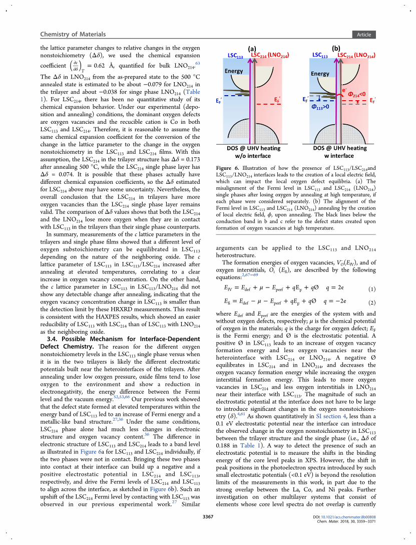
Defect Chemistry. The reason for the different oxygennonstoichiometry levels in the LSC113 single phase versus whenit is in the two trilayers is likely the different electrostaticpotentials built near the heterointerfaces of the trilayers. Afterannealing under low oxygen pressure, oxide films tend to loseoxygen to the environment and show a reduction inelectronegativity, the energy difference between the Fermilevel and the vacuum energy.52,53,66 Our previous work showedthat the defect state formed at elevated temperatures within theenergy band of LSC113 led to an increase of Fermi energy and ametallic-like band structure.27,36 Under the same conditions,LSC214 phase alone had much less changes in electronicstructure and oxygen vacancy content.36 The difference inelectronic structure of LSC113 and LSC214 leads to a band levelas illustrated in Figure 6a for LSC113 and LSC214 individually, ifthe two phases were not in contact. Bringing these two phasesinto contact at their interface can build up a negative and apositive electrostatic potential in LSC214 and LSC113,respectively, and drive the Fermi levels of LSC214 and LSC113to align across the interface, as sketched in Figure 6b). Such anupshift of the LSC214 Fermi level by contacting with LSC113 wasobserved in our previous experimental work.27 Similar
arguments can be applied to the LSC113 and LNO214heterostructure.The formation energies of oxygen vacancies, VO
·· (EfV), and ofoxygen interstitials, Oi
’’ (Efi), are described by the followingequations:2,67−69
μ= + − + + =E E E q q qE Ø 2efV def perf F (1)
μ= − − + + = −E E E qE q qØ 2efi def perf F (2)
where Edef and Eperf are the energies of the system with andwithout oxygen defects, respectively; μ is the chemical potentialof oxygen in the materials; q is the charge for oxygen defect; EFis the Fermi energy; and Ø is the electrostatic potential. Apositive Ø in LSC113 leads to an increase of oxygen vacancyformation energy and less oxygen vacancies near theheterointerface with LSC214 or LNO214. A negative Øequilibrates in LSC214 and in LNO214, and decreases theoxygen vacancy formation energy while increasing the oxygeninterstitial formation energy. This leads to more oxygenvacancies in LSC214 and less oxygen interstitials in LNO214near their interface with LSC113. The magnitude of such anelectrostatic potential at the interface does not have to be largeto introduce significant changes in the oxygen nonstoichiom-etry (δ).4,61 As shown quantitatively in SI section 4, less than a0.1 eV electrostatic potential near the interface can introducethe observed change in the oxygen nonstoichiometry in LSC113between the trilayer structure and the single phase (i.e., Δδ of0.188 in Table 1). A way to detect the presence of such anelectrostatic potential is to measure the shifts in the bindingenergy of the core level peaks in XPS. However, the shift inpeak positions in the photoelectron spectra introduced by suchsmall electrostatic potentials (<0.1 eV) is beyond the resolutionlimits of the measurements in this work, in part due to thestrong overlap between the La, Co, and Ni peaks. Furtherinvestigation on other multilayer systems that consist ofelements whose core level spectra do not overlap is currently
Figure 6. Illustration of how the presence of LSC113/LSC214andLSC113/LNO214 interfaces leads to the creation of a local electric field,which can impact the local oxygen defect equilibria. (a) Themisalignment of the Fermi level in LSC113 and LSC214 (LNO214)single phases after losing oxygen by annealing at high temperature, ifeach phase were considered separately. (b) The alignment of theFermi level in LSC113 and LSC214 (LNO214) annealing by the creationof local electric field, ϕ, upon annealing. The black lines below theconduction band in b and c refer to the defect states created uponformation of oxygen vacancies at high temperature.
Chemistry of Materials Article
DOI: 10.1021/acs.chemmater.8b00808Chem. Mater. 2018, 30, 3359−3371
3367

ongoing to confirm the existence of an electrostatic potentialnear the perovskite-related heterointerfaces. If the model aboveis correct, one should expect the LSC113 in LSC113/LSC214 andLSC113/LNO214 trilayers to lose less oxygen than the singlephase LSC113, and the LSC214 and LNO214 in trilayers to losemore oxygen than the single-phase films. This is what isobserved in Figure 5c−e. All of these results support theexplanation we proposed above, that the existence of anelectrostatic potential near the heterointerfaces can change thedefect chemistry in the thin layers of LSC113, LSC214, andLNO214. In the model we proposed above, the driving force forcharged defect distribution affects not only electron transferfrom LSC113 across the interface to LSC214 or LNO214 but alsoexchange and re-equilibration of charged oxygen defects.Consistent with the electron transfer direction, the interstitialoxygens (−2 formal charge) within LNO214 can transfer toLSC113, leading to less oxygen vacancies (+2 formal charge) inLSC113.It is also worth noting that the Sr/La ratio increased slightly
at the very top surface of the LSC113/LSC214 trilayer after 500°C annealing. Furthermore, the increase of Sr at the A-site ofLSC113 is known to increase the oxygen vacancy concentrationin LSC113.
39 The possible diffusion of Sr from LSC214 intoLSC113 might also explain the higher oxygen vacancies forLSC113 in LSC113/LSC214 than LSC113/LNO214. However, itcannot explain why LSC113 in the LSC113/LSC214 has lessoxygen vacancies than the LSC113 single phase. Therefore, webelieve the electrostatic potential near the interface as describedabove is more likely to be behind the change in oxygen defectchemistry instead of Sr interdiffusion.In the description of the oxygen defect formation energy
above, we assumed that Edef − Eperf in eqs 1 and 2 is the samefor the single phase and near the heterointerface. This quantitycan in fact also be affected by the symmetry breaking andalteration of coordination and bond energies near theinterface.70 While we do not rule out this interface chemistryrelated effect, calculating the difference in Edef − Eper for singlephase versus heterointerface is beyond the scope of this work.Furthermore, in this paper we did not consider the effect ofstrain which is known to impact the oxygen defect chemistry.51
This is because all our films are very thin (∼10 nm in total). Asa result, the in-plane lattice parameter of all the films aredetermined by the substrate, leading to very similar in-planelattice parameters for both the single phase films and thetrilayer films. Lastly, it is worth noting that besides oxygendefects, the defect chemistry for other ions can also be stronglyinfluenced by the presence of the interface, as shown by Xu etal. for the intercalation energy of Li in the LixFePO4/LixMPO4heterostructure.23
3.5. Perspectives and Implications of the Phenomen-on. The fabrication and operation of many oxide devicesinvolve elevated temperatures, reactive gas environments, andthe presence of an external bias. Such external drivers canchange the oxygen nonstoichiometry in oxides, leading todifferent electronic structures, conductivities, and magneticproperties. The ability to change the response of oxygen defectsto the environment presents a new way of tailoring thefunctionality of transition metal oxides. For example, tensilestrain is now known to be one way to lower the oxygen vacancyor interstitial formation energy, leading to the ability toaccommodate more oxygen vacancies or interstitials, andeventually faster oxygen exchange kinetics at elevated temper-atures.51,71−73
To tune the oxygen defects and functionality of oxideheterostructures, we proposed to couple the defect chemistryand electronic structure of two oxides through their interface.Depending on the neighboring oxide, one can either suppressor enhance the oxygen defect formation under an electrostaticdriving force arising internally from the interface. In this work,we showed that by placing LSC113 next to LNO214, oxygenvacancy formation in LSC113 was strongly suppressed. One keyparameter is the relative Fermi level positions of the two phasesupon the change of their oxygen nonstoichiometry due to theexternal driving force (here the annealing temperature), finallyforming an internal driving force (here the interface electro-static potential). Based on the rigid band theory of Lankhorstetal.,66 the Fermi level of LSC113 corresponds to the gradualfilling up of states in a broad electron band with electronsintroduced by oxygen vacancy formation. One needs similarmodels that correlate oxygen nonstoichiometry to the Fermilevel in different materials so that the neighboring oxides couldbe chosen for controlling the oxygen defects. Our results pointout “band engineering” as a potential way of tailoring defectchemistry and generating different functionalities in oxidesuperlattices. We are currently working on other hetero-structures that consist of different RP phase materials to furtherconfirm the impact of band engineering through hetero-interfaces on the defect chemistry of those materials.In this work, we used a combination of HAXPES and
HRXRD to probe the relative oxygen nonstoichiometry levelsin LSC113/LSC214 and LSC113/LNO214 trilayer structures. Thisapproach is nondestructive and does not require a complicatedsample preparation, and it can therefore be used for materialsthat are sensitive to electron beam damage. Several recentstudies have shown the significant impact of heterointerfaces onlocal electronic structure and transport properties. Chen et al.74
and Huijben et al.75 reported enhanced electronic mobility byinserting a single-unit cell of La1−xSrxMnO3 (x = 0, 1/8, and 1/3)74 and SrCuO3 between LaAlO3 and SrTiO3. The resultingenhanced mobility was tied to potential changes in oxygendefect chemistry induced by the inserted layer. Yajima et al.76
succeeded in tuning the band alignment in perovskite metal−semiconductor heterojunctions over a broad range of 1.7 eV byinserting a few unit cells of either LaTiO3 or SrAlOx betweenthe SrRuO3 and Nb:SrTiO3. However, the impact of theinterface on the oxygen defects accompanied by such a largechange in electrostatic field was not discussed. The methodsused in the work can be easily adapted to other systems,including those mentioned above, to clarify the role of localoxygen defects in determining the novel properties that arisenear oxide heterointerfaces, such as magneto-electric cou-pling,10 interfacial superconductivity,11,12 high electronic andionic conductivity,13,14,74−76 and catalytic activity.15−23 Fur-thermore, this approach can be extended to in situ studies atelevated temperatures, in reactive gas environments, and underelectrochemical potentials to probe more quantitatively howthe local defects evolve when the chemical potential of the gasphase is changed. This can be particularly useful for theapplications to high temperature electrochemical energyconversion or to redox-reaction based resistive memories.
4. CONCLUSIONInterfaces of perovskite and Ruddlesden−Popper type oxidespresent an interesting case for affecting the defect chemistry ofeach phase through their interfaces. We used a novelcombination of HAXPES and HRXRD to probe the local
Chemistry of Materials Article
DOI: 10.1021/acs.chemmater.8b00808Chem. Mater. 2018, 30, 3359−3371
3368

oxygen defect distribution across the buried interfaces ofLSC113/LSC214 and LSC113/LNO214 trilayer model systems.From HAXPES measurements, we found that the Co in theLSC113 adjacent to LSC214 is more reducible than the Co in theLSC113 adjacent to LNO214. This is because of a largerconcentration of oxygen vacancies that form in LSC113 of theLSC113/LSC214 than that in LSC113 of the LSC113/LNO214, afterannealing in a reducing atmosphere. The HRXRD resultsindicated consistent behavior as found from HAXPES.Reduction-induced chemical expansion of the LSC113 singlephase was larger than that of LSC113 adjacent to LSC214 or toLNO214. The comparison of lattice parameters upon reductionindicated that the Δδ in LSC113 adjacent to LNO214 was lessthan that in LSC113 adjacent to LSC214 and in the LSC113 singlephase. On the other hand, LSC214 and LNO214 were found tolose more oxygen when in contact with the LSC113 layercompared to their single phase counterparts. Our resultsdemonstrated that the oxygen defect chemistry of thesetransition metal oxides was strongly impacted by the presenceof interfaces and the properties of the adjacent phases. Weattributed this behavior to the electrostatic potentials built nearthe heterointerfaces. In this work, combining HAXPES withHRHRD, we provide a nondestructive way to qualitativelyprobe the local oxygen defect states in transition metal oxideheterostructures. Our results also point out interface engineer-ing as a potential way to control the local defect chemistry,electronic structure, and functionality for oxide supper-lattices.
■ ASSOCIATED CONTENT*S Supporting InformationThe Supporting Information is available free of charge on theACS Publications website at DOI: 10.1021/acs.chemma-ter.8b00808.
S1 Probing depth of hard X-ray photoelectron spectros-copy (HAXPES); S2 Identifying Co valence state usingphotoelectron spectroscopy (PES); S3 Quantifyingoxygen nonstoichiometrychange in LSC113/LSC214 andLSC113/LNO213 trilayers based on chemical expansion;S4 Estimating oxygen nonstoichiometry change intro-duced by the interface electrostatic potential (PDF)
■ AUTHOR INFORMATIONCorresponding Author*E-mail: [email protected] Chen: 0000-0001-6193-7508Present Address$Guangzhou Key Laboratory for Surface Chemistry of EnergyMaterials, New Energy Institute, School of Environment andEnergy, South China University of Technology, 382 East Road,University City, Guangzhou 510006, P. R. China.NotesThe authors declare no competing financial interest.
■ ACKNOWLEDGMENTSAuthors acknowledge the US-DOE - Basic Energy Sciences,Grant No. DE-SC0002633 for financial support. Y.C. acknowl-edges additional support from the Schlumberger FoundationFaculty for the Future fellowship. D.D.F. was supported by theU.S. Department of Energy (DOE), Office of Science, Office ofBasic Energy Sciences (BES), Division of Materials Science and
Engineering. The authors acknowledge useful discussions withDr. Christian Lenser and Dr. Mostafa Youssef. This work madeuse of the MRSEC Shared Experimental Facilities at MIT,supported by the National Science Foundation under AwardNumber DMR - 1419807.
■ REFERENCES(1) Ganduglia-Pirovano, M. V.; Hofmann, A.; Sauer, J. Oxygenvacancies in transition metal and rare earth oxides: Current state ofunderstanding and remaining challenges. Surf. Sci. Rep. 2007, 62, 219−270.(2) Tuller, H. L.; Bishop, S. R. Tailoring Material Properties throughDefect Engineering. Chem. Lett. 2010, 39, 1226−1231.(3) Freysoldt, C.; Grabowski, B.; Hickel, T.; Neugebauer, J.; Kresse,G.; Janotti, A.; Van de Walle, C. G. First-principles calculations forpoint defects in solids. Rev. Mod. Phys. 2014, 86, 253−305.(4) Chen, D.; Tuller, H. L. Voltage-Controlled Nonstoichiometry inOxide Thin Films: Pr0.1Ce0.9O2−δ Case Study. Adv. Funct. Mater. 2014,24, 7638−7644.(5) Kalinin, S. V.; Spaldin, N. A. Functional Ion Defects in TransitionMetal Oxides. Science 2013, 341, 858−859.(6) Kalinin, S. V.; Borisevich, A.; Fong, D. Beyond CondensedMatter Physics on the Nanoscale: The Role of Ionic and Electro-chemical Phenomena in the Physical Functionalities of OxideMaterials. ACS Nano 2012, 6, 10423−10437.(7) Kosacki, I.; Suzuki, T.; Anderson, H. U.; Colomban, P. Ramanscattering and lattice defects in nanocrystalline CeO2 thin films. SolidState Ionics 2002, 149, 99−105.(8) Rupp, J. L. M.; Fabbri, E.; Marrocchelli, D.; Han, J. W.; Chen, D.;Traversa, E.; Tuller, H. L.; Yildiz, B. Scalable Oxygen- Ion TransportKinetics in Metal- Oxide Films: Impact of Thermally Induced LatticeCompaction in Acceptor Doped Ceria Films. Adv. Funct. Mater. 2014,24, 1562−1574.(9) Schweiger, S.; Kubicek, M.; Messerschmitt, F.; Murer, C.; Rupp,J. L. M. A Microdot Multilayer Oxide Device: Let Us Tune the Strain-Ionic Transport Interaction. ACS Nano 2014, 8, 5032−5048.(10) Bauer, U.; Emori, S.; Beach, G. S. D. Voltage-controlled domainwall traps in ferromagnetic nanowires. Nat. Nanotechnol. 2013, 8, 411−416.(11) Ohtomo, A.; Muller, D. A.; Grazul, J. L.; Hwang, H. Y. Artificialcharge-modulation in atomic-scale perovskite titanate superlattices.Nature 2002, 419, 378−380.(12) Hwang, H. Y.; Iwasa, Y.; Kawasaki, M.; Keimer, B.; Nagaosa, N.;Tokura, Y. Emergent phenomena at oxide interfaces. Nat. Mater. 2012,11, 103−113.(13) Maier, J. Nanoionics: ionic charge carriers in small systems.Phys. Chem. Chem. Phys. 2009, 11, 3011−22.(14) Fabbri, E.; Pergolesi, D.; Traversa, E. Ionic conductivity in oxideheterostructures: the role of interfaces. Sci. Technol. Adv. Mater. 2010,11, 054503.(15) Li, G. H.; Gray, K. A. The solid-solid interface: Explaining thehigh and unique photocatalytic reactivity of TiO2-based nano-composite materials. Chem. Phys. 2007, 339, 173−187.(16) Sase, M.; Hermes, F.; Yashiro, K.; Sato, K.; Mizusaki, J.; Kawada,T.; Sakai, N.; Yokokawa, H. Enhancement of oxygen surface exchangeat the hetero-interface of (La,Sr)CoO3/(La,Sr)2CoO4 with PLD-Layered films. J. Electrochem. Soc. 2008, 155, B793−B797.(17) Sase, M.; Yashiro, K.; Sato, K.; Mizusaki, J.; Kawada, T.; Sakai,N.; Yamaji, K.; Horita, T.; Yokokawa, H. Enhancement of oxygenexchange at the hetero interface of (La,Sr)CoO3/(La,Sr)2CoO4 incomposite ceramics. Solid State Ionics 2008, 178, 1843−1852.(18) Hayd, J.; Yokokawa, H.; Ivers-Tiffee, E. Hetero-Interfaces atNanoscaled (La,Sr)CoO3‑δ Thin-Film Cathodes Enhancing OxygenSurface-Exchange Properties. J. Electrochem. Soc. 2013, 160, F351−F359.(19) Yashiro, K.; Nakamura, T.; Sase, M.; Hermes, F.; Sato, K.;Kawada, T.; Mizusaki, J. Composite Cathode of Perovskite-Related
Chemistry of Materials Article
DOI: 10.1021/acs.chemmater.8b00808Chem. Mater. 2018, 30, 3359−3371
3369
Oxides, (La,Sr)CoO3‑δ/(La,Sr)2CoO4‑δ, for Solid Oxide Fuel Cells.Electrochem. Solid-State Lett. 2009, 12, B135−B137.(20) Crumlin, E. J.; Mutoro, E.; Ahn, S. J.; la O, G. J.; Leonard, D. N.;Borisevich, A.; Biegalski, M. D.; Christen, H. M.; Shao-Horn, Y.Oxygen Reduction Kinetics Enhancement on a HeterostructuredOxide Surface for Solid Oxide Fuel Cells. J. Phys. Chem. Lett. 2010, 1,3149−3155.(21) Ivanov, D. V.; Pinaeva, L. G.; Isupova, L. A.; Sadovskaya, E. M.;Prosvirin, I. P.; Gerasimov, E. Y.; Yakovleva, I. S. Effect of surfacedecoration with LaSrFeO4 on oxygen mobility and catalytic activity ofLa0.4Sr0.6FeO3‑δ in high-temperature N2O decomposition, methanecombustion and ammonia oxidation. Appl. Catal., A 2013, 457, 42−51.(22) Stacchiola, D. J.; Senanayake, S. D.; Liu, P.; Rodriguez, J. A.Fundamental Studies of Well-Defined Surfaces of Mixed-MetalOxides: Special Properties of MOx/TiO2(110) {M = V, Ru, Ce, orW}. Chem. Rev. 2013, 113, 4373−4390.(23) Xu, S.; Jacobs, R.; Wolverton, C.; Kuech, T.; Morgan, D.Nanoscale Voltage Enhancement at Cathode Interfaces in Li-IonBatteries. Chem. Mater. 2017, 29, 1218−1229.(24) Herpers, A.; Lenser, C.; Park, C.; Offi, F.; Borgatti, F.;Panaccione, G.; Menzel, S.; Waser, R.; Dittmann, R. SpectroscopicProof of the Correlation between Redox-State and Charge-CarrierTransport at the Interface of Resistively Switching Ti/PCMO Devices.Adv. Mater. 2014, 26, 2730−2735.(25) Kim, Y. M.; He, J.; Biegalski, M. D.; Ambaye, H.; Lauter, V.;Christen, H. M.; Pantelides, S. T.; Pennycook, S. J.; Kalinin, S. V.;Borisevich, A. Y. Probing oxygen vacancy concentration andhomogeneity in solid-oxide fuel-cell cathode materials on thesubunit-cell level. Nat. Mater. 2012, 11, 888−894.(26) Kim, Y. M.; Morozovska, A.; Eliseev, E.; Oxley, M. P.; Mishra,R.; Selbach, S. M.; Grande, T.; Pantelides, S. T.; Kalinin, S. V.;Borisevich, A. Y. Direct observation of ferroelectric field effect andvacancy-controlled screening at the BiFeO3/LaxSr1‑xMnO3 interface.Nat. Mater. 2014, 13, 1019−1025.(27) Tsvetkov, N.; Chen, Y.; Yildiz, B. Reducibility of Co at theLa0.8Sr0.2CoO3/(La0.5Sr0.5)2CoO4 hetero-interface at elevated temper-atures. J. Mater. Chem. A 2014, 2, 14690−14695.(28) Chambers, S. A., Probing Perovskite Interfaces and Superlatticeswith X-ray Photoemission Spectroscopy. In Hard X-ray PhotoelectronSpectroscopy (HAXPES); Woicik, C. J., Ed.; Springer InternationalPublishing: Cham, 2016; pp 341−380.(29) Slooten, E.; Zhong, Z.; Molegraaf, H. J. A.; Eerkes, P. D.; deJong, S.; Massee, F.; van Heumen, E.; Kruize, M. K.; Wenderich, S.;Kleibeuker, J. E.; Gorgoi, M.; Hilgenkamp, H.; Brinkman, A.; Huijben,M.; Rijnders, G.; Blank, D. H. A.; Koster, G.; Kelly, P. J.; Golden, M. S.Hard x-ray photoemission and density functional theory study of theinternal electric field in SrTiO3/LaAlO3 oxide heterostructures. Phys.Rev. B: Condens. Matter Mater. Phys. 2013, 87, 085128.(30) Chen, Y. Z.; Bovet, N.; Trier, F.; Christensen, D. V.; Qu, F. M.;Andersen, N. H.; Kasama, T.; Zhang, W.; Giraud, R.; Dufouleur, J.;Jespersen, T. S.; Sun, J. R.; Smith, A.; Nygard, J.; Lu, L.; Buchner, B.;Shen, B. G.; Linderoth, S.; Pryds, N. A high-mobility two-dimensionalelectron gas at the spinel/perovskite interface of γ-Al2O3/SrTiO3. Nat.Commun. 2013, 4, 1371.(31) Chroneos, A.; Yildiz, B.; Tarancon, A.; Parfitt, D.; Kilner, J. A.Oxygen diffusion in solid oxide fuel cell cathode and electrolytematerials: mechanistic insights from atomistic simulations. EnergyEnviron. Sci. 2011, 4, 2774−2789.(32) Tarancon, A.; Burriel, M.; Santiso, J.; Skinner, S. J.; Kilner, J. A.Advances in layered oxide cathodes for intermediate temperature solidoxide fuel cells. J. Mater. Chem. 2010, 20, 3799−3813.(33) Han, J. W.; Yildiz, B. Mechanism for enhanced oxygen reductionkinetics at the (La,Sr)CoO3‑δ/(La,Sr)2CoO4+δ a hetero-interface.Energy Environ. Sci. 2012, 5, 8598−8607.(34) Feng, Z.; Yacoby, Y.; Gadre, M. J.; Lee, Y.-L.; Hong, W. T.;Zhou, H.; Biegalski, M. D.; Christen, H. M.; Adler, S. B.; Morgan, D.;Shao-Horn, Y. Anomalous Interface and Surface Strontium Segrega-tion in (La1−ySry)2CoO4±δ/La1−xSrxCoO3−δ Heterostructured ThinFilms. J. Phys. Chem. Lett. 2014, 5, 1027−1034.
(35) Gadre, M. J.; Lee, Y. L.; Morgan, D. Cation interdiffusion modelfor enhanced oxygen kinetics at oxide heterostructure interfaces. Phys.Chem. Chem. Phys. 2012, 14, 2606−2616.(36) Chen, Y.; Cai, Z. H.; Kuru, Y.; Ma, W.; Tuller, H. L.; Yildiz, B.Electronic Activation of Cathode Superlattices at Elevated Temper-atures - Source of Markedly Accelerated Oxygen Reduction Kinetics.Adv. Energy Mater. 2013, 3, 1221−1229.(37) Rueff, J. P.; Ablett, J. M.; Ceolin, D.; Prieur, D.; Moreno, T.;Baledent, V.; Lassalle-Kaiser, B.; Rault, J. E.; Simon, M.; Shukla, A. TheGALAXIES beamline at the SOLEIL synchrotron: inelastic X-rayscattering and photoelectron spectroscopy in the hard X-ray range. J.Synchrotron Radiat. 2015, 22, 175−179.(38) Momma, K.; Izumi, F. VESTA 3 for three-dimensionalvisualization of crystal, volumetric and morphology data. J. Appl.Crystallogr. 2011, 44, 1272−1276.(39) Mizusaki, J.; Mima, Y.; Yamauchi, S.; Fueki, K.; Tagawa, H.Nonstoichiometry of the perovskite-type oxides La1−xSrxCoO3−δ. J.Solid State Chem. 1989, 80, 102−111.(40) Madhukar, S.; Aggarwal, S.; Dhote, A. M.; Ramesh, R.;Krishnan, A.; Keeble, D.; Poindexter, E. Effect of oxygen stoichiometryon the electrical properties of La0.5Sr0.5CoO3 electrodes. J. Appl. Phys.1997, 81, 3543−3547.(41) Vashook, V. V.; Ullmann, H.; Olshevskaya, O. P.; Kulik, V. P.;Lukashevich, V. E.; Kokhanovskij, L. V. Composition and electricalconductivity of some cobaltates of the type La2−xSrxCoO4.5−x/2±δ. SolidState Ionics 2000, 138, 99−104.(42) Galakhov, V. R.; Karelina, V. V.; Kellerman, D. G.; Gorshkov, V.S.; Ovechkina, N. A.; Neumann, M. Electronic structure, X-ray spectra,and magnetic properties of the LiCoO2-delta and NaxCoO2nonstoichiometric oxides. Phys. Solid State 2002, 44, 266−273.(43) Chuang, T. J.; Brundle, C. R.; Rice, D. W. Interpretation of thex-ray photoemission spectra of cobalt oxides and cobalt oxide surfaces.Surf. Sci. 1976, 59, 413−429.(44) Petitto, S. C.; Marsh, E. M.; Carson, G. A.; Langell, M. A. Cobaltoxide surface chemistry: The interaction of CoO(100), Co3O4(110)and Co3O4(111) with oxygen and water. J. Mol. Catal. A: Chem. 2008,281, 49−58.(45) Gu, Z. J.; Xiang, X.; Fan, G. L.; Li, F. Facile Synthesis andCharacterization of Cobalt Ferrite Nanocrystals via a SimpleReduction-Oxidation Route. J. Phys. Chem. C 2008, 112, 18459−18466.(46) Brabers, V. A. M.; van Setten, F. X-ray photoelectronspectroscopy study of the ionic configuration of the spinelCuMnCoO4. J. Phys. D: Appl. Phys. 1983, 16, L169−L172.(47) Oku, M. X-ray photoelectron spectrum of low-spin Co(III) inLiCoO2. J. Solid State Chem. 1978, 23, 177−185.(48) Moses, A. W.; Flores, H. G. G.; Kim, J. G.; Langell, M. A.Surface properties of LiCoO2, LiNiO2 and LiNi1‑xCoxO2. Appl. Surf.Sci. 2007, 253, 4782−4791.(49) Galenda, A.; Natile, M. M.; Krishnan, V.; Bertagnolli, H.;Glisenti, A. LaSrCoFeO and Fe2O3/LaSrCoFeO powders: Synthesisand characterization. Chem. Mater. 2007, 19, 2796−2808.(50) Biesinger, M. C.; Payne, B. P.; Grosvenor, A. P.; Lau, L. W. M.;Gerson, A. R.; Smart, R. S. Resolving surface chemical states in XPSanalysis of first row transition metals, oxides and hydroxides: Cr, Mn,Fe, Co and Ni. Appl. Surf. Sci. 2011, 257, 2717−2730.(51) Cai, Z. H.; Kuru, Y.; Han, J. W.; Chen, Y.; Yildiz, B. SurfaceElectronic Structure Transitions at High Temperature on PerovskiteOxides: The Case of Strained La0.8Sr0.2CoO3 Thin Films. J. Am. Chem.Soc. 2011, 133, 17696−17704.(52) Deskins, N. A.; Rousseau, R.; Dupuis, M. Defining the Role ofExcess Electrons in the Surface Chemistry of TiO2. J. Phys. Chem. C2010, 114, 5891−5897.(53) Greiner, M. T.; Helander, M. G.; Tang, W. M.; Wang, Z. B.;Qiu, J.; Lu, Z. H. Universal energy-level alignment of molecules onmetal oxides. Nat. Mater. 2012, 11, 76−81.(54) Mizusaki, J.; Tabuchi, J.; Matsuura, T.; Yamauchi, S.; Fueki, K.Electrical Conductivity and Seebeck Coefficient of NonstoichiometricLa1‑xSrxCoO3‑δ. J. Electrochem. Soc. 1989, 136, 2082−2088.
Chemistry of Materials Article
DOI: 10.1021/acs.chemmater.8b00808Chem. Mater. 2018, 30, 3359−3371
3370

(55) Lee, W.; Han, J. W.; Chen, Y.; Cai, Z.; Yildiz, B. Cation SizeMismatch and Charge Interactions Drive Dopant Segregation at theSurfaces of Manganite Perovskites. J. Am. Chem. Soc. 2013, 135, 7909−7925.(56) Dulli, H.; Dowben, P. A.; Liou, S. H.; Plummer, E. W. Surfacesegregation and restructuring of colossal-magnetoresistant manganeseperovskites La0.65Sr0.35MnO3. Phys. Rev. B: Condens. Matter Mater. Phys.2000, 62, R14629−R14632.(57) Chen, Y.; Tellez, H.; Burriel, M.; Yang, F.; Tsvetkov, N.; Cai, Z.;McComb, D. W.; Kilner, J. A.; Yildiz, B. Segregated Chemistry andStructure on (001) and (100) Surfaces of (La1−xSrx)2CoO4 Overridethe Crystal Anisotropy in Oxygen Exchange Kinetics. Chem. Mater.2015, 27, 5436−5450.(58) Swallow, J. G.; Woodford, W. H.; Chen, Y.; Lu, Q.; Kim, J. J.;Chen, D.; Chiang, Y. M.; Carter, W. C.; Yildiz, B.; Tuller, H. L.; VanVliet, K. J. Chemomechanics of ionically conductive ceramics forelectrical energy conversion and storage. J. Electroceram. 2014, 32, 3−27.(59) Marrocchelli, D.; Bishop, S. R.; Tuller, H. L.; Watson, G. W.;Yildiz, B. Charge localization increases chemical expansion in cerium-based oxides. Phys. Chem. Chem. Phys. 2012, 14, 12070−12074.(60) Chen, X. Y.; Yu, J. S.; Adler, S. B. Thermal and chemicalexpansion of Sr-doped lanthanum cobalt oxide (La1‑xSrxCoO3‑δ.).Chem. Mater. 2005, 17, 4537−4546.(61) Biegalski, M. D.; Crumlin, E.; Belianinov, A.; Mutoro, E.; Shao-Horn, Y.; Kalinin, S. V. In situ examination of oxygen non-stoichiometry in La0.80Sr0.20CoO3−δ thin films at intermediate andlow temperatures by x-ray diffraction. Appl. Phys. Lett. 2014, 104,161910.(62) Nakamura, T.; Yashiro, K.; Sato, K.; Mizusaki, J. Defect chemicaland statistical thermodynamic studies on oxygen nonstoichiometricNd2‑xSrxNiO4+δ. Solid State Ionics 2009, 180, 1406−1413.(63) Nakamura, T.; Yashiro, K.; Sato, K.; Mizusaki, J. Structuralanalysis of La2‑xSrxNiO4+δ by high temperature X-ray diffraction. SolidState Ionics 2010, 181, 292−299.(64) Kharton, V. V.; Kovalevsky, A. V.; Avdeev, M.; Tsipis, E. V.;Patrakeev, M. V.; Yaremchenko, A. A.; Naumovich, E. N.; Frade, J. R.Chemically induced expansion of La(2)NiO(4+delta)-based materials.Chem. Mater. 2007, 19, 2027−2033.(65) Mogni, L. V.; Prado, F. D.; Cuello, G. J.; Caneiro, A. Study ofthe Crystal Chemistry of the n = 2 Ruddlesden-Popper PhasesSr3FeMO6+δ (M = Fe, Co, and Ni) Using in Situ High TemperatureNeutron Powder Diffraction. Chem. Mater. 2009, 21, 2614−2623.(66) Lankhorst, M. H. R.; Bouwmeester, H. J. M.; Verweij, H. Use ofthe rigid band formalism to interpret the relationship between Ochemical potential and electron concentration in La1‑xSrxCoO3-δ. Phys.Rev. Lett. 1996, 77, 2989−2992.(67) Bristowe, N. C.; Littlewood, P. B.; Artacho, E. Surface defectsand conduction in polar oxide heterostructures. Phys. Rev. B: Condens.Matter Mater. Phys. 2011, 83, 205405.(68) Ingram, B. J.; Eastman, J. A.; Chang, K. C.; Kim, S. K.; Fister, T.T.; Perret, E.; You, H.; Baldo, P. M.; Fuoss, P. H. In situ x-ray studieso f oxygen su r f a c e ex change behav io r in th in f i lmLa0.6Sr0.4Co0.2Fe0.8O3-δ. Appl. Phys. Lett. 2012, 101, 051603.(69) Yu, L. P.; Zunger, A. A polarity-induced defect mechanism forconductivity and magnetism at polar-nonpolar oxide interfaces. Nat.Commun. 2014, 5, 5118.(70) Yang, J.; Youssef, M.; Yildiz, B. Predicting point defect equilibriaacross oxide hetero-interfaces: model system of ZrO2/Cr2O3. Phys.Chem. Chem. Phys. 2017, 19, 3869−3883.(71) Kubicek, M.; Cai, Z. H.; Ma, W.; Yildiz, B.; Hutter, H.; Fleig, J.Tensile Lattice Strain Accelerates Oxygen Surface Exchange andDiffusion in La1‑xSrxCoO3-δ Thin Films. ACS Nano 2013, 7, 3276−3286.(72) Tsvetkov, N.; Lu, Q. Y.; Chen, Y.; Yildiz, B. Accelerated OxygenExchange Kinetics on Nd2NiO4+delta Thin Films with Tensile Strainalong c-Axis. ACS Nano 2015, 9, 1613−1621.
(73) Kushima, A.; Yip, S.; Yildiz, B. Competing strain effects inreactivity of LaCoO3 with oxygen. Phys. Rev. B: Condens. Matter Mater.Phys. 2010, 82, 115435.(74) Chen, Y. Z.; Trier, F.; Wijnands, T.; Green, R. J.; Gauquelin, N.;Egoavil, R.; Christensen, D. V.; Koster, G.; Huijben, M.; Bovet, N.;Macke, S.; He, F.; Sutarto, R.; Andersen, N. H.; Sulpizio, J. A.; Honig,M.; Prawiroatmodjo, G.; Jespersen, T. S.; Linderoth, S.; Ilani, S.;Verbeeck, J.; Van Tendeloo, G.; Rijnders, G.; Sawatzky, G. A.; Pryds,N. Extreme mobility enhancement of two-dimensional electron gasesat oxide interfaces by charge-transfer-induced modulation doping. Nat.Mater. 2015, 14, 801−806.(75) Huijben, M.; Koster, G.; Kruize, M. K.; Wenderich, S.;Verbeeck, J.; Bals, S.; Slooten, E.; Shi, B.; Molegraaf, H. J. A.;Kleibeuker, J. E.; van Aert, S.; Goedkoop, J. B.; Brinkman, A.; Blank, D.H. A.; Golden, M. S.; van Tendeloo, G.; Hilgenkamp, H.; Rijnders, G.Defect Engineering in Oxide Heterostructures by Enhanced OxygenSurface Exchange. Adv. Funct. Mater. 2013, 23, 5240−5248.(76) Yajima, T.; Hikita, Y.; Minohara, M.; Bell, C.; Mundy, J. A.;Kourkoutis, L. F.; Muller, D. A.; Kumigashira, H.; Oshima, M.; Hwang,H. Y. Controlling band alignments by artificial interface dipoles atperovskite heterointerfaces. Nat. Commun. 2015, 6, 6759.
Chemistry of Materials Article
DOI: 10.1021/acs.chemmater.8b00808Chem. Mater. 2018, 30, 3359−3371
3371