Embed Size (px)
Citation preview
18 AJAM 2013 Official Journal of the American Academy of Aesthetic Medicine 2013 AJAM 19Official Journal of the American Academy of Aesthetic Medicine
Eusebio Aguilar Saba, MD
Prominent ear (loP ear) is a common birth defect, occurring in 5% of the population. this malformation is inherited in an autosomal dominant pattern with variable penetrance. in most patients, two deformities coexist, determining the greatest separation of the ears – inadequate development of the antihelix fold and over-development of the concha.
more than 200 surgical techniques have been described for its correction, indicating the lack of an ideal technique.there are several techniques to help create an antihelix fold. Some of them involve conducting various types of sections in the ear cartilage, some by an anterior approach, such as that by Stenström or by a posterior approach, such as those by Converse, while others are more conservative with the cartilage, such as the technique by mustardé.
We briefly discuss the historical development of other surgical techniques for prominent ears correction and describe in detail this new procedure. Which includes closed anterior scoring of the cartilage, 2mm anterior incisions, simple percutaneous suturing of the cartilage for a natural antihelix fold, and following the patients in periods varying between 12 and 36 months.
Objectivesthe objective of the study is to introduce a modified anterior minimal incision otoplasty technique, evaluate clinical outcomes and patient satisfaction within a period of one to three years.
Settingsall the operations were performed in a private cosmetic
medical center and at the Pediatrics emergency Hospital in lima, Peru.
Methodsthis was a retrospective clinical case study of patients undergoing cosmetic otoplasty with a modified anterior scoring minimal incision technique during 2006 to 2009. the study population comprised 46 subjects desiring operative correction of prominent ears over the past three years. the subjects (26 male and 20 female) ranged in age from five to 48 years. a total of 92 otoplasties were performed with 39 bilateral and seven unilateral prominent ears. local anesthesia was used in 90% of the cases. Clinical follow-up ranged from one day to three years-two months.
the anterior scoring minimal incision technique is based on a work by Panfilov. anterior antihelix cartilage is weakened by a gentle scoring of the anterior folding. therefore, a new formed antihelix fold can be achieved without tension. the newly formed antihelix is fixed in the defined position with anterior transcutaneous 4/0 nylon sutures.
the anterior approach begins with infiltration of lidocaine 2% and epinephrine 1:100,000 on only the anterior portion of the auricular pavilion.
Modified Incisionless Otoplasty
an incision of 3mm is made in the lower third portion of the auricle, following the helix rim, where the peeler and rasp are introduced. Using a peeler to undermine the skin and perichondrium, and separate the anterior antihelix cartilage. the area is then scraped with a rasp to partially remove the anterior antihelix cartilage. a 2mm incision entry in the frontal antihelix (fossa triangularis) , then a 4/0 nylon is passed through both antihelix fold cartilage into the scaphoid fossa, then pass the 4/0 nylon needle subcutaneous to the helix rim (scaphoid fossa), enter at the same exit point to pass the needle transcutaneous through both antihelix fold to the fossa triangularis, so both ends of the suture will appear to finish the knot and create the new antihelix fold. make the same mattress sutures in the middle and lower third of the anterior portion of the antihelix. this is completed with Steri-Strip to close the 2mm skin incisions. Compression is done with cotton soaked in soap solution on the concha and antihelix-helix fold, covered with loose gauze and bandage for 24 hours. then we recommend sleeping with a specially ear-designed elastic band for three weeks. this anterior minimal technique has an average procedure time of 25 to 30 minutes.
Resultseighty two percent of 46 patients (26 male and 20 female, ranged in age from five to 48 years), were examined after a long-term period in a follow-up visit. the outcome measures were subjective analysis done by the authors and patients, after guided photographic comparisons of before and after photos and physical examination. the aesthetic result of otoplasty was rated as "very good" by 82% of patients and as "good" by 16% of patients; 1.5% of patients thought the result was "satisfactory" and 0.3% "unsatisfactory." Complete recurrence of the protrusion was seen in one patient, who went to a revision secondary anterior otoplasty technique, with satisfactory ratings.
Eusebio Aguilar Saba, MD, is a Cosmetic Surgeon and Aesthetic Medicine practitioner at the Cosmetic Surgery & Antiaging Center in Lima, Peru. He is a member of the International Confederation for Plastic, Reconstructive & Aesthetic Surgery.
References
Zambudio G, Ruiz JI, Guirao MJ, Sánchez JM, Girón O, Gutiérrez MA. Otoplastia por vía anterior para el tratamiento de las orejas prominentes. Una técnica mínimamente invasiva. Cir Pediatr. 2007;20:119-21.
Aguilar R, Soto C, Barrena S, Díaz M, López JC, Ros Z, et al. Estudio de la evolución de 238 otoplastias mediante encuesta de satisfacción. Cir Pediatr. 2008;21:104-6.
Mustarde JC. The correction of prominent eras using simple mattress sutures. Br J Plast Surg. 1963;16:170-6.
Furnas DW. Correction of prominent ears by concha-mastoid sutures. Plast Reconstr Surg. 1968;42:189-93.
Stenstrom SJ. A natural technique for correction of congeni- tally prominent ears. Plast Reconstr Surg. 1963;32:509-18. 22. Stenstrom SJ, Heftner J. The Stenstrom otoplasty. Clin Plast. Surg. 1978;5:465-70.
Dimitri E. Panfilov, Aesthetic Surgery of the facial Mosaic. 2007; 67:559-69
Mustardé, JC: The correction of Prominent Ears Using Simple Mattress Sutures: A Ten Year Survey. Plast Reconstr Surg. 39:382-386, 1967.
Furnas, DW: Correction of Prominent Ears by Conchal Mastoid Sutures. Plast Reconstr Surg. 24:189-193, 1968.
Stenstrom, SJ: A “natural” technique for correction of congenitally-prominent ears. Plast Reconst. Surg 32:509-518, 1963.
Fritsch, MH: Incisionless Otoplasty. Laryngoscope. 105: Suppl 70:1-11, 1995. (Early technique before cartilage scoring added)
Conclusionthe introduction of this modified anterior minimal incision and scoring procedure with a simple transcutaneous mattress suture for the correction of prominent ears provides permanent results. this technique also minimizes operation time to a 25- to 30-minute procedure with local anesthesia and minimal complications. this is a simple procedure to learn, with natural and permanent results. Post-operative complications were protrusion of sutures in two ears or recurrence in one ear (repeated surgery). no anterior skin necrosis, no visible irregularity of the anterior surface of the cartilage, no keloid, no infection, no hypertrophic scarring and no hematoma occurred – these are common complications on conventional posterior surgery techniques.
We recommend this technique for the routine correction of prominent ears with a poorly formed antihelical fold or deep concha bowl that will provide permanent and natural results. numerous techniques have been described for the resolution of prominent ears and all have advantages and disadvantages. in our experience, minimizing trauma to the cartilage structure leads to good and natural cosmetic results and fewer complications.
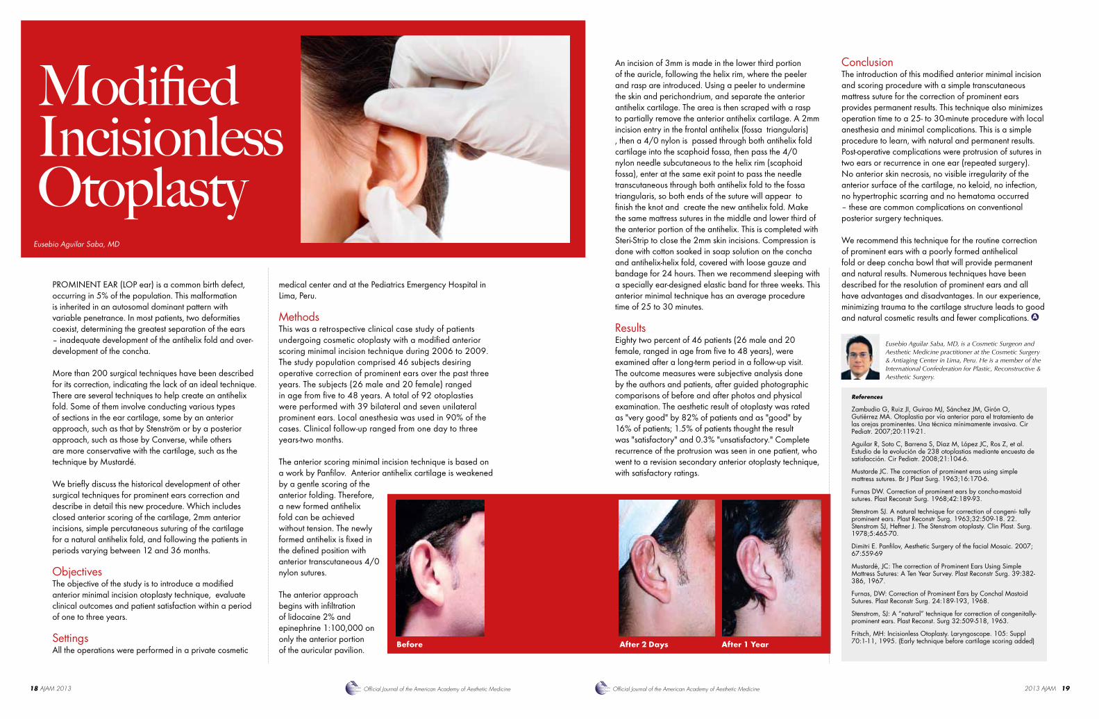
Before After 2 Days After 1 Year