Embed Size (px)
Citation preview
This journal is c The Royal Society of Chemistry 2013 Chem. Commun., 2013, 49, 8803--8805 8803
Cite this: Chem. Commun.,2013,49, 8803
Modification with hemeproteins increases the diffusivemovement of nanorods in dilute hydrogen peroxidesolutions†
Ada-Ioana Bunea, Ileana-Alexandra Pavel, Sorin David and Szilveszter Gaspar*
Nanorods were decorated with different hemeproteins that are able to
convert hydrogen peroxide. When dispersed into hydrogen peroxide
solutions, most of these nanorods are characterized by diffusion
coefficients which increase with the concentration of hydrogen
peroxide. Such a behaviour does not characterize unmodified nanorods.
Modification with biomolecules is very often used to extend theutility of nanomaterials.1 Unlike modification with antibodiesor DNA, modification with enzymes is also an interesting way toimpact the motion of nano- and microparticles. Catalase (Cat) ismost often used to enzymatically impact the motion of particles.Because this enzyme decomposes hydrogen peroxide with for-mation of oxygen, the motion of Cat-modified particles isenhanced through the so-called bubble recoil mechanism whenhydrogen peroxide is available.2 Impressive particle speeds wereobtained by modification with Cat and by using high hydrogenperoxide concentrations (>1.5%). However, it remains unclear ifmotion is also affected at low hydrogen peroxide concentrations,when the formation of oxygen bubbles does not occur. Takinginto account that most of the enzymes are not able to produceany bubbles under any experimental conditions, finding out ifmodification with enzymes can provide particles with kineticenergy through mechanisms other than bubble recoil is quiteimportant. A positive answer to this question is provided by asystem consisting of two enzymes electrically ‘‘wired’’ to a pieceof carbon fiber, which moves in the presence of glucose.3
However, this structure is quite large (0.5–1 cm in length),manually assembled, and moves for only three minutes exclu-sively at the air/liquid interface. Such behaviour makes theenzyme-modified carbon fiber suitable for a limited range ofapplications. A positive answer is also suggested (but notexperimentally demonstrated) by recent reports describing thatthe diffusive movement of urease and Cat molecules increasesin the presence of their substrates.4
In the present communication we demonstrate that it ispossible to harvest mechanical forces generated by enzymes,through mechanisms other than bubble recoil, and use theseforces to enhance the motion of nanoobjects.
Nanorods (200 nm in diameter), combining a Au segment witheither a polypyrrole (PPy) segment or a Pt segment, were fabricatedusing a variation of a previously described procedure.5 Such nano-rods were further modified with a combination of cytochrome c(Cyt c), horseradish peroxidase (HRP) and Cat (see ESI†). Thehemeprotein-modified nanorods will be denoted from now on as(Protein1)Segment1–Segment2(Protein2). For example, nanorodswith HRP immobilized onto the PPy segment, and Cyt c immobi-lized onto the gold segment, will be denoted as (HRP)PPy–Au(Cyt c).The nanorods tested in the present study are listed in Table 1.
Why were these and not other structures fabricated and thentested? First of all, the more commonly-used Pt segment wasreplaced with PPy because this polymer has electrical conductivitywhile being lighter than Pt, and a surface chemistry different enoughfrom that of Au to allow selective modification with hemeproteins.Nanorods with lengths varying from 1.5 to 2.5 mm were used in orderto determine whether the observed effects are limited to a certainsize. Finally, HRP, Cyt c and Cat were selected because of their abilityto convert hydrogen peroxide. There are three other importantfeatures of the selected hemeproteins. First, not only HRP, but alsoCat and Cyt c were reported to have peroxidase-like activity.6
Secondly, not only Cat, but also Cyt c and HRP were reported tohave catalase-like activity.7 Thirdly, HRP, Cat, and Cyt c were allreported to show direct electron transfer to and from solid sub-strates.8 These features together suggest that these hemeproteins are
Table 1 Structures and sizes of tested nanorod batches
Structure Nanorod length (mm)
(HRP)PPy–Au(Cyt c) 2.5 � 0.3; 1.5 � 0.2(HRP)PPy–Au(Cat) 1.9 � 0.2; 1.7 � 0.2; 1.6 � 0.2(HRP)PPy–Au(HRP) 1.7 � 0.2; 1.6 � 0.2(HRP)PPy–Au 2.0 � 0.3; 1.6 � 0.2PPy–Au(Cyt c) 1.9 � 0.2; 1.7 � 0.2; 1.5 � 0.2PPy–Au 2.0 � 0.3; 1.9 � 0.2; 1.7 � 0.2Pt–Au 2.5 � 0.3; 1.9 � 0.2
International Centre of Biodynamics, 1B Intrarea Portocalelor, 060101 – Bucharest,
Romania. E-mail: [email protected]; Fax: +40 21 3104354; Tel: +40 21 3104361
† Electronic supplementary information (ESI) available: Further details on materials,methods, and results. See DOI: 10.1039/c3cc44614j
Received 19th June 2013,Accepted 31st July 2013
DOI: 10.1039/c3cc44614j
www.rsc.org/chemcomm
ChemComm
COMMUNICATION
Publ
ishe
d on
31
July
201
3. D
ownl
oade
d by
Uni
vers
ity o
f V
irgi
nia
on 2
3/10
/201
4 01
:50:
49.
View Article OnlineView Journal | View Issue
8804 Chem. Commun., 2013, 49, 8803--8805 This journal is c The Royal Society of Chemistry 2013
capable of undergoing full catalytic cycles, even when only hydrogenperoxide is available, in two different ways. They are capable ofundergoing full catalytic cycles by regenerating their hydrogenperoxide-oxidized redox centre through accepting electrons fromthe nanorods and also by oxidizing hydrogen peroxide to oxygen.
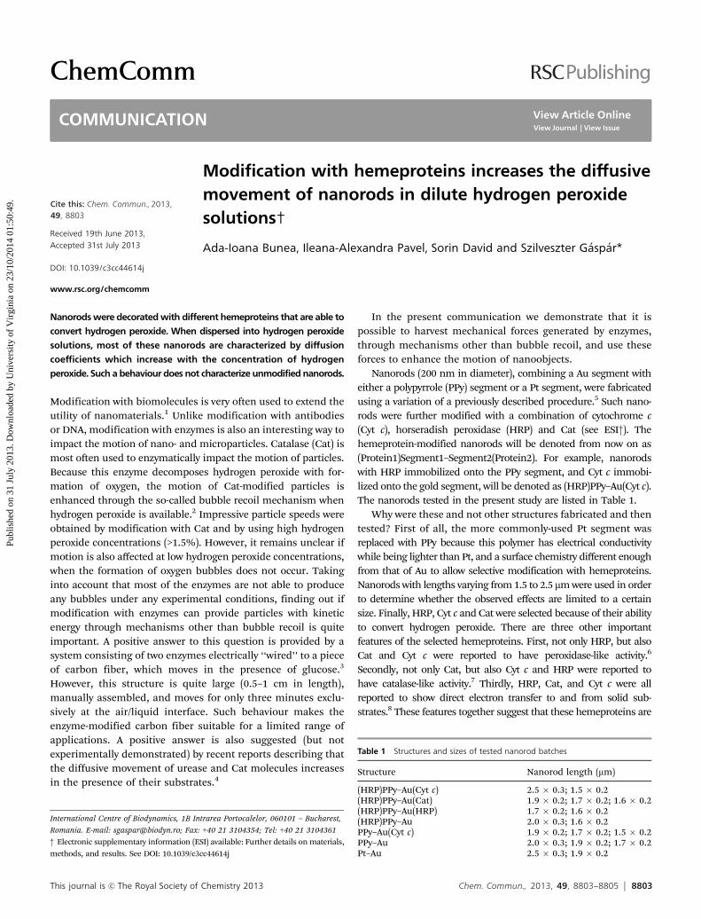
The diffusion coefficients of all nanorods were determined insolutions with different concentrations of hydrogen peroxide. Asshown in Fig. 1, the hemeprotein-modified nanorods behave inthree different ways in dilute hydrogen peroxide solutions. Thediffusion coefficient of (HRP)PPy–Au nanorods increases withhydrogen peroxide concentrations up to 10 mM and then dropsback to the values recorded in water (Fig. 1A). Interestingly, HRP isa hemeprotein that was reported to show ‘‘suicide inhibition’’ athigh concentrations of hydrogen peroxide.9 The diffusion coeffi-cient of PPy–Au(Cyt c), (HRP)PPy–Au(Cyt c) and (HRP)PPy–Au(Cat)nanorods increases with hydrogen peroxide concentrations up to25 mM, then remains quite constant (Fig. 1B). Finally, the diffu-sion coefficient of (HRP)PPy–Au(HRP) nanorods does notchange significantly (Fig. 1A). Experiments have also shown thathemeprotein-modified particles behave in a similar fashion overthe whole range of investigated particle lengths. Moreover, theobserved changes in the diffusion coefficient are not transient. Thediffusion coefficient of (HRP)PPy–Au nanorods in 10 mM hydrogenperoxide was measured for 30 minutes and it stayed elevated over
the whole time period (see ESI†). Pt–Au nanorods, while displayingautopropulsion at high concentrations of hydrogen peroxide,10
present a diffusive movement that is not significantly impacted bylow hydrogen peroxide concentrations (Fig. 1B). PPy–Au nanorodsare decelerated by hydrogen peroxide concentrations in theinvestigated range (Fig. 1B). All these features together clearlyindicate that the observed increase in the diffusion coefficientis definitely dependent on the hydrogen peroxide concentrationand mediated by the immobilized hemeproteins.
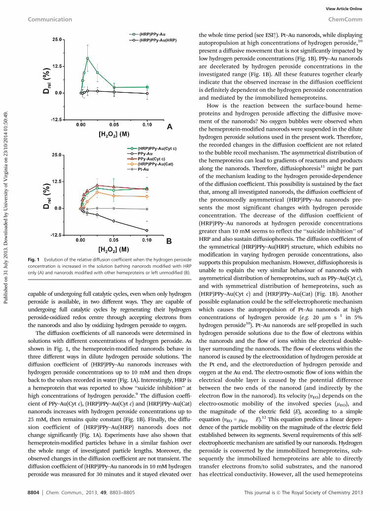
How is the reaction between the surface-bound heme-proteins and hydrogen peroxide affecting the diffusive move-ment of the nanorods? No oxygen bubbles were observed whenthe hemeprotein-modified nanorods were suspended in the dilutehydrogen peroxide solutions used in the present work. Therefore,the recorded changes in the diffusion coefficient are not relatedto the bubble recoil mechanism. The asymmetrical distribution ofthe hemeproteins can lead to gradients of reactants and productsalong the nanorods. Therefore, diffusiophoresis11 might be partof the mechanism leading to the hydrogen peroxide-dependenceof the diffusion coefficient. This possibility is sustained by the factthat, among all investigated nanorods, the diffusion coefficient ofthe pronouncedly asymmetrical (HRP)PPy–Au nanorods pre-sents the most significant changes with hydrogen peroxideconcentration. The decrease of the diffusion coefficient of(HRP)PPy–Au nanorods at hydrogen peroxide concentrationsgreater than 10 mM seems to reflect the ‘‘suicide inhibition’’ ofHRP and also sustain diffusiophoresis. The diffusion coefficient ofthe symmetrical (HRP)PPy–Au(HRP) structure, which exhibits nomodification in varying hydrogen peroxide concentrations, alsosupports this propulsion mechanism. However, diffusiophoresis isunable to explain the very similar behaviour of nanorods withasymmetrical distribution of hemeproteins, such as PPy–Au(Cyt c),and with symmetrical distribution of hemeproteins, such as(HRP)PPy–Au(Cyt c) and (HRP)PPy–Au(Cat) (Fig. 1B). Anotherpossible explanation could be the self-electrophoretic mechanismwhich causes the autopropulsion of Pt–Au nanorods at highconcentrations of hydrogen peroxide (e.g. 20 mm s�1 in 5%hydrogen peroxide10). Pt–Au nanorods are self-propelled in suchhydrogen peroxide solutions due to the flow of electrons withinthe nanorods and the flow of ions within the electrical double-layer surrounding the nanorods. The flow of electrons within thenanorod is caused by the electrooxidation of hydrogen peroxide atthe Pt end, and the electroreduction of hydrogen peroxide andoxygen at the Au end. The electro-osmotic flow of ions within theelectrical double layer is caused by the potential differencebetween the two ends of the nanorod (and indirectly by theelectron flow in the nanorod). Its velocity (vEO) depends on theelectro-osmotic mobility of the involved species (mEO), andthe magnitude of the electric field (E), according to a simpleequation (vEO = mEO � E).12 This equation predicts a linear depen-dence of the particle mobility on the magnitude of the electric fieldestablished between its segments. Several requirements of this self-electrophoretic mechanism are satisfied by our nanorods. Hydrogenperoxide is converted by the immobilized hemeproteins, sub-sequently the immobilized hemeproteins are able to directlytransfer electrons from/to solid substrates, and the nanorodhas electrical conductivity. However, all the used hemeproteins
Fig. 1 Evolution of the relative diffusion coefficient when the hydrogen peroxideconcentration is increased in the solution bathing nanorods modified with HRPonly (A) and nanorods modified with other hemeproteins or left unmodified (B).
Communication ChemComm
Publ
ishe
d on
31
July
201
3. D
ownl
oade
d by
Uni
vers
ity o
f V
irgi
nia
on 2
3/10
/201
4 01
:50:
49.
View Article Online
This journal is c The Royal Society of Chemistry 2013 Chem. Commun., 2013, 49, 8803--8805 8805
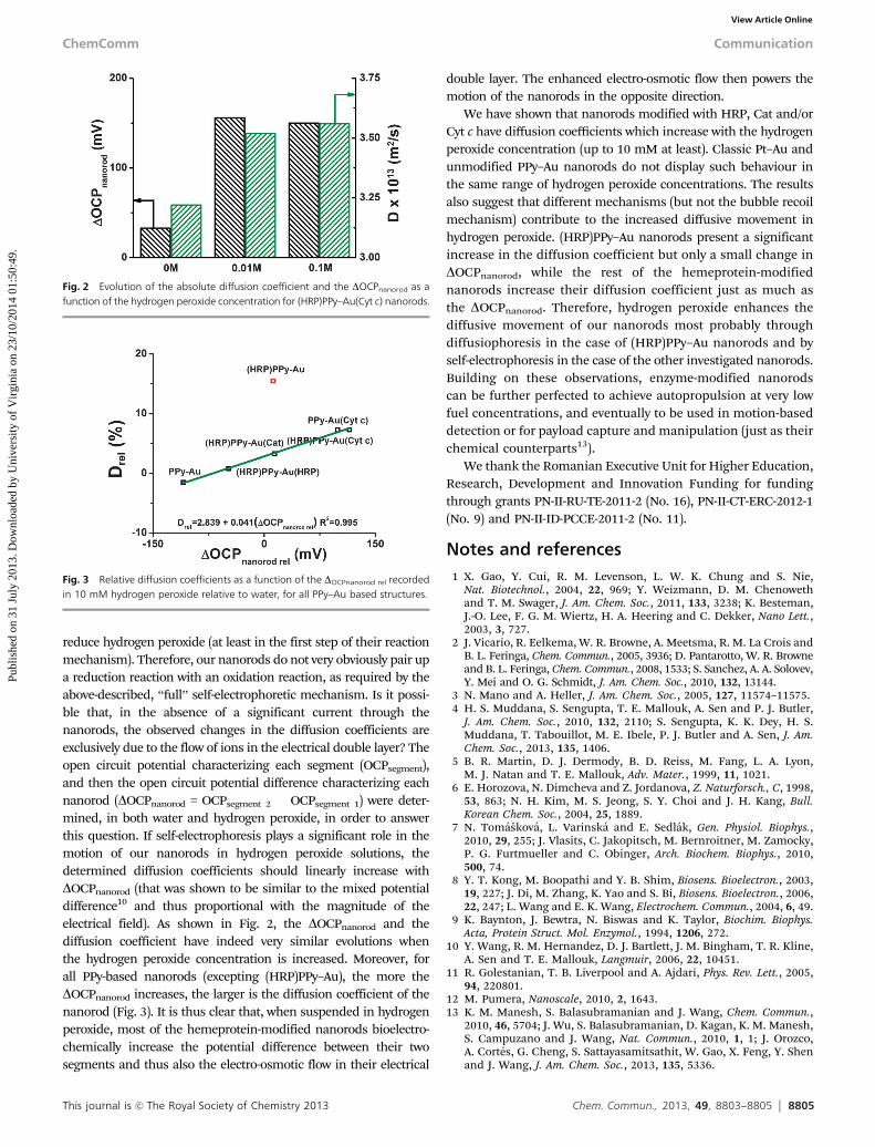
reduce hydrogen peroxide (at least in the first step of their reactionmechanism). Therefore, our nanorods do not very obviously pair upa reduction reaction with an oxidation reaction, as required by theabove-described, ‘‘full’’ self-electrophoretic mechanism. Is it possi-ble that, in the absence of a significant current through thenanorods, the observed changes in the diffusion coefficients areexclusively due to the flow of ions in the electrical double layer? Theopen circuit potential characterizing each segment (OCPsegment),and then the open circuit potential difference characterizing eachnanorod (DOCPnanorod = OCPsegment 2 � OCPsegment 1) were deter-mined, in both water and hydrogen peroxide, in order to answerthis question. If self-electrophoresis plays a significant role in themotion of our nanorods in hydrogen peroxide solutions, thedetermined diffusion coefficients should linearly increase withDOCPnanorod (that was shown to be similar to the mixed potentialdifference10 and thus proportional with the magnitude of theelectrical field). As shown in Fig. 2, the DOCPnanorod and thediffusion coefficient have indeed very similar evolutions whenthe hydrogen peroxide concentration is increased. Moreover, forall PPy-based nanorods (excepting (HRP)PPy–Au), the more theDOCPnanorod increases, the larger is the diffusion coefficient of thenanorod (Fig. 3). It is thus clear that, when suspended in hydrogenperoxide, most of the hemeprotein-modified nanorods bioelectro-chemically increase the potential difference between their twosegments and thus also the electro-osmotic flow in their electrical
double layer. The enhanced electro-osmotic flow then powers themotion of the nanorods in the opposite direction.
We have shown that nanorods modified with HRP, Cat and/orCyt c have diffusion coefficients which increase with the hydrogenperoxide concentration (up to 10 mM at least). Classic Pt–Au andunmodified PPy–Au nanorods do not display such behaviour inthe same range of hydrogen peroxide concentrations. The resultsalso suggest that different mechanisms (but not the bubble recoilmechanism) contribute to the increased diffusive movement inhydrogen peroxide. (HRP)PPy–Au nanorods present a significantincrease in the diffusion coefficient but only a small change inDOCPnanorod, while the rest of the hemeprotein-modifiednanorods increase their diffusion coefficient just as much asthe DOCPnanorod. Therefore, hydrogen peroxide enhances thediffusive movement of our nanorods most probably throughdiffusiophoresis in the case of (HRP)PPy–Au nanorods and byself-electrophoresis in the case of the other investigated nanorods.Building on these observations, enzyme-modified nanorodscan be further perfected to achieve autopropulsion at very lowfuel concentrations, and eventually to be used in motion-baseddetection or for payload capture and manipulation (just as theirchemical counterparts13).
We thank the Romanian Executive Unit for Higher Education,Research, Development and Innovation Funding for fundingthrough grants PN-II-RU-TE-2011-2 (No. 16), PN-II-CT-ERC-2012-1(No. 9) and PN-II-ID-PCCE-2011-2 (No. 11).
Notes and references1 X. Gao, Y. Cui, R. M. Levenson, L. W. K. Chung and S. Nie,
Nat. Biotechnol., 2004, 22, 969; Y. Weizmann, D. M. Chenowethand T. M. Swager, J. Am. Chem. Soc., 2011, 133, 3238; K. Besteman,J.-O. Lee, F. G. M. Wiertz, H. A. Heering and C. Dekker, Nano Lett.,2003, 3, 727.
2 J. Vicario, R. Eelkema, W. R. Browne, A. Meetsma, R. M. La Crois andB. L. Feringa, Chem. Commun., 2005, 3936; D. Pantarotto, W. R. Browneand B. L. Feringa, Chem. Commun., 2008, 1533; S. Sanchez, A. A. Solovev,Y. Mei and O. G. Schmidt, J. Am. Chem. Soc., 2010, 132, 13144.
3 N. Mano and A. Heller, J. Am. Chem. Soc., 2005, 127, 11574–11575.4 H. S. Muddana, S. Sengupta, T. E. Mallouk, A. Sen and P. J. Butler,
J. Am. Chem. Soc., 2010, 132, 2110; S. Sengupta, K. K. Dey, H. S.Muddana, T. Tabouillot, M. E. Ibele, P. J. Butler and A. Sen, J. Am.Chem. Soc., 2013, 135, 1406.
5 B. R. Martin, D. J. Dermody, B. D. Reiss, M. Fang, L. A. Lyon,M. J. Natan and T. E. Mallouk, Adv. Mater., 1999, 11, 1021.
6 E. Horozova, N. Dimcheva and Z. Jordanova, Z. Naturforsch., C, 1998,53, 863; N. H. Kim, M. S. Jeong, S. Y. Choi and J. H. Kang, Bull.Korean Chem. Soc., 2004, 25, 1889.
7 N. Tomaskova, L. Varinska and E. Sedlak, Gen. Physiol. Biophys.,2010, 29, 255; J. Vlasits, C. Jakopitsch, M. Bernroitner, M. Zamocky,P. G. Furtmueller and C. Obinger, Arch. Biochem. Biophys., 2010,500, 74.
8 Y. T. Kong, M. Boopathi and Y. B. Shim, Biosens. Bioelectron., 2003,19, 227; J. Di, M. Zhang, K. Yao and S. Bi, Biosens. Bioelectron., 2006,22, 247; L. Wang and E. K. Wang, Electrochem. Commun., 2004, 6, 49.
9 K. Baynton, J. Bewtra, N. Biswas and K. Taylor, Biochim. Biophys.Acta, Protein Struct. Mol. Enzymol., 1994, 1206, 272.
10 Y. Wang, R. M. Hernandez, D. J. Bartlett, J. M. Bingham, T. R. Kline,A. Sen and T. E. Mallouk, Langmuir, 2006, 22, 10451.
11 R. Golestanian, T. B. Liverpool and A. Ajdari, Phys. Rev. Lett., 2005,94, 220801.
12 M. Pumera, Nanoscale, 2010, 2, 1643.13 K. M. Manesh, S. Balasubramanian and J. Wang, Chem. Commun.,
2010, 46, 5704; J. Wu, S. Balasubramanian, D. Kagan, K. M. Manesh,S. Campuzano and J. Wang, Nat. Commun., 2010, 1, 1; J. Orozco,A. Cortes, G. Cheng, S. Sattayasamitsathit, W. Gao, X. Feng, Y. Shenand J. Wang, J. Am. Chem. Soc., 2013, 135, 5336.
Fig. 2 Evolution of the absolute diffusion coefficient and the DOCPnanorod as afunction of the hydrogen peroxide concentration for (HRP)PPy–Au(Cyt c) nanorods.
Fig. 3 Relative diffusion coefficients as a function of the DOCPnanorod rel recordedin 10 mM hydrogen peroxide relative to water, for all PPy–Au based structures.
ChemComm Communication
Publ
ishe
d on
31
July
201
3. D
ownl
oade
d by
Uni
vers
ity o
f V
irgi
nia
on 2
3/10
/201
4 01
:50:
49.
View Article Online