Embed Size (px)
Citation preview

REVIEW
Modern technologies and algorithms for
scaffolding assembled genomes
Jay GhuryeID, Mihai PopID*
Department of Computer Science and Center for Bioinformatics and Computational Biology, University of
Maryland, College Park, Maryland, United States of America
Abstract
The computational reconstruction of genome sequences from shotgun sequencing data has
been greatly simplified by the advent of sequencing technologies that generate long reads.
In the case of relatively small genomes (e.g., bacterial or viral), complete genome
sequences can frequently be reconstructed computationally without the need for further
experiments. However, large and complex genomes, such as those of most animals and
plants, continue to pose significant challenges. In such genomes, assembly software pro-
duces incomplete and fragmented reconstructions that require additional experimentally
derived information and manual intervention in order to reconstruct individual chromosome
arms. Recent technologies originally designed to capture chromatin structure have been
shown to effectively complement sequencing data, leading to much more contiguous recon-
structions of genomes than previously possible. Here, we survey these technologies and
the algorithms used to assemble and analyze large eukaryotic genomes, placed within the
historical context of genome scaffolding technologies that have been in existence since the
dawn of the genomic era.
Background
The increased availability and lower cost of DNA sequencing have revolutionized biomedical
research. Thousands of humans have been sequenced to date, and genome sequencing is
increasingly used in clinical practice, particularly in the context of cancer [1, 2]. Despite the
long length of sequences generated by third-generation sequencing technologies (tens of thou-
sands of base pairs), the automated reconstruction of entire genomes continues to be a formi-
dable computational task, in no small part because of genomic repeats—ubiquitous features of
eukaryotic genomes [3]. Recently, new genomic technologies have been developed that can
"bridge" across repeats or other genomic regions that are difficult to sequence or assemble.
We refer to technologies originally developed as a tool for interrogating the structure of
genomes by cross-linking adjacent genomic segments and capturing these adjacencies through
sequencing. These technologies are increasingly used to help improve genome assemblies by
"scaffolding" together large segments of the genome. We survey here recent advances in this
field, placed within the context of the technologies and algorithms that have been used for scaf-
folding throughout the entire genomic revolution. Note that our primary focus is on
PLOS Computational Biology | https://doi.org/10.1371/journal.pcbi.1006994 June 5, 2019 1 / 20
a1111111111
a1111111111
a1111111111
a1111111111
a1111111111
OPEN ACCESS
Citation: Ghurye J, Pop M (2019) Modern
technologies and algorithms for scaffolding
assembled genomes. PLoS Comput Biol 15(6):
e1006994. https://doi.org/10.1371/journal.
pcbi.1006994
Editor: Nicola Segata, University of Trento, ITALY
Published: June 5, 2019
Copyright: © 2019 Ghurye, Pop. This is an open
access article distributed under the terms of the
Creative Commons Attribution License, which
permits unrestricted use, distribution, and
reproduction in any medium, provided the original
author and source are credited.
Funding: JG and MP were supported in part by the
U.S. Naval Research Laboratory (http://www.nrl.
navy.mil), award N00173-16-2-C001 to MP. MP
was supported in part by the U.S. National
Institutes of Health (http://www.nih.gov), award
R01-AI-100947 to MP. The funders had no role in
study design, data collection and analysis, decision
to publish, or preparation of the manuscript.
Competing interests: The authors have declared
that no competing interests exist.

reconstructing the genome sequence of organisms rather than the structure of their chromo-
somes. Readers interested in the latter are referred to, e.g., [4].
Genome assembly (Fig 1)—the computational process used to reconstruct genomes from
the relatively short DNA fragments that can be sequenced—is complicated mainly by genomic
repeats [5–9]. These DNA segments that occur in two or more nearly identical copies within
genomes induce ambiguity in the reconstruction of a genome, ambiguity that cannot be
resolved with the information contained in the reads alone. Furthermore, genomes also con-
tain regions with unusual base pair composition that are difficult to sequence. As a result, typi-
cal genome assemblies of eukaryotic genomes are highly fragmented, comprising tens to
hundreds of thousands of contiguous genomic segments (contigs). This fact was recognized
from the early days of genomics, and scientists have developed techniques that can generate
information complementary to that contained in the reads. The assembly of the first living
organism to be sequenced (Haemophilus influenzae [10]) relied on paired-read data that linked
together relatively distant segments of the genome, allowing the assembled contigs to be
ordered and oriented into a "scaffold" of the H. influenzae main chromosome [11–14].
Genome assembly approaches have been extensively reviewed [11–17], including recently
[18]. Missing from this extensive body of literature is a focus on the algorithmic considerations
underlying the use of long-range linking data in the assembly process. In this review, we high-
light recent developments in the technologies used to generate long-range linking information
and describe the computational algorithms that use this information to scaffold together the
genomic segments generated by assembly algorithms. We place recent advances within the his-
torical context of genome scaffolding technologies and algorithms and show how these tech-
nologies and algorithms have moved the field forward toward assembling larger and more
Fig 1. Overview of the genome assembly process. First, genetic material is sequenced, generating a collection of
sequenced fragments (reads). These reads are processed by a computer program called an assembler, which merges the
reads based on their overlap to construct larger contigs. Contigs are then oriented and ordered with respect to each
other with a computer program called a scaffolder, relying on a variety of sources of linkage information. The scaffolds
provide information about the long-range structure of the genome without specifying the actual DNA sequence within
the gaps between contigs. The size of the gaps can also only be approximately estimated. contig, contiguous genomic
segment.
https://doi.org/10.1371/journal.pcbi.1006994.g001
PLOS Computational Biology | https://doi.org/10.1371/journal.pcbi.1006994 June 5, 2019 2 / 20

complex eukaryotic genomes. We conclude with a survey of recent projects that demonstrate
the effective combination of sequencing and scaffolding technologies to generate high-quality
genome reconstructions.
Sources of information for genome scaffolding
Broadly speaking, any type of information that hints at the relative location of genomic seg-
ments along a chromosome can be used to drive the scaffolding process. In most cases, the
information used derives from genomic technologies specifically designed to interrogate the
structure of chromosomes, though indirect inferences based on evolutionary arguments have
also been used effectively in genome scaffolding. Fig 2 shows the extent to which different
sequencing technologies can provide the linkage information for scaffolding. This linkage
information can span anywhere from several hundreds to tens of thousands of base pairs (Illu-
mina, Pacific Biosciences, and Oxford Nanopore) to hundreds of thousands of base pairs
(linked reads and optical maps) to millions of base pairs (Chicago and Hi-C).
The genomic range spanned by the linking information is directly tied to the effectiveness
of a particular technology to resolve certain classes of repeats [19]—to be effective, links must
be longer than the length of a repeat but short enough so that they do not span multiple repeat
units (which would increase the computational complexity of the scaffolding or repeat resolu-
tion process). Given the broad range of the lengths of repeats in most organisms, best results
are usually obtained from a mixture of technologies or from technologies that yield data span-
ning a broad range of distances (such as third-generation sequencing reads, optical maps, or
Fig 2. The genomic span covered by different technologies mentioned in this review. Reads and optical maps derived from the NA12878 sample (DNA
from a human individual sequenced as part of the 1000 Genomes Project) were mapped to the GRCh38 human genome reference. The histograms
represent as follows: Illumina—the separation between natively generated paired-end reads (SRX1049855); Pacbio—the length of the reads generated by the
Pacific Biosciences technology (SRX1607993); Oxford Nanopore—the length of the reads generated by the Oxford Nanopore technology (https://github.
com/nanopore-wgs-consortium/NA12878); optical maps—the length of the fragments mapped by the BioNano nanocoding technology (from BioNano
website); linked reads—the span of the region covered by reads originating from the same DNA fragment, as generated by the 10X Genomics technology
(SRX1392293); Chicago—the separation between read pairs generated by the Chicago chromosome conformation capture protocol (SRX1423027); and Hi-
C—the separation between read pairs generated by the Hi-C chromosome conformation capture protocol (SRX3651893).
https://doi.org/10.1371/journal.pcbi.1006994.g002
PLOS Computational Biology | https://doi.org/10.1371/journal.pcbi.1006994 June 5, 2019 3 / 20

Hi-C links). Sequencing reads can be viewed as providing linking information that spans any
distance within the length of the reads—as such, they provide valuable information for repeats
of a broad range of sizes, up to the length of the reads.
We structure our presentation around the key types of information that can be used to
organize genomic contigs into chromosome-wide scaffolds (Table 1).
Physical mapping
Physical-mapping technologies attempt to estimate the location of specific loci along genomic
chromosomes. The loci can be short DNA segments that are unique within the genome, as in
the case of sequence-tagged sites (STSs)[20], or the recognition sequence of a restriction
enzyme, as in the case of restriction mapping and optical mapping. The approximate location
of the markers along chromosomes can be identified through a number of techniques, from
fluorescence in situ hybridization (FISH)[21] to the analysis of the random breakage of DNA
being exposed to X-rays (radiation hybrid mapping [22]) to direct measurement of restriction
fragment sizes, as performed in restriction mapping [23]. The original use of restriction
enzymes to map chromosomes resulted in unordered information—simply the list of sizes of
the restriction fragments generated from the molecule—information that had to be converted
into an ordered map through a complex computational process. Optical maps are an enhance-
ment of restriction mapping, which provides the fragment order in addition to their size based
on imaging the fluorescence of DNA molecules immobilized on a glass slide [24] or within a
nanochannel [25] (the latter technology is called nanocoding).
Physical-mapping data is among the earliest technologies used to order genomic contigs
along a chromosome [26–28]. The computational approach used to perform this task simply
involves comparing experimentally derived maps to theoretical (in silico) maps generated
from the sequenced contigs (Fig 3). This process is easiest when the landmarks being com-
pared are distinguishable from each other (as is the case for STS and radiation hybrid maps)
and substantially more complex and error prone for restriction maps, in which all the land-
marks are identical in sequence, e.g., in the context of optical mapping [29].
The experimental maps themselves are often the result of assembling a collection of DNA
fragments (clones) that have been mapped separately, leading to similar analytical challenges
as those encountered in genome assembly [30]. In the case of unordered restriction maps, the
assembly process is guided by the probability that two clones overlap, which is computed by
Table 1. Comparison of different sequencing and mapping technologies.
Category Scaffolding Data Separation on the Genome Orientation Ordering Distance
Physical mapping Restriction maps 10–100 Kb Yes No Yes
Optical maps 10–100 Kb Yes Yes Yes
Subcloning 10x Genomics 100 Kb Yes Yes Yes
Illumina TSLR 100 Kbp Yes Yes Yes
Long-read data Pacific Biosciences 10–15 Kb Yes Yes Yes
Oxford Nanopore 15–20 Kb Yes Yes Yes
Paired read Paired-end reads 100–500 bp Yes Yes Yes
Mate pairs 1,000–10,000 bp Yes Yes Yes
Chromosome conformation Hi-C 30–100 Mb Yes Yes No
Chicago 3–100 Mb Yes Yes No
Synteny Reference genome(s) Up to genome size Yes Yes Yes
Abbreviation: TSLR, TruSeq Synthetic Long Read.
https://doi.org/10.1371/journal.pcbi.1006994.t001
PLOS Computational Biology | https://doi.org/10.1371/journal.pcbi.1006994 June 5, 2019 4 / 20

taking into account the number of restriction fragments shared by the clones [31]. The pair-
wise overlap probabilities are used to assemble the clones into a chromosome-wide structure
using a heuristic assembly algorithm (fingerprinted contigs [FPC]), which also allows for man-
ual intervention to inspect and correct the resulting layout.
Ordered restriction maps, as generated by optical or nanocoding mapping, can be aligned
using variants of dynamic programming alignment algorithms [32, 33]. In SOMA [34], frag-
ment-sizing errors are penalized through a chi-squared scoring function, and a variant of a
scheduling algorithm is used to determine the layout of contigs with ambiguous mappings.
The runtime of the alignment algorithm used in SOMA scales with the fourth power of the
number of restriction fragments, making the approach impractical for large genomes. Two
recent approaches address this limitation. TWIN [35] relies on an extension of the FM-index
[36] to speed up the alignment process, whereas Maligner [37] indexes the reference map
by simulating the effect of common mapping errors such as false cuts or missed restriction
sites.
Fig 3. Mapping-based scaffolding approaches. (a) Contigs (arrows) are aligned to a reference genome, and their order and orientation is inferred from
the alignment. (b) Long reads aligned to the ends of contigs imply their adjacency; (c) optical maps (tics represent location of restriction sites) can be
used to infer the order and orientation of contigs (arrows) by aligning the inferred restriction pattern (tics within arrows) to that of the experimental
map. contig, contiguous genomic segment.
https://doi.org/10.1371/journal.pcbi.1006994.g003
PLOS Computational Biology | https://doi.org/10.1371/journal.pcbi.1006994 June 5, 2019 5 / 20

Subcloning
Subcloning involves breaking up the genome into large fragments that are then sequenced sep-
arately, retaining the connection between the sequencing reads generated from the same frag-
ment (we refer to them as “linked reads” subsequently). The assembly process can then be run
for each fragment separately, and the resulting assemblies can be merged together to recon-
struct the full genome sequence. Initially, subcloning relied on bacterial artificial chromosomes
(BACs) grown in Escherichia coli, yielding fragments in the range of ~100 kbp in length. The
two ends of each BAC clone were sequenced first in order to construct a clone map [38] repre-
senting the relative relationship between individual BACs along the genome. From this infor-
mation, a minimal tiling path was identified in order to decide which fragments would be fully
sequenced. This strategy was used effectively in the early days of genomics, most notably dur-
ing the public effort to sequence the human genome [39].
Recently, new technologies have been developed that perform the subcloning process in vitro.
The technology from 10x Genomics partitions large DNA fragments into droplets, and the DNA is
sheared, and sequencing libraries are constructed within the droplets. The DNA within each drop-
let is tagged with a droplet-specific barcode, and these barcoded DNA molecules then undergo
sequencing, and a postprocessing algorithm parses the barcodes to group the reads originating
from the same large DNA fragment [40]. The Illumina TruSeq Synthetic Long Read (TSLR) tech-
nology [41] is based on fragmenting DNA into large segments of about 10 kb in size, which are dis-
tributed into pools such that each pool contains a relatively small number of fragments (~200–
300). Each pool is processed separately and barcoded with a unique barcode prior to sequencing.
When the original fragments have been sequenced deeply enough (which is usually the case
for the TSLR technology and most applications of BACs), the pooled reads can be assembled
together in order to create complete reconstructions of the individual fragments, effectively
generating long and highly accurate sequencing reads. Several approaches have also been
developed that rely on the unassembled pooled reads to guide the scaffolding process,
approaches that can be effective even at low depths of sequencing coverage. Fragscaff [42], a
method which was originally designed for contiguity-preserving transposition sequencing
data, creates links between the ends of contigs, which are represented in the set of reads from
the same pool. Within the resulting graph, Fragscaff then identifies a minimum spanning tree
by using as edge weights the number of pools shared between contigs. The longest path within
this tree is selected as the scaffold backbone. ARCS [43] maps the linked reads to the assembled
contigs and constructs intercontig links by identifying pairs of contigs whose ends share
sequences from the same read pool. Scaffolding is then performed with the LINKS scaffolder
[44], a tool originally developed for scaffolding assemblies with the help of long-read data. A
similar approach is used by ARKS [45], a tool that relies on k-mer matches instead of sequence
alignment to infer the assignment of the linked reads to assembled contigs. In Supernova, Wei-
senfeld and colleagues [46] relied on pool-specific barcodes to construct an adjacency graph in
which the nodes are the initial set of contigs or scaffolds and edges denote the number of pool-
specific barcodes shared between scaffolds. In this graph, Supernova finds linear paths consis-
tent with the links provided by barcodes and selects the highest-scoring path as the backbone
of the final scaffold. Linked reads have also been used to identify errors in the assembly. Tig-
mint [47] flags as misassemblies regions of contigs where the depth of coverage by read pools
(as inferred from the mapping of reads to assemblies) is lower than expected.
Long-read data
Sequencing technologies that generate long sequencing reads, such as Pacific Bioscience [48]
and Oxford Nanopore [49], can be seen as a special case of subcloning. Whereas genome
PLOS Computational Biology | https://doi.org/10.1371/journal.pcbi.1006994 June 5, 2019 6 / 20

assemblers such as CANU [50], FALCON [51], HINGE [52], MECAT [53], miniasm [54], and
Flye [55] are effective in reconstructing genomic contigs from long-read data to achieve high-
quality assemblies with only long-read data, the genome needs to be sequenced at considerably
high coverage, incurring significant costs. A more cost-effective strategy involves supplement-
ing a short-read assembly with a relatively low-coverage set of long-read data, effectively repre-
senting a collection of sparsely sampled genomic subclones. SSPACE-LongRead [56] was one
of the earliest methods able to leverage long reads for scaffolding. This approach used BLASR
[57] (an aligner tuned for the high error rates of third-generation sequencing technologies) to
align contigs to the long reads in order to infer orientation, ordering, and the distance between
contigs. SMSC [58] and BIGMAC [59] both start by using the long-read data to identify poten-
tial errors in the assembly, break the contigs at the boundaries of these errors, and then scaffold
the resulting data using linking information inferred from the long reads. Because the align-
ment of long-read data is computationally intensive, LINKS [44] proposes an alignment-free
approach that extracts pairs of k-mers separated by a predefined distance within long reads
and then treats these as if they were paired short reads, relying on traditional scaffolding
approaches. Unicycler [60] operates directly on the assembly graph generated by the SPAdes
assembler [61] from short reads, using the long-read data to disambiguate paths through the
graphs and generate longer, more accurate contigs. npScarf [62] leverages the real-time gener-
ation of data by nanopore sequencing devices to iteratively develop and improve the scaffold-
ing of a genome as more data become available. The scaffolding algorithm operates in a greedy
fashion, linking contigs together as soon as sufficient support is available in the set of reads
and breaking prior links if new ones that contradict them have stronger support. This iterative
greedy process can be stopped by the user once a sufficiently good assembly is generated,
allowing the dynamic selection of the depth of sequencing depending on the actual quality of
the resulting reconstruction.
Paired-read technologies
By far, the most common source of information for scaffolding is technologies that yield infor-
mation about the relative placement of pairs of reads along the genome being sequenced. Most
commonly, this information is derived by carefully controlling DNA shearing prior to
sequencing in order to obtain fragments of uniform sizes and by tracking the link between
DNA sequences "read" from the same fragment. Multiple protocols have been developed to
generate read-pairing information, and different names are commonly used to reflect the
experimental source: paired-end reads (pairings natively generated by Illumina sequencing
instruments, usually short range ~300–500 bp) and mate-pair or jumping libraries (pairing
information derived with the help of additional experimental assays, usually spanning thou-
sands to tens of thousands of base pairs). Here, we use these terms interchangeably, as the
information being generated is the same—pairs of reads with an approximately known relative
distance and orientation. The information provided by mate pairs can be used to link the con-
tigs and produce scaffolds [63] or to guide the assembly process itself, allowing the effective
resolution of repeats [19, 64].
The algorithms for using mate-pair data in scaffolding genomes all follow a similar work-
flow. First, mate pairs whose ends map to different contigs are used to link together the corre-
sponding contigs. Second, the pairwise linkage information is used to orient and order contigs
with respect to each other. Third, the size of the gap between adjacent contigs is estimated
from the experimentally determined size of the mate pairs, and a linear layout of the contigs
along a scaffold is generated (Fig 4). Because contig orientation and ordering are computation-
ally hard problems [7], scaffolders implement different greedy heuristics. Scaffolders such as
PLOS Computational Biology | https://doi.org/10.1371/journal.pcbi.1006994 June 5, 2019 7 / 20

MIP [65], SOPRA [66], and SCARPA [67] use integer programming to find the optimal orien-
tation and ordering of contigs. Bambus [68] and SSPACE [69] use multiple libraries in a hier-
archical manner to perform scaffolding, starting from libraries with smaller insert sizes (which
are more accurate and yield a simpler problem) and progressively expanding scaffolds using
libraries with larger insert sizes. OPERA-LG [70] uses a branch and bound search to determine
the relative placement of contigs along the chromosome. The authors show that the size of the
search space is bounded by the ratio between the library and contig size, implying that the
branch and bound heuristic is efficient for the data typically encountered in practical applica-
tions despite a theoretically exponential complexity. The scaffolder inGAP-sf [71] merges
information from both the assembly graph and paired-read data to construct scaffolds. In
addition, this scaffolder introduces a statistical model for estimating the support for a link
between two contigs, information that is used in constructing the scaffold. A number of tools
have also been developed that use RNA sequencing (RNA-seq) data for scaffolding. Because of
the long lengths of eukaryotic introns, such approaches can yield long-range genomic connec-
tions using standard short-length paired-end sequencing protocols, with the caveat that scaf-
folding is only effective in genic regions. Tools developed specifically for such data include
RNAPATH [72], L_RNA [73], Rascaf [74], AGOUTI [75], and P_RNA [76].
In the context of repeat resolution, the orientation and distance constraints imposed by
paired reads limit the number of possible traversals of the graph through a repeat region and
can link together the unique genomic regions surrounding each instance of a repeat. Assem-
blers such as Velvet [77], ABySS [78], ABySS 2.0 [79], and IDBA-UD [80] use paired-end
information to guide the walk through the assembly graph. SPAdes [81] and metaSPAdes [82]
Fig 4. Use of pairwise linkage information for scaffolding. (a) Paired-end reads are sequenced from the genome.
Depending on the technology, the approximate distance and/or relative orientation of the paired reads may not be
known. (b) The reads are aligned to contigs. Reads with their ends aligned to two different contigs provide linkage
information useful for scaffolding. (c) Linkage information is used to orient and order the contigs into scaffolds. Usually
not all constraints can be preserved, and algorithms attempt to minimize inconsistencies (marked with X).
https://doi.org/10.1371/journal.pcbi.1006994.g004
PLOS Computational Biology | https://doi.org/10.1371/journal.pcbi.1006994 June 5, 2019 8 / 20

use the ratio of expected to observed numbers of mate pairs connecting two nodes [83] in the
de Bruijn graph to check if the path traverses through a repetitive region. Wetzel and col-
leagues [19] explored the extent to which mate pairs can be used to resolve repetitive regions
in prokaryotic genomes and showed that mate-pair libraries are most effective if tuned to the
structure of the assembly graph.
Chromosomal contact data
A special type of paired-read data is generated by techniques recently developed to study the
three-dimensional structure of chromosomes inside a cell [84]. These techniques are collec-
tively referred to as chromosomal conformation capture (C3), which generate pairwise linking
information between reads that originate from genomic regions that are physically adjacent in
a cell. Unlike mate-pair data, the distance and the relative orientation between the paired reads
are not known a priori.
The two most commonly used protocols for capturing chromosome conformation are Hi-
C [84] and Chicago [85]. In the Hi-C protocol, DNA in the cell nucleus is cross-linked and cut
with a restriction enzyme. This process generates fragments of DNA that are distally located
but physically associated with each other. The sticky ends of these fragments are biotinylated
and then ligated to form a chimeric circle. The resulting circles are sheared and processed into
sequencing libraries in which individual templates are chimeras of the physically associated
DNA molecules. The Chicago protocol from Dovetail Genomics starts not with cells but with
purified DNA so that any biologically associated interactions are eliminated. Artificial nucleo-
somes with random specificity are then used to condense the DNA into chromatin, which is
then processed through the standard Hi-C protocol. The result is a collection of fragments that
is enriched for sets of paired reads that capture long-range interactions between segments of
DNA that were in contact within the artificial chromatin.
Because Hi-C and Chicago protocols do not provide estimates of the distance between the
paired reads, the data can only be used to estimate the relative order and orientation of contigs
and not the size of the gaps separating them. The scaffolding process starts by filtering the data
to eliminate artifacts such as reads aligning to multiple locations or chimeric reads derived
from the ligation junctions. Several tools have been developed for this purpose, including
HiCUP [86], HiCPro [87], Juicer [88], Juicebox [89], and HiFive [90]. These tools align reads
to the assembly using standard alignment programs [91, 92] and filter the alignments to
remove experimental artifacts, yielding the “true” alignments, which imply the contact infor-
mation. The number of paired reads linking two genomic regions (contact frequency) strongly
correlates with the one-dimensional distance between the corresponding regions, thereby
yielding an estimate of the relative placement of these segments within a genome. Further-
more, the contact frequency is much higher within a chromosome than across chromosomes,
making it possible to infer chromosome structure directly from the genome assembly. Most of
the algorithms developed to use Hi-C data for scaffolding use these properties to group contigs
into chromosome-specific bins and then orient and order the contigs within each chromo-
some by maximizing the concordance with the experimentally derived contact frequencies. A
major confounding factor in using Hi-C data for scaffolding is the nonrandom association
between topological domains [93]. DNA-to-DNA interactions within the nucleus are orga-
nized in a domain structure in which interactions are much stronger within a domain than
across domains. As a result, the Hi-C contact patterns exhibit a modular structure that can
confound the estimate of distance between contigs during the scaffolding process.
DNATri [94] and LACHESIS [95] were the earliest methods developed to use Hi-C datasets
for scaffolding. DNATri relies on a limited-memory Broyden–Fletcher–Goldfarb–Shanno
PLOS Computational Biology | https://doi.org/10.1371/journal.pcbi.1006994 June 5, 2019 9 / 20

optimization algorithm to identify the placement of contigs that best matches the contact fre-
quencies derived from the Hi-C data. LACHESIS first clusters the contigs into chromosome
groups using hierarchical clustering, matching a user-specified number of chromosomes.
Then, it orders and orients contigs in each chromosome group/cluster separately by formulat-
ing the problem as identifying the "trunk" of a minimum spanning tree of the graph that
encodes the Hi-C links between contigs. GRAAL [96] models the Hi-C data by distinguishing
between cis- contacts (occurring within the same molecule) and trans- contacts (occurring
across molecules). The contact frequency for the former are distance dependent, whereas the
latter are drawn from a uniform probability distribution. The contigs are ordered and oriented
to maximize the fit with this modeled data using a Metropolis optimization algorithm [97].
SALSA [98] relies on Hi-C data to correct misassemblies in the input contigs and then orients
and orders the contigs using a maximal matching algorithm [98]. 3D-DNA [99] also corrects
the errors in the input assembly and then iteratively orients and orders uniquely assembled
contigs (unitigs) into a single megascaffold. This megascaffold is then broken into a user-speci-
fied number of chromosomes, identifying chromosomal ends based the on a Hi-C contact
map. Putnam and colleagues [85] proposed a method called Hi-Rise that was specifically
designed for handling Chicago libraries (based on artificial chromatin). They rely on a likeli-
hood function that matches the characteristics of these data and use dynamic programming to
identify a layout of contigs that maximizes the fit with the experimental data. Recently, Zhang
and colleagues developed an approach for scaffolding polyploid genomes using Hi-C data in
an approach called ALLHIC [100]. This approach relies upon the LACHESIS algorithm
applied to Hi-C data that have been filtered to remove contacts that connect across haplotypes,
thereby yielding haplotype-specific scaffolds.
Practical considerations
The scaffolds generated with the help of the data described previously simply organize contigs
along a genome without specifying the actual DNA sequence represented within the gap
between adjacent contigs. Once the relative location of contigs is known, however, it is fre-
quently easy to reconstruct the sequence within the gaps, a process that is commonly referred
to as gap filling. Most commonly, mate-pair information is used to identify which reads could
be placed within a gap, and then those reads are assembled to fill in the sequence within the
gap, extending or even joining the adjacent contigs. Variants of this process are included in
virtually all genome assemblers, e.g., ABySS [78], ALLPATHS-LG[101], and EULER [102],
and several stand-alone solutions were also developed: GapFiller [103], SOAPdenovo GapClo-
ser [104], and Sealer [105]. The latter approach relies on Bloom filters [106] to reduce memory
usage, thereby enabling gap filling in large draft genomes. When long reads are available, gap
filling can be performed with the help of reads that could not previously be incorporated in the
assembly. The relatively higher quality of the contig sequences allows gap filling software to
identify alignments that were missed during the assembly process. This principle is used by
PBJelly [107] and GMCloser [108], approaches specifically developed for Pacific Biosciences
data. GMCloser relies on a likelihood ratio test to determine the quality of alignments and
remove poor-quality alignments that could lead to misassemblies.
Each of the data types used for scaffolding contain errors and have specific biases. Incorrect
insert size estimates in mate-pair data can lead to ordering and gap estimation errors in scaf-
folds [109]. Hi-C data cannot provide accurate orientation information at small genomic dis-
tances, yielding small inversions within the scaffolds [110]. Optical mapping data have fairly
low resolution and contain many errors, including incorrect estimates of fragment sizes and
missed or spurious restriction cuts [111]. To reduce the impact of such errors on the ultimate
PLOS Computational Biology | https://doi.org/10.1371/journal.pcbi.1006994 June 5, 2019 10 / 20
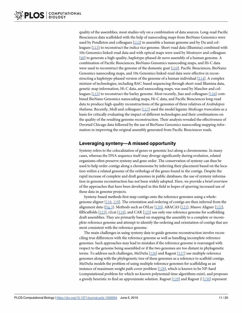
quality of the assemblies, most studies rely on a combination of data sources. Long-read Pacific
Biosciences data scaffolded with the help of nanocoding maps from BioNano Genomics were
used by Pendleton and colleagues [112] to assemble a human genome and by Du and col-
leagues [113] to reconstruct the indica rice genome. Short-read data (Illumina) combined with
10x Genomics linked-read data and with optical maps were used by Mostoyov and colleagues
[40] to generate a high-quality, haplotype-phased de novo assembly of a human genome. A
combination of Pacific Biosciences, BioNano Genomics nanocoding maps, and Hi-C data
were used to reconstruct the genome of the domestic goat [110]. Pacific Biosciences, BioNano
Genomics nanocoding maps, and 10x Genomics linked-read data were effective in recon-
structing a haplotype-phased version of the genome of a human individual [114]. A complex
mixture of technologies, including BAC-based sequencing through short-read Illumina data,
genetic map information, Hi-C data, and nanocoding maps, was used by Mascher and col-
leagues [115] to reconstruct the barley genome. Most recently, Jiao and colleagues [116] com-
bined BioNano Genomics nanocoding maps, Hi-C data, and Pacific Biosciences long-read
data to produce high-quality reconstructions of the genomes of three relatives of Arabidopsisthaliana. Recently, Moll and colleagues [117] used the model legume Medicago trunculata as a
basis for critically evaluating the impact of different technologies and their combinations on
the quality of the resulting genome reconstruction. Their analysis revealed the effectiveness of
Dovetail Chicago data followed by the use of BioNano Genomics nanocoding mapping infor-
mation in improving the original assembly generated from Pacific Biosciences reads.
Leveraging synteny—A missed opportunity
Synteny refers to the colocalization of genes or genomic loci along a chromosome. In many
cases, whereas the DNA sequence itself may diverge significantly during evolution, related
organisms often preserve synteny and gene order. The conservation of synteny can thus be
used to help order contigs along a chromosome by inferring their placement based on the loca-
tion within a related genome of the orthologs of the genes found in the contigs. Despite the
rapid increase of complete and draft genomes in public databases, the use of synteny informa-
tion in genome reconstruction has not been widely adopted. Here, we provide a brief overview
of the approaches that have been developed in this field in hopes of spurring increased use of
these data in genome projects.
Synteny-based methods first map contigs onto the reference genomes using a whole-
genome aligner [118, 119]. The orientation and ordering of contigs are then inferred from the
alignment data (Fig 3). Methods such as OSLay [120], ABACAS [121], Mauve Aligner [122],
fillScaffolds [123], r2cat [124], and CAR [125] use only one reference genome for scaffolding
draft assemblies. They are primarily based on mapping the assembly to a complete or incom-
plete reference genome and attempt to identify the ordering and orientation of contigs that are
most consistent with the reference genome.
The main challenges in using synteny data to guide genome reconstruction involve recon-
ciling true differences with the reference genome as well as handling incomplete reference
genomes. Such approaches may lead to mistakes if the reference genome is rearranged with
respect to the genome being assembled or if the two genomes are too distant in phylogenetic
terms. To address such challenges, MeDuSa [126] and Ragout [127] use multiple reference
genomes along with the phylogenetic tree of these genomes as a reference to scaffold contigs.
MeDuSa models the problem of using multiple reference genomes for scaffolding as an
instance of maximum weight path cover problem [128], which is known to be NP-hard
(computational problem for which no known polynomial time algorithms exist), and proposes
a greedy heuristic to find an approximate solution. Ragout [129] and Ragout 2 [130] represent
PLOS Computational Biology | https://doi.org/10.1371/journal.pcbi.1006994 June 5, 2019 11 / 20

the target and reference genomes as a multicolored breakpoint graph with nodes representing
the conserved synteny blocks and edges representing the adjacency of these blocks. In this
graph, Ragout finds the missing adjacencies by solving a half-breakpoint state parsimony prob-
lem on the given phylogenetic tree and then orients and orders synteny blocks to reconstruct
the target genome. Multi-CAR [131] starts by processing each reference genome separately
using CAR. Multi-CAR then reconciles the different contig orderings by constructing a graph
in which nodes are contigs and edges are the adjacencies given by different reference genomes.
A maximal weight perfect matching [132] within this graph defines the final set of scaffolds.
Perspective
As we have shown, technological advances on both the experimental and computational sides
have dramatically improved the ability of reconstructing the genomes of complex eukaryotic
organisms, including repeat-rich plants such as rice and barley. Most of the technologies used
today are evolved versions of approaches developed decades ago during the dawn of the geno-
mic era. Although read lengths have increased, the accuracy of optical and nanocoding maps
has improved, and subcloning approaches are now performed in vitro without the need for
culturing the DNA in an E. coli host; the fundamental properties of the data being generated
have not changed in a meaningful way. The exceptions are the technologies used to interrogate
the structure of chromosomes through sequencing—Hi-C and related approaches. The paired
reads generated by these technologies no longer provide per-pair distance constraints; rather,
distance information can only be reconstructed from the frequency of "contacts" between dis-
tant sections of the genome being reconstructed. In return, however, these technologies pro-
vide much longer-range linking information than provided by any other technology. Just as
long-read technologies have dramatically advanced our ability to reconstruct genomes, the
long-range linking information provided by Hi-C and similar technologies has made it possi-
ble to link together complete chromosome arms [99, 110].
In the near future, it is likely that many previously intractable genomes will be recon-
structed with the help of long-read sequencing data coupled with paired-read information
from chromosome conformation capture technologies, augmented by short-read and short
mate-pair technologies aimed at resolving the small-scale structure of genomes. This opportu-
nity is particularly relevant to scientists studying the complex genomes of plants [133] or
insects [134] for which few genomic resources are currently available.
As we have already mentioned, a largely unused source of information is the sequences of
the many genomes already sequenced and deposited in public databases. This vast source of
data can be a valuable addition to the other types of genomic data being used in genome recon-
struction, particularly in projects aiming to more densely sample particular regions of the tree
of life (e.g., the genomes of cereal crops [133, 135]).
In our review, we have omitted a discussion of experimental challenges or cost, in part
because our primary focus has been on algorithmic considerations and in part because of the
rapid changes in technologies that would make any cost estimates obsolete even before the ink
has dried on the paper. In general, such practical aspects are insufficiently discussed in current
literature, and the community would benefit from a review focused on the technical challenges
and costs of the available technologies.
Generating haplotype-phased chromosome-scale assemblies of eukaryotic genomes with
the mix of sequencing technologies has been the driving force behind the development of the
newer genome assembly methods. Koren and colleagues [136] proposed a method called “trio-
binning” that uses short and accurate Illumina reads from two parental genomes to partition
PLOS Computational Biology | https://doi.org/10.1371/journal.pcbi.1006994 June 5, 2019 12 / 20
long and noisy reads from an offspring into a haplotype-specific set of reads, and each haplo-
type is then assembled independently.
It is conceivable that in the very near future, further developments in genomic technologies
will make the automatic reconstruction of mammalian genomes possible. Recent advances in
nanopore sequencing devices are already yielding longer reads than all prior technologies,
potentially leading to the ability to assemble complete eukaryotic genomes from nanopore
data alone. Rather than the end of a road, such developments will create the opportunity for
scientists to tackle even harder challenges, such as the complete reconstruction of individual
haplotypes, particularly in the context of heterogeneous mixtures such as tumors or microbial
mixtures or polyploid genomes. Some progress is being made in haplotyping human genomes
with the help of pedigree information (specifically trios comprising two parents and a child)
[136]; however, the solution to the more complex problems posed by mixtures and polyploidy
will require further developments in both genomic technologies, such as those outlined in our
review, as well as in the design of algorithms and tools able to effectively leverage the informa-
tion provided by these technologies.
References1. Alexandrov LB, Nik-Zainal S, Wedge DC, Aparicio SAJR, Behjati S, Biankin AV, et al. Signatures of
mutational processes in human cancer. Nature. 2013; 500(7463):415–21. https://doi.org/10.1038/
nature12477 PubMed Central PMCID: PMCPMC3776390. PMID: 23945592
2. Kasar S, Kim J, Improgo R, Tiao G, Polak P, Haradhvala N, et al. Whole-genome sequencing reveals
activation-induced cytidine deaminase signatures during indolent chronic lymphocytic leukaemia evo-
lution. Nat Commun. 2015; 6:8866. https://doi.org/10.1038/ncomms9866 PubMed Central PMCID:
PMCPMC4686820. PMID: 26638776
3. Alkan C, Sajjadian S, Eichler EE. Limitations of next-generation genome sequence assembly. Nature
Methods. 2010; 8:61. https://doi.org/10.1038/nmeth.1527 PMID: 21102452
4. Barutcu AR, Fritz AJ, Zaidi SK, van Wijnen AJ, Lian JB, Stein JL, et al. C-ing the Genome: A Compen-
dium of Chromosome Conformation Capture Methods to Study Higher-Order Chromatin Organization.
2016; 231(1):31–5. https://doi.org/10.1002/jcp.25062 PMID: 26059817
5. Medvedev P, Georgiou K, Myers G, Brudno M. Computability of Models for Sequence Assembly. In:
Giancarlo R, Hannenhalli S, editors. Algorithms in Bioinformatics. WABI 2007. Lecture Notes in Com-
puter Science, vol 4645. Berlin: Springer; 2007. p. 289–301.
6. Tarhio J, Ukkonen E. A greedy algorithm for constructing shortest common superstrings. In: Gruska J,
Rovan B, Wiedermann J, editors. Mathematical Foundations of Computer Science 1986. MFCS 1986.
Lecture Notes in Computer Science, vol 233. Berlin: Springer; 1986 p. 602–10.
7. Kececioglu JD, Myers EW. Combinatorial algorithms for DNA sequence assembly. Algorithmica.
1995; 13(1–2):7–51. https://doi.org/10.1007/bf01188580
8. Schmid M, Frei D, Patrignani A, Schlapbach R, Frey JE, Remus-Emsermann MNP, et al. Pushing the
limits of de novo genome assembly for complex prokaryotic genomes harboring very long, near identi-
cal repeats. Nucleic Acids Research. 2018; 46(17):8953–65. https://doi.org/10.1093/nar/gky726
PMID: 30137508
9. Jain M, Koren S, Miga KH, Quick J, Rand AC, Sasani TA, et al. Nanopore sequencing and assembly
of a human genome with ultra-long reads. Nature Biotechnology. 2018; 36:338. https://doi.org/10.
1038/nbt.4060 PMID: 29431738
10. Fleischmann RD, Adams MD, White O, Clayton RA, Kirkness EF, Kerlavage AR, et al. Whole-genome
random sequencing and assembly of Haemophilus influenzae Rd. Science. 1995; 269(5223):496–
512. PMID: 7542800.
11. Nagarajan N, Pop M. Sequence assembly demystified. Nat Rev Genet. 2013; 14(3):157–67. https://
doi.org/10.1038/nrg3367 PMID: 23358380
12. Ghurye JS, Cepeda-Espinoza V, Pop M. Metagenomic Assembly: Overview, Challenges and Applica-
tions. Yale J Biol Med. 2016; 89(3):353–62. PubMed Central PMCID: PMCPMC5045144. PMID:
27698619
13. Miller JR, Koren S, Sutton G. Assembly algorithms for next-generation sequencing data. Genomics.
2010; 95(6):315–27. https://doi.org/10.1016/j.ygeno.2010.03.001 PubMed Central PMCID:
PMCPMC2874646. PMID: 20211242
PLOS Computational Biology | https://doi.org/10.1371/journal.pcbi.1006994 June 5, 2019 13 / 20

14. Simpson JT, Pop M. The Theory and Practice of Genome Sequence Assembly. Annu Rev Genomics
Hum Genet. 2015; 16:153–72. https://doi.org/10.1146/annurev-genom-090314-050032 PMID:
25939056
15. Schatz MC, Delcher AL, Salzberg SL. Assembly of large genomes using second-generation sequenc-
ing. 2010; 20(9):1165–73. https://doi.org/10.1101/gr.101360.109 PMID: 20508146
16. Alkan C, Coe BP, Eichler EE. Genome structural variation discovery and genotyping. Nature Reviews
Genetics. 2011; 12:363. https://doi.org/10.1038/nrg2958 PMID: 21358748
17. Chaisson MJP, Wilson RK, Eichler EE. Genetic variation and the de novo assembly of human
genomes. Nature Reviews Genetics. 2015; 16:627. https://doi.org/10.1038/nrg3933 PMID: 26442640
18. Sedlazeck FJ, Lee H, Darby CA, Schatz MC. Piercing the dark matter: bioinformatics of long-range
sequencing and mapping. Nature Reviews Genetics. 2018; 19(6):329–46. https://doi.org/10.1038/
s41576-018-0003-4 PMID: 29599501
19. Wetzel J, Kingsford C, Pop M. Assessing the benefits of using mate-pairs to resolve repeats in de
novo short-read prokaryotic assemblies. BMC Bioinformatics. 2011; 12:95. https://doi.org/10.1186/
1471-2105-12-95 PubMed Central PMCID: PMCPMC3103447. PMID: 21486487
20. Williams BD, Schrank B, Huynh C, Shownkeen R, Waterston RH. A genetic mapping system in Cae-
norhabditis elegans based on polymorphic sequence-tagged sites. Genetics. 1992; 131(3):609–24.
PubMed Central PMCID: PMCPMC1205034. PMID: 1321065
21. Wu R, Shi Z-R. Comparison of Chromogenic in situ Hybridization, Fluorescence in situ Hybridization,
and Immunohistochemistry. Handbook of Immunohistochemistry and in Situ Hybridization of Human
Carcinomas. Cambridge, MA: Elsevier Academic Press; 2002. p. 13–26.
22. Lawrence S, Morton NE, Cox DR. Radiation hybrid mapping. Proceedings of the National Academy of
Sciences. 1991; 88(17):7477–80. https://doi.org/10.1073/pnas.88.17.7477 PMID: 1881887
23. Schwartz DC, Li X, Hernandez LI, Ramnarain SP, Huff EJ, Wang YK. Ordered restriction maps of Sac-
charomyces cerevisiae chromosomes constructed by optical mapping. Science. 1993; 262
(5130):110–4. PMID: 8211116.
24. Cai W, Aburatani H, Stanton VP Jr., Housman DE, Wang YK, Schwartz DC. Ordered restriction endo-
nuclease maps of yeast artificial chromosomes created by optical mapping on surfaces. Proc Natl
Acad Sci U S A. 1995; 92(11):5164–8. https://doi.org/10.1073/pnas.92.11.5164 PMID: 7761468;
PubMed Central PMCID: PMC41869.
25. Lam ET, Hastie A, Lin C, Ehrlich D, Das SK, Austin MD, et al. Genome mapping on nanochannel
arrays for structural variation analysis and sequence assembly. Nat Biotechnol. 2012; 30(8):771–6.
https://doi.org/10.1038/nbt.2303 PubMed Central PMCID: PMCPMC3817024. PMID: 22797562
26. Fickett JW, Cinkosky MJ. A Genetic Algorithm for Assembling Chromosome Physical Maps. Bioinfor-
matics, Supercomputing and Complex Genome Analysis. Proceedings of the Second International
Conference on Bioinformatics, Supercomputing, and Complex Genome Analysis. St. Petersburg, FL:
World Scientific; 1993. p. 273–85.
27. Gillett W, Daues J, Hanks L, Capra R. Fragment collapsing and splitting while assembling high-resolu-
tion restriction maps. J Comput Biol. 1995; 2(2):185–205. https://doi.org/10.1089/cmb.1995.2.185
PMID: 7497126
28. Kohara Y, Akiyama K, Isono K. The physical map of the whole E. coli chromosome: application of a
new strategy for rapid analysis and sorting of a large genomic library. Cell. 1987; 50(3):495–508.
PMID: 3038334
29. Engler FW, Hatfield J, Nelson W, Soderlund CA. Locating sequence on FPC maps and selecting a
minimal tiling path. Genome Res. 2003; 13(9):2152–63. https://doi.org/10.1101/gr.1068603 PubMed
Central PMCID: PMCPMC403717. PMID: 12915486
30. Golumbic MC, Kaplan H, Shamir R. On the Complexity of DNA Physical Mapping. Adv Appl Math.
1994; 15(3):251–61. https://doi.org/10.1006/aama.1994.1009
31. Soderlund C, Longden I, Mott R. FPC: a system for building contigs from restriction fingerprinted
clones. Comput Appl Biosci. 1997; 13(5):523–35. PMID: 9367125
32. Anantharaman TS, Mishra B, Schwartz DC. Genomics via Optical Mapping II: Ordered Restriction
Maps. J Comput Biol. 1997; 4(2):91–118. https://doi.org/10.1089/cmb.1997.4.91 PMID: 9228610
33. Valouev A, Schwartz DC, Zhou S, Waterman MS. An algorithm for assembly of ordered restriction
maps from single DNA molecules. Proc Natl Acad Sci U S A. 2006; 103(43):15770–5. https://doi.org/
10.1073/pnas.0604040103 PubMed Central PMCID: PMCPMC1635078. PMID: 17043225
34. Nagarajan N, Read TD, Pop M. Scaffolding and validation of bacterial genome assemblies using opti-
cal restriction maps. Bioinformatics. 2008; 24(10):1229–35. https://doi.org/10.1093/bioinformatics/
btn102 PubMed Central PMCID: PMCPMC2373919. PMID: 18356192
PLOS Computational Biology | https://doi.org/10.1371/journal.pcbi.1006994 June 5, 2019 14 / 20

35. Muggli M, Puglisi S, Boucher C. Efficient Indexed Alignment of Contigs to Optical Maps. In: Brown D,
Morgenstern B, editors. Algorithms in Bioinformatics. Lecture Notes in Computer Science. Berlin:
Springer; 2014. p. 68–81.
36. Ferragina P, Manzini G. Opportunistic data structures with applications. Proceedings 41st Annual
Symposium on Foundations of Computer Science; 2000 Nov 12–14; Redondo Beach, CA. Piscat-
away, NJ: IEEE; 2000.
37. Mendelowitz LM, Schwartz DC, Pop M. Maligner: a fast ordered restriction map aligner. Bioinformat-
ics. 2016; 32(7):1016–22. https://doi.org/10.1093/bioinformatics/btv711 PubMed Central PMCID:
PMCPMC4907389. PMID: 26637292
38. Rowen L, Mahairas G, Hood L. Sequencing the human genome. Science. 1997; 278(5338):605–7.
Epub 1998/02/12. PMID: 9381170.
39. Lander ES, Linton LM, Birren B, Nusbaum C, Zody MC, Baldwin J, et al. Initial sequencing and analy-
sis of the human genome. Nature. 2001; 409(6822):860–921. https://doi.org/10.1038/35057062
PMID: 11237011.
40. Mostovoy Y, Levy-Sakin M, Lam J, Lam ET, Hastie AR, Marks P, et al. A hybrid approach for de novo
human genome sequence assembly and phasing. Nat Methods. 2016; 13(7):587–90. https://doi.org/
10.1038/nmeth.3865 PubMed Central PMCID: PMCPMC4927370. PMID: 27159086
41. Voskoboynik A, Neff NF, Sahoo D, Newman AM, Pushkarev D, Koh W, et al. The genome sequence
of the colonial chordate, Botryllus schlosseri. eLife. 2013; 2:e00569. Epub 2013/07/11. https://doi.org/
10.7554/eLife.00569 PMID: 23840927; PubMed Central PMCID: PMCPMC3699833.
42. Adey A, Kitzman JO, Burton JN, Daza R, Kumar A, Christiansen L, et al. In vitro, long-range sequence
information for de novo genome assembly via transposase contiguity. Genome Res. 2014; 24
(12):2041–9. https://doi.org/10.1101/gr.178319.114 PubMed Central PMCID: PMCPMC4248320.
PMID: 25327137
43. Yeo S, Coombe L, Warren RL, Chu J, Birol I. ARCS: scaffolding genome drafts with linked reads. Bio-
informatics. 2018; 34(5):725–31. https://doi.org/10.1093/bioinformatics/btx675 PMID: 29069293
44. Warren RL, Yang C, Vandervalk BP, Behsaz B, Lagman A, Jones SJM, et al. LINKS: Scalable, align-
ment-free scaffolding of draft genomes with long reads. 2015; 4(1):35. https://doi.org/10.1186/s13742-
015-0076-3 PMID: 26244089
45. Coombe L, Zhang J, Vandervalk BP, Chu J, Jackman SD, Birol I, et al. ARKS: chromosome-scale
scaffolding of human genome drafts with linked read kmers. 2018; 19(1):234. https://doi.org/10.1186/
s12859-018-2243-x PMID: 29925315
46. Weisenfeld NI, Kumar V, Shah P, Church DM, Jaffe DB. Direct determination of diploid genome
sequences. Genome Res. 2017; 27(5):757–67. https://doi.org/10.1101/gr.214874.116 PMID:
28381613
47. Jackman SD, Coombe L, Chu J, Warren RL, Vandervalk BP, Yeo S, et al. Tigmint: correcting assem-
bly errors using linked reads from large molecules. 2018; 19(1):393. https://doi.org/10.1186/s12859-
018-2425-6 PMID: 30367597
48. Levene MJ, Korlach J, Turner SW, Foquet M, Craighead HG, Webb WW. Zero-mode waveguides for
single-molecule analysis at high concentrations. Science. 2003; 299(5607):682–6. https://doi.org/10.
1126/science.1079700 PMID: 12560545
49. Jain M, Olsen HE, Paten B, Akeson M. The Oxford Nanopore MinION: delivery of nanopore sequenc-
ing to the genomics community. Genome Biol. 2016; 17(1):239. https://doi.org/10.1186/s13059-016-
1103-0 PubMed Central PMCID: PMCPMC5124260. PMID: 27887629
50. Koren S, Walenz BP, Berlin K, Miller JR, Bergman NH, Phillippy AM. Canu: scalable and accurate
long-read assembly via adaptive k-mer weighting and repeat separation. 2017. https://doi.org/10.
1101/gr.215087.116 PMID: 28298431
51. Chin C-S, Peluso P, Sedlazeck FJ, Nattestad M, Concepcion GT, Clum A, et al. Phased diploid
genome assembly with single-molecule real-time sequencing. Nature Methods. 2016; 13:1050.
https://doi.org/10.1038/nmeth.4035 PMID: 27749838
52. Kamath GM, Shomorony I, Xia F, Courtade T, Tse DN. HINGE: Long-read assembly achieves optimal
repeat resolution. 2017. https://doi.org/10.1101/gr.216465.116 PMID: 28320918
53. Xiao C-L, Chen Y, Xie S-Q, Chen K-N, Wang Y, Han Y, et al. MECAT: fast mapping, error correction,
and de novo assembly for single-molecule sequencing reads. Nature Methods. 2017; 14:1072. https://
doi.org/10.1038/nmeth.4432 PMID: 28945707
54. Li H. Minimap and miniasm: fast mapping and de novo assembly for noisy long sequences. Bioinfor-
matics. 2016; 32(14):2103–10. Epub 2016/05/07. https://doi.org/10.1093/bioinformatics/btw152
PubMed Central PMCID: PMCPMC4937194. PMID: 27153593
PLOS Computational Biology | https://doi.org/10.1371/journal.pcbi.1006994 June 5, 2019 15 / 20

55. Kolmogorov M, Yuan J, Lin Y, Pevzner P. Assembly of Long Error-Prone Reads Using Repeat
Graphs. BioRxiv [Preprint]. 2018. Available from: https://doi.org/10.1101/247148.
56. Boetzer M, Pirovano W. SSPACE-LongRead: scaffolding bacterial draft genomes using long read
sequence information. BMC Bioinformatics. 2014; 15:211. https://doi.org/10.1186/1471-2105-15-211
PubMed Central PMCID: PMCPMC4076250. PMID: 24950923
57. Chaisson MJ, Tesler G. Mapping single molecule sequencing reads using basic local alignment with
successive refinement (BLASR): application and theory. BMC Bioinformatics. 2012; 13:238. https://
doi.org/10.1186/1471-2105-13-238 PubMed Central PMCID: PMCPMC3572422. PMID: 22988817
58. Zhu S, Chen DZ, Emrich SJ. Single molecule sequencing-guided scaffolding and correction of draft
assemblies. BMC Genomics. 2017; 18(Suppl 10):879. Epub 2017/12/16. https://doi.org/10.1186/
s12864-017-4271-8 PMID: 29244003; PubMed Central PMCID: PMCPMC5731603.
59. Lam K-K, Hall R, Clum A, Rao SJBB. BIGMAC: breaking inaccurate genomes and merging assembled
contigs for long read metagenomic assembly. 2016; 17(1):435. https://doi.org/10.1186/s12859-016-
1288-y PMID: 27793084
60. Wick RR, Judd LM, Gorrie CL, Holt KE. Unicycler: Resolving bacterial genome assemblies from short
and long sequencing reads. PLoS Comput Biol.2017; 13(6):e1005595. https://doi.org/10.1371/journal.
pcbi.1005595 PMID: 28594827
61. Bankevich A, Nurk S, Antipov D, Gurevich AA, Dvorkin M, Kulikov AS, et al. SPAdes: A New Genome
Assembly Algorithm and Its Applications to Single-Cell Sequencing. J Comput Biol. 2012; 19(5):455–
77. Epub 2012/04/18. https://doi.org/10.1089/cmb.2012.0021 PMID: 22506599; PubMed Central
PMCID: PMC3342519.
62. Cao MD, Nguyen SH, Ganesamoorthy D, Elliott AG, Cooper MA, Coin LJ. Scaffolding and completing
genome assemblies in real-time with nanopore sequencing. Nature communications. 2017; 8:14515.
Epub 2017/02/22. https://doi.org/10.1038/ncomms14515 PMID: 28218240; PubMed Central PMCID:
PMCPMC5321748.
63. Huson DH, Reinert K, Myers EW. The greedy path-merging algorithm for contig scaffolding. J ACM.
2002; 49(5):603–15. https://doi.org/10.1145/585265.585267
64. Ghurye J, Pop M. Better Identification of Repeats in Metagenomic Scaffolding. In: Frith M, Storm
Pedersen C, editors. Algorithms in Bioinformatics. Lecture Notes in Computer Science. Berlin:
Springer; 2016. p. 174–84.
65. Salmela L, Makinen V, Valimaki N, Ylinen J, Ukkonen E. Fast scaffolding with small independent
mixed integer programs. Bioinformatics. 2011; 27(23):3259–65. https://doi.org/10.1093/
bioinformatics/btr562 PMID: 21998153; PubMed Central PMCID: PMC3223363.
66. Dayarian A, Michael TP, Sengupta AM. SOPRA: Scaffolding algorithm for paired reads via statistical
optimization. BMC Bioinformatics. 2010; 11:345. https://doi.org/10.1186/1471-2105-11-345 PubMed
Central PMCID: PMCPMC2909219. PMID: 20576136
67. Donmez N, Brudno M. SCARPA: scaffolding reads with practical algorithms. Bioinformatics. 2013; 29
(4):428–34. https://doi.org/10.1093/bioinformatics/bts716 PMID: 23274213
68. Pop M, Kosack DS, Salzberg SL. Hierarchical scaffolding with Bambus. Genome Res. 2004; 14
(1):149–59. https://doi.org/10.1101/gr.1536204 PubMed Central PMCID: PMCPMC314292. PMID:
14707177
69. Boetzer M, Henkel CV, Jansen HJ, Butler D, Pirovano W. Scaffolding pre-assembled contigs using
SSPACE. Bioinformatics. 2011; 27(4):578–9. https://doi.org/10.1093/bioinformatics/btq683 PMID:
21149342
70. Gao S, Bertrand D, Chia BKH, Nagarajan N. OPERA-LG: efficient and exact scaffolding of large,
repeat-rich eukaryotic genomes with performance guarantees. Genome Biol. 2016; 17:102. https://
doi.org/10.1186/s13059-016-0951-y PubMed Central PMCID: PMCPMC4864936. PMID: 27169502
71. Shi W, Ji P, Zhao F. The combination of direct and paired link graphs can boost repetitive genome
assembly. Nucleic Acids Research. 2017; 45(6):e43–e. https://doi.org/10.1093/nar/gkw1191 PMID:
27924003
72. Mortazavi A, Schwarz EM, Williams B, Schaeffer L, Antoshechkin I, Wold BJ, et al. Scaffolding a Cae-
norhabditis nematode genome with RNA-seq. Genome Res. 2010; 20(12):1740–7. Epub 2010/10/29.
https://doi.org/10.1101/gr.111021.110 PMID: 20980554; PubMed Central PMCID:
PMCPMC2990000.
73. Xue W, Li JT, Zhu YP, Hou GY, Kong XF, Kuang YY, et al. L_RNA_scaffolder: scaffolding genomes
with transcripts. BMC Genomics. 2013; 14:604. Epub 2013/09/10. https://doi.org/10.1186/1471-2164-
14-604 PMID: 24010822; PubMed Central PMCID: PMCPMC3846640.
PLOS Computational Biology | https://doi.org/10.1371/journal.pcbi.1006994 June 5, 2019 16 / 20

74. Song L, Shankar DS, Florea L. Rascaf: Improving Genome Assembly with RNA Sequencing Data.
Plant Genome. 2016; 9(3). Epub 2016/12/03. https://doi.org/10.3835/plantgenome2016.03.0027
PMID: 27902792.
75. Zhang SV, Zhuo L, Hahn MW. AGOUTI: improving genome assembly and annotation using transcrip-
tome data. GigaScience. 2016; 5(1):31. Epub 2016/07/21. https://doi.org/10.1186/s13742-016-0136-3
PMID: 27435057; PubMed Central PMCID: PMCPMC4952227.
76. Zhu BH, Xiao J, Xue W, Xu GC, Sun MY, Li JT. P_RNA_scaffolder: a fast and accurate genome scaf-
folder using paired-end RNA-sequencing reads. BMC Genomics. 2018; 19(1):175. Epub 2018/03/04.
https://doi.org/10.1186/s12864-018-4567-3 PMID: 29499650; PubMed Central PMCID:
PMCPMC5834899.
77. Zerbino DR, McEwen GK, Margulies EH, Birney E. Pebble and rock band: heuristic resolution of
repeats and scaffolding in the velvet short-read de novo assembler. PLoS ONE. 2009; 4(12):e8407.
https://doi.org/10.1371/journal.pone.0008407 PubMed Central PMCID: PMCPMC2793427. PMID:
20027311
78. Simpson JT, Wong K, Jackman SD, Schein JE, Jones SJ, Birol I. ABySS: A parallel assembler for
short read sequence data. Genome Res. 2009; 19(6):1117–23. https://doi.org/10.1101/gr.089532.108
PMID: 19251739
79. Jackman SD, Vandervalk BP, Mohamadi H, Chu J, Yeo S, Hammond SA, et al. ABySS 2.0:
Resource-efficient assembly of large genomes using a Bloom filter. Genome Res. 2017. https://doi.
org/10.1101/gr.214346.116 PMID: 28232478
80. Peng Y, Leung HCM, Yiu SM, Chin FYL. IDBA-UD: a de novo assembler for single-cell and metage-
nomic sequencing data with highly uneven depth. Bioinformatics. 2012; 28(11):1420–8. https://doi.
org/10.1093/bioinformatics/bts174 PMID: 22495754
81. Bankevich A, Nurk S, Antipov D, Gurevich AA, Dvorkin M, Kulikov AS, et al. SPAdes: a new genome
assembly algorithm and its applications to single-cell sequencing. J Comput Biol. 2012; 19(5):455–77.
https://doi.org/10.1089/cmb.2012.0021 PubMed Central PMCID: PMCPMC3342519. PMID:
22506599
82. Nurk S, Meleshko D, Korobeynikov A, Pevzner PA. metaSPAdes: a new versatile metagenomic
assembler. Genome Res. 2017; 27(5):824–34. https://doi.org/10.1101/gr.213959.116 PMID:
28298430
83. Prjibelski AD, Vasilinetc I, Bankevich A, Gurevich A, Krivosheeva T, Nurk S, et al. ExSPAnder: a uni-
versal repeat resolver for DNA fragment assembly. Bioinformatics. 2014; 30(12):i293–301. https://doi.
org/10.1093/bioinformatics/btu266 PubMed Central PMCID: PMCPMC4058921. PMID: 24931996
84. Lieberman-Aiden E, van Berkum NL, Williams L, Imakaev M, Ragoczy T, Telling A, et al. Comprehen-
sive mapping of long-range interactions reveals folding principles of the human genome. Science.
2009; 326(5950):289–93. https://doi.org/10.1126/science.1181369 PubMed Central PMCID:
PMCPMC2858594. PMID: 19815776
85. Putnam NH, O’Connell BL, Stites JC, Rice BJ, Blanchette M, Calef R, et al. Chromosome-scale shot-
gun assembly using an in vitro method for long-range linkage. Genome Res. 2016; 26(3):342–50.
https://doi.org/10.1101/gr.193474.115 PMID: 26848124
86. Wingett S, Ewels P, Furlan-Magaril M, Nagano T, Schoenfelder S, Fraser P, et al. HiCUP: pipeline for
mapping and processing Hi-C data. F1000Res. 2015. https://doi.org/10.12688/f1000research.7334.1
PMID: 26835000
87. Servant N, Varoquaux N, Lajoie BR, Viara E, Chen C-J, Vert J-P, et al. HiC-Pro: an optimized and flex-
ible pipeline for Hi-C data processing. Genome Biol. 2015; 16:259. https://doi.org/10.1186/s13059-
015-0831-x PubMed Central PMCID: PMCPMC4665391. PMID: 26619908
88. Durand NC, Shamim MS, Machol I, Rao SSP, Huntley MH, Lander ES, et al. Juicer Provides a One-
Click System for Analyzing Loop-Resolution Hi-C Experiments. Cell Syst. 2016; 3(1):95–8. https://doi.
org/10.1016/j.cels.2016.07.002 PMID: 27467249
89. Durand NC, Robinson JT, Shamim MS, Machol I, Mesirov JP, Lander ES, et al. Juicebox Provides a
Visualization System for Hi-C Contact Maps with Unlimited Zoom. Cell Syst. 2016; 3(1):99–101. Epub
2016/07/29. https://doi.org/10.1016/j.cels.2015.07.012 PMID: 27467250; PubMed Central PMCID:
PMCPMC5596920.
90. Sauria MEG, Phillips-Cremins JE, Corces VG, Taylor J. HiFive: a tool suite for easy and efficient HiC
and 5C data analysis. Genome Biol. 2015; 16:237. https://doi.org/10.1186/s13059-015-0806-y
PubMed Central PMCID: PMCPMC5410870. PMID: 26498826
91. Langmead B, Salzberg SL. Fast gapped-read alignment with Bowtie 2. Nat Methods. 2012; 9(4):357–
9. https://doi.org/10.1038/nmeth.1923 PMID: 22388286
PLOS Computational Biology | https://doi.org/10.1371/journal.pcbi.1006994 June 5, 2019 17 / 20

92. Li H, Durbin R. Fast and accurate short read alignment with Burrows-Wheeler transform. Bioinformat-
ics. 2009; 25(14):1754–60. https://doi.org/10.1093/bioinformatics/btp324 PubMed Central PMCID:
PMCPMC2705234. PMID: 19451168
93. Dixon JR, Selvaraj S, Yue F, Kim A, Li Y, Shen Y, et al. Topological domains in mammalian genomes
identified by analysis of chromatin interactions. Nature. 2012; 485:376. https://doi.org/10.1038/
nature11082 PMID: 22495300
94. Kaplan N, Dekker J. High-throughput genome scaffolding from in vivo DNA interaction frequency. Nat
Biotechnol. 2013; 31(12):1143–7. https://doi.org/10.1038/nbt.2768 PubMed Central PMCID:
PMCPMC3880131. PMID: 24270850
95. Burton JN, Adey A, Patwardhan RP, Qiu R, Kitzman JO, Shendure J. Chromosome-scale scaffolding
of de novo genome assemblies based on chromatin interactions. Nat Biotechnol. 2013; 31(12):1119–
25. https://doi.org/10.1038/nbt.2727 PubMed Central PMCID: PMCPMC4117202. PMID: 24185095
96. Marie-Nelly H, Marbouty M, Cournac A, Flot J-F, Liti G, Parodi DP, et al. High-quality genome (re)
assembly using chromosomal contact data. Nat Commun. 2014; 5:5695. https://doi.org/10.1038/
ncomms6695 PubMed Central PMCID: PMCPMC4284522. PMID: 25517223
97. Metropolis N. Monte Carlo: In the beginning and some great expectations. In: Alcouffe R, Dautray R,
Forster A, Ledanois G, Mercier B, editors. Monte-Carlo Methods and Applications in Neutronics, Pho-
tonics and Statistical Physics. Lecture Notes in Physics, vol 240. Springer: Berlin; 1985. p. 62–70.
98. Ghurye J, Pop M, Koren S, Bickhart D, Chin C-S. Scaffolding of long read assemblies using long
range contact information. BMC Genomics. 2017; 18(1):527. https://doi.org/10.1186/s12864-017-
3879-z PMID: 28701198; PubMed Central PMCID: PMCPMC5508778.
99. Dudchenko O, Batra SS, Omer AD, Nyquist SK, Hoeger M, Durand NC, et al. De novo assembly of the
genome using Hi-C yields chromosome-length scaffolds. Science. 2017; 356(6333):92–5. https://doi.
org/10.1126/science.aal3327 PubMed Central PMCID: PMCPMC5635820. PMID: 28336562
100. Zhang J, Zhang X, Tang H, Zhang Q, Hua X, Ma X, et al. Allele-defined genome of the autopolyploid
sugarcane Saccharum spontaneum L. Nature Genetics. 2018; 50(11):1565–73. https://doi.org/10.
1038/s41588-018-0237-2 PMID: 30297971
101. Gnerre S, Maccallum I, Przybylski D, Ribeiro FJ, Burton JN, Walker BJ, et al. High-quality draft assem-
blies of mammalian genomes from massively parallel sequence data. Proc Natl Acad Sci U S A. 2011;
108(4):1513–8. https://doi.org/10.1073/pnas.1017351108 PubMed Central PMCID:
PMCPMC3029755. PMID: 21187386
102. Pevzner PA, Tang H, Waterman MS. An Eulerian path approach to DNA fragment assembly. Proc
Natl Acad Sci U S A. 2001; 98(17):9748–53. https://doi.org/10.1073/pnas.171285098 PubMed Central
PMCID: PMCPMC55524. PMID: 11504945
103. Boetzer M, Pirovano W. Toward almost closed genomes with GapFiller. Genome Biol. 2012; 13(6):
R56. https://doi.org/10.1186/gb-2012-13-6-r56 PubMed Central PMCID: PMCPMC3446322. PMID:
22731987
104. Luo R, Liu B, Xie Y, Li Z, Huang W, Yuan J, et al. SOAPdenovo2: an empirically improved memory-effi-
cient short-read de novo assembler. Gigascience. 2012; 1(1):18. https://doi.org/10.1186/2047-217X-
1-18 PubMed Central PMCID: PMCPMC3626529. PMID: 23587118
105. Paulino D, Warren RL, Vandervalk BP, Raymond A, Jackman SD, Birol I. Sealer: a scalable gap-clos-
ing application for finishing draft genomes. BMC Bioinformatics. 2015; 16:230. https://doi.org/10.1186/
s12859-015-0663-4 PubMed Central PMCID: PMCPMC4515008. PMID: 26209068
106. Bloom BH. Space/time trade-offs in hash coding with allowable errors. Commun ACM. 1970; 13
(7):422–6. https://doi.org/10.1145/362686.362692
107. English AC, Richards S, Han Y, Wang M, Vee V, Qu J, et al. Mind the gap: upgrading genomes with
Pacific Biosciences RS long-read sequencing technology. PLoS ONE. 2012; 7(11):e47768. https://
doi.org/10.1371/journal.pone.0047768 PubMed Central PMCID: PMCPMC3504050. PMID:
23185243
108. Kosugi S, Hirakawa H, Tabata S. GMcloser: closing gaps in assemblies accurately with a likelihood-
based selection of contig or long-read alignments. Bioinformatics. 2015; 31(23):3733–41. https://doi.
org/10.1093/bioinformatics/btv465 PMID: 26261222
109. Murphy RR, O’Connell J, Cox AJ, Schulz-Trieglaff O. NxRepair: error correction in de novo sequence
assembly using Nextera mate pairs. PeerJ. 2015; 3:e996. https://doi.org/10.7717/peerj.996 PubMed
Central PMCID: PMCPMC4458127. PMID: 26056623
110. Bickhart DM, Rosen BD, Koren S, Sayre BL, Hastie AR, Chan S, et al. Single-molecule sequencing
and chromatin conformation capture enable de novo reference assembly of the domestic goat
genome. Nat Genet. 2017; 49(4):643–50. https://doi.org/10.1038/ng.3802 PMID: 28263316
PLOS Computational Biology | https://doi.org/10.1371/journal.pcbi.1006994 June 5, 2019 18 / 20

111. Mendelowitz L, Pop M. Computational methods for optical mapping. GigaScience. 2014; 3(1):33.
https://doi.org/10.1186/2047-217X-3-33 PMID: 25671093; PubMed Central PMCID: PMC4323141.
112. Pendleton M, Sebra R, Pang AWC, Ummat A, Franzen O, Rausch T, et al. Assembly and diploid archi-
tecture of an individual human genome via single-molecule technologies. Nat Methods. 2015; 12
(8):780–6. https://doi.org/10.1038/nmeth.3454 PubMed Central PMCID: PMCPMC4646949. PMID:
26121404
113. Du H, Yu Y, Ma Y, Gao Q, Cao Y, Chen Z, et al. Sequencing and de novo assembly of a near complete
indica rice genome. Nat Commun. 2017; 8:15324. https://doi.org/10.1038/ncomms15324 PMID:
28469237
114. Seo J-S, Rhie A, Kim J, Lee S, Sohn M-H, Kim C-U, et al. De novo assembly and phasing of a Korean
human genome. Nature. 2016; 538(7624):243–7. https://doi.org/10.1038/nature20098 PMID:
27706134
115. Mascher M, Gundlach H, Himmelbach A, Beier S, Twardziok SO, Wicker T, et al. A chromosome con-
formation capture ordered sequence of the barley genome. Nature. 2017; 544(7651):427–33. https://
doi.org/10.1038/nature22043 PMID: 28447635
116. Jiao W-B, Accinelli GG, Hartwig B, Kiefer C, Baker D, Severing E, et al. Improving and correcting the
contiguity of long-read genome assemblies of three plant species using optical mapping and chromo-
some conformation capture data. Genome Res. 2017; 27(5):778–86. https://doi.org/10.1101/gr.
213652.116 PMID: 28159771
117. Moll KM, Zhou P, Ramaraj T, Fajardo D, Devitt NP, Sadowsky MJ, et al. Strategies for optimizing Bio-
Nano and Dovetail explored through a second reference quality assembly for the legume model, Medi-
cago truncatula. BMC genomics. 2017; 18(1):578. https://doi.org/10.1186/s12864-017-3971-4 PMID:
28778149.
118. Delcher AL, Salzberg SL, Phillippy AM. Using MUMmer to identify similar regions in large sequence
sets. Curr Protoc Bioinformatics. 2003;Chapter 10:Unit 10.3. https://doi.org/10.1002/0471250953.
bi1003s00 PMID: 18428693
119. Rahmani A-M, Liljeberg P, Plosila J, Tenhunen H. LastZ: An Ultra Optimized 3D Networks-on-Chip
Architecture. Proceedings of the 2011 14th Euromicro Conference on Digital System Design; 2011;
Oulu, Finland. Piscataway, NJ: IEEE; 2011.
120. Richter DC, Schuster SC, Huson DH. OSLay: optimal syntenic layout of unfinished assemblies. Bioin-
formatics. 2007; 23(13):1573–9. https://doi.org/10.1093/bioinformatics/btm153 PMID: 17463020
121. Assefa S, Keane TM, Otto TD, Newbold C, Berriman M. ABACAS: algorithm-based automatic conti-
guation of assembled sequences. Bioinformatics. 2009; 25(15):1968–9. https://doi.org/10.1093/
bioinformatics/btp347 PubMed Central PMCID: PMCPMC2712343. PMID: 19497936
122. Rissman AI, Mau B, Biehl BS, Darling AE, Glasner JD, Perna NT. Reordering contigs of draft genomes
using the Mauve aligner. Bioinformatics. 2009; 25(16):2071–3. https://doi.org/10.1093/bioinformatics/
btp356 PubMed Central PMCID: PMCPMC2723005. PMID: 19515959
123. Munoz A, Zheng C, Zhu Q, Albert VA, Rounsley S, Sankoff D. Scaffold filling, contig fusion and com-
parative gene order inference. BMC Bioinformatics. 2010; 11(1):304. https://doi.org/10.1186/1471-
2105-11-304 PMID: 20525342
124. Husemann P, Stoye J. r2cat: synteny plots and comparative assembly. Bioinformatics. 2010; 26
(4):570–1. https://doi.org/10.1093/bioinformatics/btp690 PubMed Central PMCID: PMCPMC2820676.
PMID: 20015948
125. Lu CL, Chen K-T, Huang S-Y, Chiu H-T. CAR: contig assembly of prokaryotic draft genomes using
rearrangements. BMC Bioinformatics. 2014; 15:381. https://doi.org/10.1186/s12859-014-0381-3
PubMed Central PMCID: PMCPMC4253983. PMID: 25431302
126. Bosi E, Donati B, Galardini M, Brunetti S, Sagot M-F, Lio P, et al. MeDuSa: a multi-draft based scaf-
folder. Bioinformatics. 2015; 31(15):2443–51. https://doi.org/10.1093/bioinformatics/btv171 PMID:
25810435
127. Kolmogorov M, Raney B, Paten B, Pham S. Ragout-a reference-assisted assembly tool for bacterial
genomes. Bioinformatics. 2014; 30(12):i302–9. https://doi.org/10.1093/bioinformatics/btu280 PubMed
Central PMCID: PMCPMC4058940. PMID: 24931998
128. Zeng F, Yao L, Chen Z, Qi H. A Distributed and Shortest-Path-Based Algorithm for Maximum Cover
Sets Problem in Wireless Sensor Networks. Proceedigns of the 10th International Conference on
Trust, Security and Privacy in Computing and Communications; 2011; Changsha, China. Piscataway,
NJ: IEEE; 2011.
129. Alekseyev MA, Pevzner PA. Breakpoint graphs and ancestral genome reconstructions. Genome Res.
2009; 19(5):943–57. https://doi.org/10.1101/gr.082784.108 PubMed Central PMCID:
PMCPMC2675983. PMID: 19218533
PLOS Computational Biology | https://doi.org/10.1371/journal.pcbi.1006994 June 5, 2019 19 / 20

130. Kolmogorov M, Armstrong J, Raney BJ, Streeter I, Dunn M, Yang F, et al. Chromosome assembly of
large and complex genomes using multiple references. Genome Res. 2018. https://doi.org/10.1101/
gr.236273.118 PMID: 30341161
131. Bertoni A, Valentini G. Discovering multi–level structures in bio-molecular data through the Bernstein
inequality. BMC Bioinformatics. 2008; 9(Suppl 2):S4. https://doi.org/10.1186/1471-2105-9-s2-s4
PMID: 18387206
132. Kolmogorov V. Blossom V: a new implementation of a minimum cost perfect matching algorithm. Math
Program Comput. 2009; 1(1):43–67. https://doi.org/10.1007/s12532-009-0002-8
133. Yuan Y, Bayer PE, Batley J, Edwards D. Improvements in Genomic Technologies: Application to Crop
Genomics. Trends in Biotechnology. 2017; 35(6):547–58. https://doi.org/10.1016/j.tibtech.2017.02.
009 PMID: 28284542
134. Wachi N, Matsubayashi KW, Maeto K. Application of next-generation sequencing to the study of non-
model insects. 2018; 21(1):3–11. https://doi.org/10.1111/ens.12281
135. Tello-Ruiz MK, Naithani S, Stein JC, Gupta P, Campbell M, Olson A, et al. Gramene 2018: unifying
comparative genomics and pathway resources for plant research. Nucleic Acids Res. 2018; 46(D1):
D1181–D9. Epub 2017/11/23. https://doi.org/10.1093/nar/gkx1111 PMID: 29165610; PubMed Central
PMCID: PMCPMC5753211.
136. Koren S, Rhie A, Walenz BP, Dilthey AT, Bickhart DM, Kingan SB, et al. De novo assembly of haplo-
type-resolved genomes with trio binning. Nature Biotechnology. 2018; 36:1174. https://doi.org/10.
1038/nbt.4277 PMID: 30346939
PLOS Computational Biology | https://doi.org/10.1371/journal.pcbi.1006994 June 5, 2019 20 / 20