Embed Size (px)
Citation preview

Journal of Computer-Aided Molecular Design, 11 (1997) 317–332. 317
*H.B. and S.T.-K. contributed equally to this work.**To whom correspondence should be addressed.
© 1997 Kluwer Academic Publishers. Printed in The Netherlands.
J-CAMD 405
Modelling of the binding site of the human m1 muscarinic receptor:Experimental validation and refinement
Hélène Bourdona,*, Susanne Trumpp-Kallmeyerb,*, Herman Schreuderb, Jan Hoflackb,Marcel Hibertb and Camille-Georges Wermutha,**
aLaboratoire de Pharmacochimie Moleculaire, Centre de Neurochimie du CNRS, 5 rue Blaise Pascal, F-67084 Strasbourg, FrancebSynthélabo Biomoléculaire, BP 447 R/9, 16 rue d’Ankara, F-67080 Strasbourg, France
Received 25 July 1996Accepted 4 April 1997
Keywords: m1 muscarinic receptor; Molecular modelling; Receptor–ligand interactions
Summary
Our model of the human m1 muscarinic receptor has been refined on the basis of the recently publishedprojection map of bovine rhodopsin. The refined model has a slightly different helix arrangement, whichreveals the presence of an extra hydrophobic pocket located between helices 3, 4 and 5. The interactionof series of agonists and antagonists with the m1 muscarinic receptor has been studied experimentallyby site-directed mutagenesis. In order to account for the observed results, three-dimensional models ofm1 ligands docked in the target receptor are proposed. Qualitatively, the obtained models are in goodagreement with the experimental observations. Agonists and partial agonists have a relatively small size.They can bind to the same region of the receptor using, however, different anchoring receptor residues.Antagonists are usually larger molecules, filling almost completely the same pocket as agonists. Theycan usually produce much stronger interactions with aromatic residues. Experimental data combinedwith molecular modelling studies highlight how subtle and diverse receptor–ligand interactions couldbe.
Introduction
Muscarinic receptors belong to the family of G-pro-tein-coupled receptors (GPCRs) activated by acetylcholine[1]. Five distinct subtypes termed m1–m5 [2,3] have beenidentified. They are found in different areas of the brainand the periphery, and are likely to be involved in manydifferent physiological processes. Agonists and antagon-ists able to discriminate receptor subtypes would representvery valuable pharmacological tools and potentially usefultherapeutic agents. Particularly, m1-selective agonists maybe useful to enhance cognitive function in the treatmentof Alzheimer’s disease, whereas m1-selective antagonistsmay behave as antiulcer drugs. The design of such com-pounds still remains a challenge. Molecular modellingcombined with experimental structure–function relation-ship studies by site-directed mutagenesis might help tounderstand receptor–ligand recognition. We describe here
three-dimensional (3D) models of agonists and antagon-ists binding to the human m1 muscarinic receptor subtype.These models are qualitatively in good agreement withrecent site-directed mutagenesis experiments on human m1
muscarinic receptors (J.L. Paquet et al., personal com-munication).
Materials and Methods
Receptor modellingModelling was achieved with the commercially avail-
able SYBYL 6.04 and 6.1 software package [4]. The inter-active modelling and display were performed on a SiliconGraphics 4D/280 computer.
The initial 3D model of the m1 muscarinic receptor hasbeen defined with the strategy and methods previouslydescribed [5]. Recently, a low-resolution map of bovinerhodopsin has been published [6] which confirms the

318
seven-helix topology of GPCRs. Nevertheless, the relative
a b
Fig. 1. (a) Electron density map of bovine rhodopsin. (Reprinted with permission from the concerned authors and from Nature [6]. Copyright1993 Macmillan Magazines Limited.) (b) Theoretical electron density map of our refined 3D model of GPCRs, generated by X-PLOR. The twofootprints are viewed from the extracellular surface.
positioning and tilt angles of the helices seem to be some-what different from those found in bacteriorhodopsin(bR) [7,8]. Therefore, the existing human muscarinic m1
receptor model was refined by repositioning by hand theseven transmembrane α-helices in order to obtain anoptimal fit with the bovine rhodopsin footprint. Forcomparison with the published bovine rhodopsin foot-print, a theoretical electron density map for the refinedmodel was calculated with X-PLOR [9]. The completemodel was placed in an artificial unit cell with axes of 100Å and angles of 90°. Structure factors and phases werecalculated between infinity and 8.0 Å and were then usedto calculate the electron density map. The map was exam-ined using FRODO [10]. The calculated electron densityof the refined human m1 receptor model shows a goodmatch with the bovine rhodopsin projection map (Fig. 1).The relative tilt of the seven helices is now in good agree-ment with their 3D arrangement in the refined modelproposed by Baldwin [8].
In the present study, an additional step in the mod-elling of the human m1 muscarinic receptor was the build-ing of the extra- and intracellular loops which connect theseven transmembrane-spanning α-helices of the receptor.The extracellular N-terminal end of the receptor, thethree extracellular loops, and the first and second intra-cellular loops were built using the ‘LOOP SEARCH’command available from the BIOPOLYMER module ofSYBYL. We also built the disulfide bridge between Cys304
and Cys431 (the numbering of the residues of the humanm1 muscarinic receptor is depicted in bold in Fig. 2).Unlike the helical transmembrane domains that were fitwith those of the rhodopsin footprint, the loops of thehuman m1 receptor could not be fit with those of bRbecause of the difference in the sequence length. There-fore, they were built using a ‘sequence homology’ criteri-on and assembled in such a way that they had no badsteric interactions. The geometry of the complete structure
was then optimized, first by rotating manually the di-hedral angles of the side chains and subsequently byenergy minimization for 2000 steps using the conjugategradient minimizer and the Kollman ‘All Atom’ parame-ters of the AMBER force field installed in SYBYL [11].In order to account to some extent for the membraneenvironment, a dielectric constant of 5 was chosen duringthe optimization procedure.
Small-molecule modellingThree-dimensional models of the ligands used in the
mutagenesis experiments (Fig. 3 and Table 1) were builtfrom X-ray coordinates when they were available [12,13]or by using the standard fragments available in SYBYL(secondary and tertiary amines have been protonated).Regarding the m1 receptor antagonists, the mutagenesisexperiments were performed on three of the most potentones: pirenzepine, NMS (N-methylscopolamine) and 4-DAMP (4-diphenylacetoxy-N-methylpyperidin methio-dide). Some other antagonists could have been modelled,like the recently published FC 20-94 [14], but since theaim of our modelling study was to account for experi-mental data we restricted our computations to the testedcompounds. In order to find a lowest energy conforma-tion for each ligand, the conformational space of eachcompound was explored by a molecular dynamics simula-tion as follows. First, the charge distributions of the li-gand structures were obtained with the semiempiricalmolecular orbital package MOPAC using the AM1 ap-proach [15] and Mulliken population analyses. Eachcharged structure was then minimized, and after chargerecalculation of the optimized conformation the dynamicconformational search was performed at increasing tem-peratures and a constant pressure of 0 atm, with integra-tion steps of 1 fs, using the Tripos force field. The struc-tures were initialized at 10 K starting from a Boltzmanndistribution followed by 1 ps of equilibration and 104 psof simulation up to 1000 K. The conformations were

319
recorded every 1 ps and the charges were then recalcul-
Fig. 2. Residue numbering in each of the seven transmembrane do-mains (TM) of the human m1 muscarinic receptor: the first digitcorresponds to the helical transmembrane domain and the next twodigits indicate the position of the residue in the helix.
ated before the minimization. In this way, the conforma-tion with the lowest energy minimum was identified foreach compound.
A more accurate method would consist in dynamiccharge sets that follow the molecule as it changes confor-mation. In our computations however, by superimposingthe different structures generated by our dynamic simula-tion method, we checked that, for each ligand, the con-formational space was broadly covered, thus producing arepresentative sample of all the possible conformations.
DockingIn order to find common conformations of the studied
compounds to help the docking procedure, we first re-defined a pharmacophore for muscarinic agonists as de-scribed in the literature [16]. Using the ‘active analogueapproach’ and the agonist oxadiazole 1 [17] as a refer-ence, we superposed cationic heads and polar areas com-mon to our set of muscarinic agonists (Fig. 3). We ac-cepted a difference of 15 kcal/mol between the energy ofthe superposed conformer and the calculated lowest mini-mum-energy conformer of each compound, assuming thatthis additional energy can easily be compensated by thespecific interactions established between the ligand andthe receptor. Regarding the antagonists, no pharmaco-phore was identified since, in contrast to agonists, nomuscarinic antagonist was rigid enough to be used as atemplate.
Conformers extracted from the superposition of theagonists and low-energy conformers of the antagonistswere then separately and manually docked in the putativebinding site, taking into account mutagenesis results(Table 1) and ligand structure–activity relationshipstudies. Thus, after establishing the ionic pair between thecharged head group of the ligand and Asp311, we system-atically tried to establish one H-bond with Asn617 locatedon helix 6, for which the mutation to Ala was shown to
decrease the affinity of agonists and antagonists (Table1). H-bonds between Thr504 and agonists were not builtdue to the lack of effect seen by the mutation to an Ala.Side chains of the interacting residues were oriented inorder to respect the putative active conformation of theligands, as well as H-bond geometry (i.e. an angle of120°–180° between the donor atom, the H-atom and theacceptor atom of the H-bond) and orientation of the lonepairs of electrons. The receptor–ligand complex was thenminimized within a sphere of 10 Å around the ligandaccording to the optimization procedure described above.During this step, a dielectric constant of 1 was chosen,and the H-bonds were constrained by a distance range of1.8–3 Å, and a force constant of 100. Our purpose was toaccount for mutagenesis data, especially the fact thatmutation of Asn617 to Ala had a strong influence on thebinding of agonists and antagonists. Therefore, this ratherlarge value of 100 for the force constant was chosen toensure the stability of the H-bond established betweeneach ligand and Asn617. No constraint was set on the ionicbond, to allow a more flexible positioning of the ligandduring the optimization. Furthermore, no constraint wasapplied to the angular part of the H-bond, to checkwhether the correct geometry (i.e. an ‘acceptor-H-donor’angle of between 120° and 180°) was maintained afterminimization of the complex, thus indicating the plausi-bility of the H-bond.
Results and Discussion
G-protein-coupled receptors play a critical role inmany physiological processes. It is of prime importanceto better understand their ligand structure–activity rela-tionships and the receptor structure–function relationshipsin order to be able to modulate signal transduction phe-nomena.
Ideally, these relationships should be derived fromhigh-resolution crystallographic structures of GPCR–li-gand complexes to minimize speculations regarding thereceptor–ligand interaction mode. However, access tosuch structural information still remains a challenge.Instead of waiting until such data become available, wehave decided to generate novel hypotheses from theoreti-cal structural models and to probe them experimentallywith a combination of complementary techniques such assite-directed mutagenesis, chemical labelling, spectro-scopy, mapping with ligands, etc.
Hence, we have proposed in 1991 [18] a general modelfor GPCRs based on the helix packing of bR and experi-mental data then available [19]. Most related experimentaldata published since are in good qualitative agreementwith these first models.
The 3D structure of bR has recently been refined [20]to an R-factor of 28% at 3.5 Å resolution. In the refinedcoordinates, three of the six surface loops are clearly
320
resolved. The relative positioning of the helices is the
Fig. 3. Structure of the muscarinic ligands used for the site-directed mutagenesis experiments.
same as in the low-resolution structure, except for helixD (TM IV), which is in fact located 4 Å closer to theexternal side of the lipid bilayer.
Four years ago, the first experimental two-dimension-al (2D) model of a GPCR, bovine rhodopsin, becameavailable [6]. It confirmed the existence of seven trans-membrane helices, but suggested that their packing isslightly different from bR [8]. Meanwhile, the putativebinding site of human muscarinic m1 receptor ligands
proposed from the model has systematically been ex-plored by site-directed mutagenesis (Table 1). We reporthere our refined model of GPCRs and how it accountsqualitatively well for new structural and biological ex-perimental data.
Before entering into the presentation and discussion ofthe results, it might be useful to briefly review the poten-tial and limitations of such an approach. Regarding theexperimental data, site-directed mutagenesis results mustbe analyzed cautiously since the effect of a mutation can

321
be interpreted as the consequence of a direct interaction
TABLE 1KI MUTANT/KI WILD TYPE RATIO OF THE DIFFERENT TESTED AGONISTS AND ANTAGONISTS
Ligand Trp307
→PheTrp307
→AlaTyr312
→PheAsn316
→AlaTrp410
→PheThr504
→AlaThr504
→Alaa
Trp613
→PheTrp613
→AlaAsn617
→AlaSer718
→AlaThr719
→Ala
AgonistsCarbachol 0.3b 00.9b 3.6b 08.3b 006b 2.5b 31 0.9b 000.2b >100 1.5b 1.3b
Oxo-M 0.6b 19.1b 2.3b 31.7b 007.4b 2.7b 0.1b 000.8b >100 2.1b 1b
Partial agonistsPilocarpine 00.6 03.2 0.8 4.8Oxotremorine 00.8 13.3 1 1.6McN-A343 00.7 00.7 0.3 0.1Arecoline 00.7 03.8 0.5 0.6
AntagonistsPirenzepine 2.2b 28.9b 0.8b 06.4 066.1b 0.6 1.2b 034.6b >100 1.5 0.94-DAMP 0.8b 13.2b 1.3b 10.4 131.9b 1.3 01 4.8b 288.2b >100 1.1 0.5NMSc 1.4 09.7 2.8 07.2 096.7 0.8 01 1.8 115 >105 1.6 0.7
a Taken from Ref. 27, measured in the m3 receptor subtype.b Ratio IC50 mutant/IC50 wild type (KI values could not be determined because the Hill coefficient was lower than 0.8).c Ratio KD mutant/KD wild type.
of the ligand with the mutated residue, or as an indirecteffect via a conformational change of the receptor. More-over, a fact that we cannot take into account is that thevarious single-point mutations might affect the equilib-rium between the quiescent and activated states of thereceptor [21]. Indeed, it was shown that active receptorstates can be induced by mutations. It was also shownthat an active receptor state activates G-protein withoutan agonist being bound and has a higher affinity foragonists but not for antagonists. Therefore, the decreasein the binding observed for antagonists could also berelated to their lower affinity for the active receptor state,which can be induced by the mutation.
We have tried to minimize these problems by probinga whole region of the receptor and by focusing our inter-pretation to the differential effects of mutations on aseries of ligands. The 3D models of GPCRs suffer fromsome obvious limitations. They have been derived byhomology modelling from low-resolution experimentalstructures and cannot pretend to have a higher resolutionthan the original template (which is 3.5 Å parallel and 10Å perpendicular to the membrane); the ligand affinity canbe affected by many factors (coupling to G-protein; ex-pression system and level; allosteric modulators; solva-tion; etc.) which cannot be taken into account; the dy-namics of the receptor backbone cannot be simulatedwithout empirical approximations; etc.
Taken together, these limitations indicate that theseGPCR models have a qualitative value only and cannotquantitatively account for subtle differences in affinityand efficacy. Conscious of these issues but convinced ofthe usefulness of this approach, we report below our re-fined model of the muscarinic m1 receptor–ligand com-plexes.
Muscarinic m1 receptor model based on the bovine rhod-opsin projection map
The model of the human muscarinic m1 receptor basedon the bovine rhodopsin projection map has a helix pack-ing which is somewhat different from that observed forbR (Figs. 4a and b). In the bovine rhodopsin-derivedmodels like in our human m1 muscarinic receptor model(Fig. 4b), helices 4, 5, 6 and 7 are parallel to each other,with helices 1, 2 and 3 tilted by approximately 15°. Heli-ces 3 and 5 lie closer together than in the bR-derivedmodel, helix 4 being behind them. In contrast, in bR andhence in GPCR models based on it (Fig. 4a), helix 4 isroughly parallel to helices 2 and 3 rather than to helices5, 6 and 7. In addition, helices 3 and 5 are further separ-ated because helix 4 is positioned between them.
The different helix arrangement in bovine rhodopsin-derived models leads to a different size and shape of thebinding pocket compared to bR. In GPCR models basedon the bovine rhodopsin projection map, the binding cleftis wider and possesses an additional pocket defined byhelices 3, 4 and 5. On the contrary, the binding cleft inthe bR-derived GPCR models is narrower with one majorpocket, defined by helices 2, 3, 4, 5, 6 and 7. The addi-tional pocket is missing due to the different orientation ofhelices 3, 4 and 5.
In the refined human m1 receptor model, helix 3 pres-ents a slightly wider face to the inside of the pocket. Inaddition, only the last third of helix 1 (near the intra-cellular side) seems to participate in the transmembranecleft, whereas the extracellular half has contact essentiallywith helices 2 and 7 and the lipid bilayer.
Despite these differences, the proposed binding residuesof acetylcholine and other neurotransmitters [18] are thesame in GPCR models based on bovine rhodopsin and onbR and are found at a similar depth in the extracellular

322
part of the transmembrane region. The amino acids con-
a
b
Fig. 4. Stereoviews, from the extracellular side, of (a) the refined 3D structure of bR (retrieved from the current release version of the PDB,available on the Internet: www.pdb.bnl.gov, id=2brd), and (b) our model of the human m1 muscarinic receptor based on the bovine rhodopsindensity map.
served across all GPCRs are found essentially located inthe cytoplasmic half of the receptor.
Like all cationic neurotransmitter receptors, the humanm1 receptor contains a highly conserved Asp residue onhelix 3: Asp311 (see the alignment of GPCRs in Ref. 5)[22]. In the model, the ammonium-aspartate ion pair issurrounded by a series of conserved aromatic residues onhelices 3, 4, 5, 6 and 7. The top of the binding cleft isformed by Trp307 located one helix turn above Asp311, andTyr432 located in the second extracellular loop after Cys431,whereas Trp410, Trp613 and Phe509 form the bottom of thecleft. The sides of this hydrophobic cluster are composedof residues Tyr616, Trp707, Tyr711, Trp712 and Tyr312. Allthese aromatic residues can form π-cation interactionswith the positive head of the ligand, thus stabilizing theionic pair and slowing down the ligand release. Further-more, Tyr711, Tyr715 and Tyr616 could also interact by theirhydroxyl moiety with the charged nitrogen [5]. Particular-ly, the Tyr711→Ala mutation has been shown to induce adecrease in affinity in the binding of acetylcholine, piren-zepine, NMS and 4-DAMP [23].
Besides this series of highly conserved aromatic resi-dues, a number of conserved hydrogen bonding residuesare also present in this pocket: Asn617, Thr501, Thr504,Gln434 and Tyr432. Among them, Gln434 and Tyr432 belongto the second extracellular loop. Indeed, the building ofthis loop shows that the disulfide bridge between Cys304
and Cys431 creates a connection between helix 3 and heli-
ces 4 and 5. This connection brings some additional rigid-ity to the system, especially around the binding site. Inthis way, some residues of the second extracellular loop,like Gln434 and Tyr432, are in a favorable orientation to-wards the binding cleft. Although Gln434 is highly con-served in all muscarinic receptor subtypes, it was shownthat mutation of this residue has no effect on the bindingcharacteristics of the human m2 receptor [24]. Tyr432 ispresent both in the human m1 and m2 receptor sequencesand is conservatively replaced by a Phe residue in thehuman m3 and m4 receptor subtypes. Pointing closer tothe binding site than Gln434, Tyr432 could be one of theresidues responsible for the ligand affinity. This hypoth-esis can be supported by the fact that there is mountingevidence that the second extracellular loop is also in-volved in the binding of agonists to small ligand GPCRs,such as adenosine receptors [25].
Site-directed mutagenesis experiments on muscarinic re-ceptors
In the case of muscarinic receptors, site-directed muta-genesis experiments have shown that the residues respon-sible for the ligand anchoring are mainly the highly con-served Asp on helix 3, Asp311 (helix numbering), andpolar residues located on helices 5 and 6. Indeed, the sidechain of Asp311 was labelled by [3H]propylbenzylcholinemustard [22], and its mutation to Asn, which abolishedthe charge property, elicited reduction of the affinity ofboth agonists and antagonists [26]. Regarding the second-

323
ary anchoring residues of muscarinic receptors, differentstudies have been performed essentially on the m3 and m2
subtypes.In a study of the rat m3 muscarinic receptor subtype,
the hypothesis was made by Wess et al. [27] that the OHgroups present in the side chains of the conserved hydro-philic residues may specifically interact via H-bonds withthe electron-rich moieties common to all muscarinic li-gands. In order to validate this hypothesis, the authorshave created a series of single-point mutants in the rat m3
receptor by converting serine and threonine residues toalanine, and tyrosine residues to phenylalanine. All theseOH-group-deleting mutations led to a 10–40-fold decreasein agonist binding affinity compared to the wild-typereceptor. The same mutations had only little or no effecton the binding affinity of the antagonists NMS and tri-hexylphenidyl, suggesting that muscarinic agonists andantagonists may bind to their receptor by different mol-ecular mechanisms. Particularly, the two residues Thr504
and Tyr616, which show the strongest decrease in agonistbinding affinity after mutation, were proposed to interactvia H-bonds with agonists, whereas a serine residue,Ser214, seemed to be involved in an H-bond with antagon-ists. The corresponding mutant Ser214→Ala bound bothNMS and trihexylphenidyl with about 10-fold lower affin-ity than the wild-type receptor, while the binding of agon-ists was not affected by this mutation.
As for the rat m3 receptor, in the case of the human m2
receptor the threonine mutations on helix 5 (Thr501 andThr504) alter the binding of most agonists but not of ant-agonists [24].
A series of single-point mutations were performed onthe human m1 receptor by Matsui et al. [23] to probe theallosteric site where gallamine can bind and allostericallychange the binding affinity of agonists and antagonistsfor the competitive site. From these studies, it was sug-gested that the allosteric site may be located close to andjust extracellular to the antagonist binding site.
In order to probe the agonist competitive site com-pared to the antagonist binding site, various single-pointmutations were performed on cloned human muscarinicm1 receptors. Some amino acids thought to be involved inagonist binding were systematically converted into phen-ylalanine or alanine in the case of the conserved trypto-phan and tyrosine, or only to alanine in the case of thehydrophilic asparagine, threonine and serine residues. Thebinding affinity of agonists, antagonists and partial agon-ists for the mutant receptors has been studied by competi-tion with [3H]NMS (Table 1).
From the mutation of Trp307 and Trp613, it appearedthat the binding of carbachol and arecoline is only slight-ly affected. In contrast, this was not the case for theagonist oxotremorine-M (oxo-M), whose affinity for theTrp307→Ala mutant was decreased 19-fold. In addition,oxo-M and McNeil-A343 (McN-A343) bound to the
Trp613→Phe mutant receptor with higher affinity. Thislatter observation is in agreement with Wess’s results [28],which showed that conversion of Trp613 to Phe in the ratm3 receptor resulted in a mutant receptor with an acetyl-choline binding affinity that was 20 times lower than thatof the wild-type receptor. Thus, as was predicted by theformer model of muscarinic receptors, Trp307 and Trp613
are most probably located in the agonist binding site. Inaddition, mutation of these residues to Ala strongly de-creased the affinity of the three antagonists pirenzepine,4-DAMP and NMS. Similarly, in a recent article [29], itwas shown that mutation of Trp307 to Ala decreased theaffinity of PZ by a factor of 155. It has also been report-ed by Matsui et al. [23] that acetylcholine, NMS, 4-DAMP and pirenzepine show a decreased affinity for theTrp307→Ala mutant receptor. Thus, in agreement with themodel, Trp307 and Trp613 may indeed interact with thecationic head of agonists and antagonists by formingcation-π complexes, as mentioned previously, or they maydirectly form hydrophobic interactions with the aromaticrings of antagonists. Mutation of Trp410 into Phe led to amoderate decrease in the binding of agonists. This is inagreement with a weak hydrophobic interaction betweenTrp410 and agonists, which is not changed much throughthe conservative mutation to a Phe residue. On the con-trary, this mutation led to a strong decrease in the bind-ing of pirenzepine, 4-DAMP and NMS, suggesting thatthe considered antagonists have much stronger interac-tions with Trp410 than agonists.
The most interesting result from this series of experi-ments is the finding that Asn617 plays a crucial role in thebinding of antagonists. From the model, it was foundthat this residue, conserved in at least 32 GPCRs, includ-ing the muscarinic receptors [5], was ideally located tointeract with the ester group of acetylcholine. In receptorsactivated by aromatic ligands such as dopaminergic,adrenergic, serotoninergic and histaminergic receptors,Asn617 is replaced by a highly conserved Phe. As predicted[18], site-directed mutagenesis studies demonstrated thatthis Phe is important for the binding of serotonin andadrenaline [30]. Asn617 was therefore proposed to be acrucial element in the binding of muscarinic ligands. Oursite-directed mutagenesis experiments on the human mus-carinic m1 receptor show that the Asn617→Ala mutantreceptor displays a 100-fold decreased affinity for[3H]NMS and that no reliable monophasic competitivedisplacement could be obtained with the considered agon-ists and antagonists. Interestingly, mutation of Thr504 toAla in the m1 receptor does not affect the affinity ofagonists and antagonists. These results are different fromthose obtained by Wess on the rat m3 receptor, where theThr504→Ala mutation induced a 40-fold decrease in thebinding of agonists [27] whereas the Asn617→Ala mutationhad only a slight effect [31]. Although the binding affin-ities of (−)-QNB, 4-DAMP and trihexylphenidyl for the

324
Asn617→Ala and Asn617→Ser mutants were only moder-
TABLE 2BINDING AFFINITIES OF THE MUSCARINIC LIGANDSFOR THE PARENT MUSCARINIC m1 RECEPTOR
Muscarinic ligand KI (µM) [3H]NMS
AgonistsCarbachol 125Arecoline 017.6Oxo-M 009.9McN-A343 008.69Pilocarpine 005.1Oxotremorine 000.967
AntagonistsPirenzepine 000.01164-DAMP 000.000896NMS 000.000120a
a KD (µM).
ately decreased (8–43-fold), the binding of subclasses ofantagonists, including atropine-like agents and pirenzepi-ne, was also drastically decreased (235–28 300-fold) by theAsn617→Ala mutation in the m3 receptor subtype [31]. Inthe human m2 receptor subtype, it was shown thatAsn617→Ala is unable to bind [3H]NMS and [3H]QNB[22]. Thus, from these experimental results it can be con-cluded that, in the m1, m2 and m3 receptor subtypes, theAsn617 residue on helix 6 clearly plays a role in the bind-ing of antagonists. This residue is not involved in thebinding of agonists in the m3 receptor subtype and itscontribution to the binding of agonists in the m1 and m2
receptor subtypes could not be proven, because of the lowaffinity of the tritiated ligand for the mutant receptor inthe competitive experiments. However, as was alreadyproposed in the case of the m2 receptor subtype [5], in the3D model of the m1 receptor Asn617 is ideally located inthe agonist environment. Due to this very favorable posi-tion in the binding site, and since Thr504 is not involved inthe binding of agonists and antagonists, we have specu-lated that, in the case of the human m1 receptor subtype,Asn617 plays a key role in the binding of both agonistsand antagonists.
More generally, we can conclude that the consideredagonists and antagonists bind to the same region of thehuman m1, m2 and m3 receptors consisting of helices 2, 3,4, 5, 6 and 7. However, different agonists may bind todifferent subregions and may form a different hydrogenbonding network, depending on their chemical structureand the receptor subtype.
It should be noted that the results of the differentstudies must be compared with extreme caution. It cannotbe excluded that the different assay conditions used in themutagenesis studies are responsible for the different re-sults obtained for agonists. Moreover, different radio-labelled ligands have been used which might induce differ-ent conformational states of the receptors. Finally, thecell types and the incubation times were also different inthe two experiments. Hence, it is not clear yet whetherthe differences observed are related to the receptor sub-types and to species differences only.
Muscarinic ligand docking: Agreement with experimentaldata
Taking into account our former model and the mostrecent results of Wess on the rat m3 receptor, Nordvalland Hacksell [32] have proposed a model for the bindingsite of the human m1 muscarinic receptor. Using a combi-nation of homology-based modelling and indirect ap-proaches, the authors proposed Asn617 and Thr504 forestablishing H-bonds with the considered agonists. Thismodel was attractive since it was able to accommodateagonists which were structurally different from those usedto generate it and since it was able to differentially ac-
commodate the two stereoisomers of muscarine. However,it does not fully account for the more recently disclosedexperimental data on GPCRs in general [6], and on them1 receptor in particular (Table 1).
The most recent description of the interactions of thehuman m1 receptor with a series of muscarinic ligandswas made by Fanelli et al. [33]. In this article, a set of 34agonists, partial agonists, weak partial agonists and ant-agonists were fitted into the binding cleft of the m1 re-ceptor model using Wess’s mutagenesis results. Agonistsand antagonists were docked in two different sites: thearomatic moiety of the tricyclic antagonists was dockednear helices 2, 3 and 7 whereas agonists were fitted in thepocket built by helices 3, 4, 5 and 6. Partial agonists werefitted in between the agonist and antagonist binding do-mains, their orientation depending on the relative valuesof the NMS/oxo-M affinity ratio. Thus, in this model,agonist and antagonist binding sites are obviously differ-ent and share only Asp311 as a common anchoring point.The proposed QSAR model shows a good linear correla-tion between the intermolecular interaction descriptors(interaction energies) and the pharmacological action.However, this latest model is not consistent with morerecent mutagenesis experiments (Table 1). Taking theminto account, we can now put forward the hypothesis thatthe tested agonists and antagonists bind to the sameregion in the transmembrane cleft of the receptor. In thepresent work, our aim is to account qualitatively for theserecent data by fitting the considered ligands in the activesite of our refined 3D model of the human m1 receptor.
As described, the pharmacophoric conformations ofthe nine compounds used in the site-directed mutagenesisexperiments were fitted into the binding site according tothe docking procedure described above. These conforma-tions were not drastically changed after minimization ofthe ligand–receptor complex.
For all compounds, an ionic interaction of the cationichead with Asp311 could be established, together with one

325
H-bond formed by a heteroatom of the molecule with the
Fig. 5. Stereoplot of carbachol docked in the binding site of the m1 muscarinic receptor. Only residues of the binding site and the main chain aredisplayed.
Asn617 residue. This finding is consistent with the electro-static features proposed in the pharmacophore models[16,34]. It can be noted that in the minimized complexes,the geometry of the H-bonds was not changed very much,the angle between the donor, the hydrogen and the ac-ceptor atoms still being in the range of 120°–180°. Thisindicates that, assuming the validity of our model of thehuman m1 muscarinic receptor, our hypothesis of H-bonds between ligands and the Asn617 residue was a realis-tic one.
Regarding the ionic interaction, there are observabledifferences in the figures in the distance between the cat-ionic head group of the ligand and Asp311. This distanceranges from 3.9 to 5.1 Å. These differences have beenallowed by the minimization procedure and depend onthe presence of an additional H-bond with Asp311 (whichshortens the ionic bond in the case of the protonatedamines) and also on the structure of each ligand (whichforces it to adopt specific interaction geometries anddistances).
A second general remark that we can make from theobservation of the different ligand–receptor complexes isthe contrast between the variety of binding modes of theagonists and the more rigid and similar binding modes ofthe antagonists. This is actually supported by the bindingaffinities of the muscarinic ligands for the parent recep-tor, reported in Table 2. Indeed, there is an increase inthe affinity from the agonists (KI in the µM range) to theantagonists (KI in the nM range), whereas the affinity ofthe partial agonists is between those of the agonists andthose of the antagonists. These values suggest strongerinteractions of the antagonists with the residues of thebinding site.
Accordingly, in the model, the considered agonists,which are much smaller and more flexible than the antag-onists, do not fill the whole agonist binding site of thereceptor. Therefore, different agonists are able to interactwith different subsites in the agonist binding pocket. Asan example, carbachol can establish two H-bonds be-tween its carboxamide function and the carboxamidefunction of the Asn617 side chain on helix 6 (Fig. 5). Notsurprisingly, due to its small size, carbachol cannot estab-lish strong interactions with the aromatic residues Trp307,Tyr312, Trp410 and Trp613. Indeed, mutations of these resi-dues only affect the binding affinity of carbachol by0.2–6-fold (Table 1).
As a second example, oxo-M, which is larger thancarbachol, is much more affected by the same mutations(Fig. 6). The Trp307→Ala mutation was tested and effec-tively decreased the affinity 19-fold. In addition, theTrp613→Phe mutation increased the affinity 10-fold andthe Trp410→Phe mutation decreased the affinity 7-fold. Inthe refined model, the carbonyl oxygen of oxo-M formsan H-bond with Asn617 and the quaternary ammoniumgroup of oxo-M can form π-cation interactions withTrp307. Trp410 can form additional π-cation and aromaticinteractions with the molecule. The latter interactions arelikely to be weaker, due to the larger interaction distance.The increase in affinity for the Trp613→Phe mutation israther difficult to understand: it might be due to a moreindirect interaction.
Interestingly, the Trp307→Ala mutation strongly de-creased the affinity of oxo-M (19-fold), whereas it did notaffect the binding of oxotremorine in spite of the ligandstructural similarity. Once again, the 3D model of thereceptor could qualitatively account for this difference inaffinity. In the case of oxo-M, due to a larger interaction

326
distance between the quaternary ammonium head group
Fig. 6. Stereoplot of oxo-M docked in the binding site of the m1 muscarinic receptor.
Fig. 7. Stereoplot of oxotremorine docked in the binding site of the m1 muscarinic receptor.
and Asp311 as compared to oxotremorine, the Trp307 sidechain is located near the cationic head of the agonist. Onthe other hand, in addition to the ionic bond with Asp311,the protonated pyrrolidine ring of oxotremorine can forman H-bond with Asp311. Therefore, the pyrrolidine head ofoxotremorine is positioned in a subsite different fromoxo-M, closer to Asp311 and far away from Trp307 (Fig. 7).
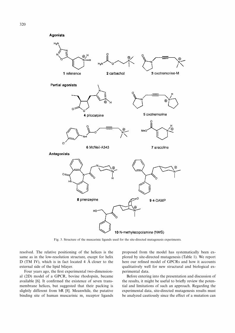
Docking of the partial agonist McN-A343 was particu-larly interesting. Although it is much larger than theformer agonists, each of its chemical moieties can beaccommodated easily, producing complementary interac-tions with specific residues in the receptor (Fig. 8). Thefirst interaction consists of an H-bond between the car-
bonyl oxygen of McN-A343 and Asn617. Next, the chloro-substituted phenyl moiety can interact with the extrahydrophobic pocket near the extracellular surface, be-tween helices 3, 4 and 5. This extra hydrophobic pocketis defined by the aromatic residues Phe502 located on helix5, Tyr432 on the second extracellular loop and Trp417,which is conserved in at least 85 GPCRs, including themuscarinic receptors. This observation justifies the refine-ment of the 3D model of the m1 receptor: in the originalmodel, because of the tilt of helix 4, it was difficult tofind a hydrophobic pocket large enough to accommodatethe phenyl ring of McN-A343. This pocket, which is notfilled in the case of full agonists, may constitute a pos-sible molecular feature that leads to partial agonism.
327
However, this finding needs to be supported by the study
Fig. 8. Stereoplot of McN-A343 docked in the binding site of the m1 muscarinic receptor.
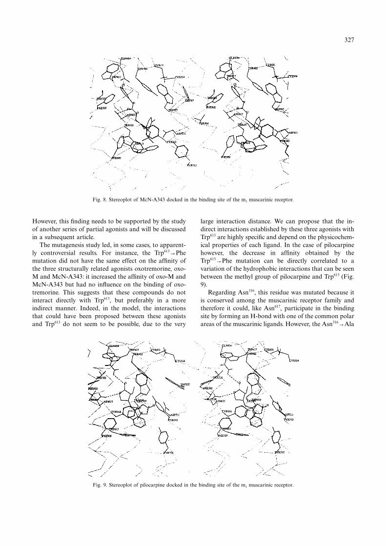
Fig. 9. Stereoplot of pilocarpine docked in the binding site of the m1 muscarinic receptor.
of another series of partial agonists and will be discussedin a subsequent article.
The mutagenesis study led, in some cases, to apparent-ly controversial results. For instance, the Trp613→Phemutation did not have the same effect on the affinity ofthe three structurally related agonists oxotremorine, oxo-M and McN-A343: it increased the affinity of oxo-M andMcN-A343 but had no influence on the binding of oxo-tremorine. This suggests that these compounds do notinteract directly with Trp613, but preferably in a moreindirect manner. Indeed, in the model, the interactionsthat could have been proposed between these agonistsand Trp613 do not seem to be possible, due to the very
large interaction distance. We can propose that the in-direct interactions established by these three agonists withTrp613 are highly specific and depend on the physicochem-ical properties of each ligand. In the case of pilocarpinehowever, the decrease in affinity obtained by theTrp613→Phe mutation can be directly correlated to avariation of the hydrophobic interactions that can be seenbetween the methyl group of pilocarpine and Trp613 (Fig.9).
Regarding Asn316, this residue was mutated because itis conserved among the muscarinic receptor family andtherefore it could, like Asn617, participate in the bindingsite by forming an H-bond with one of the common polarareas of the muscarinic ligands. However, the Asn316→Ala

328
mutation does not produce the same drastic effect onevery tested agonist and antagonist as Asn617→Ala (themost significant change in affinity was a decrease of 31-fold in the affinity of oxo-M). In the 3D model, Asn316
can form interhelical H-bonds, thus participating in theoverall conformation of the receptor. Thus, its mutationto Ala is likely to lead to a more indirect effect on thebinding of ligands. As small agonists do not have thesame hindrance in the receptor, they are not affected inthe same way by a change in the conformation of thereceptor. This might be responsible for the differentbinding affinities observed in the Asn316→Ala mutantreceptor.
Figure 10 shows a superposition of all the tested agon-ists in their receptor-bound conformation. We can seethat although they are concentrated in the same pocketand show similar structures, the different agonists presentdiverse binding modes. Particularly, there is no exactsuperposition of the cationic heads and of the two polarareas, in contrast to the muscarinic agonist pharmaco-phore models proposed by Trummlitz or Hoffmann,where the common electrostatic features can be superim-posed [16,34]. We can conclude that the indirect approachused to identify receptor–ligand interactions (i.e. the phar-macophore identification) cannot be considered as aunique mode of investigation. Although the receptormodel possesses some specific residues that were experi-mentally shown to bind the pharmacophoric elements, thedifferent agonists do not bind to the receptor in exactlythe same way. Although the conformations extractedfrom the superimposition are approximately conserved,their orientation in the binding pocket can vary relativelyto each other. In fact, the particular structure of eachagonist is responsible for its distinct binding mode andfor the varying orientation of the side chains of the bind-ing residues that accommodate it.
The antagonists are much larger molecules than theagonists (Fig. 11). They present, in the model, a moresimilar and rigid binding mode than agonist molecules,which is in agreement with their higher affinity values forthe parent receptor (Table 2). Due to their size, the antag-onists pirenzepine and 4-DAMP (Figs. 12a and b) fill thewhole binding pocket. Indeed, one of their aromatic ringscan be located in the hydrophobic pocket at the top ofthe binding cleft, interacting with residues Phe502, Trp417
and Tyr432. The second ring can form strong hydrophobicinteractions with Trp410 and Tyr613 located at the bottomof the binding cleft. This is consistent with the high de-crease in affinity obtained by the Trp613→Ala andTrp410→Phe mutations (Table 1). Furthermore, the cat-ionic head of these compounds is located close to Trp307,for which the mutation into Ala decreased the affinity bya factor of 13–29.
Structure–activity relationship studies have shown thatin the case of pirenzepine the endocyclic amide function
is essential for the interaction with the muscarinic recep-tor [35]. Indeed, in our model the endocyclic amide groupof pirenzepine can form very favorable H-bonds withAsn617. Mutation of this Asn to Ala in the m1 and m3
receptors produced mutant receptors which showed adrastically decreased affinity (>100-fold) for pirenzepine.Similarly, in a model of the binding mode of pirenzepineto the human m1 receptor, an H-bond between the endo-cyclic function of pirenzepine and Asn617 was proposed,on the basis that the Asn617→Asp mutation decreased theaffinity of pirenzepine by a factor of 612 [29].
Besides these H-bonds with Asn617, in our model theN4 nitrogen atom of pirenzepine is ideally located toestablish an ionic interaction with Asp311. This observa-tion is in agreement with experimental data, whichshowed that, at physiological pH, pirenzepine is mainlymonoprotonated, and this protonation occurs on the N4nitrogen [35]. Moreover, some analogues of pirenzepinehave been synthesized. These molecules only possessed theN4 nitrogen in their structure and were shown to be asactive as pirenzepine itself [36].
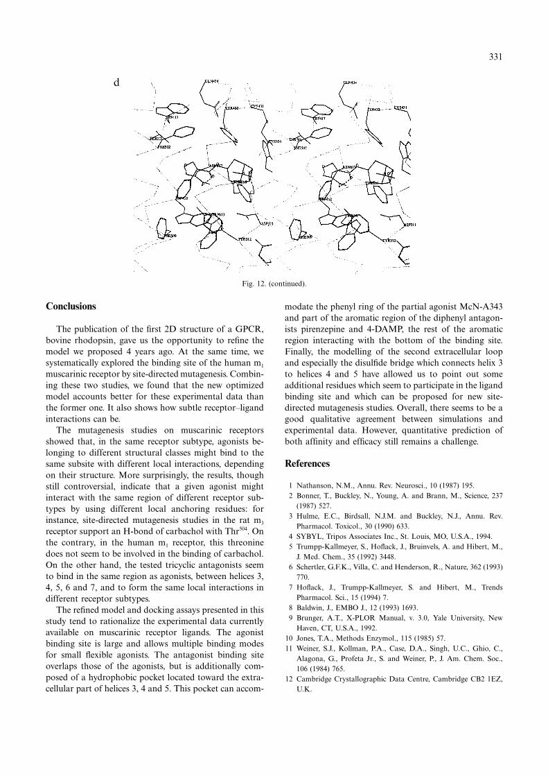
Like the two other antagonists, NMS has a muchlarger volume than agonists. Nevertheless, the moleculestill has some flexibility and, thus, two possible bindingmodes can be identified. In the first one (Fig. 12c), thecarbonyl oxygen of NMS is involved in an H-bond withAsn617. The phenyl ring of NMS is located at the top ofthe extra hydrophobic pocket between helices 3, 4 and 5,forming hydrophobic interactions with Phe502, Trp417 andTyr432. In the second binding mode (Fig. 12d), NMS canform two H-bonds with the Asn617 residue, with both itscarbonyl and hydroxyl functions. The importance of thehydroxyl function in the binding of NMS-like com-pounds has been demonstrated by Barlow and Ramtoola[37]. In this second possible binding mode, the phenylring of NMS forms strong hydrophobic interactions withTrp410 and Trp613 located at the bottom of the agonistbinding site. Actually, mutations of these two residues(Trp410→Phe and Trp613→Ala) lead to a relatively strongdecrease in affinity (97- and 115-fold, respectively). Inagreement with these findings, the second binding modeseems to be much more plausible.
In both cases, an H-bond could be established betweenthe epoxide moiety of NMS and Tyr616 on helix 6. Thisadditional interaction could account for the increase inaffinity obtained when the epoxide moiety is added toatropine.
It should be noted that the binding pocket for tricyclicand diphenyl molecules is probably also present in theneurokinin-1 receptor: site-directed mutagenesis studiesshowed that a histidine residue located on helix 5 of thisreceptor interacts directly with the benzhydryl of CP-96345, a substance P antagonist, suggesting that othernearby residues within this transmembrane domain shouldalso interact with the antagonist [38].

329
Fig. 10. Superposition view of the complexes made by the tested agonists with the human m1 receptor model. The color code is as follows:carbachol, pink; oxo-M, orange; pilocarpine, yellow; oxotremorine, red; McN-A343, cyan; arecoline, blue.
Fig. 11. Superposition view of the complexes made by the tested antagonists with the m1 receptor. The color code is as follows: pirenzepine, red;4-DAMP, yellow; NMS, blue.

330
a
b
c
Fig. 12. Stereoplots of the tested antagonists docked in the binding site of the m1 muscarinic receptor: (a) pirenzepine; (b) 4-DAMP; (c) NMS (firstbinding mode); (d) NMS (second binding mode).
331
Conclusions
d
Fig. 12. (continued).
The publication of the first 2D structure of a GPCR,bovine rhodopsin, gave us the opportunity to refine themodel we proposed 4 years ago. At the same time, wesystematically explored the binding site of the human m1
muscarinic receptor by site-directed mutagenesis. Combin-ing these two studies, we found that the new optimizedmodel accounts better for these experimental data thanthe former one. It also shows how subtle receptor–ligandinteractions can be.
The mutagenesis studies on muscarinic receptorsshowed that, in the same receptor subtype, agonists be-longing to different structural classes might bind to thesame subsite with different local interactions, dependingon their structure. More surprisingly, the results, thoughstill controversial, indicate that a given agonist mightinteract with the same region of different receptor sub-types by using different local anchoring residues: forinstance, site-directed mutagenesis studies in the rat m3
receptor support an H-bond of carbachol with Thr504. Onthe contrary, in the human m1 receptor, this threoninedoes not seem to be involved in the binding of carbachol.On the other hand, the tested tricyclic antagonists seemto bind in the same region as agonists, between helices 3,4, 5, 6 and 7, and to form the same local interactions indifferent receptor subtypes.
The refined model and docking assays presented in thisstudy tend to rationalize the experimental data currentlyavailable on muscarinic receptor ligands. The agonistbinding site is large and allows multiple binding modesfor small flexible agonists. The antagonist binding siteoverlaps those of the agonists, but is additionally com-posed of a hydrophobic pocket located toward the extra-cellular part of helices 3, 4 and 5. This pocket can accom-
modate the phenyl ring of the partial agonist McN-A343and part of the aromatic region of the diphenyl antagon-ists pirenzepine and 4-DAMP, the rest of the aromaticregion interacting with the bottom of the binding site.Finally, the modelling of the second extracellular loopand especially the disulfide bridge which connects helix 3to helices 4 and 5 have allowed us to point out someadditional residues which seem to participate in the ligandbinding site and which can be proposed for new site-directed mutagenesis studies. Overall, there seems to be agood qualitative agreement between simulations andexperimental data. However, quantitative prediction ofboth affinity and efficacy still remains a challenge.
References
1 Nathanson, N.M., Annu. Rev. Neurosci., 10 (1987) 195.2 Bonner, T., Buckley, N., Young, A. and Brann, M., Science, 237
(1987) 527.3 Hulme, E.C., Birdsall, N.J.M. and Buckley, N.J., Annu. Rev.
Pharmacol. Toxicol., 30 (1990) 633.4 SYBYL, Tripos Associates Inc., St. Louis, MO, U.S.A., 1994.5 Trumpp-Kallmeyer, S., Hoflack, J., Bruinvels, A. and Hibert, M.,
J. Med. Chem., 35 (1992) 3448.6 Schertler, G.F.K., Villa, C. and Henderson, R., Nature, 362 (1993)
770.7 Hoflack, J., Trumpp-Kallmeyer, S. and Hibert, M., Trends
Pharmacol. Sci., 15 (1994) 7.8 Baldwin, J., EMBO J., 12 (1993) 1693.9 Brunger, A.T., X-PLOR Manual, v. 3.0, Yale University, New
Haven, CT, U.S.A., 1992.10 Jones, T.A., Methods Enzymol., 115 (1985) 57.11 Weiner, S.J., Kollman, P.A., Case, D.A., Singh, U.C., Ghio, C.,
Alagona, G., Profeta Jr., S. and Weiner, P., J. Am. Chem. Soc.,106 (1984) 765.
12 Cambridge Crystallographic Data Centre, Cambridge CB2 1EZ,U.K.
332
13 Allen, F.H., Kennard, O. and Taylor, R., Acc. Chem. Res., 16(1983) 146.
14 Melchiorre, C., Minarini, A., Budriesi, R., Chiarini, A., Spam-pinato, S. and Tumiatti, V., Life Sci., 11/12 (1995) 837.
15 Dewar, M.J.S., Zoebish, E.G., Healy, E.F. and Stewart, J.J.P., J.Am. Chem. Soc., 107 (1985) 3902.
16 Hoffmann, R., Bourguignon, J.J. and Wermuth, C.G., Pharmaco-chem. Libr., 16 (1991) 283.
17 Showell, G.A., Tracey, L.G., Kneen, C.O., MacLeod, A.M., Mer-chant, K.J., Saunders, J., Freedman, S.B., Patel, S. and Baker, R.,J. Med. Chem., 34 (1991) 1086.
18 Hibert, M., Trumpp-Kallmeyer, S., Bruinvels, A. and Hoflack, J.,Mol. Pharmacol., 40 (1991) 8.
19 Henderson, R., Baldwin, J.M. and Ceska, T.A., J. Mol. Biol., 213(1990) 899.
20 Grigorieff, N., Ceska, T.A., Downing, K.H., Baldwin, J.M. andHenderson, R., J. Mol. Biol., 259 (1996) 393.
21 Kenakin, T., Trends Pharmacol. Sci., 16 (1995) 188.22 Curtis, C.A.M., Wheatley, M., Bansal, S., Birdsall, N.J.M.,
Eveleigh, P., Pedder, E.K., Poyner, D. and Hulme, E.C., J. Biol.Chem., 264 (1989) 489.
23 Matsui, H., Lazareno, S. and Birdsall, N.J.M., Mol. Pharmacol.,47 (1995) 88.
24 P9 poster, 7th Swiss Workshop of Methodology in ReceptorResearch, 1996.
25 Olah, M.E., Jacobson, K.A. and Stiles, G.L., J. Mol. Biol., 269(1994) 24692.
26 Hulme, E.C., Curtis, C.A.M., Page, K.M. and Jones, P.G., LifeSci., 11/12 (1995) 891.
27 Wess, J., Gdula, D. and Brann, M.R., EMBO J., 10 (1991)3729.
28 Wess, J., Nanavati, S., Vogel, Z. and Maggio, R., EMBO J., 12(1993) 331.
29 Murgolo, N.J., Kozlowski, J., Tice, M.A.B., Hollinger, F.P.,Brown, J.E., Zhou, G., Taylor, L.A. and McQuade, R.D., Bioorg.Med. Chem. Lett., 6 (1996) 785.
30 Dixon, R.A.F., Strader, C.D. and Sigal, I.S., Annu. Rep. Med.Chem., 23 (1988) 221.
31 Bluml, K., Mutschler, E. and Wess, J., J. Biol. Chem., 29 (1994)18870.
32 Nordvall, G. and Hacksell, U., J. Med. Chem., 36 (1993) 967.33 Fanelli, F., Menziani, M.C., Carotti, A. and De Benedetti, P.G.,
Bioorg. Med. Chem., 3 (1994) 195.34 Kier, L.B., Mol. Pharmacol., 3 (1967) 487.35 Eberlein, W.G., Trummlitz, G., Engel, W.W., Schmidt, G., Pelzer,
H. and Mayer, N., J. Med. Chem., 30 (1987) 1378.36 Quintero, M.G., Donetti, A., Giachetti, A., Ladinsky, H.,
Limonta, P., Micheletti, R. and Schiavi, G.B., IXth InternationalSymposium on Medicinal Chemistry, West Berlin, Abstract Book,1986.
37 Barlow, R.B. and Ramtoola, S., Br. J. Pharmacol., 71 (1980)31.
38 Fong, T.M., Cascieri, M.A., Yu, H., Bansal, A., Swain, C. andStrader, C.D., Nature, 362 (1993) 350.