Embed Size (px)
Citation preview
Modelling acid-mediated tumour invasion
Antonio FasanoDipartimento di Matematica U. Dini, Firenze
Levico, sept. 2008
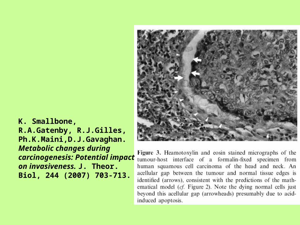
K. Smallbone, R.A.Gatenby, R.J.Gilles, Ph.K.Maini,D.J.Gavaghan. Metabolic changes during carcinogenesis: Potential impact on invasiveness. J. Theor. Biol, 244 (2007) 703-713.
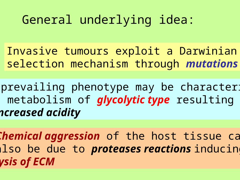
General underlying idea:
Invasive tumours exploit a Darwinianselection mechanism through mutations
The prevailing phenotype may be characterizedby a metabolism of glycolytic type resulting in an increased acidity
Chemical aggression of the host tissue canalso be due to proteases reactions inducinglysis of ECM
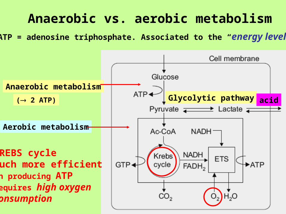
KREBS cycleMuch more efficient in producing ATPRequires high oxygen consumption
Aerobic metabolism
Glycolytic pathway
Anaerobic metabolism
Anaerobic vs. aerobic metabolism
( 2 ATP)
ATP = adenosine triphosphate. Associated to the “energy level”
acid
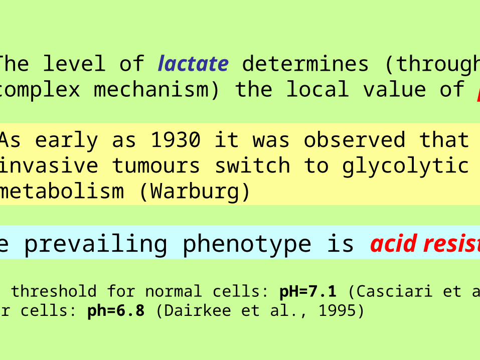
The level of lactate determines (through acomplex mechanism) the local value of pH
As early as 1930 it was observed that invasive tumours switch to glycolyticmetabolism (Warburg)
The prevailing phenotype is acid resistant
Apoptosis threshold for normal cells: pH=7.1 (Casciari et al., 1992)For tumour cells: ph=6.8 (Dairkee et al., 1995)
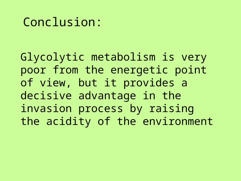
Conclusion:
Glycolytic metabolism is very poor from the energetic point of view, but it provides a decisive advantage in the invasion process by raising the acidity of the environment
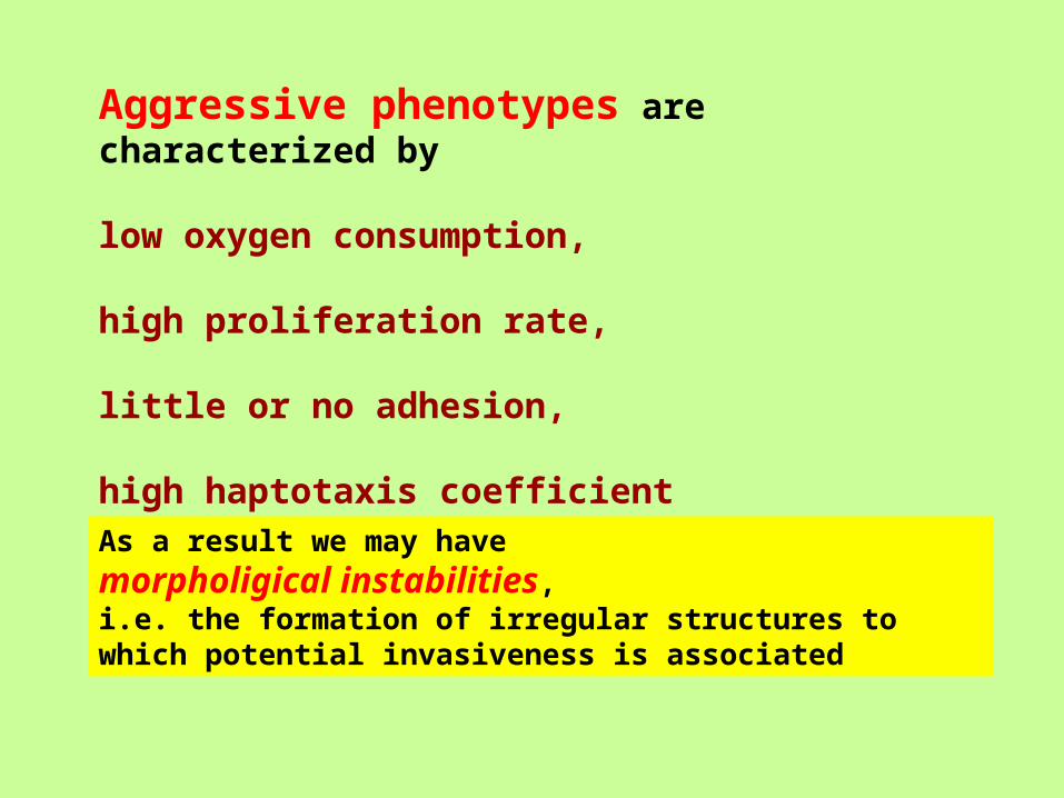
Aggressive phenotypes are characterized by
low oxygen consumption, high proliferation rate,
little or no adhesion,
high haptotaxis coefficient
As a result we may have
morpholigical instabilities, i.e. the formation of irregular structures to which potential invasiveness is associated
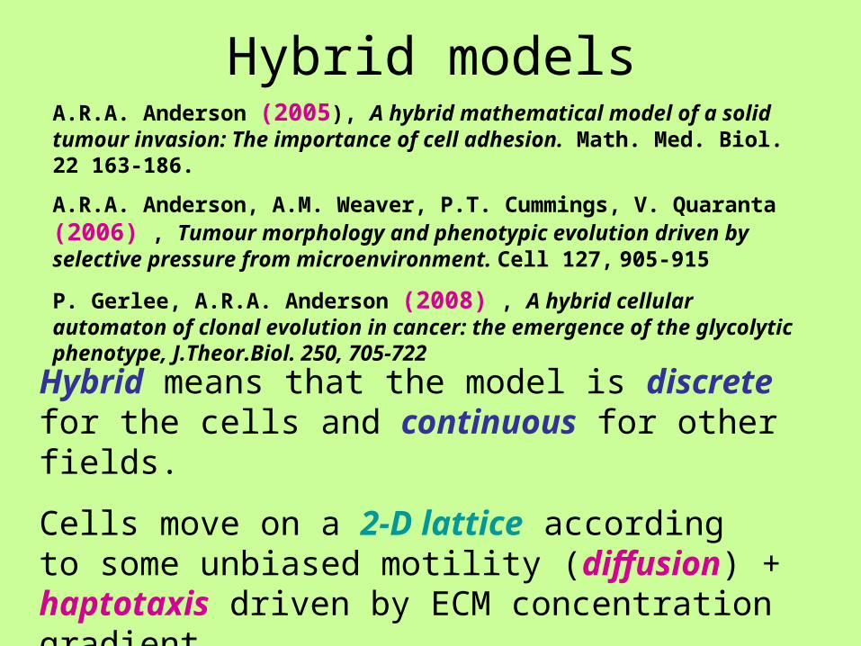
Hybrid modelsA.R.A. Anderson (2005), A hybrid mathematical model of a solid tumour invasion: The importance of cell adhesion. Math. Med. Biol. 22 163-186.
A.R.A. Anderson, A.M. Weaver, P.T. Cummings, V. Quaranta (2006) , Tumour morphology and phenotypic evolution driven by selective pressure from microenvironment. Cell 127, 905-915
P. Gerlee, A.R.A. Anderson (2008) , A hybrid cellular automaton of clonal evolution in cancer: the emergence of the glycolytic phenotype, J.Theor.Biol. 250, 705-722
Hybrid means that the model is discrete for the cells and continuous for other fields.
Cells move on a 2-D lattice according to some unbiased motility (diffusion) + haptotaxis driven by ECM concentration gradient
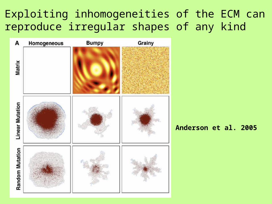
Exploiting inhomogeneities of the ECM canreproduce irregular shapes of any kind
Anderson et al. 2005
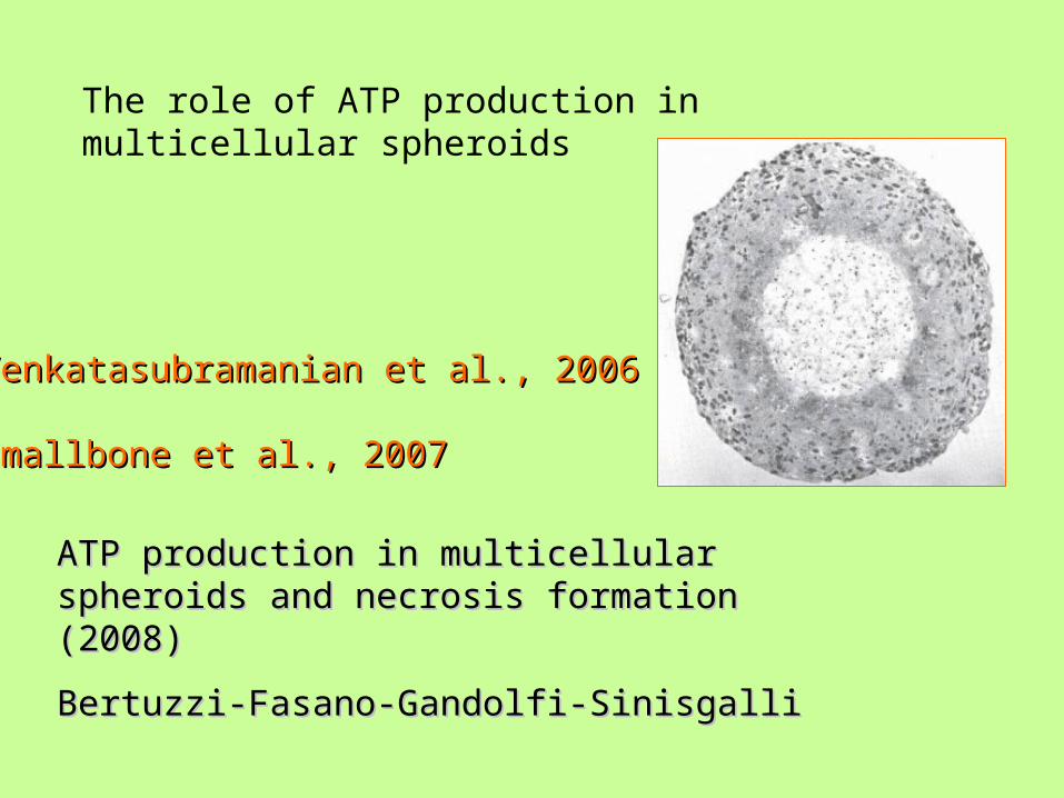
Venkatasubramanian et al., 2006Venkatasubramanian et al., 2006
Smallbone et al., 2007Smallbone et al., 2007
ATP production in multicellular spheroids and ATP production in multicellular spheroids and necrosis formation (2008)necrosis formation (2008)
Bertuzzi-Fasano-Gandolfi-SinisgalliBertuzzi-Fasano-Gandolfi-Sinisgalli
The role of ATP production in multicellular spheroids
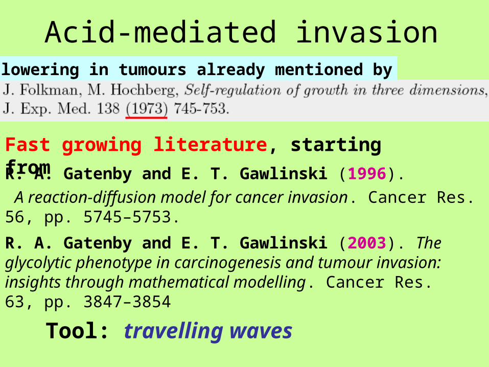
Acid-mediated invasion
Fast growing literature, starting from
R. A. Gatenby and E. T. Gawlinski (1996).
A reaction-diffusion model for cancer invasion. Cancer Res. 56, pp. 5745–5753.
R. A. Gatenby and E. T. Gawlinski (2003). The glycolytic phenotype in carcinogenesis and tumour invasion: insights through mathematical modelling. Cancer Res. 63, pp. 3847–3854
Tool: travelling waves
pH lowering in tumours already mentioned by
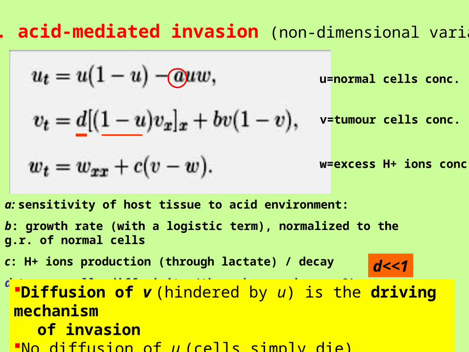
G.G. acid-mediated invasion (non-dimensional variables)
a: sensitivity of host tissue to acid environment:
b: growth rate (with a logistic term), normalized to the g.r. of normal cells
c: H+ ions production (through lactate) / decay
d: tumour cells diffusivity (through gap, i.e. u=0) d<<1
w=excess H+ ions conc.
v=tumour cells conc.
u=normal cells conc.
Diffusion of v (hindered by u) is the driving mechanism of invasion
No diffusion of u (cells simply die)
The model has several limitations concerning the biological mechanisms involved
• no extracellular fluid
• instantaneous removal of dead cells
• metabolism is ignored
Therefore is goal is simply to show that there is a mathematcal structure able to reproduce invasion
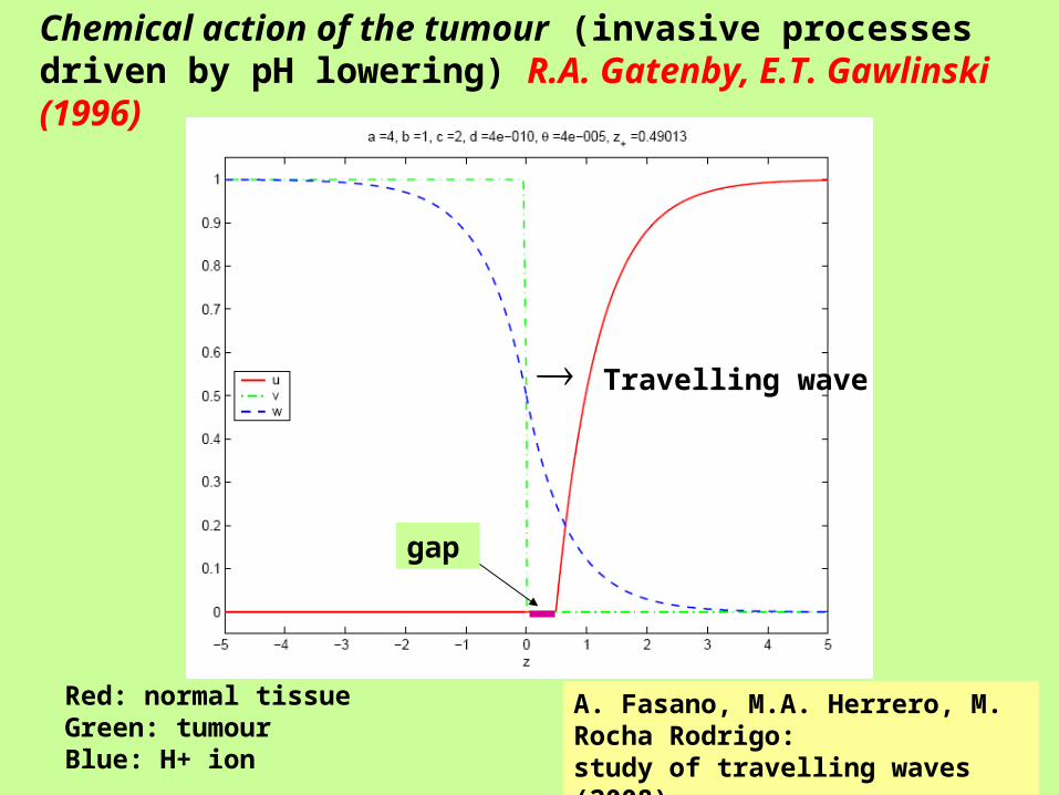
Chemical action of the tumour (invasive processes driven by pH lowering) R.A. Gatenby, E.T. Gawlinski (1996)
Red: normal tissueGreen: tumourBlue: H+ ion
A. Fasano, M.A. Herrero, M. Rocha Rodrigo:study of travelling waves (2008)
Travelling wave
gap
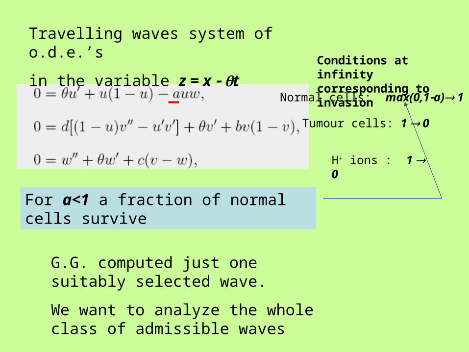
Travelling waves system of o.d.e.’s
in the variable z = x t
Normal cells: max(0,1a) 1
Tumour cells: 1 0
H+ ions : 1 0
Conditions at infinity corresponding to invasion
For a<1 a fraction of normal cells survive
G.G. computed just one suitably selected wave.
We want to analyze the whole class of admissible waves
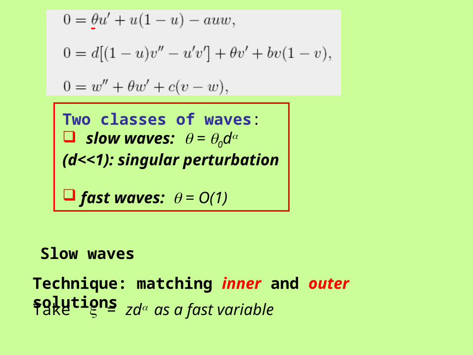
Two classes of waves: slow waves: = 0d (d<<1): singular perturbation
fast waves: = O(1)
Technique: matching inner and outer solutions
Take = zd as a fast variable
Slow waves
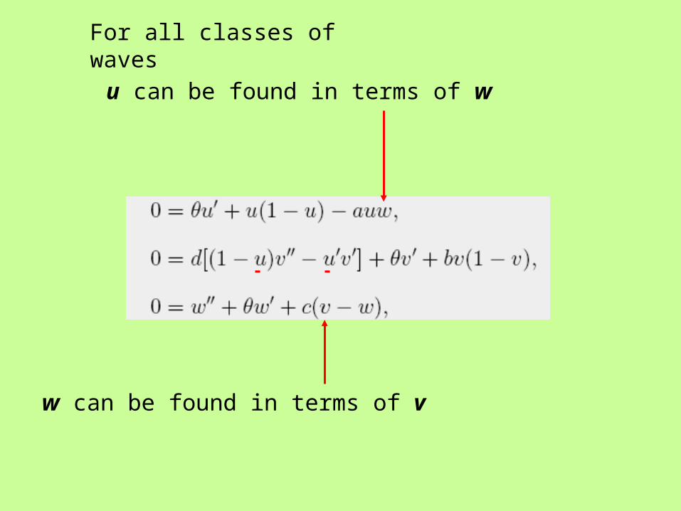
u can be found in terms of w
w can be found in terms of v
For all classes of waves
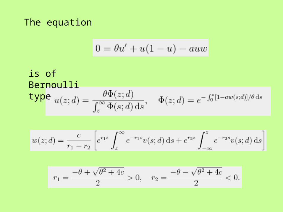
The equation
is of Bernoulli type
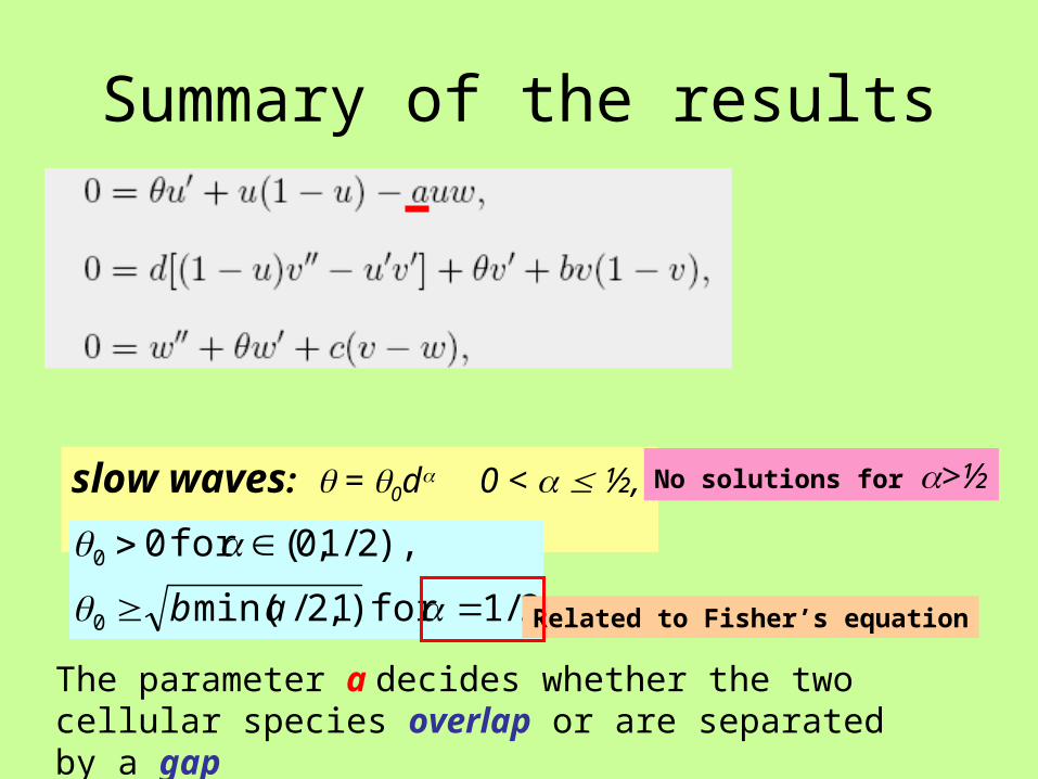
Summary of the results
slow waves: = 0d 0 < ½,
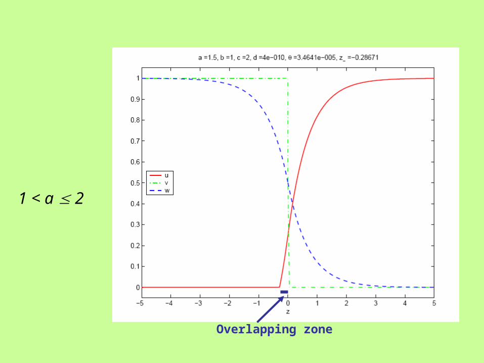
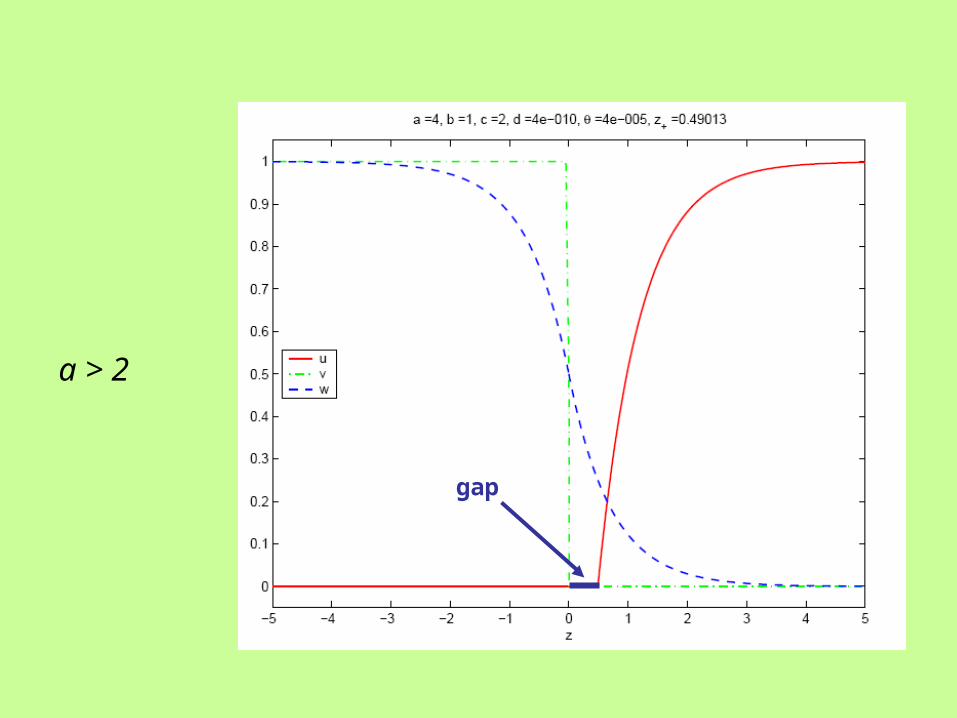
The parameter a decides whether the two cellular species overlap or are separated by a gap
2/1for )1,2/min(
),2/1,0(for 0
0
0
ab
No solutions for >½
Related to Fisher’s equation
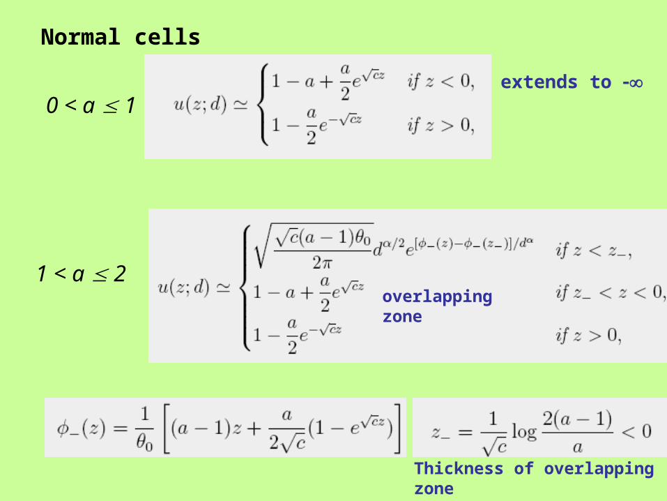
0 < a 1
1 < a 2overlapping zone
extends to
Thickness of overlapping zone
Normal cells
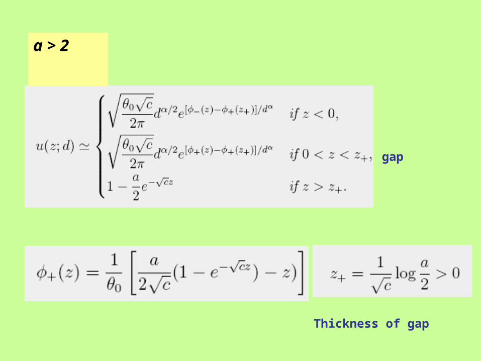
a > 2
gap
Thickness of gap
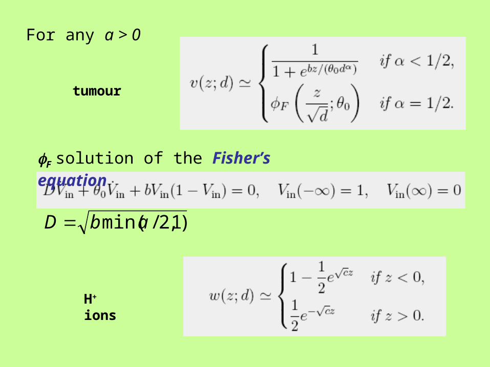
For any a > 0
F solution of the Fisher’s equation
)1,2/min(abD
tumour
H+ ions
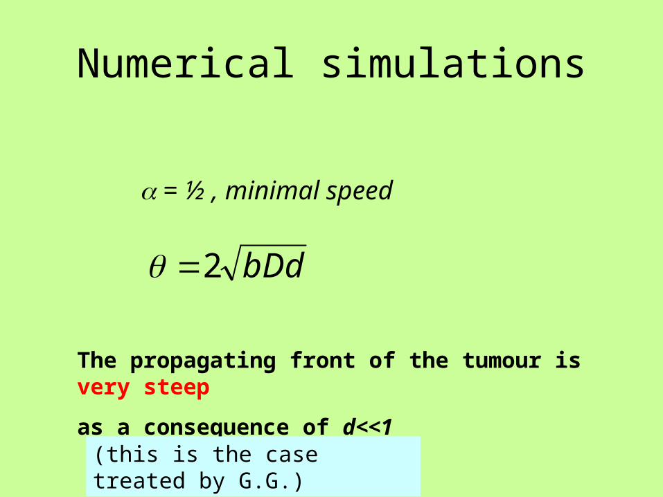
Numerical simulations
= ½ , minimal speed
bDd2
The propagating front of the tumour is very steep
as a consequence of d<<1
(this is the case treated by G.G.)
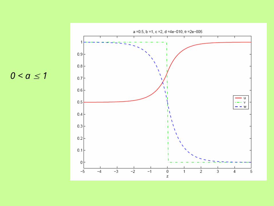
0 < a 1
1 < a 2
Overlapping zone
a > 2
gap
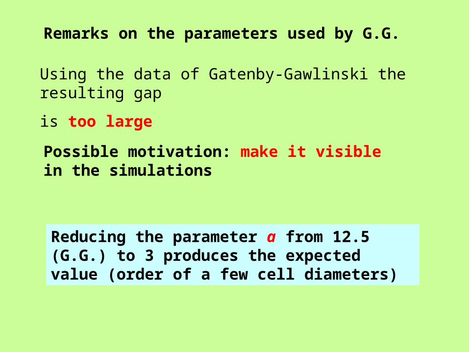
Using the data of Gatenby-Gawlinski the resulting gap
is too large
Possible motivation: make it visible in the simulations
Reducing the parameter a from 12.5 (G.G.) to 3 produces the expected value (order of a few cell diameters)
Remarks on the parameters used by G.G.
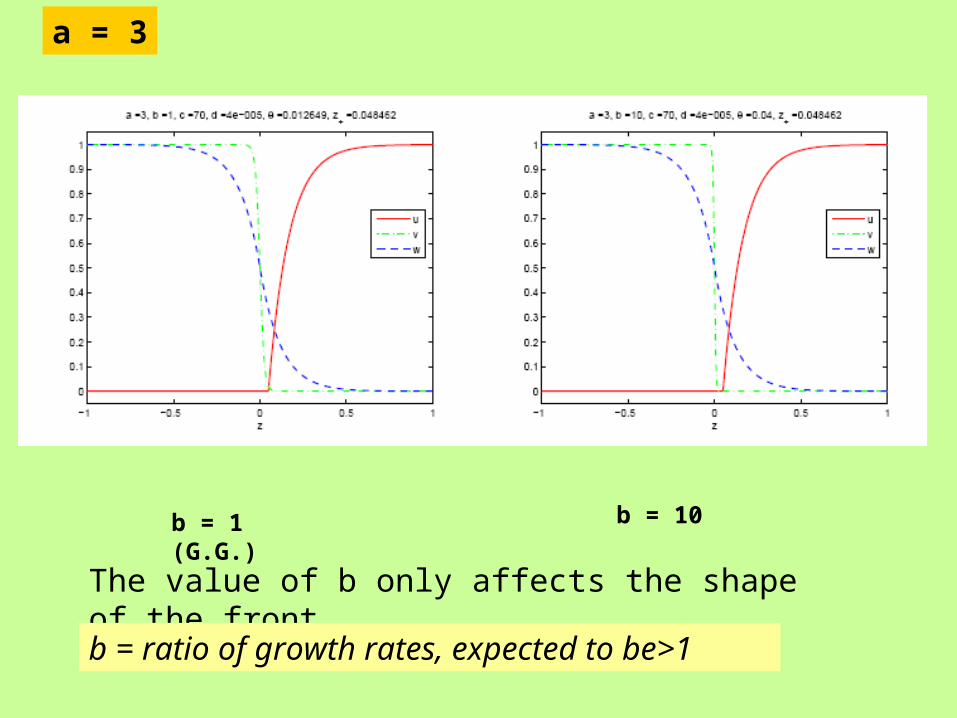
a = 3
b = 1 (G.G.) b = 10
The value of b only affects the shape of the front
b = ratio of growth rates, expected to be>1
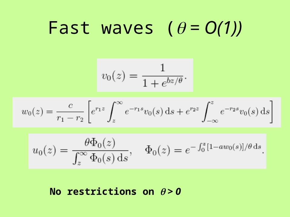
Fast waves ( = O(1))
No restrictions on > 0
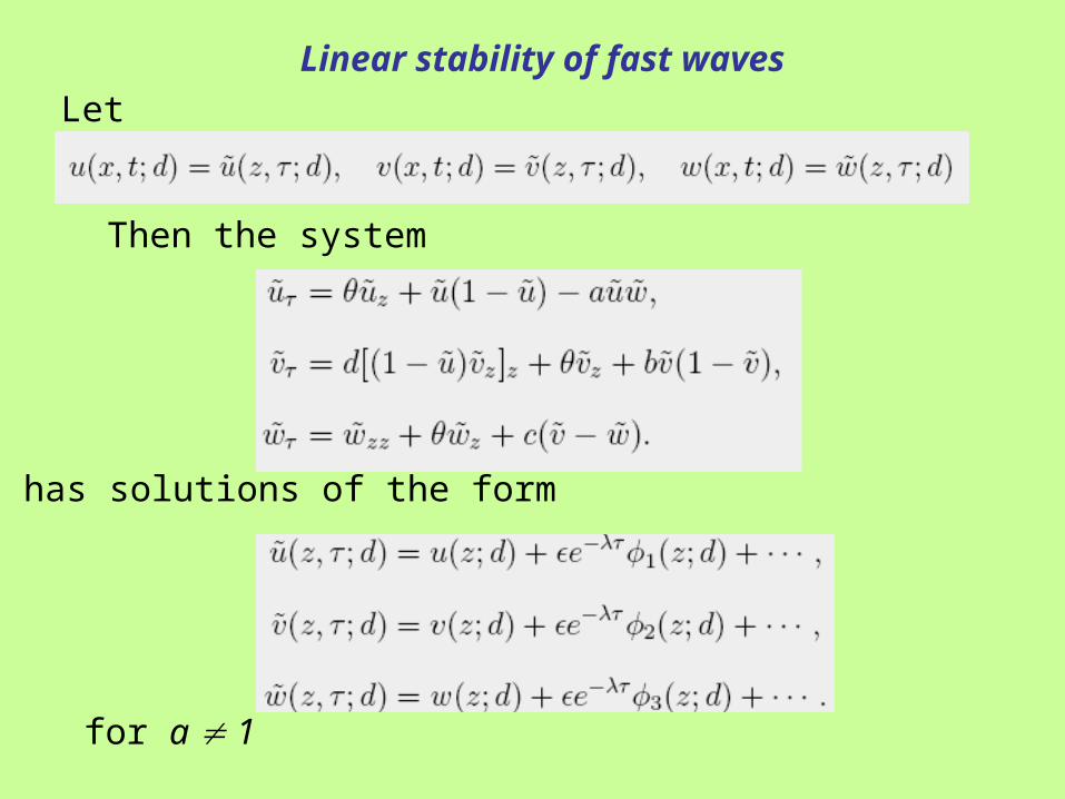
Let
Then the system
has solutions of the form
for a 1
Linear stability of fast waves
Other invasion models are based on a combined mechanism of ECM lysis and haptotaxis
(still based on the analysis of travelling waves)
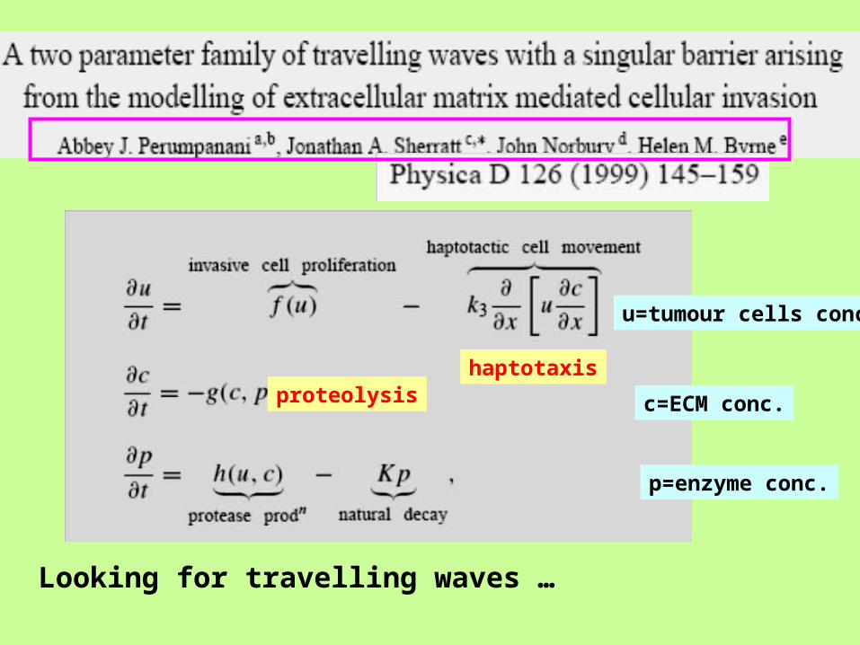
haptotaxisproteolysis
Looking for travelling waves …
u=tumour cells conc.
c=ECM conc.
p=enzyme conc.
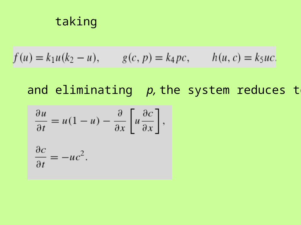
taking
and eliminating p, the system reduces to
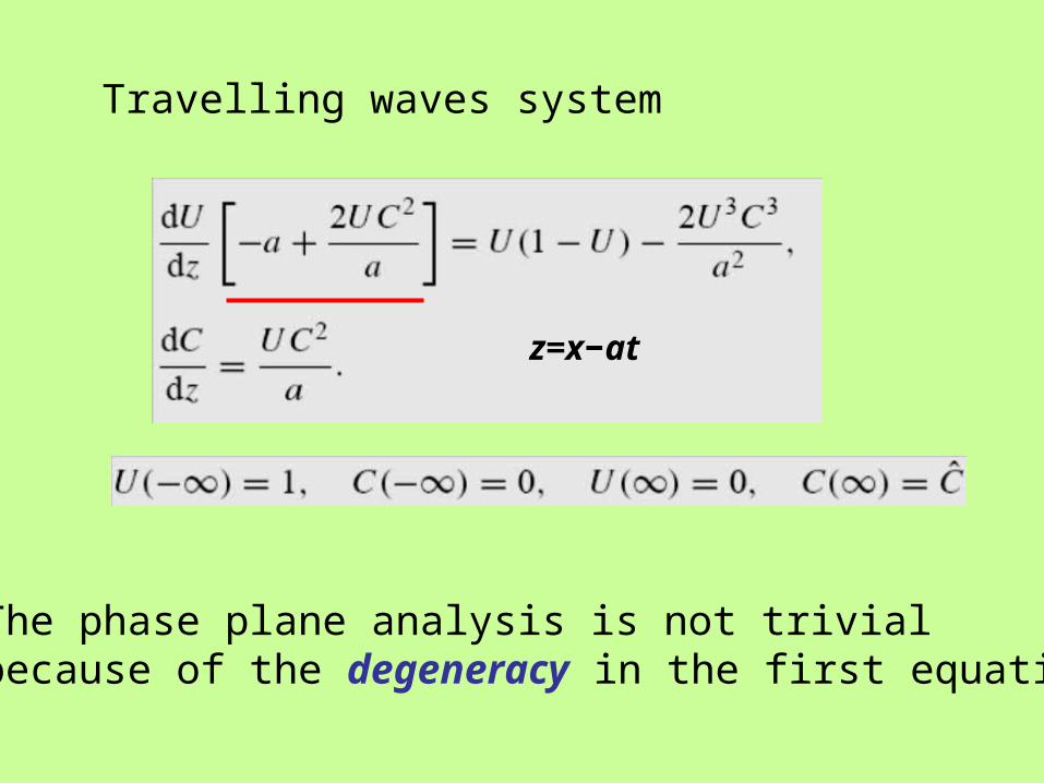
Travelling waves system
z=x−at
The phase plane analysis is not trivialbecause of the degeneracy in the first equation
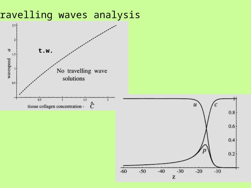
travelling waves analysis
t.w.
tumour cells
ECM
enzyme
diffusion haptotaxis
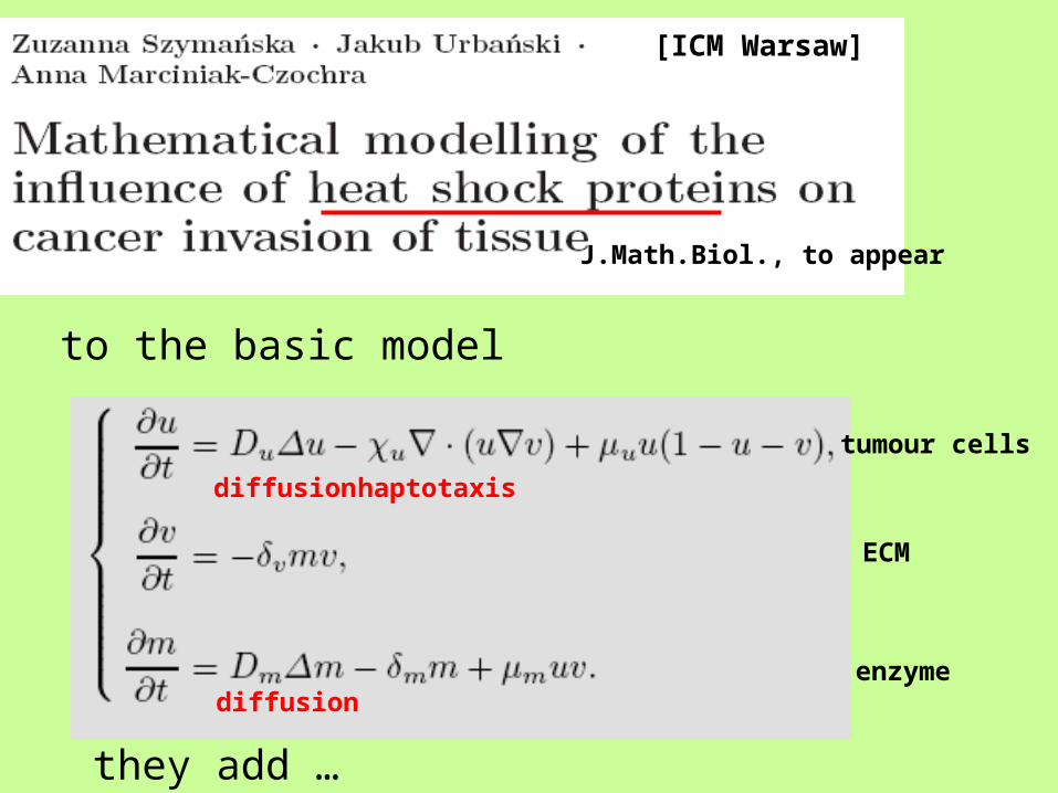
to the basic model
J.Math.Biol., to appear
they add …
diffusion
[ICM Warsaw]
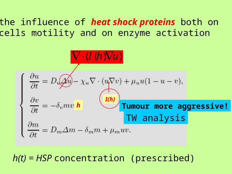
hI(h)
the influence of heat shock proteins both on cells motility and on enzyme activation
))(( uhI
h(t) = HSP concentration (prescribed)
Tumour more aggressive!
TW analysis
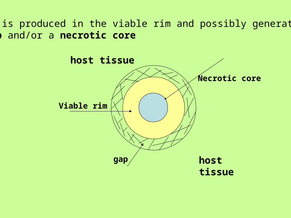
Viable rim
Necrotic core
gap
host tissue
host tissue
Acid is produced in the viable rim and possibly generate a gap and/or a necrotic core
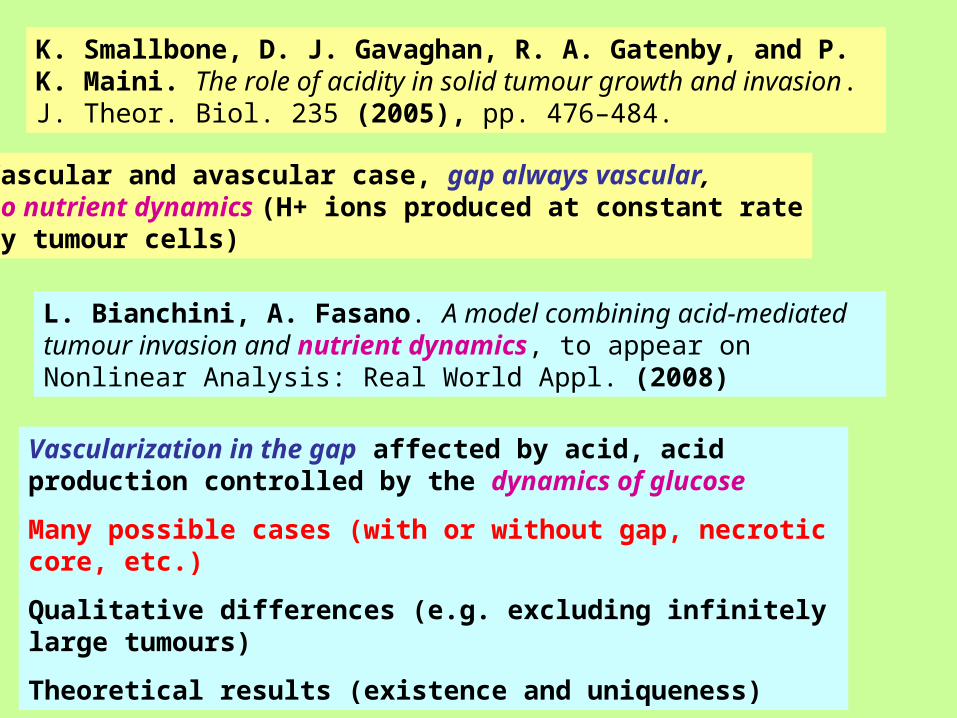
K. Smallbone, D. J. Gavaghan, R. A. Gatenby, and P. K. Maini. The role of acidity in solid tumour growth and invasion. J. Theor. Biol. 235 (2005), pp. 476–484.
L. Bianchini, A. Fasano. A model combining acid-mediated tumour invasion and nutrient dynamics, to appear on Nonlinear Analysis: Real World Appl. (2008)
Vascular and avascular case, gap always vascular,no nutrient dynamics (H+ ions produced at constant rateby tumour cells)
Vascularization in the gap affected by acid, acid production controlled by the dynamics of glucose
Many possible cases (with or without gap, necrotic core, etc.)
Qualitative differences (e.g. excluding infinitely large tumours)
Theoretical results (existence and uniqueness)