Embed Size (px)
Citation preview
Implementing the probe beam deflection
technique for acoustic sensing in
photoacoustic and ultrasound imaging
Ronald A. Barnes Jr.
The University of Texas at San Antonio
This work is a collaboration between The University of Texas at San Antonio and The University of Texas
Health Science Center.
Outline• Introduction
• Background
• Modeling (MATLAB)
– Acoustic Wave Propagation
– Ray Tracing
• Simulation (MATLAB)
– Optimum Sensor Topology
– Optimum Beam Topology
– Quadrant Photodiode Simulation
– Acoustic Wave Directionality Measurement
– Sensor Frequency Response
• Visualization (ParaView and MATLAB)
• Conclusion
Introduction
• What is Photoacoustic Tomography?Photoacoustic Tomography (PAT) is accomplished by measuring the propagating acoustic energy
radiated from a sample of tissue whose thermal expansion is invoked by a pulse laser. An image of the tissue composition is reconstructed based on the measurement of the of this acoustic energy.
• What is the Probe Beam Deflection Technique?The Probe Beam Deflection Technique (PBDT) is sensing topology that uses probe beam lasers and
there deflection and refraction to measure the properties of the propagating acoustic wave, through the implementation of a Quadrant Photodiode (QPD).
• Why is Modeling and Simulation important for this project?
To develop an efficient algorithm for reconstruction of a tissue composition image, one must understand the interaction between probe beam and propagating acoustic wave front. A ray tracing simulation in combination with an acoustic wave simulation will allow for the prediction of beam deflection or refraction for various experimental topologies and implementations.

Background (PAT)
• Light enters a scattering medium (Ex. Tissue Phantom) where a portion of the energy is absorbed by the tissue in the form of heat, this produces thermal expansion.
• If the temperature increase inside the phantom occurs at a faster rate then the thermal relaxation time of the tissue, an acoustic wave will propagate as a result of the photo-acoustic effect.
• This acoustic wave produced is a wideband ultrasonic transmission and to date is measured with piezoelectric transducers.
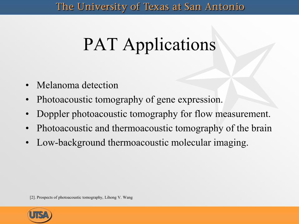
PAT Applications
• Melanoma detection
• Photoacoustic tomography of gene expression.
• Doppler photoacoustic tomography for flow measurement.
• Photoacoustic and thermoacoustic tomography of the brain
• Low-background thermoacoustic molecular imaging.
[2]. Prospects of photoacoustic tomography, Lihong V. Wang
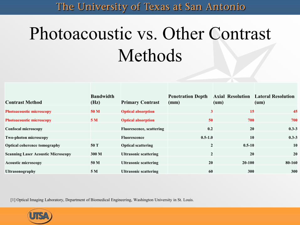
Photoacoustic vs. Other Contrast
Methods
Contrast Method
Bandwidth
(Hz) Primary Contrast
Penetration Depth
(mm)
Axial Resolution
(um)
Lateral Resolution
(um)
Photoacoustic microscopy 50 M Optical absorption 3 15 45
Photoacoustic microscopy 5 M Optical absorption 50 700 700
Confocal microscopy Fluorescence, scattering 0.2 20 0.3-3
Two-photon microscopy Fluorescence 0.5-1.0 10 0.3-3
Optical coherence tomography 50 T Optical scattering 2 0.5-10 10
Scanning Laser Acoustic Microscopy 300 M Ultrasonic scattering 2 20 20
Acoustic microscopy 50 M Ultrasonic scattering 20 20-100 80-160
Ultrasonography 5 M Ultrasonic scattering 60 300 300
[1] Optical Imaging Laboratory, Department of Biomedical Engineering, Washington University in St. Louis.
Background (PBDT)
• PBDT is implemented by focusing probe beams through an enclosure filled with a propagation medium. As an acoustic wave travels through the medium the refractive index is changed relative to the pressure gradient produced by the wave. The probe beam deflects and refracts as it interacts with the refractive index profile along its beam path.
• The probe beam deflection technique offers various advantages when compared to transducers, these include: Wave front directionality measurement, passive sensing, and low implementation cost.

Development of a Model
• Step 1: Produce a model of acoustic wave propagation in homogeneous and heterogeneous mediums based on the 2nd order PDE governing acoustic wave propagation.
• Step 2: Modify this model in such a way that all parameters are adjustable. This includes: Initial acoustic wave magnitude, propagation medium properties, acoustic wave frequency, etc.
• Step 3: Convert the pressure values in the four dimensional dataset (3 dim. for space and 1 for time) to refractive index using the lorentz-lorenz relation.
• Step 4: Develop a ray tracing simulation to trace a bundle of rays through the previously created dataset using the vector form of Snells law. This simulation should have adjustable parameters which include: initial ray origin (for all rays that make up beam), initial ray intensity, and initial ray direction.
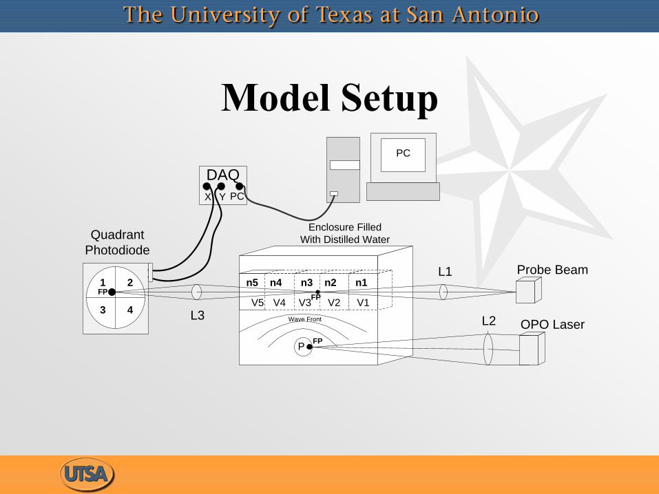
Model Setup
n1n2n3n4n51 2
3 4
Quadrant
Photodiode
L1
DAQ
OPO Laser
P
L2
FP
L3
Enclosure Filled
With Distilled Water
X Y
V1V2V3V4V5
FP
PC
Wave Front
Probe Beam
FP
PC
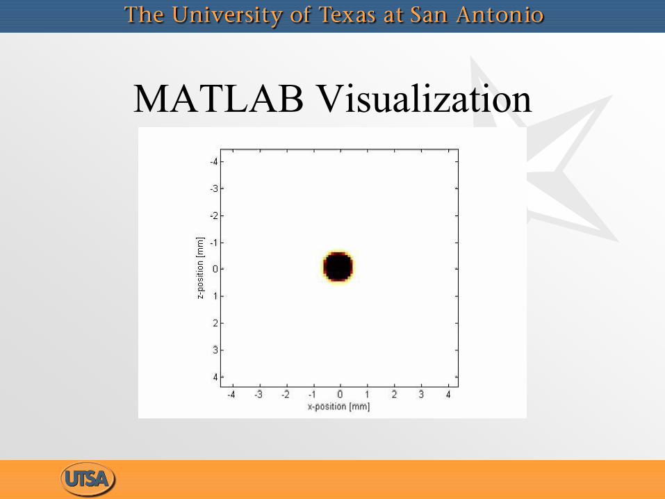
MATLAB Visualization
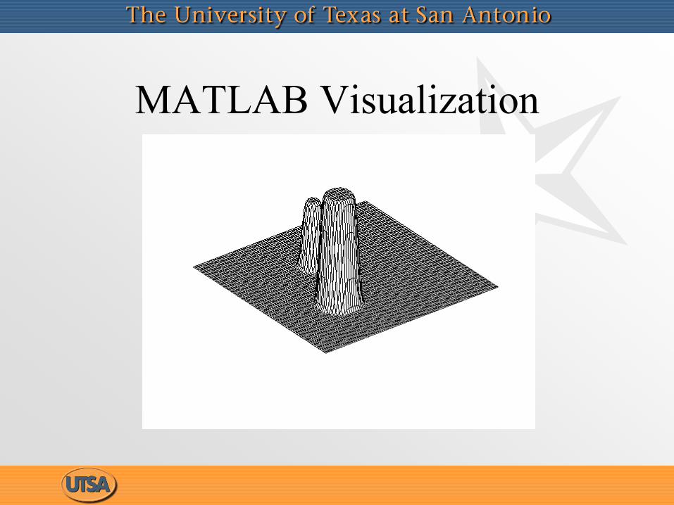
MATLAB Visualization
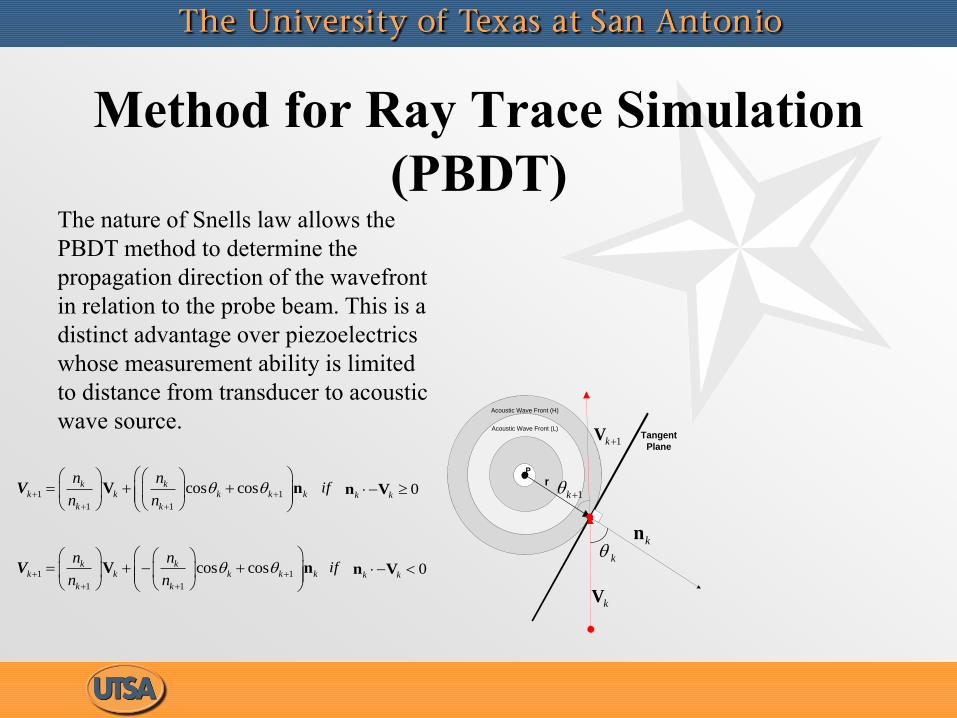
Method for Ray Trace Simulation
(PBDT)The nature of Snells law allows the
PBDT method to determine the
propagation direction of the wavefront
in relation to the probe beam. This is a
distinct advantage over piezoelectrics
whose measurement ability is limited
to distance from transducer to acoustic
wave source.
r
k
P
1k
Tangent
Plane
Acoustic Wave Front (H)
Acoustic Wave Front (L)
kn
kV
1kV
1 1
11
cos coskk k
kk k k
kk
n n
n n
V nV if 0k k n V
1 1
1 1
cos cosk kk k k
k k
k k
n n
n n
V nV if 0k k n V
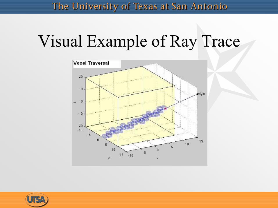
Visual Example of Ray Trace
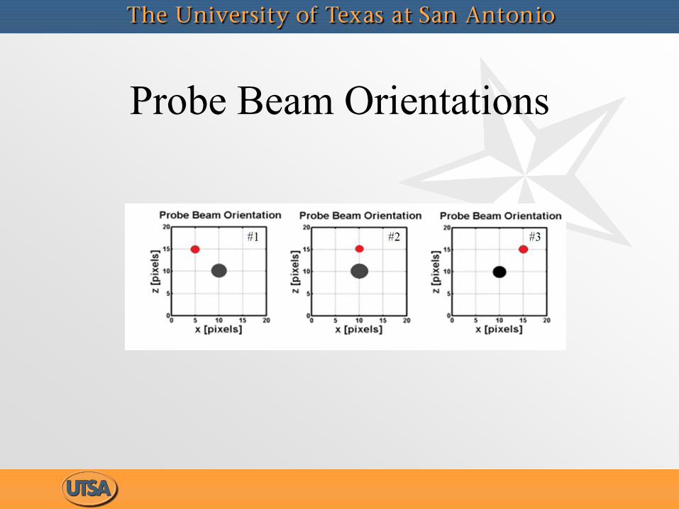
Probe Beam Orientations
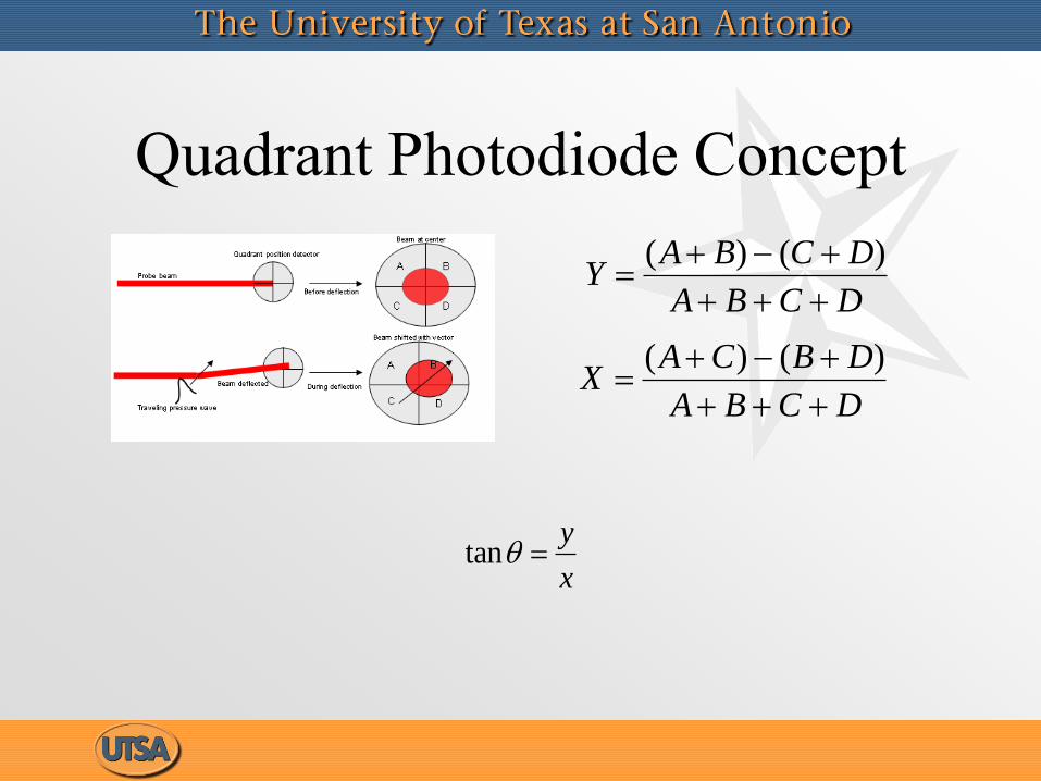
Quadrant Photodiode Concept
DCBA
DBCAX
)()(
DCBA
DCBAY
)()(
tany
x
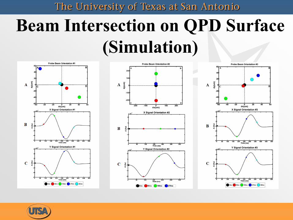
Beam Intersection on QPD Surface
(Simulation)
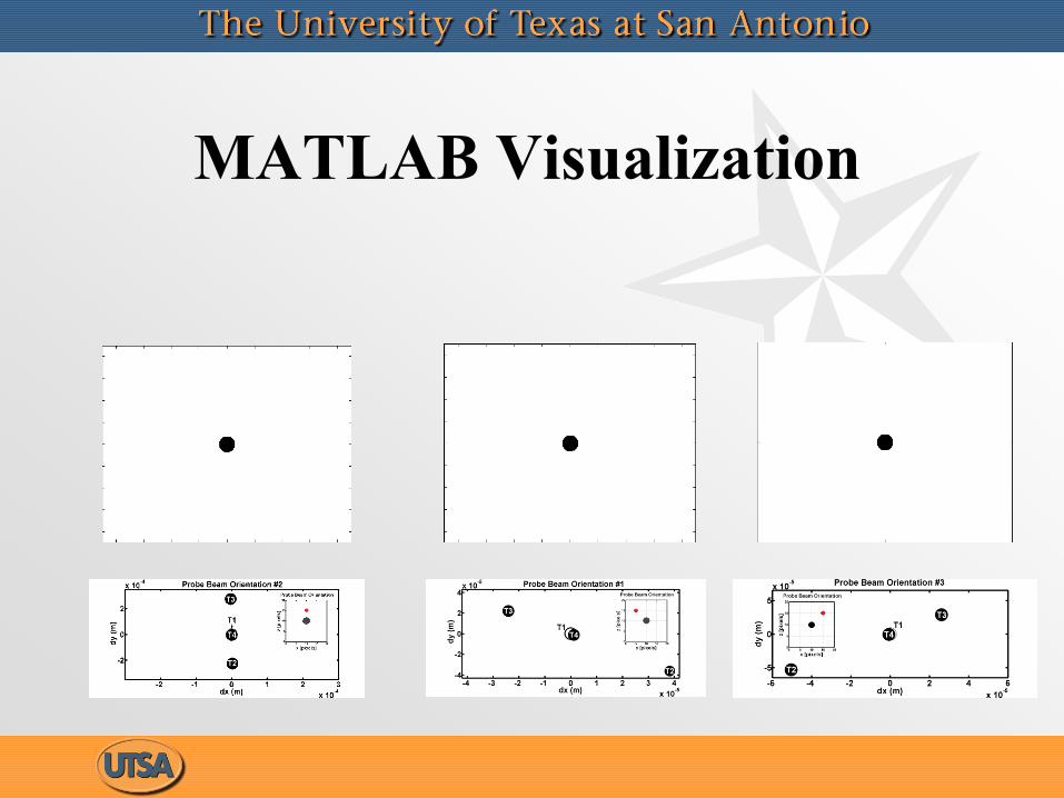
MATLAB Visualization
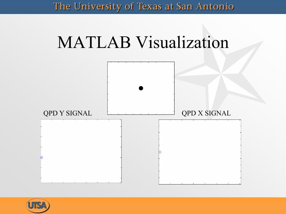
MATLAB Visualization
QPD X SIGNALQPD Y SIGNAL
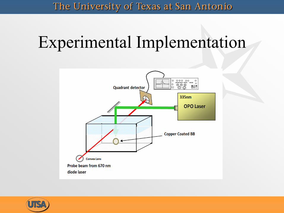
Experimental Implementation

Probe Beam Orientations
(Experiment)
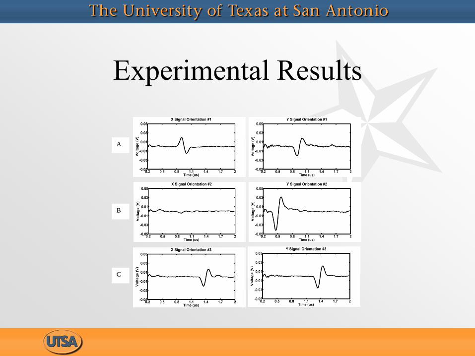
Experimental Results
A
B
C
Future Work• Define optimum beam and sensor
topologies for experimental implementation
of PBDT derived from simulation.
• Define the frequency response of PBDT and
compare to the frequency response to
commercially available transducers.
• Develop reconstruction algorithm based on
integrating line detectors as proposed by G.
Paltauf but with added angular information.
Acknowledgments
• NSF grant (HRD-0932339), Drs. Demetris Kazakos and
Richard Smith, project managers.
• PREM Grant # DMR- 0934218.

References• André Conjusteau, Saher Maswadi, Sergey Ermilov, Hans-Peter Brecht, Norman Barsalou, Randolph D. Glickman
and Alexander A. Oraevsky, "Detection of gold-nanorod targeted pathogens using optical and piezoelectric
optoacoustic sensors: comparative study", Proc. SPIE 7177, 71771P (2009); doi:10.1117/12.813434
• B. E. Treeby and B. T. Cox, "A k-space Green's function solution for acoustic initial value problems in homogeneous
media with power law absorption," J. Acoust. Soc. Am., vol. 129, no. 6, pp. 3652-3660, 2011.
• B. E. Treeby, E. Z. Zhang, and B. T. Cox, "Photoacoustic tomography in absorbing acoustic media using time
reversal," Inverse Prob., vol. 26, no. 11, p. 115003, 2010.
• B. E. Treeby and B. T. Cox, "Modeling power law absorption and dispersion for acoustic propagation using the
fractional Laplacian," J. Acoust. Soc. Am., vol. 127, no. 5, pp. 2741-2748, 2010
• B. E. Treeby and B. T. Cox, "k-Wave: MATLAB toolbox for the simulation and reconstruction of photoacoustic
wave-fields," J. Biomed. Opt., vol. 15, no. 2, p. 021314, 2010.
• B. T. Cox and B. E. Treeby, "Artifact trapping during time reversal photoacoustic imaging for acoustically
heterogeneous media," IEEE Trans. Med. Imaging, vol. 29, no. 2, pp. 387-396, 2010.
• B. T. Cox, S. Kara, S. R. Arridge, and P. C. Beard, "k-space propagation models for acoustically heterogeneous
media: Application to biomedical photoacoustics," J. Acoust. Soc. Am., vol. 121, no. 6, pp. 3453-3464, 2007
• B. T. Cox and P. C. Beard, "Fast calculation of pulsed photoacoustic fields in fluids using k-space methods," J.
Acoust. Soc. Am., vol. 177, no. 6, pp. 3616-3627, 2005.
• Wang, L. V., Ed. . Photoacoustic Imaging and Spectroscopy. Taylor & Francis, 2009.

Questions??