Embed Size (px)
Citation preview
Modeling Protein Folding Pathways
Christopher Bystroff, Yu Shao
Dept of Biology
Rensselaer Polytechnic Institute, Troy, NY.
e-mail:{bystrc, shaoy}@rpi.edu
Summary
Proteins fold through a series of intermediate states called a pathway. Protein
folding pathways have been modeled using either simulations or a heirarchy ofstatistical models. Here we present a series of related statistical models that at-
tempt to predict early, middle and late intermediates along the folding pathway. I-
sites motifs are discrete models for folding initiation sites. HMMSTR is a modelfor local structure patterns composed of I-sites motifs. HMMSTR-CM is an ap-
proach toward assembling motifs and groups of motifs in a contact map represen-tation, using heuristic rules to predict contact maps either with or without the use
of templates. We also discuss the I-sites/ROSETTA server, which is a foldingsimulation algorithm that uses a fragment library as input. The results of blind
structure prediction experiments are discussed. Pathway-based predictions some-
times lead to an unambiguous prediction of the fold topology, even without usingtemplates.
1. Introduction: Darwin versus Boltzmann
All computational models that predict something have certain underlying as-
sumptions that constitute the physical basis for the model. In protein structure pre-diction, there are two physical/biological processes that can be modeled: the proc-
ess of evolution, or the process of folding. We may give these two paradigms
names, Darwin and Boltzmann, after the scientists who defined the fundamentalprinciples of evolutionary biology and statistical thermodynamics, respectively.
2 Christopher Bystroff, Yu Shao
Most of the work in protein structure prediction is Darwin-based, using thewell-known premise that sequences that have a common ancestor have similar
folds, and they strive to extrapolate this principle to increasingly distant sequencerelationships. Methods that use multiple sequence alignment, structural alignment,
or "threading potentials" are implicitly searching for a common ancestor. Despite
the oft-used “energy-like” scoring functions, these methods do not address thephysical process of folding. Evolution happens on the time scale of millions of
years, folding on the time scale of fractions of a second.Protein structure prediction of the Boltzmann kind is perceived to be a very dif-
ficult problem. Many have tried their hand at it over the last thirty years, and an
equal number have failed to improve upon Darwin-based methods. The problemof predicting folding pathways may be perceived to be even harder, since it should
depend on first solving the protein folding problem. But this is not true, as weshall see. Prediction of the protein folding pathway may be evaluated by looking
at the success in predicting sub-segments or substructures of proteins. If the com-putational model has the right underlying assumptions about what comes first in
the pathway, and what comes next, and so on, then blind predictions, such as those
done as part of CASP, the Critical Assessment of Protein Structure Prediction bi-annual worldwide experiment (Moult et al. 2001), may validate that model. And
the pathway model that eventually arises from this process will tell us more thanjust final answer.
In this chapter we present a series of bioinformatics and simulation experimentsrelated to predicting protein structure by modeling the folding pathway. We will
conclude that ab initio predictions can be done either by simulations or by a rule-
based fragment assembly method, and that it is possible to find folds that are notpresent in the database of structures. We will discuss issues of accuracy and
resolution and present some possible directions for the future.
1.1 Protein Folding Pathway History
The early work of Levinthal and Anfinsen established that a protein chain foldsspontaneously and reproducibly to a unique three dimensional structure when
placed in aqueous solution. Levinthal proved beyond the shadow of a doubt that
the folding process cannot occur by random diffusion. Anfinsen proposed thatproteins must form intermediate structures in a time-ordered sequence of events,
or "pathway" (Anfinsen and Scheraga 1975). The nature of the pathways, specifi-cally whether they are restricted to partially native states or whether they might
Modeling Protein Folding Pathways 3
include non-specific interactions, such as an early collapse driven by the hydro-phobic effect, was left unanswered.
Over the years, the theoretical models for folding have converged somewhat(Baldwin 1995, Colon and Roder 1996, Oliveberg et al. 1998, Pande et al. 1998),
in part due to a better understanding of the structure of the so-called “unfolded
state" (Dyson and Wright 1996, Gillespie and Shortle 1997, Mok et al. 1999) andto a more detailed description of kinetic and equilibrium folding intermediates
(Eaton et al. 1996, Gulotta et al. 2001, Houry et al. 1996). An image of the transi-tion state of folding can now be mapped out by point mutations, or "phi-value
analysis" (Fersht et al. 1992, Grantcharova et al. 2000, Heidary and Jennings
2002, Mateu et al. 1999, Nolting et al. 1997). The "folding funnel" model (Chan etal. 1995, Onuchic et al. 1997) has reconciled hydrophobic collapse with the alter-
native nucleation-condensation model (Nolting and Andert 2000) by envisioning adistorted, funicular energy landscape (Laurents and Baldwin 1998) and a "mini-
mally frustrated" pathway (Nymeyer et al. 2000, Shoemaker and Wolynes 1999)through this landscape. The view remains of a channeled, counter-entropic search
for the hole in the funnel as the predominant barrier to folding (Zwanzig 1997).
Simulations using various simplified representations of the protein chain, in-cluding lattice models, have clarified the basic nature of folding pathways
(Kolinski and Skolnick 1997, Mirny and Shakhnovich 2001, Shakhnovich 1998,Thirumalai and Klimov 1998). The topology of the fold plays a dominant role in
defining the critical positions that effect the folding rate (Ortiz and Skolnick 2000,Shea and Brooks 2001). Models that represent the chain in atomistic detail show
that minimally frustrated, low-energy pathways may involve the propagation of
structure along the chain like a zipper (Alm and Baker 1999, Munoz et al. 1998).All-atom, explicit solvent molecular dynamics simulations have reproduced the
experimentally determined conformations for short peptides (Cavalli et al. 2002,Duan and Kollman 1998, Garcia and Sanbonmatsu 2001, Krueger and Kollman
2001, Shao and Bystroff 2003). This large body of work is still inconclusive, butclearly folding is best represented by an ensemble rather than a single pathway.
2. Knowledge-based Models for Folding Pathways
The approach that began with I-sites is an attempt to build a hierarchical series
of models mirroring the hierarchy of folding events, from initiation to nucleationto propagation and condensation. The hierarchy can be roughly described as “lo-
4 Christopher Bystroff, Yu Shao
5 10
5 5
10 1010
5
a. b.
cal to global.” Each model builds on the model before it. At each point the resultsare an ensemble of conformational states.
"Local structure" is a generic term for the conformations of short pieces of theprotein chain, usually 3-20 residue pieces. Local structure motifs include the two
common forms (alpha helix and beta strand) along with a few dozen turns, half-turns, caps, bulges and coils. The role of local structure motifs with regard to the
initiation of folding has been discussed by Baldwin, Rooman and others (Baldwin
and Rose 1999, Efimov 1993, Rooman et al. 1990).
2.1. I-sites: A Library of Folding Initiation Site Motifs
I-sites is a library of 262 sequence patterns that map to local structures. A se-quence pattern is expressed as a position-specific scoring matrix (PSSM). Recur-
rent sequence patterns had been previously used for prediction of structural motifs,including the Schellman motif (Schellman 1980), the hydrophobic staple (Munoz
et al. 1995), and various types of coiled coil (Woolfson and Alber 1995). Recur-
rent sequence patterns of various lengths were found by exhaustively clusteringshort segments of sequence profiles for proteins in a non-redundant database of
known structures (Bystroff et al. 1996, Han and Baker 1996, 1995, Han et al.1997). Bystroff and Baker mapped recurrent sequence patterns to their predomi-
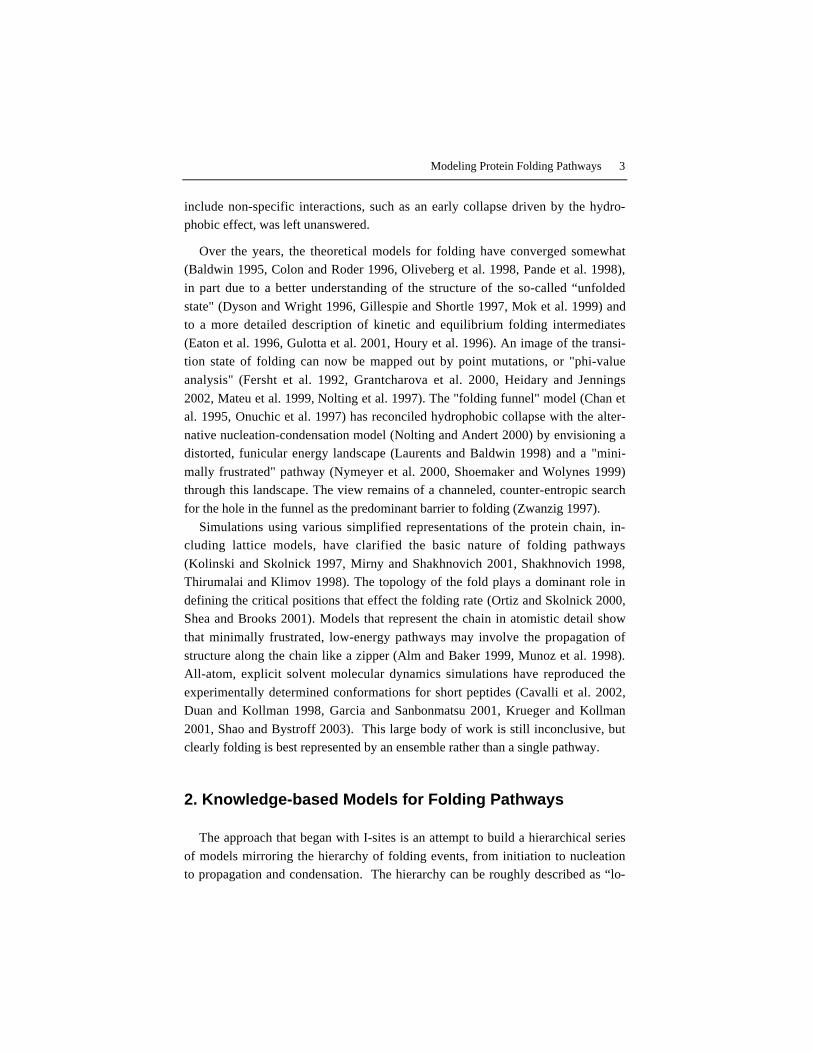
nant structural motifs and used reinforcement learning to optimize the sequence-structure correlation (Bystroff and Baker 1998). The resulting I-sites Library (Fig.
1) has been used in various prediction experiments (Bystroff and Baker 1997, By-
stroff and Shao 2002) and has inspired numerous experimental studies since its
Fig. 1. a. I-sites profile for alpha-alpha corner motif. Boxes are shaded lighter in propor-tion to the log-likelihood ratio of each amino acid at each position relative to the start ofthe motif. b. Stereo image of the alpha-alpha corner motif.
Modeling Protein Folding Pathways 5
1
2
3
4
7
11
12 13
14
15
16 17
18
19 20
21
22
23
24
25
26
27 28 29
30 31 32
33 34 35 36
37
38
39
40
41
42
43
44
45 46 47
48
49 50 51 52
53
54
55
56 57
58 59
60 61
62
63
64
65 6667
68 69
70
71
73
74
75
80 81
82
83
84
85 86
87
88
89
90
94 95 96 97
98 99
100
101
102 103 104
105
106
107
108
109 110 111 112
113
114
115 116
117
118 119 120
121
122
123
124
125
126
127
129 130
131 132
133
134
135
136
138 139
140
141
142
143 144 145
146
147 148 149
151 152 153
154 155
156
157
158
159
160
161 162 163
164
165
166
169
170
171 172 173 174
175 176 177
178
179
180 181 182 183
184
185
186
187
188
189 190
191
192
193
194
195 196
197
198
199
200
201
204 205
206
207
208
209
215
216 217
218
219 220
221
222
223
224
225
226 227 228
231 232
236 237
238 239
240
241 242 243
244
245
246
247
248
249
250
254
255
256
257 258
259 260
261
262
263
264
265
266
267
268
269 270 271 272 273
274
275
277
278
279 280
281
publication (Jacchieri 2000, Mendes et al. 2002, Northey et al. 2002b, Skolnick
and Kolinski 2002, Steward and Thornton 2002). I-sites motifs have been linked
to local structure stability in both NMR studies (Blanco et al. 1994, Munoz et al.1995, Viguera and Serrano 1995, Yi et al. 1998) and molecular dynamics simula-
tions (Bystroff and Garde 2003, Gnanakaran and Garcia 2002, Krueger and Koll-man 2001). Mutations in high-confidence I-sites motif regions are found to have
dramatic effects on folding (Mok et al. 2001, Northey et al. 2002a). About one-third of all residues in all proteins are found in high-confidence (>70%) I-sites
motif regions and these sites are predicted to be conformationally stable and early-
folding.
2.2. HMMSTR: A Hidden Markov Model for Grammatical Structure
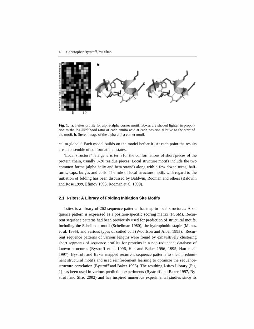
The I-sites library was condensed to a single, non-linear hidden Markov model(HMM), called HMMSTR ("hamster"). This model, trained on a large database of
protein structures and multiple sequence alignments, removes the fragment lengthdependence of I-sites motif predictions, models the adjacencies of motifs in pro-
teins, and puts all of the motifs on the same probability scale. Unlike profile
HMMs (Eddy 1996, Gough and Chothia 2002, Karplus et al. 1998), HMMSTRhas a highly branched and cyclic connectivity, containing for example a 7-residue
cycle of helix states representing the amphipathic helix heptad repeat motif. Bymodeling the adjacencies of motifs, HMMSTR is a model for the ways that local
structure can be arranged along the sequence, similar to the ways that words can
Fig. 2. HMMSTR represented as a directed graph. The symbol shape represents the secon-dary structure type; circles: helix; rectangles: beta sheet; diamonds: other motifs. Shadingrepresents the amino acid preference; dark grey: non-polar; grey: polar; light-grey: proline;lightest grey: glycine; white: no preference. Only high-probability transitions are shown.

6 Christopher Bystroff, Yu Shao
be arranged in a sentence. This is, in a simple way, a model for the grammaticalstructure of protein sequences, from words to phrases.
The result of a HMMSTR prediction is like that of any HMM, an ensemble ofMarkov state strings. Each string of states, one state for each position in the se-
quence, represents a probable arrangement of mutually-compatible local structure
motifs. A single prediction may be obtained from the ensemble by either selectingthe most probable state string, or better, by a voting procedure over the whole en-
semble (Bystroff et al. 2000). HMMSTR improved the overall accuracy in localstructure prediction over the I-sites method from 43% to 60% for 8-residue frag-
ments with RMSD < 1.4Å (Bystroff et al. 2000). HMMSTR has been used for lo-
cal and secondary structure prediction (Bystroff et al. 2000, Rost 2001), inter-residue contact prediction (Zaki et al. 2000), and as the source of a fragment li-
brary for Rosetta simulations (Bystroff and Shao 2002). Previous HMMs havemodeled proteins globally, not as fragments (Eddy 1996, Gough and Chothia
2002, Karplus et al. 1998).
3. ROSETTA: Folding Simulations Using a FragmentLibrary
The ROSETTA folding simulation algorithm uses Monte Carlo Fragment In-sertion (MCFI) to predict the 3D structures of small proteins or protein fragments
without the use of structural templates (Bonneau and Baker 2001, Bonneau et al.2001, Simons et al. 1999a, Simons et al. 1997, Simons et al. 1999b). MCFI is a
mostly downhill search in a knowledge-based energy landscape. Each MCFI move
consists of replacing the backbone angles of segments of the chain with fragmentsin a library. ROSETTA has been successful in prediction experiments (CASP
(Moult et al. 2001)) either using fragments from the database, from HMMSTR, orfrom the I-sites motif library.
In the version of ROSETTA that runs as a public server(www.bioinfo.rpi.edu), the fragment library is derived from I-sites fragment pre-
dictions, and the highest confidence I-sites were restrained to their predicted back-
bone angles to increase efficiency. Fragment insertion was allowed in the re-strained regions, but moves were constrained to deviate by more then 60° from the
I-sites prediction. Also, long sequences were simulated as overlapping short frag-ments of approximately 50 residues each, again for efficiency. The resulting pre-
dictions are spliced together at the end, using a genetic algorithm in conjunction
with the ROSETTA knowledge-based energy function. Detailed descriptions of
Modeling Protein Folding Pathways 7
each of the algorithms have been previously published (Bystroff and Shao 2002,Simons et al. 1997, Simons et al. 1999b).
3.1 Results of Fully Automated I-SITES/ROSETTA Simulations
3.1.1 Summary
A web server was used to predict 31 protein structures in the CASP4 experi-ment (2000) and 44 in the CASP5 experiment (2002). The successes and failures
of the server may be summarized in a few broad statements. The statistics andconclusions presented here refer to bona fide blind predictions sent automatically
to the CAFASP site as part of their “Fully-Automated” satellite experiment(Fischer et al. 2001). A more detailed analysis of this and other methods can be
obtained from the associated publications (Bystroff and Shao 2002, Shao and By-
stroff 2003).Over the 75 targets, 64% of the residues were found in "topologically correct"
large fragments, defined as fragments of 30 residues or more with RMSD < 6Å.At 6A RMSD, the correct overall chain trace has been reproduced, but not the
finer details of structure. Occasionally beta strand may be out of order in a sheet,and strands may be substituted for helices.
A smaller percentage of all 30-residue fragments, 44%, were predicted with a
5Å RMSD. At 5Å precision, secondary structure is occasionally mispredicted,loop structures may be wrong in detail, and axial rotations of secondary structure
units are possible. However, much or most of the non-local packing interactionsare faithfully though roughly reproduced at this level of accuracy, and strand mis-
pairing is not observed.
In practice, the details of the local structure are often correctly predicted when afragment was globally correct, but the RMSD measure is insensitive to this.
Therefore, another measure is used to evaluate the local accuracy of the predic-tions. The maximum deviation in backbone angles (mda) over a window of 8 resi-
dues is usually ~180° or small, and serves as a strictly local measure of correct-ness. 8-residue peptides that have mda < 90° and obey all of the stereochemical
constraints of a polypeptide, have an RMSD of 1.4Å at most (Bystroff and Baker
1998). Unfortunately, when mda is plotted alongside RMSD, it is immediately ob-vious that the good local structure predictions do not always coincide with the
good, large fragment predictions.
8 Christopher Bystroff, Yu Shao
3.1.2 Topologically correct large fragment predictions are found
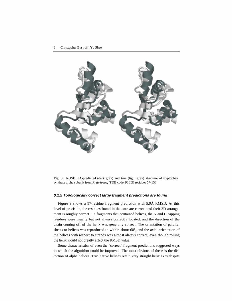
Figure 3 shows a 97-residue fragment prediction with 5.9Å RMSD. At this
level of precision, the residues found in the core are correct and their 3D arrange-ment is roughly correct. In fragments that contained helices, the N and C capping
residues were usually but not always correctly located, and the direction of thechain coming off of the helix was generally correct. The orientation of parallel
sheets to helices was reproduced to within about 60°, and the axial orientation of
the helices with respect to strands was almost always correct, even though rollingthe helix would not greatly effect the RMSD value.
Some characteristics of even the "correct" fragment predictions suggested waysin which the algorithm could be improved. The most obvious of these is the dis-
tortion of alpha helices. True native helices retain very straight helix axes despite
Fig. 3. ROSETTA-predicted (dark grey) and true (light grey) structure of tryptophansynthase alpha subunit from P. furiosus, (PDB code 1GEQ) residues 57-153.
Modeling Protein Folding Pathways 9
variability in the backbone angles. Helices in the predictions, however, were oftendistorted, sometimes bending the axis by 90° over its length. A combination of
factors produces these errors. ROSETTA has no energy penalty for helix distor-tion, while it gives a large energetic bonus for packing hydrophobic residues in the
core and for maintaining a low radius of gyration. Bent helices are found to re-
place helix kinks and alpha-alpha corners. Adding a penalty for helix distortionmight fix this problem.
Topological correctness is a weak criterion for usefulness, since it means thatonly the handedness of the chain reversals and most of the secondary structure are
right. However, these fragmentary predictions may narrow the search space for a
structural analog or remote homolog, and may therefore be useful in combinationwith other methods. The I-sites Server correctly identified the overall anti-parallel
β topology of one of the CASP5 targets, F-actin capping protein (PDB code
1IZN), a new fold at the time.
3.1.3. Good local structure correlates weakly with good tertiarystructure
If the ROSETTA simulations followed a "local structure first" pathway, thenwe would expect to see good super-secondary structure predictions coinciding
with good local structure predictions. However, this is not always the case. Fre-quently, the topologically correct large fragments have the wrong local structure.
This is true despite the fact that at least 90% of the target sequences are coveredby at least one fragment with the correct local structure in the fragment library.
Three-state secondary structure (SS) predictions were made using a version of
HMMSTR that was trained on a large dataset of proteins of known structure withSS states assigned using DSSP (Kabsch and Sander 1983). The accuracy of these
predictions over the 31 targets was 73.3%, only slightly lower than the state of theart in SS prediction (Jones 1998). SS predictions based on tertiary structure (TS)
predictions from ROSETTA had the potential of benefiting from the added TS in-formation, however this did not improve the prediction accuracy.
Using SS assignments derived from the TS predictions using DSSP or STRIDE
(Frishman and Argos 1995), the prediction accuracy was low (50-60% Q3) be-cause these programs depend on precise positioning of the hydrogen-bonding resi-
dues in assigning the strand state (E). Instead, the SS predictions were derivedfrom the fragments in the fragment library, using SS assignments from their native
proteins. Using this method, the overall Q3 score improved to 72.4%, but this isstill no better than the SS predictions that use sequence alone without running a
simulation.
10 Christopher Bystroff, Yu Shao
If the simulation were reproducing the folding process, one might expect thatthe correctly-predicted tertiary interactions would add information to the secon-
dary structure prediction. One explanation for the lack of improvement in secon-dary structure, despite some success in tertiary packing, is that topologically cor-
rect tertiary structures are possible even when the wrong local structure is used to
build it.
3.1.4. Average contact order is too low.
Relative contact order (Plaxco et al. 1998) is calculated from the coordinates as
follows:
COL N
Sij
N
=• ∑1 ∆ , (1)
where ∆Sij is the sequence separation |i-j| ≥ 5, for residues, ij, that are in contact
(Cα-Cα distance < 8Å). N is the number of contacts, and L is the length of the se-
quence. The overall average CO in the targets was 0.252, while the CO for the 32
predictions was 0.119. The lower CO is mostly the result of an increased numberof beta hairpins. Contacts that are local, such as those in beta hairpins, are easier to
find in a conformational search, and thus may represent kinetic intermediates,trapped at the end of the simulation. Kinetic trapping may be exacerbated by the
more computationally efficient server protocol. A possible solution is to do more
replicates and rely on cluster analysis to identify the global energy minimum.Practical limitations currently stand in the way of implementing this.
Alternatively, the predominance of beta hairpins may reflect an error in the en-ergy function with regard to the backbone angles. Positive φ angles, favored only
in glycine residues and usually required for turns, are found in the same proportionin the targets (8%) and in the predictions (7%), but in the targets, 44% of these
turn residues are glycines, while in the prediction only 16% are glycines. This
suggests that a larger energetic penalty for positive φ angles in non-glycine resi-
dues might correct the overabundance of hairpin turns.
3.1.5 How could Automated ROSETTA be improved?
Our results suggest that a combination of improvements in efficiency may in-crease the potential of the ROSETTA algorithm as a high-throughput engine for
tertiary structure prediction at the 30-100 residues length scale. We suggest that acombination of structure comparison metrics be used for the evaluation of correct-
Modeling Protein Folding Pathways 11
ness; a low RMSD in the context of low backbone angle deviations is shown toidentify predictions that were "correct for the right reasons".
Secondary structure assignments were not improved by the use of tertiarystructure predictions, partly because it was possible to obtain a globally correct
tertiary structure prediction by inserting fragments of the wrong local structure.
An overall low contact order was observed in the predictions relative to the truestructures. This is at least partly due to the absence of an energetic penalty for un-
favorable backbone torsion angles. These may also represent kinetically trappedintermediate structures from a simulation that was too short.
4. HMMSTR-CM: Folding Pathways Using Contact Maps
HMMSTR-CM is a pathway-based method for predicting protein structure us-
ing contact maps. Contact maps are square symmetrical Boolean matrices that rep-resent protein tertiary structures in a two-dimensional format. The 2D format has
simplified the process of developing a rule-based algorithm for folding pathways.Contact maps may be projected into three-dimensions using existing methods
(Aszodi et al. 1997, Brunger et al. 1986, Crippen 1988, Vendruscolo et al. 1997).Two-dimensional flat images are more readily discernable to the eye and more
memorable than complex, rotating three-dimensional images. With only a little
training, a student can learn to quickly distinguish a contact map for an α/β barrel
from a 3-layer α/β fold, different topologies which are very similar in their secon-
dary structures. Similarities between distant homologs or analogs of α/β and all βfolds can be seen easily in contact maps, even when the 3D structures superimpose
poorly. It makes sense that if our eyes can recognize protein folds from 2D pat-
terns, we should be able to program a computer to do so, and thereby create a newtool for learning the rules of folding.
Previous contact map prediction methods have used neural nets (Fariselli andCasadio 1999, Pollastri and Baldi 2002), correlated mutations (Olmea and Valen-
cia 1997, Ortiz et al. 1998, Singer et al. 2002), and association rules (Hu et al.2002, Zaki et al. 2000). Neural net based predictions had an average accuracy of
about 21% overall (Fariselli et al. 2001), while higher accuracies were reported for
local contacts (Pollastri and Baldi 2002), but the accuracy is lower for all-α pro-
teins.
Our earlier work (Zaki et al. 2000) led us to believe that two important factorswere missing in contact map predictions. First, typical predicted contact maps
were structurally ambiguous or physically impossible, representing either multiple
12 Christopher Bystroff, Yu Shao
or zero possible folds when projected into three dimensions. Second, the order ofappearance of contacts (i.e. the pathway) was not considered, even though much is
known about the general character of folding pathways (Baldwin 1995, Fersht1995, Galzitskaya et al. 2001, Nolting and Andert 2000). In the new approach we
tried to enforce “physicality” and protein-like characteristics by using proteintemplates and simple rules. The rules consist of common sense facts for the pack-
ing of secondary structures (Table 1). Rules for the order of appearance were de-
rived from the general assumptions of a nucleation/propagation pathway (Noltingand Andert 2000).
4.1 A knowledge-based potential for motif-motif interactions
The first step in predicting a contact map is to assign an energy to each poten-
tial contact. The energy in this case is the database-derived likelihood of contactbetween any two local structure motifs. This implies that local structure forms
first, then these sub-structures condense to form larger units, subject to a free en-
ergy of interaction, similar to a binding energy. But like its predecessors I-sitesand HMMSTR, HMMSTR-CM is a Bayesian ensemble approach; each residue is
represented as a probability distribution of motifs, rather than as a single motif.
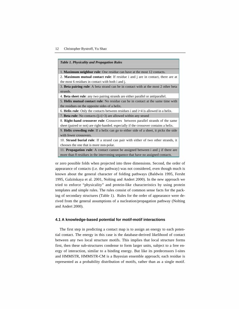
Table 1. Physicality and Propagation Rules
1. Maximum neighbor rule: One residue can have at the most 12 contacts.
2. Maximum mutual contact rule: If residue i and j are in contact, there are at
the most 6 residues in contact with both i and j.
3. Beta pairing rule: A beta strand can be in contact with at the most 2 other beta
strands.
4. Beta sheet rule: any two pairing strands are either parallel or antiparallel.
5. Helix mutual contact rule: No residue can be in contact at the same time with
the residues on the opposite sides of a helix.
6. Helix rule: Only the contacts between residues i and i+4 is allowed in a helix.
7. Beta rule: No contacts (|j-i|>3) are allowed within any strand
8. Right-hand crossover rule: Crossovers between parallel strands of the same
sheet (paired or not) are right-handed. especially if the crossover contains a helix.
9. Helix crowding rule: If a helix can go to either side of a sheet, it picks the side
with fewer crossovers.
10. Strand burial rule: If a strand can pair with either of two other strands, it
chooses the one that is more non-polar.
11. Propagation rule: A contact cannot be assigned between i and j if there are
more than 8 residues in the intervening sequence that have no assigned contacts.
Modeling Protein Folding Pathways 13
Thus, each contact potential models a pair of flickering local structures, interact-ing in proportion to their structural content.
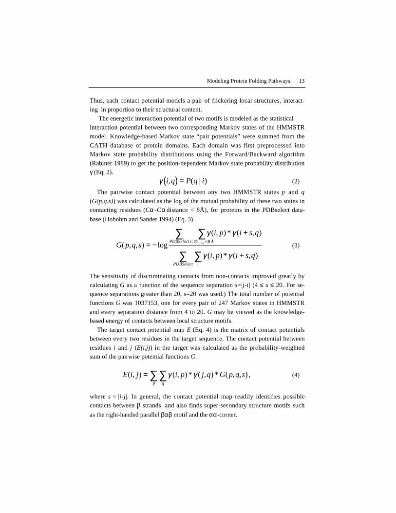
The energetic interaction potential of two motifs is modeled as the statisticalinteraction potential between two corresponding Markov states of the HMMSTR
model. Knowledge-based Markov state “pair potentials” were summed from the
CATH database of protein domains. Each domain was first preprocessed intoMarkov state probability distributions using the Forward/Backward algorithm
(Rabiner 1989) to get the position-dependent Markov state probability distributionγ (Eq. 2).
γ i q P q i, ( | )( ) = (2)
The pairwise contact potential between any two HMMSTR states p and q
(G(p,q,s)) was calculated as the log of the mutual probability of these two states incontacting residues (Cα -Cα distance < 8Å), for proteins in the PDBselect data-
base (Hobohm and Sander 1994) (Eq. 3).
G p q s
i p i s q
i p i s q
i D ÅPDBSelect
iPDBSelect
i i s( , , ) log
( , ) * ( , )
( , ) * ( , )
,= −+
+
∋ <+
∑∑
∑∑
γ γ
γ γ
8 (3)
The sensitivity of discriminating contacts from non-contacts improved greatly by
calculating G as a function of the sequence separation s=|j-i| (4 ≤ s ≤ 20. For se-quence separations greater than 20, s=20 was used.) The total number of potential
functions G was 1037153, one for every pair of 247 Markov states in HMMSTR
and every separation distance from 4 to 20. G may be viewed as the knowledge-based energy of contacts between local structure motifs.
The target contact potential map E (Eq. 4) is the matrix of contact potentialsbetween every two residues in the target sequence. The contact potential between
residues i and j (E(i,j)) in the target was calculated as the probability-weightedsum of the pairwise potential functions G.
E i j i p j q G p q sqp
( , ) ( , ) * ( , ) * ( , , )= ∑∑ γ γ , (4)
where s = |i-j|. In general, the contact potential map readily identifies possiblecontacts between β strands, and also finds super-secondary structure motifs such
as the right-handed parallel βαβ motif and the αα -corner.

14 Christopher Bystroff, Yu Shao
Target sequence
Psi-Blast
Multiple Sequencealignment
Profiler
HMMSTR
Target/templateGamma matrices BayesAligner
Target/templatealignments
Heuristics
Candidatecontact maps
Pathwayfolding
Predicted contact map
rulesPDB
Templatesequences
Templatecontact maps E map
SumGamma SumEmapSequenceprofile
Forward/backward
CijALI
Heuristics,consensus
Gpqs
4.2. Fold recognition using contact potential maps
The flowchart in Fig. 4 summarizes the steps in a contact map prediction using
HMMSTR-CM. Target sequences were aligned to database sequences using PSI-
BLAST (Altschul et al. 1997). The resulting multiple sequence alignment wasconverted to an amino acid probability distribution or sequence profile, as de-
scribed previously (Bystroff and Baker 1998). The target sequence profile and
Fig. 4. Flowchart for HMMST-CM contact map prediction. Rectangles represent algo-rithms, ovals are data, and rounded rectangles are models. Dashed lines apply to trainingset data (templates) and solid lines apply to both templates and targets. Light gray itemsare describe in referenced material Dark gray items are described in this text as follows:HMMSTR, section 2.2; Gamma matrices, Eq. 2; SumGamma, Gpqs, Eq. 3; SumEmap, Emap, Eq. 4; Rules,, Pathway folding, section 4.4, Table 1; BayesAligner, Target/templatealignments, section 4.2, Eq. 5, Fig. 5a; Heuristics, Eq. 6; CijALI, section 4.2, Eq. 7; Heu-ristics, consensus, section 4.3, Fig. 6.

Modeling Protein Folding Pathways 15
1239 template profiles from the PDBselect database (Hobohm and Sander 1994)were converted to HMMSTR γ-matrices (Eq. 2), and γtarget was aligned against
each γtemplate using Bayesian adaptive alignment (Zhu et al. 1998). The alignment
matrix in this case was the sum over all joint probabilities of Markov states (Eq.5). The alignments were evaluated using contact potential maps to choose the best
template.
Aij = γ γiqt et
jqtemplate
q
arg∑ (5)
Candidate target contact maps were generated for each alignment, and each was
evaluated by the contact free energy (CFE), as described below, and other meas-
ures. The BayesAligner produced a single score and any number of alignments.Templates with low alignment scores were rejected. Otherwise, 100 alignments
were selected at random for further evaluation.
BayesAligner produces a probability distribution over all possible alignmentswith no more than k gaps (k depends on the sequence lengths). The quality of the
alignment distribution (see Fig. 5a) was a strong indicator of the quality of the
template. Templates and/or alignments were removed from this set if they werehighly fragmented. This was assessed using a "compactness score" which is sim-
ply the length of the longest contiguously aligned region, ignoring small gaps ( ≤ 3residues). The template distance at the ends of the aligned blocks was enforced to
be physically possible values (Eq. 6) by trimming the aligned blocks if necessary.
D Å i ji j' ' .≤ × −3 8 (6)
Candidate contact maps (C) were generated using the alignments and the con-tact maps of each of the templates that had the top 10 compactness scores was
scored using the "contact free energy" (CFE, Eq. 7). CFE was calculated by sum-ming the relative contact potential E over all contacts, C. Contacts with sequence
separations |j-i| less than 4 were ignored.
CFE E i j Ei j C j iij
= −∋ = ∩ > +
∑ ( , ), ( ( ))1 3
, (7)
where <E> is the mean contact potential for the target. For each template, we cal-
culated the CFE for all contact map candidates and chose the one with the best en-
ergy as the best alignment to that template.After we carried out the above procedure for every template in our dataset, we
usually accumulated several hundred target contact map predictions. How toevaluate them and choose one as the final prediction became a problem itself. The
16 Christopher Bystroff, Yu Shao
decision was made by considering four parameters: CFE, the BayesAligner score,the compactness score and the similarity between sequence lengths of the target
and the template. The primary parameter was the CFE since it represented the freeenergy of the sequence when folded to the template structure. But we observed
that better alignments and similar lengths improved the perceived prediction qual-
ity.The automated selection of templates was sometimes overridden by our ab ini-
tio analysis, described below. If the propagation rules favored one topology overanother and a template of the favored topology was present in our list of top scor-
ers, we would select that template over a higher scoring one.
4.3. Consensus and composite contact map predictions
Often several of the top-scoring templates contained the same fold or substruc-
ture. Consensus was considered a strong indicator, especially if the fold was un-common. Multiple candidates were sometimes used to construct a single compos-
ite map. In practice, consensus similarity between many structures is difficult tosee in a 3D multiple superposition, but is easy to see in superimposed contact
maps.This prediction can be done in different ways when the top scoring templates
share a similar fold. When they disagree on some contacts, the consensus contacts
(not necessarily those from the best scoring template) are used; when some tem-plates aligned well in one region and other templates aligned well in another re-
gion, the predictions from these templates were spliced to maximize the coverage.For some recurrent contact patterns, e.g. the parallel βαβ motif, the parallel βcontacts or the helix contacts were sometimes incomplete because of misalign-
ment of the template. By combining the top scoring predictions, we could “grow”the incomplete pattern into a complete one.
Simply combining the contact maps introduces “noise” – contacts that make theprediction non-physical. (A “non-physical” contact map cannot be projected into
3-dimensions.) Manual post-processing, including pathway-based editing (dis-cussed next) was needed to enforce the physicality of the final contact map.
4.4. Ab initio rule-based pathway predictions
The fold-recognition methods described above have their roots in evolution, but
contact maps as a representation of protein structures were chosen not with the

Modeling Protein Folding Pathways 17
intention of building a Darwin-based prediction strategy, but with the intention ofmodeling the folding pathway. Contact maps simplify the conformational search.
However, as we have pointed out, not all contact maps represent physically-possible three-dimensional objects. Therefore, external information about proteins
must be included. A set of aligned templates is one source of external information.
Here we present a set of fundamental rules (Table 1) and energies (Eq. 4) thatserve the same purpose – to restrict the conformational search to contact maps that
are physically possible and protein-like. A rule-based structure propagation model was used either in conjunction with
templates or ab initio (without templates). In CASP5, ab initio predictions were
sometimes done on targets found later to be remote homologs by CASP5 asses-sors, but because our alignment method was not always able to recognize remote
homology, we treated them as potential new folds. The procedure is as follows.Starting from a contact potential map, E, we kept the contacts that were better
than a cutoff value. The cutoff value was chosen such that blocks of contacts werefound between most secondary structural units, especially between β strands. As a
result, the initial contact map was often characterized by dense blocks of contacts
between β strands and sparse contacts to helices and between helices.
If we kept all of these contacts, clearly the map would be physically impossible.
For example, a β strand element cannot be paired with more than two other βstrands. A set of common-sense rules were compiled to weed out the possible
contacts from the impossible or unlikely, and to enforce protein-like characteris-
tics, such as right-handed crossovers and exposed reverse turns (Table 1). Theserules were enforced as the prediction was propagated.
The folding pathway consisted of “assigning” or “erasing” contacts. Contactswere assigned if the energy E(i,j) passed a threshold and the corresponding contact
Cij = 1 did not violate any of the rules, otherwise they were erased. Blocks of po-tential contacts were considered together, and the order in which blocks were con-
sidered depended on their proximity to previously assigned blocks of contacts
(Table 1, Rule 11), following the principles of the nucleation/condensation foldingmechanism.
To start the folding pathway, we selected one or more local regions with highconfidence contacts as the “nucleation site(s)". We then propagated the prediction
in both directions by assigning or erasing blocks of contacts around and betweenthe nucleation site(s), subject to our set of rules. TOPS diagrams (Sternberg and
Thornton 1976) were drawn for the growing structure as a visual aid, since some
rules applied to the topology. The pathway, and the prediction, was complete

18 Christopher Bystroff, Yu Shao
b.
c.
a.
when all of the remaining contacts were rejected. The method is best described
using examples, as in the next section.
4.5. Selected Results of HMMSTR-CM Blind Structure Predictions
HMMSTR-CM was used to predict contact maps as part of the CASP5 experi-
ment. Targets in the FR (fold recognition) and NF (new fold) categories were pre-dicted using the three methods described above: threading, consensus and ab ini-
tio, collectively called HMMSTR-CM. In all these three methods, the overallaccuracy of the contact map prediction depends on the accuracy of the secondary
structure prediction, which was done using HMMSTR.
4.5.1. A prediction using templates and a pathway
YqgF, a hypothetical protein from E. coli, was successfully predicted using the
template-based approach in conjunction with a pathway prediction. All visible
secondary structure units are correctly predicted (note that the 17 residues from102 to 118 are missing in the crystal structure), and all of the true contacts have
better-than-average E(i,j) score. After aligning the contact potential matrix, E, to
Fig. 5. a . BayesAligner summary of most probable alignments between YqgF (X-axis)and 1HJR (Y-axis). b. Contact potential map for YqgF; darker is lower energy E(i,j). Pre-dicted contacts are outlined in white. c. Contact map from crystal structure of YqgF, hy-pothetical protein from E. coli.

Modeling Protein Folding Pathways 19
each of the 1258 templates, a consensus contact map was plotted using the top-scoring six templates. This map was used to construct a folding pathway. Nucle-
ating the pathway at β4α2β5 and propagating produced a TOPS diagram that
agreed with only one of the templates, 1HJR, and this template was therefore cho-sen to construct the consensus contact map. 1HJR had the third highest CFE score.
In the prediction based on 1HJR, the N-terminal 3-strand β meander is slightly
under-predicted, and a contact between helices 1 and 2 is slightly over-predicted.
Nonetheless, the topology is correct throughout (Fig. 5b). The two higher-scoringtemplates that were not chosen had very different, and incorrect, topologies.
4.5.2. A prediction using several templates
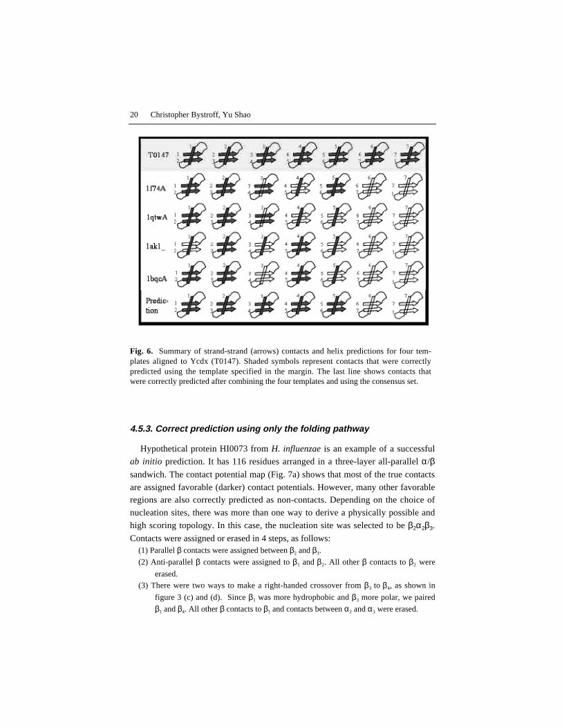
Ycdx, another hypothetical protein from E. coli, was successfully predictedusing multiple templates. The threading approach found 4 templates that had high
CFE scores and also shared common structural components. Three of those tem-
plates were 8-stranded α/β barrels and the other consisted of two parallel α/β do-
mains. Ycdx turned out to be an αβ barrel with 7 parallel β strands (PDB code
1M65). Templates with good CFE scores existed but none of them predicted allof the first five helices and the parallel β strand contacts correctly. However, by
combining the results from the top scoring templates, we made a consensus pre-
diction that was better than any of the contact maps made from the single tem-plates. In particular, we correctly found parallel contacts between the first 6 βstrands (Fig. 6).
The sixth helix and the contacts between the sixth and the seventh strands were
predicted but misaligned. Our method mispredicted the C-terminus to be a parallel
βαβ motif, as in a standard 8-stranded TIM barrel, but the true structure is three
short helices connected by loops. Visual inspection of the templates confirmed
that they share the same topology, and a consensus fold prediction would havebeen obvious given this result. But finding structural similarity and combining
structures is more easily automated in the 2D contact map format than in 3D coor-dinate space. Consensus in contact maps provides a way to merge and “grow” the
incomplete contact maps of different targets into a more complete contact map.
Ycdx also revealed a weakness of the method. HMMSTR, which is trained torecognize recurrent super-secondary motifs, does not recognize the unusual sub-
structure at the C-terminus of this protein, 3 short helices instead of the usual βαβmotif. The consensus method, as we have defined it, tends to bias the prediction
toward the more common folds. In fact, this is a problem with any template-basedmethod.
20 Christopher Bystroff, Yu Shao
4.5.3. Correct prediction using only the folding pathway
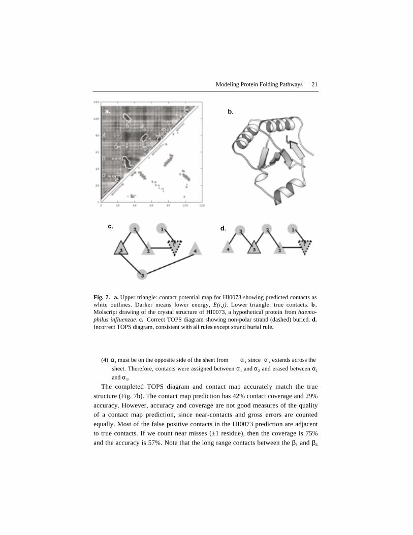
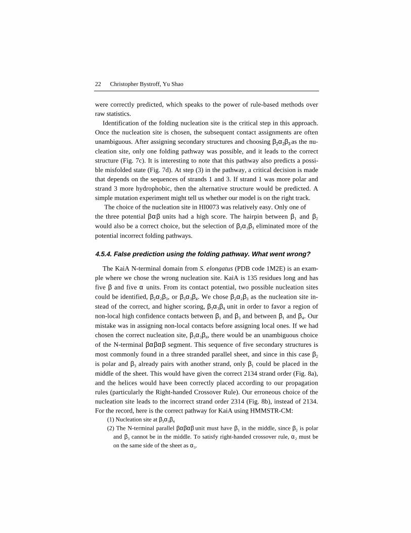
Hypothetical protein HI0073 from H. influenzae is an example of a successful
ab initio prediction. It has 116 residues arranged in a three-layer all-parallel α/βsandwich. The contact potential map (Fig. 7a) shows that most of the true contacts
are assigned favorable (darker) contact potentials. However, many other favorableregions are also correctly predicted as non-contacts. Depending on the choice of
nucleation sites, there was more than one way to derive a physically possible and
high scoring topology. In this case, the nucleation site was selected to be β2α2β3.
Contacts were assigned or erased in 4 steps, as follows:(1) Parallel β contacts were assigned between β2 and β3.
(2) Anti-parallel β contacts were assigned to β1 and β2. All other β contacts to β2 were
erased.
(3) There were two ways to make a right-handed crossover from β3 to β4, as shown in
figure 3 (c) and (d). Since β1 was more hydrophobic and β3 more polar, we paired
β1 and β4. All other β contacts to β1 and contacts between α2 and α3 were erased.
Fig. 6. Summary of strand-strand (arrows) contacts and helix predictions for four tem-plates aligned to Ycdx (T0147). Shaded symbols represent contacts that were correctlypredicted using the template specified in the margin. The last line shows contacts thatwere correctly predicted after combining the four templates and using the consensus set.
Modeling Protein Folding Pathways 21
a. b.
c. d.
(4) α1 must be on the opposite side of the sheet from α3, since α3 extends across the
sheet. Therefore, contacts were assigned between α1 and α2 and erased between α1
and α3.
The completed TOPS diagram and contact map accurately match the true
structure (Fig. 7b). The contact map prediction has 42% contact coverage and 29%
accuracy. However, accuracy and coverage are not good measures of the qualityof a contact map prediction, since near-contacts and gross errors are counted
equally. Most of the false positive contacts in the HI0073 prediction are adjacentto true contacts. If we count near misses (±1 residue), then the coverage is 75%
and the accuracy is 57%. Note that the long range contacts between the β1 and β4
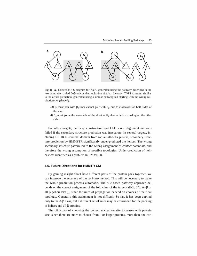
Fig. 7. a. Upper triangle: contact potential map for HI0073 showing predicted contacts aswhite outlines. Darker means lower energy, E(i,j). Lower triangle: true contacts. b.Molscript drawing of the crystal structure of HI0073, a hypothetical protein from haemo-philus influenzae. c. Correct TOPS diagram showing non-polar strand (dashed) buried. d.Incorrect TOPS diagram, consistent with all rules except strand burial rule.
22 Christopher Bystroff, Yu Shao
were correctly predicted, which speaks to the power of rule-based methods overraw statistics.
Identification of the folding nucleation site is the critical step in this approach.Once the nucleation site is chosen, the subsequent contact assignments are often
unambiguous. After assigning secondary structures and choosing β2α2β3 as the nu-
cleation site, only one folding pathway was possible, and it leads to the correctstructure (Fig. 7c). It is interesting to note that this pathway also predicts a possi-
ble misfolded state (Fig. 7d). At step (3) in the pathway, a critical decision is madethat depends on the sequences of strands 1 and 3. If strand 1 was more polar and
strand 3 more hydrophobic, then the alternative structure would be predicted. A
simple mutation experiment might tell us whether our model is on the right track. The choice of the nucleation site in HI0073 was relatively easy. Only one of
the three potential βαβ units had a high score. The hairpin between β1 and β2
would also be a correct choice, but the selection of β2α1β3 eliminated more of the
potential incorrect folding pathways.
4.5.4. False prediction using the folding pathway. What went wrong?
The KaiA N-terminal domain from S. elongatus (PDB code 1M2E) is an exam-
ple where we chose the wrong nucleation site. KaiA is 135 residues long and has
five β and five α units. From its contact potential, two possible nucleation sites
could be identified, β2α2β3, or β3α3β4. We chose β2α2β3 as the nucleation site in-
stead of the correct, and higher scoring, β3α3β4 unit in order to favor a region of
non-local high confidence contacts between β1 and β3 and between β1 and β4. Our
mistake was in assigning non-local contacts before assigning local ones. If we had
chosen the correct nucleation site, β3α3β4, there would be an unambiguous choice
of the N-terminal βαβαβ segment. This sequence of five secondary structures is
most commonly found in a three stranded parallel sheet, and since in this case β2
is polar and β3 already pairs with another strand, only β1 could be placed in the
middle of the sheet. This would have given the correct 2134 strand order (Fig. 8a),
and the helices would have been correctly placed according to our propagationrules (particularly the Right-handed Crossover Rule). Our erroneous choice of the
nucleation site leads to the incorrect strand order 2314 (Fig. 8b), instead of 2134.For the record, here is the correct pathway for KaiA using HMMSTR-CM:
(1) Nucleation site at β3α3β4
(2) The N-terminal parallel βαβαβ unit must have β1 in the middle, since β2 is polar
and β3 cannot be in the middle. To satisfy right-handed crossover rule, α2 must be
on the same side of the sheet as α3.
Modeling Protein Folding Pathways 23
1
2 3 4
5
54132
1
2 3 4
5
12 3 4 5
b.a.
(3) β5 must pair with β4 since cannot pair with β2, due to crossovers on both sides of
the sheet.
4) α5 must go on the same side of the sheet as α1, due to helix crowding on the other
side.
For other targets, pathway construction and CFE score alignment methods
failed if the secondary structure prediction was inaccurate. In several targets, in-cluding HIP1R N-terminal domain from rat, an all-helix protein, secondary struc-
ture prediction by HMMSTR significantly under-predicted the helices. The wrongsecondary structure pattern led to the wrong assignment of contact potentials, and
therefore the wrong assumption of possible topologies. Under-prediction of heli-
ces was identified as a problem in HMMSTR.
4.6. Future Directions for HMMTR-CM
By gaining insight about how different parts of the protein pack together, wecan improve the accuracy of the ab initio method. This will be necessary to make
the whole prediction process automatic. The rule-based pathway approach de-pends on the correct assignment of the fold class of the target (all-α, α/β, α+β or
all β (Zhou 1998)), since the rules of propagation depend on choices of the final
topology. Generally this assignment is not difficult. So far, it has been appliedonly to the α/β class, but a different set of rules may be envisioned for the packing
of helices and all β proteins.
The difficulty of choosing the correct nucleation site increases with protein
size, since there are more to choose from. For larger proteins, more than one cor-
Fig. 8. a. Correct TOPS diagram for KaiA, generated using the pathway described in thetext using the shaded βαβ unit as the nucleation site. b. Incorrect TOPS diagram, similarto the actual prediction, generated using a similar pathway but starting with the wrong nu-cleation site (shaded).

24 Christopher Bystroff, Yu Shao
rect choice may be required. One possible approach could be a recursive algorithmto exhaust all the possible topologies by starting with each potential nucleation
site, and then evaluate the topologies using the contact potential.Another improvement might be to attempt to make the contact map prediction
more protein like. Our predictions have many false contacts adjacent to true con-
tacts, e.g. a “fat” β-hairpin prediction – even though it is predicted at the right po-
sition. Rules to prune this type of false contacts – in other words, to beautify the
predicted contact blocks – would increase the accuracy of our prediction. This willrequire better secondary structure predictions.
5. Conclusions
We have developed methods for calculating an inter-residue contact potential
map for a protein sequence, for aligning that map to templates, and for pruningthat map using a folding pathway model. Results on CASP5 targets reveal that the
folding pathways for some α/β proteins are unambiguous given the correct choice
of the folding nucleation site. Pathway predictions improved the selection of a re-
mote homolog for one threading target. Consensus contact maps are more com-plete than maps from single templates. The contact map representation of protein
structure is a useful intermediate-level of detail that facilitates rule-based algo-
rithm development.

Modeling Protein Folding Pathways 25
References
Alm E & Baker D. (1999). Prediction of protein-folding mechanisms from free-
energy landscapes derived from native structures. Proc Natl Acad Sci U S
A 96, 11305-10.Altschul SF, Madden TL, Schaffer AA, Zhang J, Zhang Z, Miller W & Lipman
DJ. (1997). Gapped BLAST and PSI-BLAST: a new generation of pro-tein database search programs. Nucleic Acids Res 25, 3389-402.
Anfinsen CB & Scheraga HA. (1975). Experimental and theoretical aspects ofprotein folding. Adv Protein Chem 29, 205-300.
Aszodi A, Munro RE & Taylor WR. (1997). Distance geometry based compara-
tive modelling. Fold Des 2, S3-6.Baldwin RL. (1995). The nature of protein folding pathways: the classical versus
the view. J Biomol NMR 5, 103-9.Baldwin RL & Rose GD. (1999). Is protein folding hierarchic? I. Local structure
and peptide folding. Trends Biochem Sci 24, 26-33.
Blanco FJ, Rivas G & Serrano L. (1994). A short linear peptide that folds into anative stable beta-hairpin in aqueous solution. Nat Struct Biol 1, 584-90.
Bonneau R & Baker D. (2001). Ab initio protein structure prediction: progress andprospects. Annu Rev Biophys Biomol Struct 30, 173-89.
Bonneau R, Strauss CE & Baker D. (2001). Improving the performance of Rosettausing multiple sequence alignment information and global measures of
hydrophobic core formation. Proteins 43, 1-11.
Brunger AT, Clore GM, Gronenborn AM & Karplus M. (1986). Three-dimensional structure of proteins determined by molecular dynamics with
interproton distance restraints: application to crambin. Proc Natl Acad
Sci U S A 83, 3801-5.
Bystroff C & Baker D. (1997). Blind predictions of local protein structure inCASP2 targets using the I-sites library. Proteins Suppl 1, 167-71.
Bystroff C & Baker D. (1998). Prediction of local structure in proteins using a li-
brary of sequence-structure motifs. J Mol Biol 281, 565-77.Bystroff C & Garde S. (2003). Helix propensities of short peptides: Molecular dy-
namics versus bioinformatics. Proteins 50, 552-62.

26 Christopher Bystroff, Yu Shao
Bystroff C & Shao Y. (2002). Fully automated ab initio protein structure predic-tion using I-SITES, HMMSTR and ROSETTA. Bioinformatics 18 Suppl
1, S54-61.Bystroff C, Simons KT, Han KF & Baker D. (1996). Local sequence-structure
correlations in proteins. Curr Opin Biotechnol 7, 417-21.
Bystroff C, Thorsson V & Baker D. (2000). HMMSTR: A hidden markov modelfor local sequence-structure correlations in proteins. Journal of Molecu-
lar Biology 301, 173-90.Cavalli A, Ferrara P & Caflisch A. (2002). Weak temperature dependence of the
free energy surface and folding pathways of structured peptides. Proteins
47, 305-14.Chan HS, Bromberg S & Dill KA. (1995). Models of cooperativity in protein
folding. Philos Trans R Soc Lond B Biol Sci 348, 61-70.Colon W & Roder H. (1996). Kinetic intermediates in the formation of the cyto-
chrome c molten globule. Nat Struct Biol 3, 1019-25.Crippen GM, Havel, T.F. (1988). Distance Geometry and Molecular Conforma-
tion. Chemometrics Series, 15, John Wiley & Sons.
Duan Y & Kollman PA. (1998). Pathways to a protein folding intermediate ob-served in a 1-microsecond simulation in aqueous solution [see com-
ments]. Science 282, 740-4.Dyson HJ & Wright PE. (1996). Insights into protein folding from NMR. Annu
Rev Phys Chem 47, 369-95.Eaton WA, Thompson PA, Chan CK, Hage SJ & Hofrichter J. (1996). Fast events
in protein folding. Structure 4, 1133-9.
Eddy SR. (1996). Hidden Markov models. Curr Opin Struct Biol 6, 361-5.Efimov AV. (1993). Standard structures in proteins. Prog Biophys Mol Biol 60,
201-39.Fariselli P & Casadio R. (1999). A neural network based predictor of residue
contacts in proteins. Protein Eng 12, 15-21.Fariselli P, Olmea O, Valencia A & Casadio R. (2001). Progress in predicting in-
ter-residue contacts of proteins with neural networks and correlated mu-
tations. Proteins Suppl 5, 157-62.Fersht AR. (1995). Optimization of rates of protein folding: the nucleation-
condensation mechanism and its implications. Proc Natl Acad Sci U S A
92, 10869-73.
Fersht AR, Matouschek A & Serrano L. (1992). The folding of an enzyme. I. The-ory of protein engineering analysis of stability and pathway of protein
folding. J Mol Biol 224, 771-82.
Modeling Protein Folding Pathways 27
Fischer D, Elofsson A, Rychlewski L, Pazos F, Valencia A, Rost B, Ortiz AR &Dunbrack RL, Jr. (2001). CAFASP2: the second critical assessment of
fully automated structure prediction methods. Proteins Suppl 5, 171-83.Frishman D & Argos P. (1995). Knowledge-based protein secondary structure as-
signment. Proteins 23, 566-79.
Galzitskaya OV, Ivankov DN & Finkelstein AV. (2001). Folding nuclei in pro-teins. FEBS Lett 489, 113-8.
Garcia AE & Sanbonmatsu KY. (2001). Exploring the energy landscape of a betahairpin in explicit solvent. Proteins 42, 345-54.
Gillespie JR & Shortle D. (1997). Characterization of long-range structure in the
denatured state of staphylococcal nuclease. II. Distance restraints fromparamagnetic relaxation and calculation of an ensemble of structures. J
Mol Biol 268, 170-84.Gnanakaran S & Garcia AE. (2002). Folding of a Highly Conserved Diverging
Turn Motif from the SH3 Domain. Biophys J.Gough J & Chothia C. (2002). SUPERFAMILY: HMMs representing all proteins
of known structure. SCOP sequence searches, alignments and genome
assignments. Nucleic Acids Res 30, 268-72.Grantcharova VP, Riddle DS & Baker D. (2000). Long-range order in the src SH3
folding transition state. Proc Natl Acad Sci U S A 97, 7084-9.Gulotta M, Gilmanshin R, Buscher TC, Callender RH & Dyer RB. (2001). Core
formation in apomyoglobin: probing the upper reaches of the folding en-ergy landscape. Biochemistry 40, 5137-43.
Han KF & Baker D. (1996). Global properties of the mapping between local
amino acid sequence and local structure in proteins. Proc Natl Acad Sci
U S A 93, 5814-8.
Han KF & Baker D. (1995). Recurring local sequence motifs in proteins. J Mol
Biol 251, 176-87.
Han KF, Bystroff C & Baker D. (1997). Three-dimensional structures and con-texts associated with recurrent amino acid sequence patterns. Protein Sci
6, 1587-90.
Heidary DK & Jennings PA. (2002). Three topologically equivalent core residuesaffect the transition state ensemble in a protein folding reaction. J Mol
Biol 316, 789-98.Hobohm U & Sander C. (1994). Enlarged representative set of protein structures.
Protein Sci 3, 522-4.

28 Christopher Bystroff, Yu Shao
Houry WA, Rothwarf DM & Scheraga HA. (1996). Circular dichroism evidencefor the presence of burst-phase intermediates on the conformational
folding pathway of ribonuclease A. Biochemistry 35, 10125-33.Hu J, Shen X, Shao Y, Bystroff C & Zaki MJ. (2002). BIOKDD 2002, Edmonton,
Canada.
Jacchieri SG. (2000). Mining combinatorial data in protein sequences and struc-tures. Molecular Diversity 5, 145-152.
Jones DT. (1998). Critical Assessment of Protein Structure Prediction 3, Asilo-
mar, CA.
Kabsch W & Sander C. (1983). Dictionary of protein secondary structure: pattern
recognition of hydrogen-bonded and geometrical features. Biopolymers
22, 2577-637.
Karplus K, Barrett C & Hughey R. (1998). Hidden Markov models for detectingremote protein homologies. Bioinformatics 14, 846-56.
Kolinski A & Skolnick J. (1997). High coordination lattice models of proteinstructure, dynamics and thermodynamics. Acta Biochim Pol 44, 389-422.
Krueger BP & Kollman PA. (2001). Molecular dynamics simulations of a highly
charged peptide from an SH3 domain: possible sequence-function rela-tionship. Proteins 45, 4-15.
Laurents DV & Baldwin RL. (1998). Protein folding: matching theory and ex-periment. Biophys J 75, 428-34.
Mateu MG, Sanchez Del Pino MM & Fersht AR. (1999). Mechanism of foldingand assembly of a small tetrameric protein domain from tumor suppres-
sor p53. Nat Struct Biol 6, 191-8.
Mendes J, Guerois R & Serrano L. (2002). Energy estimation in protein design.Current Opinion in Structural Biology 12, 441-446.
Mirny L & Shakhnovich E. (2001). Protein folding theory: from lattice to all-atommodels. Annu Rev Biophys Biomol Struct 30, 361-96.
Mok YK, Elisseeva EL, Davidson AR & Forman-Kay JD. (2001). Dramatic stabi-lization of an SH3 domain by a single substitution: roles of the folded
and unfolded states. J Mol Biol 307, 913-28.
Mok YK, Kay CM, Kay LE & Forman-Kay J. (1999). NOE data demonstrating acompact unfolded state for an SH3 domain under non-denaturing condi-
tions. J Mol Biol 289, 619-38.Moult J, Fidelis K, Zemla A & Hubbard T. (2001). Critical assessment of methods
of protein structure prediction (CASP): round IV. Proteins Suppl 5, 2-7.
Modeling Protein Folding Pathways 29
Munoz V, Blanco FJ & Serrano L. (1995). The hydrophobic-staple motif and arole for loop-residues in alpha- helix stability and protein folding. Nat
Struct Biol 2, 380-5.Munoz V, Henry ER, Hofrichter J & Eaton WA. (1998). A statistical mechanical
model for beta-hairpin kinetics. Proc Natl Acad Sci U S A 95, 5872-9.
Nolting B & Andert K. (2000). Mechanism of protein folding. Proteins 41, 288-98.
Nolting B, Golbik R, Neira JL, Soler-Gonzalez AS, Schreiber G & Fersht AR.(1997). The folding pathway of a protein at high resolution from micro-
seconds to seconds. Proc Natl Acad Sci U S A 94, 826-30.
Northey JG, Di Nardo AA & Davidson AR. (2002a). Hydrophobic core packing inthe SH3 domain folding transition state. Nat Struct Biol 9, 126-30.
Northey JGB, Maxwell KL & Davidson AR. (2002b). Protein folding kinetics be-yond the Phi value: Using multiple amino acid substitutions to investigate
the structure of the SH3 domain folding transition state. Journal of Mo-
lecular Biology 320, 389-402.
Nymeyer H, Socci ND & Onuchic JN. (2000). Landscape approaches for deter-
mining the ensemble of folding transition states: success and failurehinge on the degree of frustration. Proc Natl Acad Sci U S A 97, 634-9.
Oliveberg M, Tan YJ, Silow M & Fersht AR. (1998). The changing nature of theprotein folding transition state: implications for the shape of the free-
energy profile for folding. J Mol Biol 277, 933-43.Olmea O & Valencia A. (1997). Improving contact predictions by the combination
of correlated mutations and other sources of sequence information. Fold
Des 2, S25-32.Onuchic JN, Luthey-Schulten Z & Wolynes PG. (1997). Theory of protein fold-
ing: the energy landscape perspective. Annu Rev Phys Chem 48, 545-600.Ortiz AR, Kolinski A & Skolnick J. (1998). Fold assembly of small proteins using
monte carlo simulations driven by restraints derived from multiple se-quence alignments. J Mol Biol 277, 419-48.
Ortiz AR & Skolnick J. (2000). Sequence evolution and the mechanism of protein
folding. Biophys J 79, 1787-99.Pande VS, Grosberg A, Tanaka T & Rokhsar DS. (1998). Pathways for protein
folding: is a new view needed? Curr Opin Struct Biol 8, 68-79.Plaxco KW, Simons KT & Baker D. (1998). Contact order, transition state place-
ment and the refolding rates of single domain proteins. J Mol Biol 277,985-94.

30 Christopher Bystroff, Yu Shao
Pollastri G & Baldi P. (2002). Prediction of contact maps by GIOHMMs and re-current neural networks using lateral propagation from all four cardinal
corners. Bioinformatics 18 Suppl 1, S62-S70.Rabiner LR. (1989). A tutorial on Hidden Markov Models and selected applica-
tions in speech recognition. Proc IEEE 77, 257-286.
Rooman MJ, Rodriguez J & Wodak SJ. (1990). Automatic definition of recurrentlocal structure motifs in proteins. J Mol Biol 213, 327-36.
Rost B. (2001). Review: protein secondary structure prediction continues to rise. JStruct Biol 134, 204-18.
Schellman C. (1980). Protein folding: The proceedings of the 28th Conference of
the German Biochemical Society, University of Regensburg, Sept 10-12,
1979, Regensburg, West Germany.
Shakhnovich EI. (1998). Folding nucleus: specific or multiple? Insights from lat-tice models and experiments. Fold Des 3, R108-11; discussion R107.
Shao Y & Bystroff C. (2003). Predicting inter-residue contacts using templatesand pathways. Proteins, Structure, Function and Genetics in press.
Shea JE & Brooks CL, 3rd. (2001). From folding theories to folding proteins: a
review and assessment of simulation studies of protein folding and un-folding. Annu Rev Phys Chem 52, 499-535.
Shoemaker BA & Wolynes PG. (1999). Exploring structures in protein foldingfunnels with free energy functionals: the denatured ensemble. J Mol Biol
287, 657-74.Simons KT, Bonneau R, Ruczinski I & Baker D. (1999a). Ab initio protein struc-
ture prediction of CASP III targets using ROSETTA. Proteins Suppl 3,
171-6.Simons KT, Kooperberg C, Huang E & Baker D. (1997). Assembly of protein ter-
tiary structures from fragments with similar local sequences using simu-lated annealing and Bayesian scoring functions. J Mol Biol 268, 209-25.
Simons KT, Ruczinski I, Kooperberg C, Fox BA, Bystroff C & Baker D. (1999b).Improved recognition of native-like protein structures using a combina-
tion of sequence-dependent and sequence-independent features of pro-
teins. Proteins 34, 82-95.Singer MS, Vriend G & Bywater RP. (2002). Prediction of protein residue con-
tacts with a PDB-derived likelihood matrix. Protein Eng 15, 721-5.Skolnick J & Kolinski A. (2002). A unified approach to the prediction of protein
structure and function. In Computational Methods for Protein Folding,Vol. 120, pp. 131-192.
Modeling Protein Folding Pathways 31
Sternberg MJ & Thornton JM. (1976). On the conformation of proteins: the hand-edness of the beta-strand-alpha-helix-beta-strand unit. J Mol Biol 105,
367-82.Steward RE & Thornton JM. (2002). Prediction of strand pairing in antiparallel
and parallel beta- sheets using information theory. Proteins-Structure
Function and Genetics 48, 178-191.Thirumalai D & Klimov DK. (1998). Fishing for folding nuclei in lattice models
and proteins. Fold Des 3, R112-8; discussion R107.Vendruscolo M, Kussell E & Domany E. (1997). Recovery of protein structure
from contact maps. Fold Des 2, 295-306.
Viguera AR & Serrano L. (1995). Experimental analysis of the Schellman motif. JMol Biol 251, 150-60.
Woolfson DN & Alber T. (1995). Predicting oligomerization states of coiled coils.Protein Sci 4, 1596-607.
Yi Q, Bystroff C, Rajagopal P, Klevit RE & Baker D. (1998). Prediction andstructural characterization of an independently folding substructure in the
src SH3 domain. J Mol Biol 283, 293-300.
Zaki MJ, Shan J & Bystroff C. (2000). Proceedings IEEE International Sympo-
sium on Bio-Informatics and Biomedical Engineering, Arlington, VA,
USA.Zhou GP. (1998). An intriguing controversy over protein structural class predic-
tion. J Protein Chem 17, 729-38.Zhu J, Liu JS & Lawrence CE. (1998). Bayesian adaptive sequence alignment al-
gorithms. Bioinformatics 14, 25-39.
Zwanzig R. (1997). Two-state models of protein folding kinetics. Proc Natl Acad
Sci U S A 94, 148-50.


![Predicting Experimental Quantities in Protein Folding Kinetics ...ai.stanford.edu/~apaydin/recomb06.pdfplied to ligand-protein docking [17], protein folding [3,2], and RNA folding](https://img.pdfslide.us/doc/110x75/60d6bde9a1a7162f153e3cd1/predicting-experimental-quantities-in-protein-folding-kinetics-ai-apaydinrecomb06pdf.jpg)