Embed Size (px)
DESCRIPTION
Dejan Krstić , Darko Zigar , Dejan Petković , Nenad Cvetković , Vera Marković , Nataša Đinđić , Boris Đinđić. * University of Niš , Faculty of Occupational Safety in Niš , Čarnojevića 10a, Niš , Serbia, e-mail: [email protected] - PowerPoint PPT Presentation
Citation preview
MODELING OF PENETRATING ELECTROMAGNETIC FIELDS OF MOBILE PHONES IN EXPERIMENTAL ANIMALS
Dejan Krstić, Darko Zigar, Dejan Petković, Nenad Cvetković, Vera Marković, Nataša Đinđić, Boris Đinđić
* University of Niš, Faculty of Occupational Safety in Niš, Čarnojevića 10a, Niš, Serbia, e-mail: [email protected]** University of Niš, Faculty of Electronic Engeenering Nis, Aleksandra Medvedeva 14, Niš *** University of Niš, Medical Faculty in Niš, Bulevar Zorana Đinđića 81, Niš
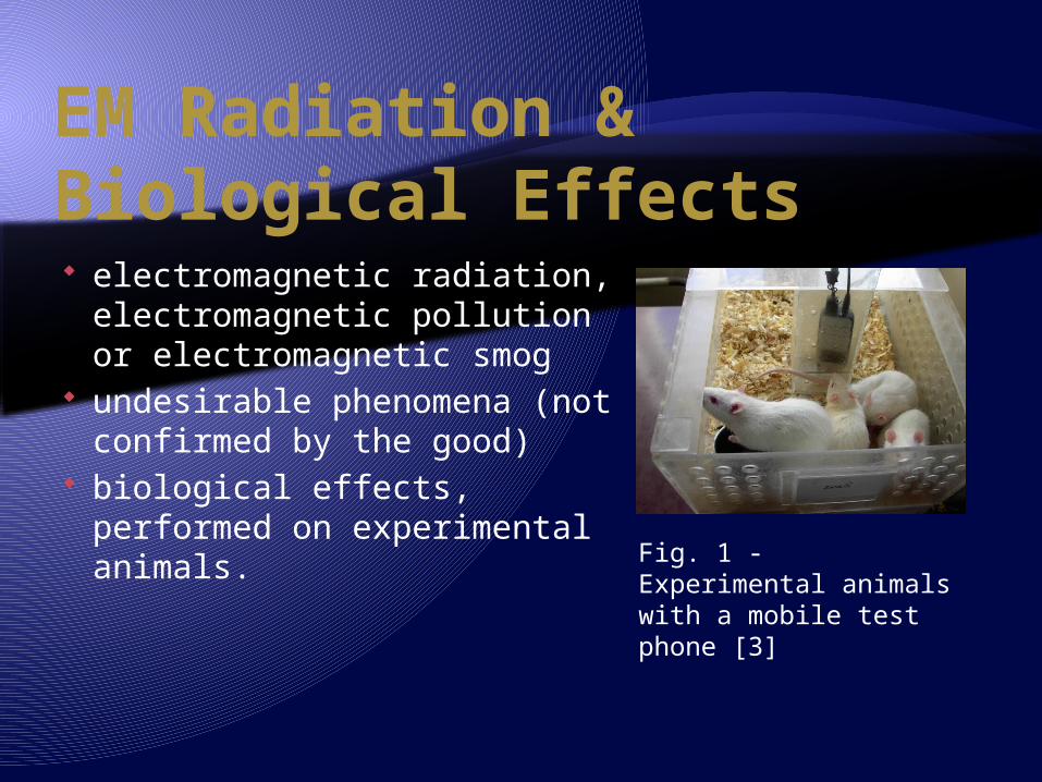
EM Radiation & Biological Effects electromagnetic radiation,
electromagnetic pollution or electromagnetic smog
undesirable phenomena (not confirmed by the good)
biological effects, performed on experimental animals. Fig. 1 - Experimental
animals with a mobile test phone [3]
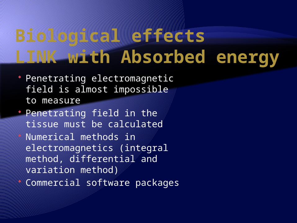
Biological effects LINK with Absorbed energy Penetrating electromagnetic
field is almost impossible to measure
Penetrating field in the tissue must be calculated
Numerical methods in electromagnetics (integral method, differential and variation method)
Commercial software packages
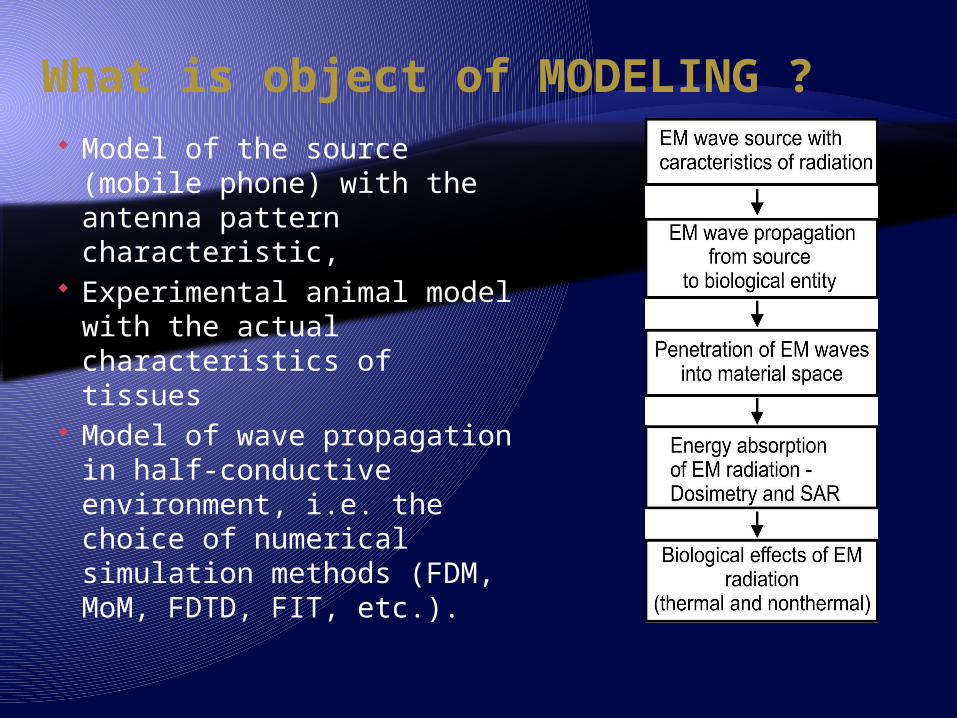
What is object of MODELING ? Model of the source
(mobile phone) with the antenna pattern characteristic,
Experimental animal model with the actual characteristics of tissues
Model of wave propagation in half-conductive environment, i.e. the choice of numerical simulation methods (FDM, MoM, FDTD, FIT, etc.).
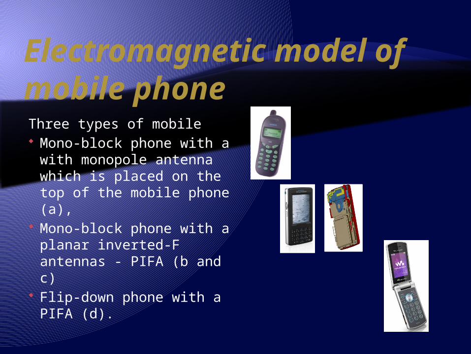
Electromagnetic model of mobile phoneThree types of mobile Mono-block phone with a
with monopole antenna which is placed on the top of the mobile phone (a),
Mono-block phone with a planar inverted-F antennas - PIFA (b and c)
Flip-down phone with a PIFA (d).
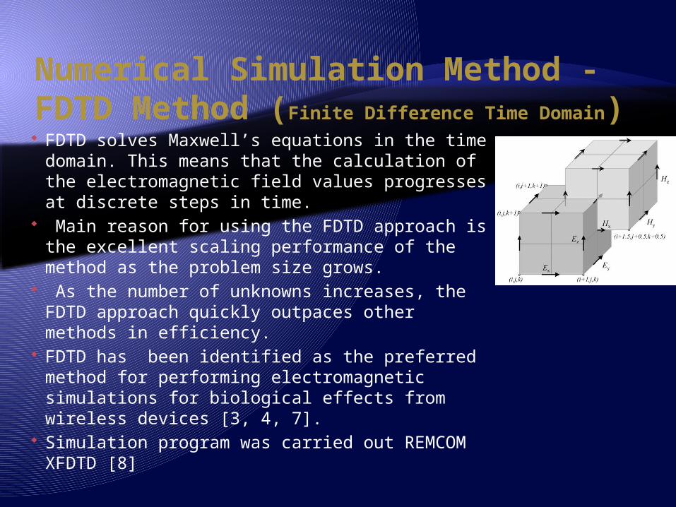
Numerical Simulation Method - FDTD Method (Finite Difference Time Domain) FDTD solves Maxwell’s equations in the time
domain. This means that the calculation of the electromagnetic field values progresses at discrete steps in time.
Main reason for using the FDTD approach is the excellent scaling performance of the method as the problem size grows.
As the number of unknowns increases, the FDTD approach quickly outpaces other methods in efficiency.
FDTD has been identified as the preferred method for performing electromagnetic simulations for biological effects from wireless devices [3, 4, 7].
Simulation program was carried out REMCOM XFDTD [8]
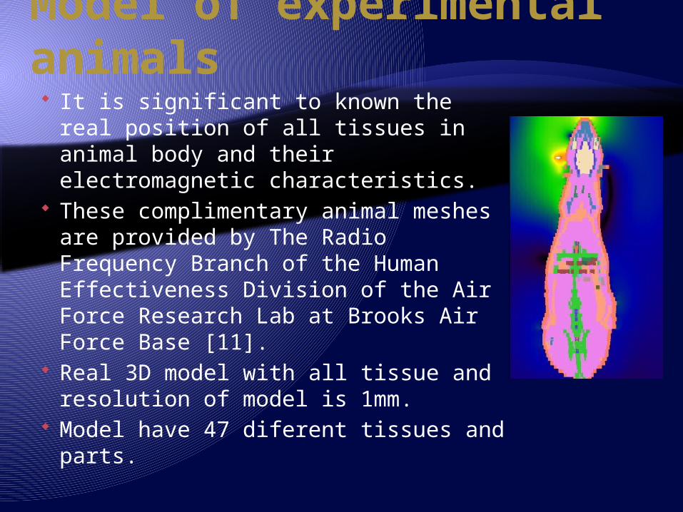
Model of experimental animals It is significant to known the real
position of all tissues in animal body and their electromagnetic characteristics.
These complimentary animal meshes are provided by The Radio Frequency Branch of the Human Effectiveness Division of the Air Force Research Lab at Brooks Air Force Base [11].
Real 3D model with all tissue and resolution of model is 1mm.
Model have 47 diferent tissues and parts.
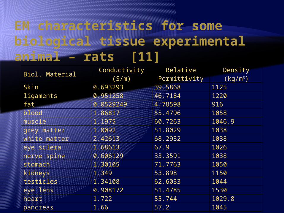
EM characteristics for some biological tissue experimental animal – rats [11]
Biol. MaterialConductivity
(S/m)Relative Permittivity
Density(kg/m3)
Skin 0.693293 39.5868 1125ligaments 0.951258 46.7184 1220fat 0.0529249 4.78598 916blood 1.86817 55.4796 1058muscle 1.1975 60.7263 1046.9grey matter 1.0092 51.8029 1038white matter 2.42613 68.2932 1038eye sclera 1.68613 67.9 1026nerve spine 0.606129 33.3591 1038stomach 1.30105 71.7763 1050kidneys 1.349 53.898 1150testicles 1.34108 62.6033 1044eye lens 0.908172 51.4785 1530heart 1.722 55.744 1029.8pancreas 1.66 57.2 1045body fluid 2.899 67.24 1010liver 1.33 43.4 1030
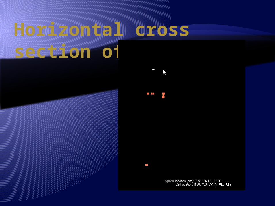
Horizontal cross section of Rat model
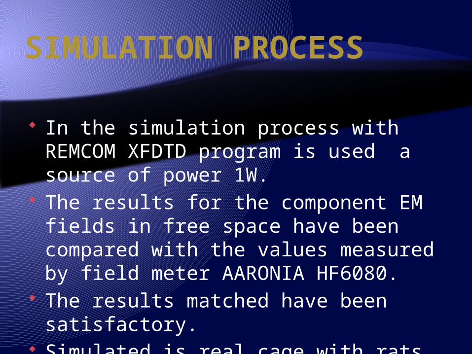
SIMULATION PROCESS
In the simulation process with REMCOM XFDTD program is used a source of power 1W.
The results for the component EM fields in free space have been compared with the values measured by field meter AARONIA HF6080.
The results matched have been satisfactory.
Simulated is real cage with rats and mobile phone in two possition.
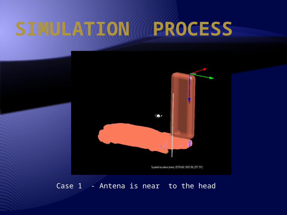
SIMULATION PROCESS
Case 1 - Antena is near to the head
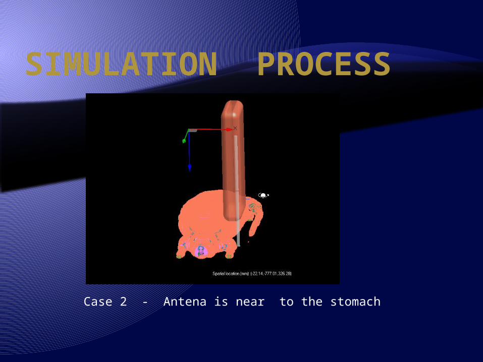
SIMULATION PROCESS
Case 2 - Antena is near to the stomach
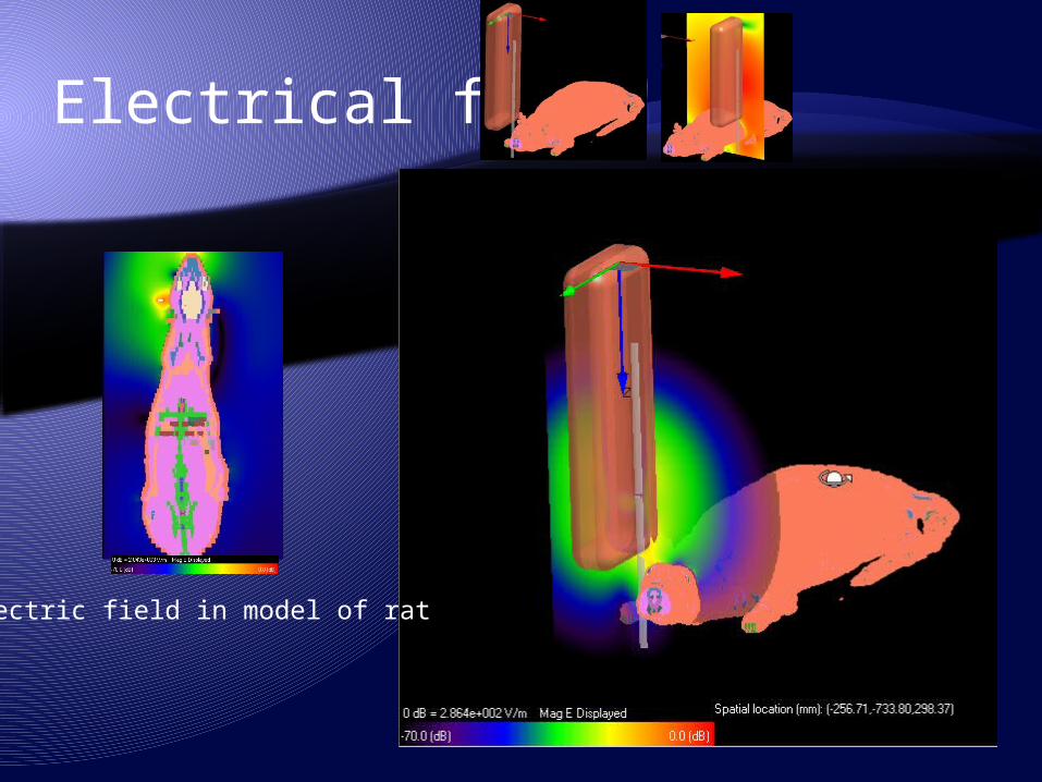
Electrical field
Electric field in model of rat
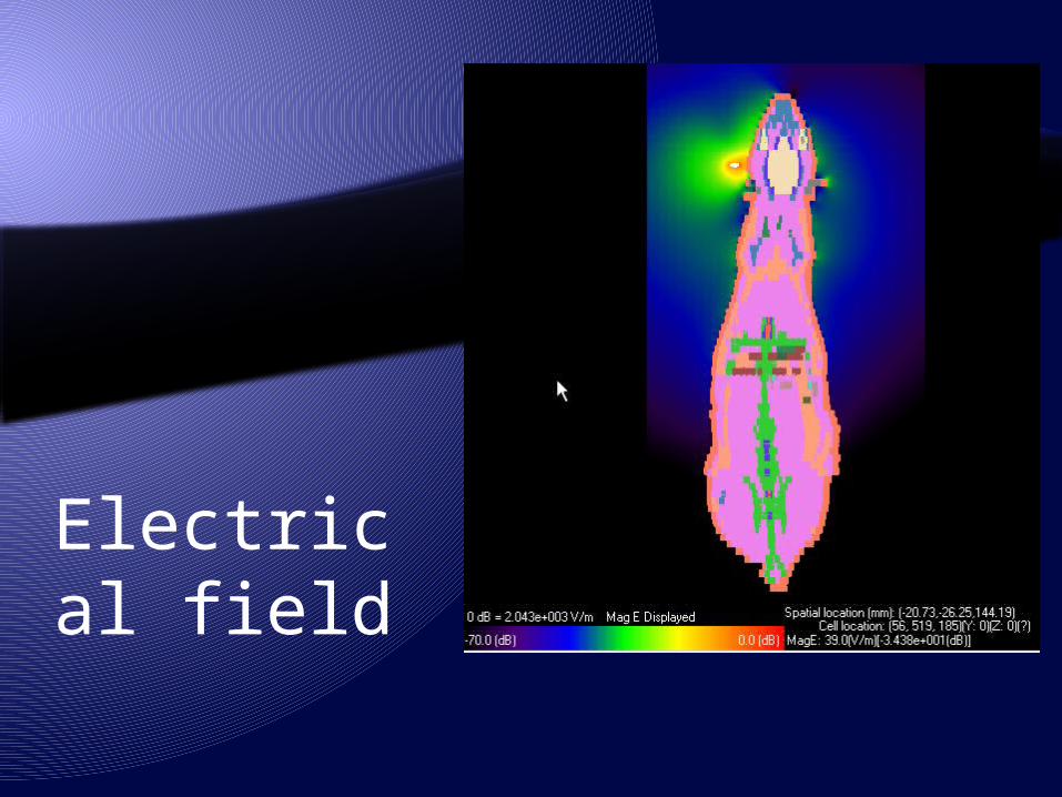
Electrical field
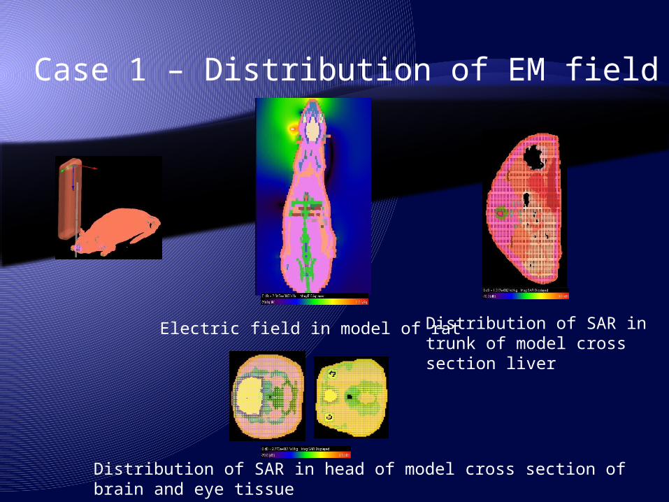
Case 1 – Distribution of EM field
Electric field in model of rat
Distribution of SAR in head of model cross section of brain and eye tissue
Distribution of SAR in trunk of model cross section liver
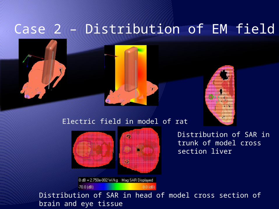
Case 2 – Distribution of EM field
Electric field in model of rat
Distribution of SAR in head of model cross section of brain and eye tissue
Distribution of SAR in trunk of model cross section liver
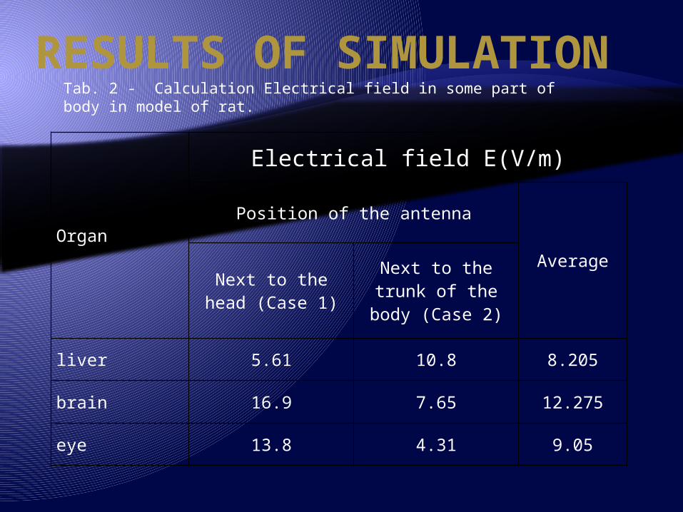
RESULTS OF SIMULATION
Organ
Electrical field E(V/m)
Position of the antenna
AverageNext to the head
(Case 1)Next to the trunk of the body (Case 2)
liver 5.61 10.8 8.205
brain 16.9 7.65 12.275
eye 13.8 4.31 9.05
Tab. 2 - Calculation Electrical field in some part of body in model of rat.
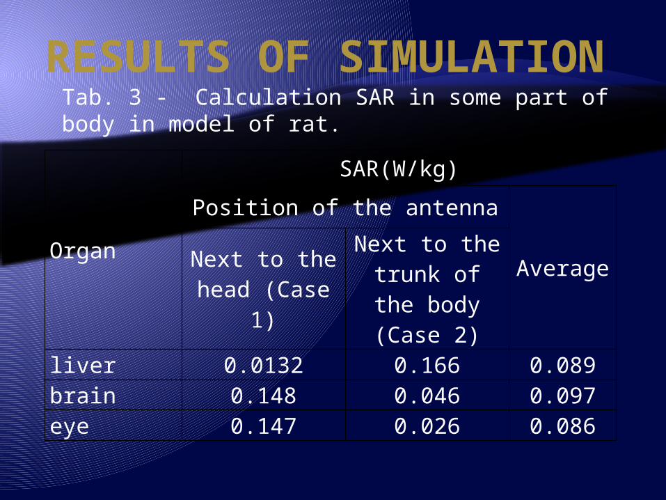
RESULTS OF SIMULATION
Organ
SAR(W/kg)
Position of the antenna
AverageNext to the head (Case 1)
Next to the trunk of the body
(Case 2)
liver 0.0132 0.166 0.089brain 0.148 0.046 0.097eye 0.147 0.026 0.086
Tab. 3 - Calculation SAR in some part of body in model of rat.
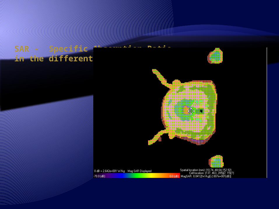
SAR - Specific Absorption Ratioin the different cross section of head
CONCLUSION The results of electric field distribution in the rats bodies suggest that there is an
unequal distribution of the fields, which depends on the position of the sources and characteristics of each tissue.
It is important to note that there are tissues which absorb 10 times higher amounts of energy the tissues adjacent to them.
Precise locate point of maximum SAR indicate the possible biological effects of radiation on these tissues.
These effects were presented and discussed in various papers which analyzed biochemical indicators of the effects of electromagnetic fields.
Such calculation enables us to develop the biological quantifiers of the effects of electromagnetic fields, which is studied by dosimetry.
Thus obtained quantifiers could be applied on the human tissue.
Acknowledgement: This work was supported by the project III43011 and III43012 of the Serbian Ministry of Education and Science.