Embed Size (px)
Citation preview
1
MODELING FUNCTIONS OF STRIATAL DOPAMINE MODULATION INLEARNING AND PLANNING
Suri R. E.1,*, Bargas J.2, and Arbib M.A. 1
1USC Brain Project, Los Angeles CA 90089-25202Departamento de Biofisica, Instituto de Fisiologia Cellular, Universidad Nacional Autonoma de Mexico,
Mexico City DFVersion from 24 February, 2000
The activity of midbrain dopamine neurons is strikingly similar to the reward prediction error of TDreinforcement learning models. Experimental evidence and simulation studies suggest that dopamineneuron activity serves as an effective reinforcement signal for learning of sensorimotor associations instriatal matrisomes.
In the current study, we simulate dopamine neuron activity with the Extended TD model (Suri andSchultz, submitted) and examine the influence of this signal on medium spiny neurons in striatalmatrisomes. This model includes transient membrane effects of dopamine, dopamine-dependent long-term adaptations of corticostriatal transmission, and rhythmic fluctuations of the membrane potentialbetween an elevated “up-state” and a hyperpolarized “down-state.” The most dominant activity in thestriatal matrisomes elicits behaviors via projections from the basal ganglia to the thalamus and the cortex.
This “standard model” performs successfully when tested for sensorimotor learning and goal-directed behavior (planning). To investigate the contributions of these model assumptions to learning andplanning, we test the performance of several model variants that lack one of these mechanisms. Thesesimulations show that the adaptation of the dopamine-like signal is necessary for planning and forsensorimotor learning. Lack of dopamine-like novelty responses decreases the number of exploratory acts,which deteriorates planning capabilities. Sensorimotor learning requires dopamine-dependent long-termadaptation of corticostriatal transmission. The model loses its planning capabilities if the dopamine-likesignal is simulated with the original TD model. The capability for planning is improved by transientdopamine membrane effects, dopamine-dependent long-term effects on corticostriatal transmission,dopamine- and input-dependent influences on the durations of membrane potential fluctuations, andmanipulations that prolong the reaction time of the model. These simulation results suggest that striataldopamine is important for sensorimotor learning, exploration, and planning.
INTRODUCTIONMidbrain dopamine neurons are phasically activated by unpredicted rewards or by the first sensory eventthat allows the animal to predict the reward but do not respond to predicted rewards. When a predictedreward is omitted, their activity is depressed at the time when the reward fails to occur (Schultz, 1998).The reward prediction error of temporal difference models (TD models) reproduces these features ofdopamine neuron activity (Sutton and Barto, 1990; Montague et al., 1996; Schultz et al., 1997; Suri andSchultz, 1999, submitted). In addition, dopamine neurons respond to novel, physically salient stimuli(Schultz, 1998). Such stimuli elicit action potential bursts followed by activity decreases below baseline
* Present Address: Roland Suri, Computational Neurobiology Laboratory,Salk Institute, Post Office Box 85800, San Diego CA 92186-5800; [email protected],http://www.cnl.salk.edu/~suri
2
levels. These biphasic novelty responses diminish with repeated stimulus presentation. TD modelsreproduce these characteristics of dopamine novelty responses if the associative weights of stimulusonsets are initialized with positive values (Suri and Schultz, 1999).
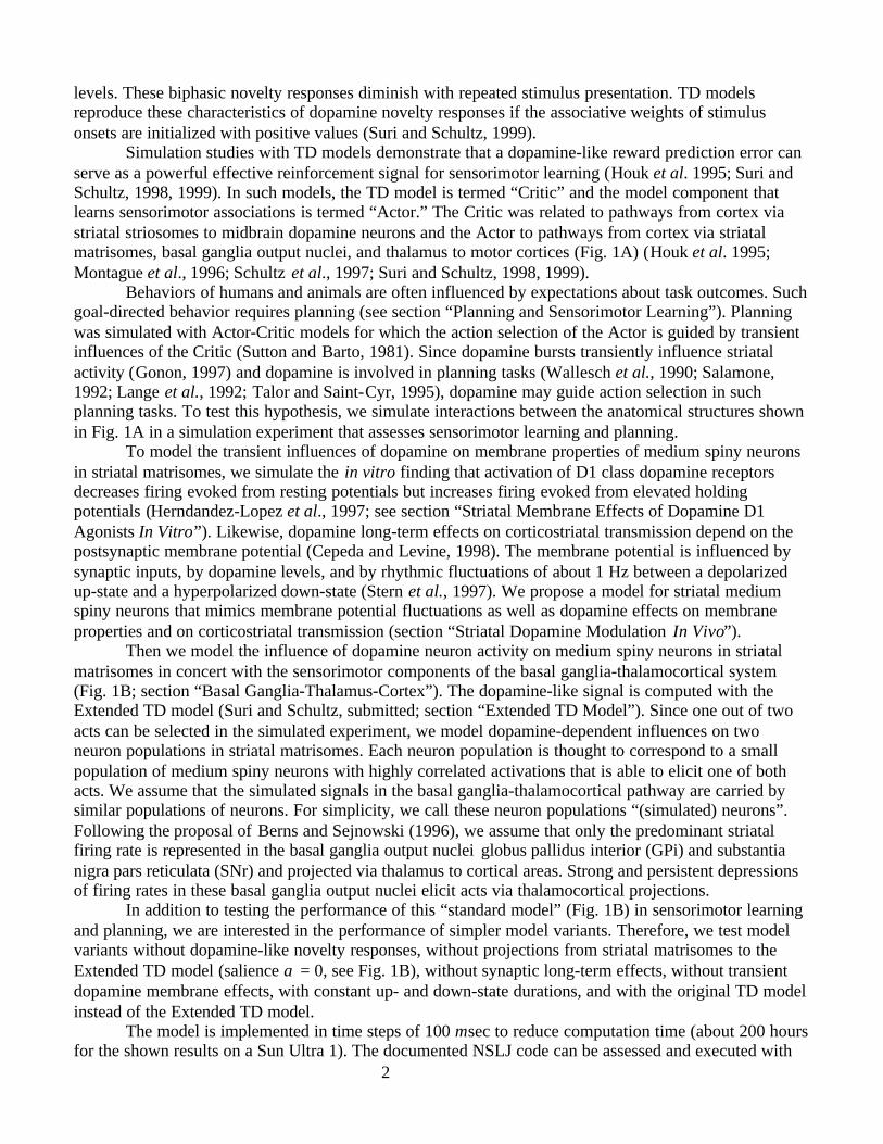
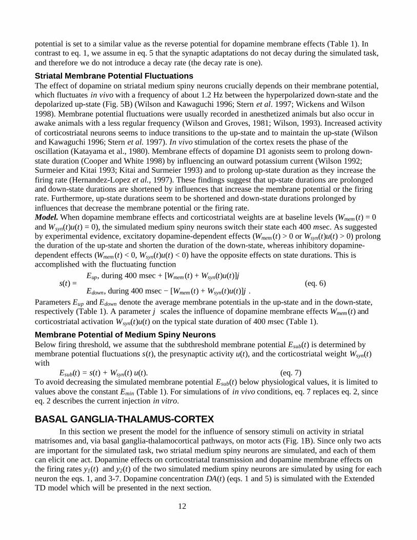
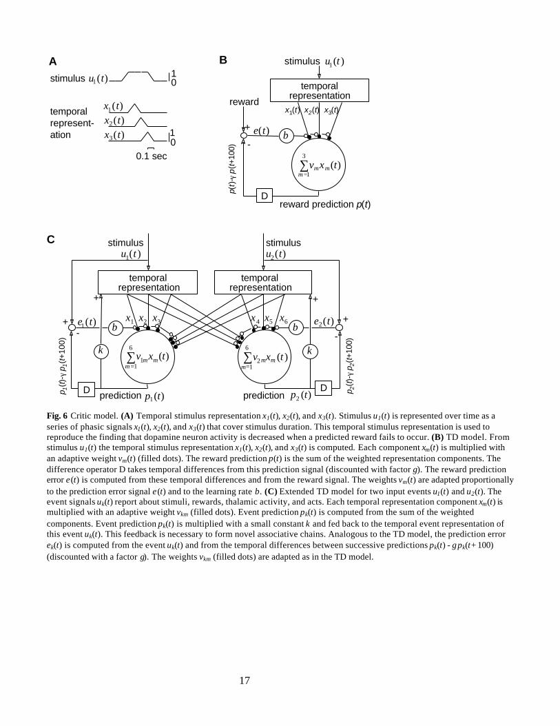
Simulation studies with TD models demonstrate that a dopamine-like reward prediction error canserve as a powerful effective reinforcement signal for sensorimotor learning (Houk et al. 1995; Suri andSchultz, 1998, 1999). In such models, the TD model is termed “Critic” and the model component thatlearns sensorimotor associations is termed “Actor.” The Critic was related to pathways from cortex viastriatal striosomes to midbrain dopamine neurons and the Actor to pathways from cortex via striatalmatrisomes, basal ganglia output nuclei, and thalamus to motor cortices (Fig. 1A) (Houk et al. 1995;Montague et al., 1996; Schultz et al., 1997; Suri and Schultz, 1998, 1999).
Behaviors of humans and animals are often influenced by expectations about task outcomes. Suchgoal-directed behavior requires planning (see section “Planning and Sensorimotor Learning”). Planningwas simulated with Actor-Critic models for which the action selection of the Actor is guided by transientinfluences of the Critic (Sutton and Barto, 1981). Since dopamine bursts transiently influence striatalactivity (Gonon, 1997) and dopamine is involved in planning tasks (Wallesch et al., 1990; Salamone,1992; Lange et al., 1992; Talor and Saint-Cyr, 1995), dopamine may guide action selection in suchplanning tasks. To test this hypothesis, we simulate interactions between the anatomical structures shownin Fig. 1A in a simulation experiment that assesses sensorimotor learning and planning.
To model the transient influences of dopamine on membrane properties of medium spiny neuronsin striatal matrisomes, we simulate the in vitro finding that activation of D1 class dopamine receptorsdecreases firing evoked from resting potentials but increases firing evoked from elevated holdingpotentials (Herndandez-Lopez et al., 1997; see section “Striatal Membrane Effects of Dopamine D1Agonists In Vitro”). Likewise, dopamine long-term effects on corticostriatal transmission depend on thepostsynaptic membrane potential (Cepeda and Levine, 1998). The membrane potential is influenced bysynaptic inputs, by dopamine levels, and by rhythmic fluctuations of about 1 Hz between a depolarizedup-state and a hyperpolarized down-state (Stern et al., 1997). We propose a model for striatal mediumspiny neurons that mimics membrane potential fluctuations as well as dopamine effects on membraneproperties and on corticostriatal transmission (section “Striatal Dopamine Modulation In Vivo”).
Then we model the influence of dopamine neuron activity on medium spiny neurons in striatalmatrisomes in concert with the sensorimotor components of the basal ganglia-thalamocortical system(Fig. 1B; section “Basal Ganglia-Thalamus-Cortex”). The dopamine-like signal is computed with theExtended TD model (Suri and Schultz, submitted; section “Extended TD Model”). Since one out of twoacts can be selected in the simulated experiment, we model dopamine-dependent influences on twoneuron populations in striatal matrisomes. Each neuron population is thought to correspond to a smallpopulation of medium spiny neurons with highly correlated activations that is able to elicit one of bothacts. We assume that the simulated signals in the basal ganglia-thalamocortical pathway are carried bysimilar populations of neurons. For simplicity, we call these neuron populations “(simulated) neurons”.Following the proposal of Berns and Sejnowski (1996), we assume that only the predominant striatalfiring rate is represented in the basal ganglia output nuclei globus pallidus interior (GPi) and substantianigra pars reticulata (SNr) and projected via thalamus to cortical areas. Strong and persistent depressionsof firing rates in these basal ganglia output nuclei elicit acts via thalamocortical projections.
In addition to testing the performance of this “standard model” (Fig. 1B) in sensorimotor learningand planning, we are interested in the performance of simpler model variants. Therefore, we test modelvariants without dopamine-like novelty responses, without projections from striatal matrisomes to theExtended TD model (salience α = 0, see Fig. 1B), without synaptic long-term effects, without transientdopamine membrane effects, with constant up- and down-state durations, and with the original TD modelinstead of the Extended TD model.
The model is implemented in time steps of 100 msec to reduce computation time (about 200 hoursfor the shown results on a Sun Ultra 1). The documented NSLJ code can be assessed and executed with
3
standard web browsers (Suri, Marmol, and Arbib, in preparation) and a Matlab code athttp://www.cnl.salk.edu/~suri. A study proposal was presented in abstract form (Suri and Arbib, 1998).
firing rate firing rate
stimuli
Extended TD Model
- -+ +
striatalmatrisomes
GPi/SNr
thalamus
-
α
reward
B
A stimuli
GPi/SNr
GPe
STN-
reward-
-
acts
DA(t)
matrisomes
dopamineneurons
striosomes
)(1 ts )(2 ts
)(1 ty )(2 ty
leaky integratorsthresholds
cortex-striatalstriosomes-
midbrain dopamineneurons
GPe/STN
α act1(t)act2(t)
thalamus-
-
DA(t)
Cortex
Actor
Critic
striatum
Fig. 1 (A) Interactions between cortex, basal ganglia, and midbrain dopamine neurons mimicked by the model. Corticalpyramidal neurons project to the striatum, which can be divided in striosomes (patches) and matrisomes (matrix) (Graybiel,1990). Prefrontal and insular cortices project chiefly to striosomes, whereas sensory and motor cortices project chiefly tomatrisomes (Graybiel, 1990). Midbrain dopamine neurons are contacted by medium spiny neurons in striosomes and project toboth striatal compartments (Graybiel, 1990; Smith and Bolam, 1990). Striatal matrisomes directly inhibit the basal gangliaoutput nuclei globus pallidus interior (GPi) and substantia nigra pars reticulata (SNr), whereas they indirectly disinhibit theseoutput nuclei via globus pallidus exterior (GPe) and subthalamic nucleus (STN) (Albin et al., 1989; Alexander and Crutcher,1990). The basal ganglia output nuclei project via thalamic nuclei to motor, occulomotor, prefrontal, and limbic cortical areas(Alexander and Crutcher, 1990). The structures shown as grey boxes correspond to the Critic and those shown as white boxesto the Actor. (B) Model architecture. The Extended TD model serves as the Critic component (grey box), and the Actorcomponent (remaining architecture) elicits acts. Actor: Sensory stimuli influence the membrane potentials of two mediumspiny projection neurons in striatal matrisomes (large circles). The membrane potentials of these neurons are also influenced byfluctuations between an elevated up-state and a hyperpolarised down-state simulated with the functions s1(t) and s2(t).Adaptations in corticostriatal weights (filled dots) and dopamine membrane effects are influenced by the membrane potentialand the dopamine-like signal DA(t) (open dots). The firing rates y1(t) and y2(t) of both striatal neurons inhibit the basal gangliaoutput nuclei substantia nigra pars reticulata (SNr) and globus pallidus interior (GPi). An indirect disinhibitory pathway fromstriatum to GPi/SNr suppresses insignificant inhibitions in the basal ganglia output nuclei (Berns and Sejnowski, 1996). Thewinning inhibition disinhibits the thalamus. These signals in the thalamus lead only to acts, coded by the signals act1(t) andact2(t), if they are sufficiently strong and persistent. This is accomplished by integrating the cortical signal and eliciting actswhen it reaches a threshold. Critic: The Critic and computes the dopamine-like reward prediction error DA(t) from the sensorystimuli, the reward signal, the thalamic signals (multiplied with the salience α), and the act signals act1(t) and act2(t).
4
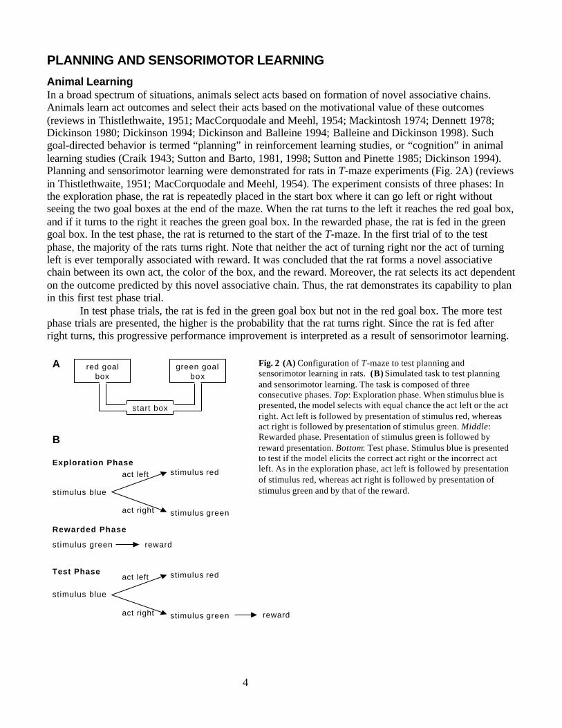
PLANNING AND SENSORIMOTOR LEARNING
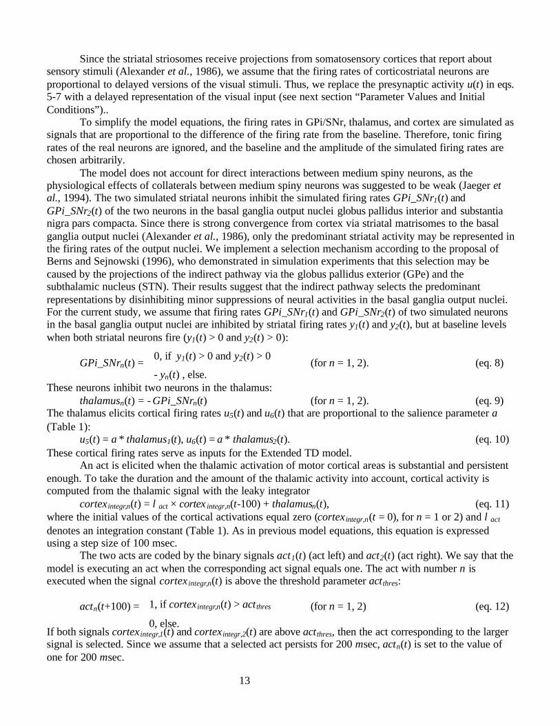
Animal LearningIn a broad spectrum of situations, animals select acts based on formation of novel associative chains.Animals learn act outcomes and select their acts based on the motivational value of these outcomes(reviews in Thistlethwaite, 1951; MacCorquodale and Meehl, 1954; Mackintosh 1974; Dennett 1978;Dickinson 1980; Dickinson 1994; Dickinson and Balleine 1994; Balleine and Dickinson 1998). Suchgoal-directed behavior is termed “planning” in reinforcement learning studies, or “cognition” in animallearning studies (Craik 1943; Sutton and Barto, 1981, 1998; Sutton and Pinette 1985; Dickinson 1994).Planning and sensorimotor learning were demonstrated for rats in T-maze experiments (Fig. 2A) (reviewsin Thistlethwaite, 1951; MacCorquodale and Meehl, 1954). The experiment consists of three phases: Inthe exploration phase, the rat is repeatedly placed in the start box where it can go left or right withoutseeing the two goal boxes at the end of the maze. When the rat turns to the left it reaches the red goal box,and if it turns to the right it reaches the green goal box. In the rewarded phase, the rat is fed in the greengoal box. In the test phase, the rat is returned to the start of the T-maze. In the first trial of to the testphase, the majority of the rats turns right. Note that neither the act of turning right nor the act of turningleft is ever temporally associated with reward. It was concluded that the rat forms a novel associativechain between its own act, the color of the box, and the reward. Moreover, the rat selects its act dependenton the outcome predicted by this novel associative chain. Thus, the rat demonstrates its capability to planin this first test phase trial.
In test phase trials, the rat is fed in the green goal box but not in the red goal box. The more testphase trials are presented, the higher is the probability that the rat turns right. Since the rat is fed afterright turns, this progressive performance improvement is interpreted as a result of sensorimotor learning.
Exploration Phase
stimulus blue
act left
act right
stimulus red
stimulus green
reward
Rewarded Phase
stimulus green
Test Phase
stimulus blue
act left
act right
stimulus red
stimulus green reward
B
A
start box
red goalbox
green goalbox
Fig. 2 (A) Configuration of T-maze to test planning andsensorimotor learning in rats. (B) Simulated task to test planningand sensorimotor learning. The task is composed of threeconsecutive phases. Top: Exploration phase. When stimulus blue ispresented, the model selects with equal chance the act left or the actright. Act left is followed by presentation of stimulus red, whereasact right is followed by presentation of stimulus green. Middle:Rewarded phase. Presentation of stimulus green is followed byreward presentation. Bottom: Test phase. Stimulus blue is presentedto test if the model elicits the correct act right or the incorrect actleft. As in the exploration phase, act left is followed by presentationof stimulus red, whereas act right is followed by presentation ofstimulus green and by that of the reward.
5
Animal Learning ModelsBased on such experimental findings, animal learning theorists suggest that animals form an internalmodel of their environment that allows them to predict the sensory consequences of their acts and to formnovel associative chains. Furthermore, animals seem to use these predictions to select their acts (Suttonand Pinette, 1985; Sutton and Barto 1981; Dickinson and Balleine 1994; Balleine and Dickinson 1998).This insight led to the implementation of neural network architectures in which an internal model, servingas the Critic, transiently influences the Actor to elicit acts (Sutton and Barto, 1981). In such Actor-Criticarchitectures, the Actor computes small random variations in act preparation signals and executes actswhen these preparation signals reach a threshold. The Critic component learns associations betweensensory stimuli, rewards, and act preparation signals and uses these associations to form novel associativechains. The output of the Critic is a signal that reflects the value of the predicted outcome and reinforcesor attenuates the act preparation signals. In this manner, the effective reinforcement signal of the Criticselects the act that predicts the optimal outcome. This animal learning model resembles the model thatwill be proposed in the current study.
Task SimulationIn the current study, we test model performance in a task analogous to the T-maze task. In the explorationphase (Fig. 2B, top), each trial starts with presentation of a stimulus called “blue” (stimulus blue) thatrepresents sensory features of the start box. When either the act right or the act left is executed duringpresentation of stimulus blue, the stimulus is extinguished and either stimulus green or stimulus red ispresented, respectively. The act right and the act left represent the rat’s right and left turn, respectively,whereas the stimuli green and red correspond to the colors of the goal boxes. If no act is selected, stimulusblue is extinguished after 600 msec. This exploration phase is simulated for a time span corresponding to80 sec (exclusive of intertrial intervals), during which stimulus blue is presented about 100 times. Thesubsequent rewarded phase consists of only one trial (Fig. 2B, middle), in which presentation of stimulusgreen is followed by reward presentation. The beginning of the test phase (Fig. 2B, bottom) is equal to theexploration phase. Stimulus green but not stimulus red is followed by reward presentation. In all threephases, the stimuli green and red are presented for 300 msec and the reward for 100 msec. Planning isassessed in the first trial of the test phase and the progress of sensorimotor learning in subsequent trials.
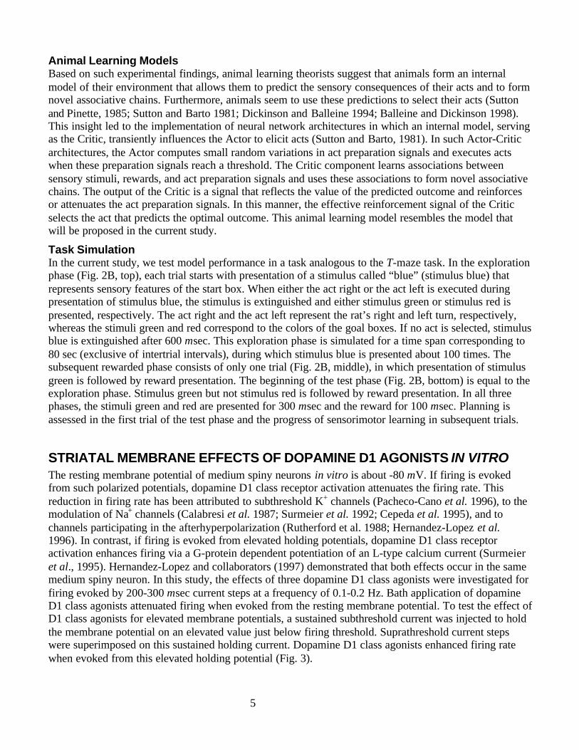
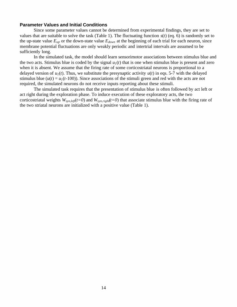
STRIATAL MEMBRANE EFFECTS OF DOPAMINE D1 AGONISTS IN VITROThe resting membrane potential of medium spiny neurons in vitro is about -80 mV. If firing is evokedfrom such polarized potentials, dopamine D1 class receptor activation attenuates the firing rate. Thisreduction in firing rate has been attributed to subthreshold K+ channels (Pacheco-Cano et al. 1996), to themodulation of Na+ channels (Calabresi et al. 1987; Surmeier et al. 1992; Cepeda et al. 1995), and tochannels participating in the afterhyperpolarization (Rutherford et al. 1988; Hernandez-Lopez et al.1996). In contrast, if firing is evoked from elevated holding potentials, dopamine D1 class receptoractivation enhances firing via a G-protein dependent potentiation of an L-type calcium current (Surmeieret al., 1995). Hernandez-Lopez and collaborators (1997) demonstrated that both effects occur in the samemedium spiny neuron. In this study, the effects of three dopamine D1 class agonists were investigated forfiring evoked by 200-300 msec current steps at a frequency of 0.1-0.2 Hz. Bath application of dopamineD1 class agonists attenuated firing when evoked from the resting membrane potential. To test the effect ofD1 class agonists for elevated membrane potentials, a sustained subthreshold current was injected to holdthe membrane potential on an elevated value just below firing threshold. Suprathreshold current stepswere superimposed on this sustained holding current. Dopamine D1 class agonists enhanced firing ratewhen evoked from this elevated holding potential (Fig. 3).
6
Control
Injected Current
Dopamine D1 AgonistSKF81297
Fig. 3 Dopamine D1 class receptor agonist SKF 81297 enhances or attenuates evoked firing depending on the holding potential(Figure adapted with permission from Hernandez-Lopez et al., 1997). (A) Firing was evoked with a current step from theresting potential of -82 mV (top, eight action potentials). 1 µM of D1 receptor agonist SKF 81297 attenuated evoked firing(middle, three action potentials). Injected current was maintained for both conditions (bottom). (B) For the same neuron, firingwas evoked from a holding potential of -57 mV (top, 10 action potentials). 1 µM of D1 receptor agonist SKF81297 increasedevoked firing (middle, 14 action potentials). Injected current was again maintained for both conditions (bottom).
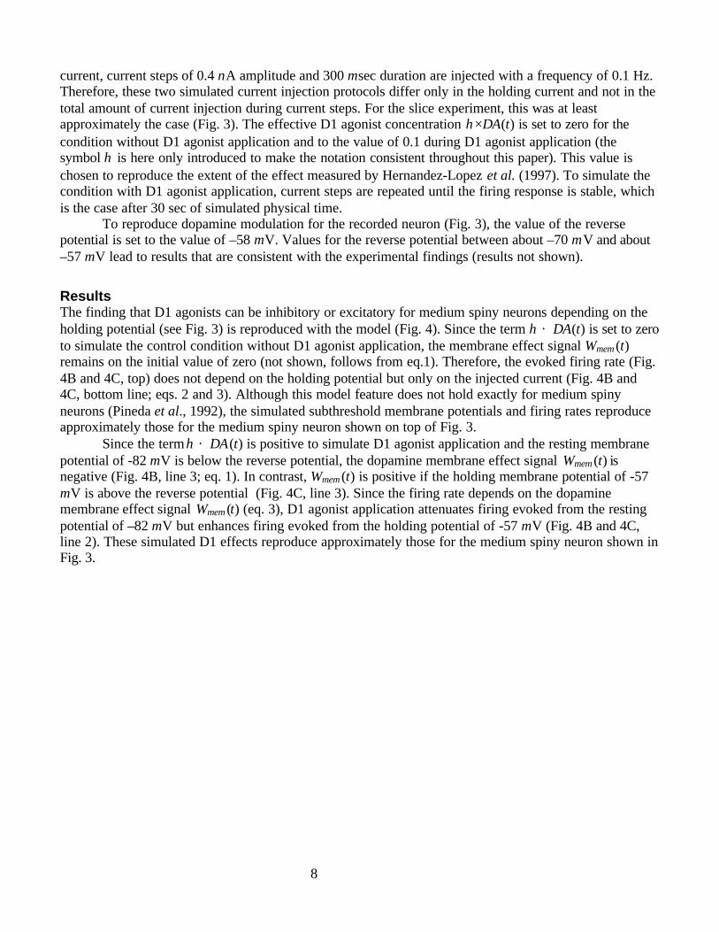
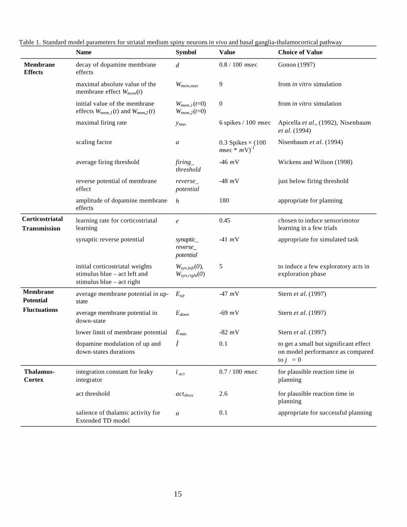
ModelEffects of neuromodulators on neuronal membrane properties have been simulated with various modelingtechniques (reviewed in Fellous and Linster, 1998). To simulate the findings of Hernandez-Lopez et al.(1997), we propose a phenomenological model for membrane effects mediated by dopamine D1 classreceptors (Fig. 4A). Since dopamine D1 class receptor activation enhances or attenuates evoked firingdepending on the holding potential, we introduce a reverse potential (Table 1). This reverse potentialcorresponds to the hypothetical holding potential between resting potential and firing threshold for whichthe effect of D1 activation on evoked firing vanishes as it reverses its sign. The term reverse potentialshould not be confused with the biophysically defined term reversal potential that is defined by a currentor voltage reversal. The influence of the holding potential on the firing rate during the current step ismodeled using a slowly varying parameter Wmem (t) that is initialized with a value of zero and then adaptedwith
Wmem (t) = δ Wmem(t-100)+ηDA(t-100)[E(t-100)-reverse_potential]. (eq. 1)The time t is given in units of msec throughout this paper. A constant δ denotes the decay rate of the D1effects. Since the D1 agonist effects decay to a value of 40% over 10 minutes (see figure 4B inHernandez-Lopez et al., 1997), we estimate the value of the decay rate δ of the dopamine membraneeffects to be 0.99985, which corresponds to 0.015 % decrease for each 100 msec. Note that this valuedepends on the mode of the D1 agonist application, since the D1 agonist effects decay faster if theagonists are applied directly on isolated cells (Surmeier et al., 1995) and much faster if dopamine neuronsare activated in vivo (Gonon, 1997). Therefore, we use the value δ =0.99985 to reproduce the experimentof Hernandez-Lopez et al. (1997) but will adjust this value for the in vivo model (see next section). Thesignal DA(t) corresponds to the dopamine D1 agonist concentration and the parameter η is a scalingfactor. A signal E(t) denotes the membrane potential in mV and is defined below. The absolute value of
7
the dopamine membrane effects Wmem (t) is limited to Wmem,max = 9, as this value is appropriate toreproduce the maximal amplitude of the D1 agonist effects.
The subthreshold membrane potential Esub(t) is computed from the resting membrane potentialErest, from the resistance R, and from the injected current I(t) according to Ohm’s law with
Esub(t) = Erest + R I(t). (eq. 2)For the neuron shown in Fig. 3, the value of the resting potential is estimated to Erest = -82 mV and the value ofthe resistance R to 27 MOhm. The latter value does not correspond to a biophysical property of the neuron butdepends on the used electrode. Since the seal between the used intracellular sharp electrode and the neuralmembrane is far from being complete, a major part of the injected current I(t) does not reach the inside ofthe neuron. For membrane potentials below firing threshold, Esub(t) approximates the membrane potential.For values above firing threshold, the membrane potential Esub(t) does not have a direct biologicalcorrespondence, as it is only defined for time steps of 100 msec and therefore cannot reflect the quicklyvarying time course of the membrane potential for the spiking neuron. Since we assume that the firing rateincreases with increasing values of the membrane potential Esub(t) and of the adaptive parameter Wmem (t),we compute the firing rate y(t) of the striatal neuron with
y(t) = ymax × tanh{a × [Esub(t) + Wmem(t) –firing_threshold]/ ymax}. (eq. 3)From Fig. 3, the firing threshold is estimated to be −56 mV. This value does not correspond to the averagefiring threshold in vivo and will be adjusted for the in vivo model. The hyperbolic tangent tanh is asigmoid function, and the function ymax × tanh{ . / ymax } smoothly limits the firing rate y(t) to valuesbelow the maximal firing rate ymax of medium spiny neurons. Medium spiny neurons fire with a maximalfiring rate ymax of about 6 Spikes per 100 msec (Apicella et al., 1992; Pineda et al., 1992; Nisenbaum et al.1994). A factor a = 0.3 Spikes/(100 msec * mV) is used to scale the firing rate to experimental data and isestimated from firing rates for constant current injections in the absence of dopamine agonists (estimatedfrom figure 1 in Nisenbaum et al. 1994; similar in figure 3 in Pineda et al., 1992). Eq. 3 does not take intoaccount that firing rate adaptations occur during a few hundreds of milliseconds after current stepinjections (Fig. 3; Pineda et al., 1992). The dopamine-dependent signal Wmem (t) influences the firing rate(eq. 3) rather than the subthreshold membrane potential Esub(t) (eq. 2), as the membrane potential beforeand after current step injections is not substantially influenced by D1 agonist application (Hernandez-Lopez et al., 1997).
Eq. 2 computes the subthreshold membrane potential Esub(t) but not the membrane potential forthe firing neuron. Since the 100 msec step size of our implementation is too long to simulate single spikes,we approximate the membrane potential E(t) for a certain firing rate with the average membrane potentialof real neurons with this firing rate. This is achieved by computing the contribution of measured actionpotentials to the average membrane potential. From intracellular voltage recordings of spontaneouslyactive striatal medium spiny neurons (figure 3F in Wickens and Wilson, 1998), we estimate for a singlespike an area of 6 mV × 100 msec between the firing threshold and the membrane potential. Using thisvalue, the membrane potential is computed with
E(t) =
(eq. 4)
For subthreshold membrane potentials, this equation sets the membrane potential E(t) to Esub(t), whereasfor the firing neuron, E(t) is set to the average membrane potential of real neurons with this firing rate.
SimulationTo test the proposed model, we simulate the experimental conditions of Hernandez-Lopez et al. (1997)(Fig. 3). To mimic the conditions for the result shown in Fig. 3A, we simulate current step injections of1.3 nA amplitude and 300 msec duration with a frequency of 0.1 Hz. To reproduce Fig. 3B, the holdingpotential of -57 mV is induced with a sustained current I(t) of 0.9 nA. Superimposed on this holding
firing_threshold + y(t) × 6 mV, if Esub(t) > firing_threshold
Esub(t), else.
8
current, current steps of 0.4 nA amplitude and 300 msec duration are injected with a frequency of 0.1 Hz.Therefore, these two simulated current injection protocols differ only in the holding current and not in thetotal amount of current injection during current steps. For the slice experiment, this was at leastapproximately the case (Fig. 3). The effective D1 agonist concentration η×DA(t) is set to zero for thecondition without D1 agonist application and to the value of 0.1 during D1 agonist application (thesymbol η is here only introduced to make the notation consistent throughout this paper). This value ischosen to reproduce the extent of the effect measured by Hernandez-Lopez et al. (1997). To simulate thecondition with D1 agonist application, current steps are repeated until the firing response is stable, whichis the case after 30 sec of simulated physical time.
To reproduce dopamine modulation for the recorded neuron (Fig. 3), the value of the reversepotential is set to the value of –58 mV. Values for the reverse potential between about –70 mV and about–57 mV lead to results that are consistent with the experimental findings (results not shown).
ResultsThe finding that D1 agonists can be inhibitory or excitatory for medium spiny neurons depending on theholding potential (see Fig. 3) is reproduced with the model (Fig. 4). Since the term η × DA(t) is set to zeroto simulate the control condition without D1 agonist application, the membrane effect signal Wmem (t)remains on the initial value of zero (not shown, follows from eq.1). Therefore, the evoked firing rate (Fig.4B and 4C, top) does not depend on the holding potential but only on the injected current (Fig. 4B and4C, bottom line; eqs. 2 and 3). Although this model feature does not hold exactly for medium spinyneurons (Pineda et al., 1992), the simulated subthreshold membrane potentials and firing rates reproduceapproximately those for the medium spiny neuron shown on top of Fig. 3.
Since the term η × DA(t) is positive to simulate D1 agonist application and the resting membranepotential of -82 mV is below the reverse potential, the dopamine membrane effect signal Wmem (t) isnegative (Fig. 4B, line 3; eq. 1). In contrast, Wmem (t) is positive if the holding membrane potential of -57mV is above the reverse potential (Fig. 4C, line 3). Since the firing rate depends on the dopaminemembrane effect signal Wmem (t) (eq. 3), D1 agonist application attenuates firing evoked from the restingpotential of –82 mV but enhances firing evoked from the holding potential of -57 mV (Fig. 4B and 4C,line 2). These simulated D1 effects reproduce approximately those for the medium spiny neuron shown inFig. 3.
9
Control
D1 Agonist
weight Wmem(t)
injectedcurrent I(t)
Control
D1 Agonist
B C
100 msec
0 nA
1 nA
2 nA
-10
0
10
D1 Agonist D1 Agonist
firing threshold
firing threshold
-80-70-60
1
5 firing rate
[spikes/100 msec]
membranepotential
[mV]
-80-70-60
1
5 firing rate
[spikes/100 msec]
membranepotential
[mV]
membranepotential E(t)
A
I (t)
dopamine D1agonist DA(t)
injectedcurrent
firing ratey(Esub(t), Wmem (t))
Esub(t)
= Erest + RI(t)
membranepotential E(t)
Wmem(t)
Fig. 4 (A) Model for effects of dopamine D1 class receptor activation on the firing rate of a medium spiny neuron in vitro . Thesubthreshold membrane potential Esub(t) depends on the constant resting membrane potential Erest and on the product of theinjected current I(t) with a resistance R. The subthreshold membrane potential Esub(t) and dopamine D1 agonist concentrationDA(t) influence the value of the signal Wmem(t). The firing rate y(t) is a monotonically increasing function of the subthresholdmembrane potential Esub(t) and the signal Wmem(t). (B ,C) Simulation of the experimental result shown in Fig. 3. Note that forthe four lines on top (B and C, line 1 and line 2), the signal E(t) [mV] denotes the membrane potential averaged over the 100msec step size of the model. Above firing threshold, values of E(t) also correspond to firing rates [spikes/100 msec]. (B)Current injection of 1.3 nA for 300 msec (bottom line). Current injection without D1 agonist application (line 1, η×DA(t) = 0)leads to a firing rate of about 3 spikes/100 msec. The signal coding for the dopamine membrane effects Wmem(t) remains on theinitial value of zero (not shown, follows from eq. 1). With dopamine D1 agonist application (line 2, η×DA(t) = 0.1), evokedfiring is attenuated to less than 1 spike/100 msec because the value of the dopamine membrane effect signal Wmem(t) is negative(line 3). (C) Current injection of 1.3 nA for 300 msec from a sustained holding current of 0.9 nA (bottom line). Withoutdopamine D1 agonist application (line 1), the rate of evoked firing does not depend on the holding current (line 1 in B) becausethe dopamine membrane effect signal Wmem(t) remains on the value of zero (not shown). With dopamine D1 agonist application(line 2, η×DA(t) = 0.1), evoked firing is increased to 4.5 spikes/100 msec because the dopamine membrane effect signalWmem(t) is positive (line 3).
10
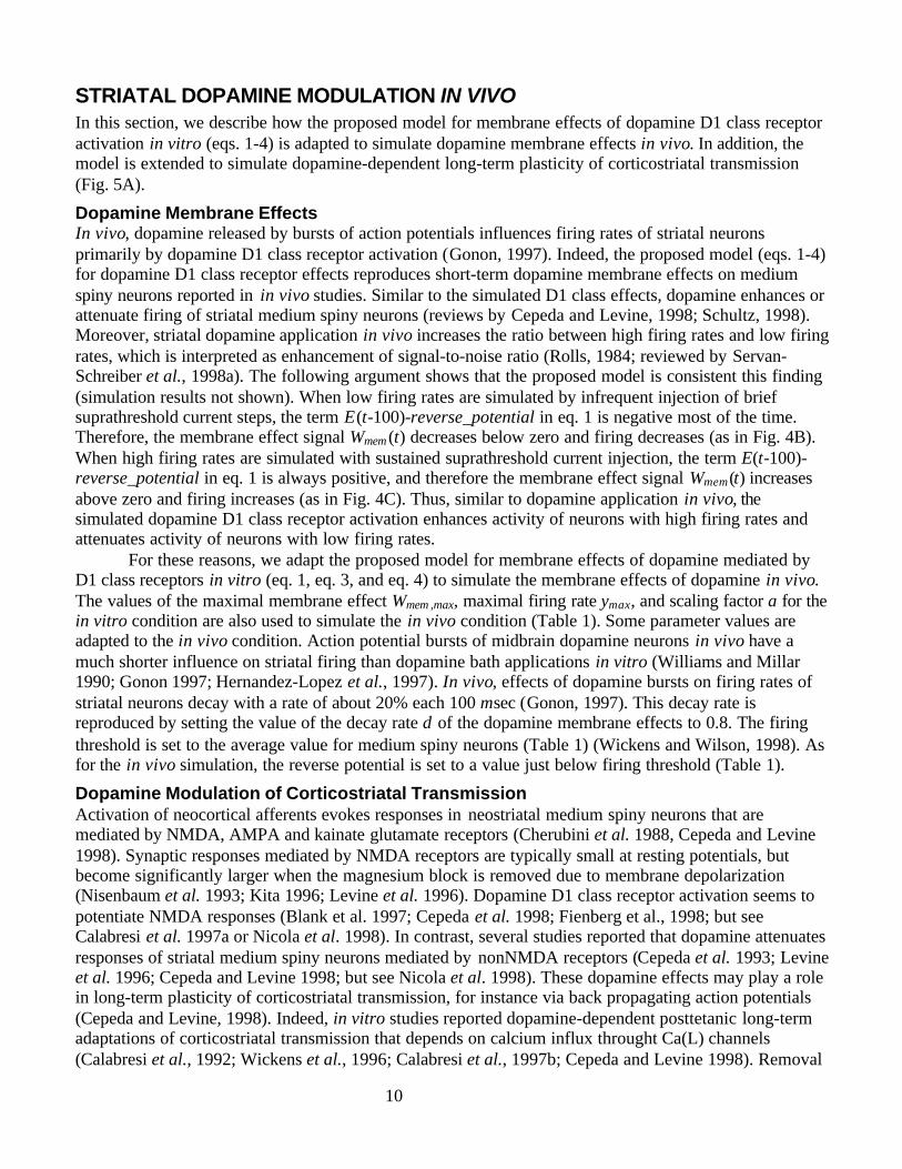
STRIATAL DOPAMINE MODULATION IN VIVOIn this section, we describe how the proposed model for membrane effects of dopamine D1 class receptoractivation in vitro (eqs. 1-4) is adapted to simulate dopamine membrane effects in vivo. In addition, themodel is extended to simulate dopamine-dependent long-term plasticity of corticostriatal transmission(Fig. 5A).
Dopamine Membrane EffectsIn vivo, dopamine released by bursts of action potentials influences firing rates of striatal neuronsprimarily by dopamine D1 class receptor activation (Gonon, 1997). Indeed, the proposed model (eqs. 1-4)for dopamine D1 class receptor effects reproduces short-term dopamine membrane effects on mediumspiny neurons reported in in vivo studies. Similar to the simulated D1 class effects, dopamine enhances orattenuate firing of striatal medium spiny neurons (reviews by Cepeda and Levine, 1998; Schultz, 1998).Moreover, striatal dopamine application in vivo increases the ratio between high firing rates and low firingrates, which is interpreted as enhancement of signal-to-noise ratio (Rolls, 1984; reviewed by Servan-Schreiber et al., 1998a). The following argument shows that the proposed model is consistent this finding(simulation results not shown). When low firing rates are simulated by infrequent injection of briefsuprathreshold current steps, the term E(t-100)-reverse_potential in eq. 1 is negative most of the time.Therefore, the membrane effect signal Wmem (t) decreases below zero and firing decreases (as in Fig. 4B).When high firing rates are simulated with sustained suprathreshold current injection, the term E(t-100)-reverse_potential in eq. 1 is always positive, and therefore the membrane effect signal Wmem(t) increasesabove zero and firing increases (as in Fig. 4C). Thus, similar to dopamine application in vivo, thesimulated dopamine D1 class receptor activation enhances activity of neurons with high firing rates andattenuates activity of neurons with low firing rates.
For these reasons, we adapt the proposed model for membrane effects of dopamine mediated byD1 class receptors in vitro (eq. 1, eq. 3, and eq. 4) to simulate the membrane effects of dopamine in vivo.The values of the maximal membrane effect Wmem ,max, maximal firing rate ymax, and scaling factor a for thein vitro condition are also used to simulate the in vivo condition (Table 1). Some parameter values areadapted to the in vivo condition. Action potential bursts of midbrain dopamine neurons in vivo have amuch shorter influence on striatal firing than dopamine bath applications in vitro (Williams and Millar1990; Gonon 1997; Hernandez-Lopez et al., 1997). In vivo, effects of dopamine bursts on firing rates ofstriatal neurons decay with a rate of about 20% each 100 msec (Gonon, 1997). This decay rate isreproduced by setting the value of the decay rate δ of the dopamine membrane effects to 0.8. The firingthreshold is set to the average value for medium spiny neurons (Table 1) (Wickens and Wilson, 1998). Asfor the in vivo simulation, the reverse potential is set to a value just below firing threshold (Table 1).
Dopamine Modulation of Corticostriatal TransmissionActivation of neocortical afferents evokes responses in neostriatal medium spiny neurons that aremediated by NMDA, AMPA and kainate glutamate receptors (Cherubini et al. 1988, Cepeda and Levine1998). Synaptic responses mediated by NMDA receptors are typically small at resting potentials, butbecome significantly larger when the magnesium block is removed due to membrane depolarization(Nisenbaum et al. 1993; Kita 1996; Levine et al. 1996). Dopamine D1 class receptor activation seems topotentiate NMDA responses (Blank et al. 1997; Cepeda et al. 1998; Fienberg et al., 1998; but seeCalabresi et al. 1997a or Nicola et al. 1998). In contrast, several studies reported that dopamine attenuatesresponses of striatal medium spiny neurons mediated by nonNMDA receptors (Cepeda et al. 1993; Levineet al. 1996; Cepeda and Levine 1998; but see Nicola et al. 1998). These dopamine effects may play a rolein long-term plasticity of corticostriatal transmission, for instance via back propagating action potentials(Cepeda and Levine, 1998). Indeed, in vitro studies reported dopamine-dependent posttetanic long-termadaptations of corticostriatal transmission that depends on calcium influx throught Ca(L) channels(Calabresi et al., 1992; Wickens et al., 1996; Calabresi et al., 1997b; Cepeda and Levine 1998). Removal
11
of the magnesium block was reported to reverse postetanic long-term depression to D1 receptor-dependent long-term potentiation (Calabresi et al. 1992; Calabresi et al. 1997b). Thus, long-termpotentiation of corticostriatal transmission may results from dopamine D1 class receptor activation duringdepolarized postsynaptic membrane potentials, whereas long-term depression may results from dopaminereceptor activation during hyperpolarized membrane potentials (Cepeda and Levine, 1998).
In addition to the postsynaptic membrane potential, dopamine concentration seems to influencethe direction of long term adaptation in corticostriatal transmission. Tetanic stimulation of corticostriatalfibers in slices that lack dopamine agonists in the bath produces long-term depression of excitatorypostsynaptic potentials, which is reversed by simultaneous pulsative dopamine application (Wickens etal., 1996; Calabresi et al. 1997b).
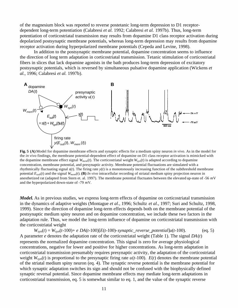
A B
u(t)presynapticactivity
dopamineDA(t)
Esub(t)
= s(t) + Wsyn(t)u(t)
Wmem(t)
firing ratey(Esub(t), Wmem (t))
Wsyn(t)
s(t)
Fig. 5 (A) Model for dopamine membrane effects and synaptic effects for a medium spiny neuron in vivo. As in the model forthe in vivo findings, the membrane potential-dependent effect of dopamine on D1 class receptor activation is mimicked withthe dopamine membrane effect signal Wmem(t). The corticostriatal weight Wsyn(t) is adapted according to dopamineconcentration, membrane potential, and presynaptic activity. Membrane potential fluctuations are simulated with arhythmically fluctuating signal s(t). The firing rate y(t) is a monotonously increasing function of the subthreshold membranepotential Esub(t) and the signal Wmem(t). (B) In vivo intracellular recording of striatal medium spiny projection neuron inanesthetized rat (adapted from Stern et. al. 1997). The membrane potential fluctuates between the elevated up-state of -56 mVand the hyperpolarized down-state of -79 mV.
Model. As in previous studies, we express long-term effects of dopamine on corticostriatal transmissionin the dynamics of adaptive weights (Montague et al., 1996; Schultz et al., 1997; Suri and Schultz, 1998,1999). Since the direction of dopamine long-term effects depends both on the membrane potential of thepostsynaptic medium spiny neuron and on dopamine concentration, we include these two factors in theadaptation rule. Thus, we model the long-term influence of dopamine on corticostriatal transmission withthe corticostriatal weight
Wsyn(t) = Wsyn(t-100)+ ε DA(t-100)[E(t-100)-synaptic_reverse_potential]u(t-100). (eq. 5)A parameter ε denotes the adaptation rate of the corticostriatal weight (Table 1). The signal DA(t)represents the normalized dopamine concentration. This signal is zero for average physiologicalconcentrations, negative for lower and positive for higher concentrations. As long-term adaptation incorticostriatal transmission presumably requires presynaptic activity, the adaptation of the corticostriatalweight Wsyn(t) is proportional to the presynaptic firing rate u(t-100). E(t) denotes the membrane potentialof the striatal medium spiny neuron (eq. 4). The synaptic reverse potential is the membrane potential forwhich synaptic adaptation switches its sign and should not be confused with the biophysically definedsynaptic reversal potential. Since dopamine membrane effects may mediate long-term adaptations incorticostriatal transmission, eq. 5 is somewhat similar to eq. 1, and the value of the synaptic reverse
12
potential is set to a similar value as the reverse potential for dopamine membrane effects (Table 1). Incontrast to eq. 1, we assume in eq. 5 that the synaptic adaptations do not decay during the simulated task,and therefore we do not introduce a decay rate (the decay rate is one).
Striatal Membrane Potential FluctuationsThe effect of dopamine on striatal medium spiny neurons crucially depends on their membrane potential,which fluctuates in vivo with a frequency of about 1.2 Hz between the hyperpolarized down-state and thedepolarized up-state (Fig. 5B) (Wilson and Kawaguchi 1996; Stern et al. 1997; Wickens and Wilson1998). Membrane potential fluctuations were usually recorded in anesthetized animals but also occur inawake animals with a less regular frequency (Wilson and Groves, 1981; Wilson, 1993). Increased activityof corticostriatal neurons seems to induce transitions to the up-state and to maintain the up-state (Wilsonand Kawaguchi 1996; Stern et al. 1997). In vivo stimulation of the cortex resets the phase of theoscillation (Katayama et al., 1980). Membrane effects of dopamine D1 agonists seem to prolong down-state duration (Cooper and White 1998) by influencing an outward potassium current (Wilson 1992;Surmeier and Kitai 1993; Kitai and Surmeier 1993) and to prolong up-state duration as they increase thefiring rate (Hernandez-Lopez et al., 1997). These findings suggest that up-state durations are prolongedand down-state durations are shortened by influences that increase the membrane potential or the firingrate. Furthermore, up-state durations seem to be shortened and down-state durations prolonged byinfluences that decrease the membrane potential or the firing rate.Model. When dopamine membrane effects and corticostriatal weights are at baseline levels (Wmem (t) = 0and Wsyn(t)u(t) = 0), the simulated medium spiny neurons switch their state each 400 msec. As suggestedby experimental evidence, excitatory dopamine-dependent effects (Wmem (t) > 0 or Wsyn(t)u(t) > 0) prolongthe duration of the up-state and shorten the duration of the down-state, whereas inhibitory dopamine-dependent effects (Wmem (t) < 0, Wsyn(t)u(t) < 0) have the opposite effects on state durations. This isaccomplished with the fluctuating function
s(t) = (eq. 6)
Parameters Eup and Edown denote the average membrane potentials in the up-state and in the down-state,respectively (Table 1). A parameter ϕ scales the influence of dopamine membrane effects Wmem (t) andcorticostriatal activation Wsyn(t)u(t) on the typical state duration of 400 msec (Table 1).
Membrane Potential of Medium Spiny NeuronsBelow firing threshold, we assume that the subthreshold membrane potential Esub(t) is determined bymembrane potential fluctuations s(t), the presynaptic activity u(t), and the corticostriatal weight Wsyn(t)with
Esub(t) = s(t) + Wsyn(t) u(t). (eq. 7)To avoid decreasing the simulated membrane potential Esub(t) below physiological values, it is limited tovalues above the constant Emin (Table 1). For simulations of in vivo conditions, eq. 7 replaces eq. 2, sinceeq. 2 describes the current injection in vitro.
BASAL GANGLIA-THALAMUS-CORTEXIn this section we present the model for the influence of sensory stimuli on activity in striatal
matrisomes and, via basal ganglia-thalamocortical pathways, on motor acts (Fig. 1B). Since only two actsare important for the simulated task, two striatal medium spiny neurons are simulated, and each of themcan elicit one act. Dopamine effects on corticostriatal transmission and dopamine membrane effects onthe firing rates y1(t) and y2(t) of the two simulated medium spiny neurons are simulated by using for eachneuron the eqs. 1, and 3-7. Dopamine concentration DA(t) (eqs. 1 and 5) is simulated with the ExtendedTD model which will be presented in the next section.
Eup, during 400 msec + [Wmem (t) + Wsyn(t)u(t)]ϕ
Edown, during 400 msec − [Wmem (t) + Wsyn(t)u(t)]ϕ.
13
Since the striatal striosomes receive projections from somatosensory cortices that report aboutsensory stimuli (Alexander et al., 1986), we assume that the firing rates of corticostriatal neurons areproportional to delayed versions of the visual stimuli. Thus, we replace the presynaptic activity u(t) in eqs.5-7 with a delayed representation of the visual input (see next section “Parameter Values and InitialConditions”)..
To simplify the model equations, the firing rates in GPi/SNr, thalamus, and cortex are simulated assignals that are proportional to the difference of the firing rate from the baseline. Therefore, tonic firingrates of the real neurons are ignored, and the baseline and the amplitude of the simulated firing rates arechosen arbitrarily.
The model does not account for direct interactions between medium spiny neurons, as thephysiological effects of collaterals between medium spiny neurons was suggested to be weak (Jaeger etal., 1994). The two simulated striatal neurons inhibit the simulated firing rates GPi_SNr1(t) andGPi_SNr2(t) of the two neurons in the basal ganglia output nuclei globus pallidus interior and substantianigra pars compacta. Since there is strong convergence from cortex via striatal matrisomes to the basalganglia output nuclei (Alexander et al., 1986), only the predominant striatal activity may be represented inthe firing rates of the output nuclei. We implement a selection mechanism according to the proposal ofBerns and Sejnowski (1996), who demonstrated in simulation experiments that this selection may becaused by the projections of the indirect pathway via the globus pallidus exterior (GPe) and thesubthalamic nucleus (STN). Their results suggest that the indirect pathway selects the predominantrepresentations by disinhibiting minor suppressions of neural activities in the basal ganglia output nuclei.For the current study, we assume that firing rates GPi_SNr1(t) and GPi_SNr2(t) of two simulated neuronsin the basal ganglia output nuclei are inhibited by striatal firing rates y1(t) and y2(t), but at baseline levelswhen both striatal neurons fire (y1(t) > 0 and y2(t) > 0):
GPi_SNrn(t) = (for n = 1, 2). (eq. 8)
These neurons inhibit two neurons in the thalamus:thalamusn(t) = - GPi_SNrn(t) (for n = 1, 2). (eq. 9)
The thalamus elicits cortical firing rates u5(t) and u6(t) that are proportional to the salience parameter α(Table 1):
u5(t) = α* thalamus1(t), u6(t) = α* thalamus2(t). (eq. 10)These cortical firing rates serve as inputs for the Extended TD model.
An act is elicited when the thalamic activation of motor cortical areas is substantial and persistentenough. To take the duration and the amount of the thalamic activity into account, cortical activity iscomputed from the thalamic signal with the leaky integrator
cortex integr,n(t) = λact × cortex integr,n(t-100) + thalamusn(t), (eq. 11)where the initial values of the cortical activations equal zero (cortex integr,n(t = 0), for n = 1 or 2) and λact
denotes an integration constant (Table 1). As in previous model equations, this equation is expressedusing a step size of 100 msec.
The two acts are coded by the binary signals act1(t) (act left) and act2(t) (act right). We say that themodel is executing an act when the corresponding act signal equals one. The act with number n isexecuted when the signal cortex integr,n(t) is above the threshold parameter actthres:
actn(t+100) = (for n = 1, 2) (eq. 12)
If both signals cortex integr,1(t) and cortex integr,2(t) are above actthres, then the act corresponding to the largersignal is selected. Since we assume that a selected act persists for 200 msec, actn(t) is set to the value ofone for 200 msec.
0, if y1(t) > 0 and y2(t) > 0
- yn(t) , else.
1, if cortex integr,n(t) > actthres
0, else.
14
Parameter Values and Initial ConditionsSince some parameter values cannot be determined from experimental findings, they are set to
values that are suitable to solve the task (Table 1). The fluctuating function s(t) (eq. 6) is randomly set tothe up-state value Eup or the down-state value Edown at the beginning of each trial for each neuron, sincemembrane potential fluctuations are only weakly periodic and intertrial intervals are assumed to besufficiently long.
In the simulated task, the model should learn sensorimotor associations between stimulus blue andthe two acts. Stimulus blue is coded by the signal u1(t) that is one when stimulus blue is present and zerowhen it is absent. We assume that the firing rate of some corticostriatal neurons is proportional to adelayed version of u1(t). Thus, we substitute the presynaptic activity u(t) in eqs. 5-7 with the delayedstimulus blue (u(t) = u1(t-100)). Since associations of the stimuli green and red with the acts are notrequired, the simulated neurons do not receive inputs reporting about these stimuli.
The simulated task requires that the presentation of stimulus blue is often followed by act left oract right during the exploration phase. To induce execution of these exploratory acts, the twocorticostriatal weights Wsyn,left(t=0) and Wsyn,right(t=0) that associate stimulus blue with the firing rate ofthe two striatal neurons are initialized with a positive value (Table 1).
15
Table 1. Standard model parameters for striatal medium spiny neurons in vivo and basal ganglia-thalamocortical pathway
Name Symbol Value Choice of Value
MembraneEffects
decay of dopamine membraneeffects
δ 0.8 / 100 msec Gonon (1997)
maximal absolute value of themembrane effect Wmem(t)
Wmem,max 9 from in vitro simulation
initial value of the membraneeffects Wmem,1(t) and Wmem,2 (t)
Wmem,1(t=0)Wmem,2(t=0)
0 from in vitro simulation
maximal firing rate ymax 6 spikes / 100 msec Apicella et al., (1992), Nisenbaumet al. (1994)
scaling factor a 0.3 Spikes × (100msec * mV)-1
Nisenbaum et al. (1994)
average firing threshold firing_threshold
-46 mV Wickens and Wilson (1998)
reverse potential of membraneeffect
reverse_potential
-48 mV just below firing threshold
amplitude of dopamine membraneeffects
η 180 appropriate for planning
learning rate for corticostriatallearning
ε 0.45 chosen to induce sensorimotorlearning in a few trials
synaptic reverse potential synaptic_reverse_potential
-41 mV appropriate for simulated task
initial corticostriatal weightsstimulus blue – act left andstimulus blue – act right
Wsyn,left(0),Wsyn,right(0)
5 to induce a few exploratory acts inexploration phase
average membrane potential in up-state
Eup -47 mV Stern et al. (1997)
average membrane potential indown-state
Edown -69 mV Stern et al. (1997)
lower limit of membrane potential Emin -82 mV Stern et al. (1997)
dopamine modulation of up anddown-states durations
ϕ 0.1 to get a small but significant effecton model performance as comparedto ϕ = 0
Thalamus-Cortex
integration constant for leakyintegrator
λact 0.7 / 100 msec for plausible reaction time inplanning
act threshold actthres 2.6 for plausible reaction time inplanning
salience of thalamic activity forExtended TD model
α 0.1 appropriate for successful planning
MembranePotentialFluctuations
CorticostriatalTransmission
16
EXTENDED TD MODEL
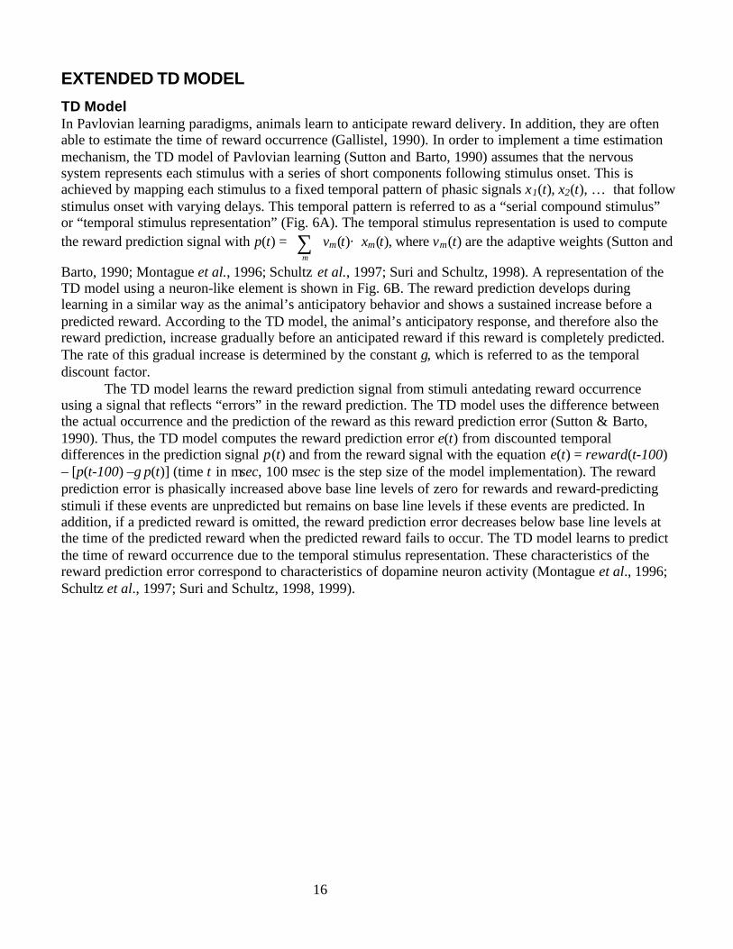
TD ModelIn Pavlovian learning paradigms, animals learn to anticipate reward delivery. In addition, they are oftenable to estimate the time of reward occurrence (Gallistel, 1990). In order to implement a time estimationmechanism, the TD model of Pavlovian learning (Sutton and Barto, 1990) assumes that the nervoussystem represents each stimulus with a series of short components following stimulus onset. This isachieved by mapping each stimulus to a fixed temporal pattern of phasic signals x1(t), x2(t), … that followstimulus onset with varying delays. This temporal pattern is referred to as a “serial compound stimulus”or “temporal stimulus representation” (Fig. 6A). The temporal stimulus representation is used to computethe reward prediction signal with p(t) = ∑
m
vm(t)× xm(t), where vm(t) are the adaptive weights (Sutton and
Barto, 1990; Montague et al., 1996; Schultz et al., 1997; Suri and Schultz, 1998). A representation of theTD model using a neuron-like element is shown in Fig. 6B. The reward prediction develops duringlearning in a similar way as the animal’s anticipatory behavior and shows a sustained increase before apredicted reward. According to the TD model, the animal’s anticipatory response, and therefore also thereward prediction, increase gradually before an anticipated reward if this reward is completely predicted.The rate of this gradual increase is determined by the constant γ, which is referred to as the temporaldiscount factor.
The TD model learns the reward prediction signal from stimuli antedating reward occurrenceusing a signal that reflects “errors” in the reward prediction. The TD model uses the difference betweenthe actual occurrence and the prediction of the reward as this reward prediction error (Sutton & Barto,1990). Thus, the TD model computes the reward prediction error e(t) from discounted temporaldifferences in the prediction signal p(t) and from the reward signal with the equation e(t) = reward(t-100)– [p(t-100) –γ p(t)] (time t in msec, 100 msec is the step size of the model implementation). The rewardprediction error is phasically increased above base line levels of zero for rewards and reward-predictingstimuli if these events are unpredicted but remains on base line levels if these events are predicted. Inaddition, if a predicted reward is omitted, the reward prediction error decreases below base line levels atthe time of the predicted reward when the predicted reward fails to occur. The TD model learns to predictthe time of reward occurrence due to the temporal stimulus representation. These characteristics of thereward prediction error correspond to characteristics of dopamine neuron activity (Montague et al., 1996;Schultz et al., 1997; Suri and Schultz, 1998, 1999).
17
01
0.1 sec
stimulus u1(t)
temporalrepresent-ation
01
A
C
temporal representation
κ
stimulus
prediction p1(t)
e1(t)
u1(t )
κ
stimulus
prediction p2 (t)
u2(t)
+ +e2(t)
x1(t)x2(t)x3(t)
x1 x2 x3 x4 x5 x6
temporal representation
+ +
v1mxm(t)m =1
6
∑ v2 mxm (t )m=1
6
∑
β β
B
D
-
Dp 1(t)
-γ p
1(t+
100)
p 2(t)
-γ p
2(t+
100)
temporal representation
stimulus
β
reward
reward prediction p(t) D
vmxm(t)m =1
3
∑
e(t)
u1(t )
x1(t) x2(t) x3(t)
p(t)
-γ p
(t+10
0) -
+
-
Fig. 6 Critic model. (A) Temporal stimulus representation x1(t), x2(t), and x3(t). Stimulus u1(t) is represented over time as aseries of phasic signals x1(t), x2(t), and x3(t) that cover stimulus duration. This temporal stimulus representation is used toreproduce the finding that dopamine neuron activity is decreased when a predicted reward fails to occur. (B) TD model. Fromstimulus u1(t) the temporal stimulus representation x1(t), x2(t), and x3(t) is computed. Each component xm(t) is multiplied withan adaptive weight vm(t) (filled dots). The reward prediction p(t) is the sum of the weighted representation components. Thedifference operator D takes temporal differences from this prediction signal (discounted with factor γ). The reward predictionerror e(t) is computed from these temporal differences and from the reward signal. The weights vm(t) are adapted proportionallyto the prediction error signal e(t) and to the learning rate β. (C) Extended TD model for two input events u1(t) and u2(t). Theevent signals uk(t) report about stimuli, rewards, thalamic activity, and acts. Each temporal representation component xm(t) ismultiplied with an adaptive weight vkm (filled dots). Event prediction pk(t) is computed from the sum of the weightedcomponents. Event prediction pk(t) is multiplied with a small constant κ and fed back to the temporal event representation ofthis event uk(t). This feedback is necessary to form novel associative chains. Analogous to the TD model, the prediction errorek(t) is computed from the event uk(t) and from the temporal differences between successive predictions pk(t) - γ pk(t+100)(discounted with a factor γ). The weights vkm (filled dots) are adapted as in the TD model.
18
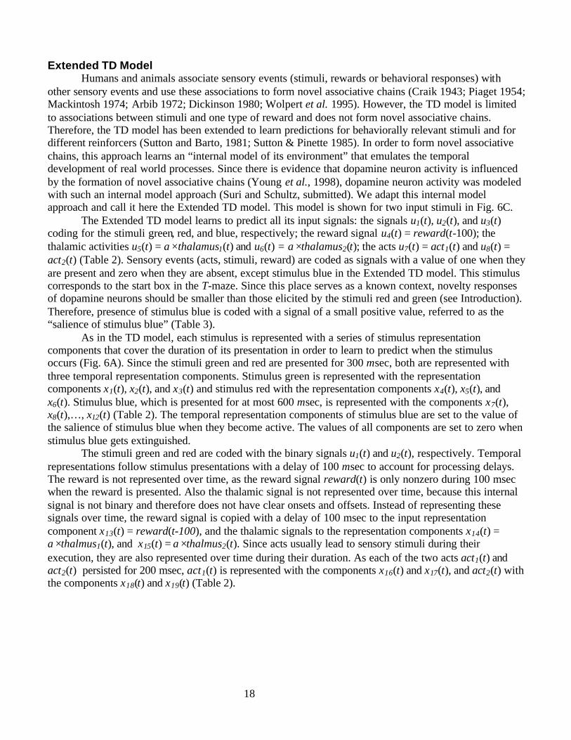
Extended TD ModelHumans and animals associate sensory events (stimuli, rewards or behavioral responses) with
other sensory events and use these associations to form novel associative chains (Craik 1943; Piaget 1954;Mackintosh 1974; Arbib 1972; Dickinson 1980; Wolpert et al. 1995). However, the TD model is limitedto associations between stimuli and one type of reward and does not form novel associative chains.Therefore, the TD model has been extended to learn predictions for behaviorally relevant stimuli and fordifferent reinforcers (Sutton and Barto, 1981; Sutton & Pinette 1985). In order to form novel associativechains, this approach learns an “internal model of its environment” that emulates the temporaldevelopment of real world processes. Since there is evidence that dopamine neuron activity is influencedby the formation of novel associative chains (Young et al., 1998), dopamine neuron activity was modeledwith such an internal model approach (Suri and Schultz, submitted). We adapt this internal modelapproach and call it here the Extended TD model. This model is shown for two input stimuli in Fig. 6C.
The Extended TD model learns to predict all its input signals: the signals u1(t), u2(t), and u3(t)coding for the stimuli green, red, and blue, respectively; the reward signal u4(t) = reward(t-100); thethalamic activities u5(t) = α×thalamus1(t) and u6(t) = α×thalamus2(t); the acts u7(t) = act1(t) and u8(t) =act2(t) (Table 2). Sensory events (acts, stimuli, reward) are coded as signals with a value of one when theyare present and zero when they are absent, except stimulus blue in the Extended TD model. This stimuluscorresponds to the start box in the T-maze. Since this place serves as a known context, novelty responsesof dopamine neurons should be smaller than those elicited by the stimuli red and green (see Introduction).Therefore, presence of stimulus blue is coded with a signal of a small positive value, referred to as the“salience of stimulus blue” (Table 3).
As in the TD model, each stimulus is represented with a series of stimulus representationcomponents that cover the duration of its presentation in order to learn to predict when the stimulusoccurs (Fig. 6A). Since the stimuli green and red are presented for 300 msec, both are represented withthree temporal representation components. Stimulus green is represented with the representationcomponents x1(t), x2(t), and x3(t) and stimulus red with the representation components x4(t), x5(t), andx6(t). Stimulus blue, which is presented for at most 600 msec, is represented with the components x7(t),x8(t),…, x12(t) (Table 2). The temporal representation components of stimulus blue are set to the value ofthe salience of stimulus blue when they become active. The values of all components are set to zero whenstimulus blue gets extinguished.
The stimuli green and red are coded with the binary signals u1(t) and u2(t), respectively. Temporalrepresentations follow stimulus presentations with a delay of 100 msec to account for processing delays.The reward is not represented over time, as the reward signal reward(t) is only nonzero during 100 msecwhen the reward is presented. Also the thalamic signal is not represented over time, because this internalsignal is not binary and therefore does not have clear onsets and offsets. Instead of representing thesesignals over time, the reward signal is copied with a delay of 100 msec to the input representationcomponent x13(t) = reward(t-100), and the thalamic signals to the representation components x14(t) =α×thalmus1(t), and x15(t) = α×thalmus2(t). Since acts usually lead to sensory stimuli during theirexecution, they are also represented over time during their duration. As each of the two acts act1(t) andact2(t) persisted for 200 msec, act1(t) is represented with the components x16(t) and x17(t), and act2(t) withthe components x18(t) and x19(t) (Table 2).
19
Table 2. Definitions of Critic input signals uk(t) and their temporal representation components xm(t) (seesection “Extended TD Model”).
Signal Temporal representationcomponents
Green goal box (stimulus green) u1(t) x1(t), x2(t), x3(t)Red goal box (stimulus red) u2(t) x4(t), x5(t), x6(t)Start box (stimulus blue) u3(t) x7(t), x8(t),…, x12(t)Reward u4(t) = reward(t-100) x13(t)Thalamic activity related to act 1 u5(t) = α×thalamus1(t) x14(t)Thalamic activity related to act 2 u6(t) = α×thalamus2(t) x15(t)Act left (act 1) u7(t) = act1(t) x16(t), x17(t)Act right (act 2) u8(t) = act2(t) x18(t), x19(t)
To form novel associative chains, a predicted event should elicit similar prediction signals as doesthe experience of this event. More precisely, a predicted stimulus should produce internal representationsignals that resemble the representation of the stimulus itself. Therefore, Suri and Schultz (submitted)proposed that the prediction of each stimulus is fed back to the temporal stimulus representation of thisstimulus and used to estimate further prediction signals. The loop time τ of this feedback is assumed to bemuch shorter than the usual 100 msec step size of the model because the feedback is computed twicewithin each time step.
The prediction pk(t − 2τ) for the input signal uk(t) (k = 1, 2,…, 8) is computed from the product ofthe adaptive weights vkm(t) with the components of the temporal representation components xm(t):
pk(t – 2τ) = ∑=
19
1m
vkm(t)× xm(t). (eq. 13a)
This prediction is fed back twice to the temporal stimulus representation with the two equations:
pk(t - τ) = ∑=
19
1m
vkm(t)× [xm(t) + κ skm pk(t - 2τ)], (eq. 13b)
pk(t) = ∑=
19
1m
vkm(t)× [xm(t) + κ skm pk(t - τ)]. (eq. 13c)
To avoid very large absolute values of the prediction signals pk(t), these signals are limited to valuesbetween –pmax and +pmax (Table 3). We do not assign a value to the small time constant τ, as τ occurs onlyin eqs. 13a and 13b and prediction signals will be shown in the figures for time steps of 100 msec. Thefeedback constant κ (Table 3) determines the gain of the feedback loop and therefore the impact of apredicted stimulus on further stimulus predictions. The number of these update equations seems tocorrespond to the number of novel links in the associative chain the model can compute (unpublishedresult). We are neither aware of mathematical considerations nor of experimental evidence that wouldindicate which components of the temporal stimulus representation xm(t) should be influenced by thisfeedback. For simplicity, we assume that the feedback influences only the first component of the temporalstimulus representation. Feedbacks to further temporal stimulus representation components do notinfluence most simulation results but lead to slightly different time courses of prediction signals insimulations that test the formation of novel associative chains (unpublished result). Feedback to the firstcomponent of the temporal stimulus representation is accomplished by setting the factor skm to one for thefirst component of the temporal stimulus representation of each stimulus or act. Also for the reward andthe two thalamic signals skm is set to 1. Otherwise, the factor skm is set to zero (s1,1 = 1, s2,4 = 1, s3,7 = 1, s4,13=1, s5,14 = 1, s6,15 = 1, s7,16 = 1, s8,18 = 1; skm = 0 otherwise).
The following equations of the Extended TD model are analogous to those of the TD model.Therefore, the proposed Extended TD model with the parameter κ = 0 is equivalent to a set of eight
20
independent TD models. For this case, the equations are for each k equivalent to those of the TD model.The prediction errors ek(t) are computed from discounted temporal differences between successivepredictions (differencer D in Fig. 6C) and from the input signals uk(t) with
ek(t) = uk(t-100) - [pk(t-100) - γpk(t)], (eq. 14)where γ is the discount factor and the input signals uk(t) (k = 1, 2, …, 8) denote the three stimuli, thereward, the thalamic signals (multiplied with salience α), and the two act signals. The value of thediscount factor γ is set to 0.98, because this value was estimated from dopamine neuron activity (Suri andSchultz, 1999). To minimize the prediction error signals, the weights vkm(t) are incrementally adaptedaccording to the product of the input prediction errors ek(t) with the eligibility traces of the temporal inputrepresentation x m (t) with
vkm(t+100) = vkm(t) + βek(t) )(txm . (eq. 15)The three weights vkm that associate the first component of the temporal representations of the threestimuli with the reward are initiated with the positive value v (Table 3) to reproduce dopamine noveltyresponses (Suri and Schultz, 1999). The other values of matrix vkm are initialized with zeros (v4,1(t=0) = v,v4,4(t=0) = v, v4,7(t=0) = v; vkm(t=0) = 0 otherwise). For the current study, the positive value v is chosen assmall as possible to reduce the number of errors due to exploration, but large enough to significantlyincrease the number of acts in the exploration phase.
The traces mx (t) are slowly decaying versions of the input representation components xm(t). Sucheligibility traces were introduced to explain how animals learn to associate sensory events that areseparated by a delay period (Sutton and Barto, 1990). Although TD models with temporal stimulusrepresentations learn to associate sensory events over a delay without representation traces (Montague etal., 1996), traces accelerate learning (Sutton & Barto, 1998; Kearns and Singh, submitted). At thebeginning of an experiment, mx (t) is set to the initial condition mx (0) = 0. Then, the traces are computedwith
mx (t) = λc mx (t-100) + (1-λc) xm(t). (eq. 16)The parameter λc is set to the value of 0.3, as this value guarantees fast learning. With this parametervalue, the eligibility traces increase with a rate of 30% each 100 msec during presentation of the event anddecrease 70% each 100 msec after event presentation.
The output signal of the Extended TD model is the reward prediction error e4(t) (eq. 14) thatresembles the firing rate of dopamine neurons (Suri and Schultz, submitted). Since dopamineconcentration in extracellular space is closely time-correlated with the firing rate of dopamine neurons(Gonon, 1997), we compute the dopamine concentration DA(t) with
DA(t) = e4(t). (eq. 17)The simulated dopamine concentration DA(t) is used to simulate the effect of dopamine on medium spinyneurons in striatal matrisomes (eqs. 1 and 5).
21
Table 3 Standard model parameters in Extended TD modelParameter Name Symbol Valueinitial weights for noveltyresponses
v 0.001
Critic learning rate β 0.5
feedback constant κ 0.8
maximal value of predictionsignals
pmax 10
discount factor γ 0.98
decay of eligibility trace λc 0.3
salience of stimulus blue forCritic
0.05
RESULTSThe proposed model was tested in the experiment described above. Since each trial started with a randomstate of the striatal membrane potentials, the model performed differently in each experiment. We showthe model performance for a typical experiment and then present the statistical analysis of 1000experiments.
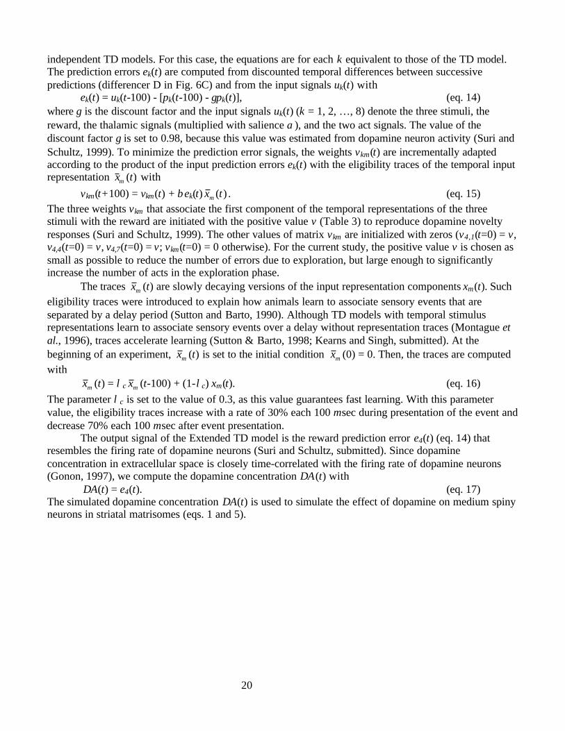
In the exploration phase, the model learns to associate act left with stimulus red and act right withstimulus green. In the first trial (Fig. 7A), stimulus blue was presented and the model executed the act left(bottom line) that led to presentation of stimulus red (line 1). Since certain associative weights of theExtended TD model had been initialized with positive values, this novel stimulus phasically activated thereward prediction signal (line 2; eq. 13). This led to a biphasic response of the dopamine-like rewardprediction error signal (line 3; eq. 14) resembling dopamine novelty responses. Since the salience ofstimulus blue had been set to a smaller value than that of the stimuli red and green (see section “ExtendedTD Model” and Table 3), onset of the stimulus blue led to very small activations of the reward predictionsignal and the reward prediction error signal (hardly visible). Since stimulus green was not presented, theprediction signal for stimulus green remained zero (line 4). The simulated striatal membrane potentialsEleft(t) and Eright(t) of the two striatal medium spiny neurons in the matrisome compartment fluctuatedeach 400 msec between the elevated up-state and the hyperpolarised down-state (line 5; eqs. 4 and 7). Themembrane potentials were slightly increased during presentation of stimulus blue, since corticostriatalweights associating stimulus blue with striatal activity were set to positive initial values (compare section“Parameter Values and Initial Conditions”). As action potentials are much shorter than the 100 msec timestep, the averaged membrane potential is shown (eq. 4, as in Fig. 4B and 4C). The membrane potential ofthe striatal neuron coding act left was increased above firing threshold for 500 msec. This persistent firingwas integrated by two neurons in motor cortex (line 6; eq. 11). When the firing rate of the neuron codingfor act left reached the act threshold actthres, this act was elicited (bottom line; eq. 12). The signal codingfor the act right remained on the value of zero (not shown).
22
Fig. 7 Model performance during exploration phase. (A) First trial. When stimulus blue was presented (line 1), the modelelicited the act left (bottom line) that led to presentation of stimulus red (line 1). Since stimulus red was presented for the firsttime, its onset phasically activated the reward prediction signal (line 2) and biphasically activated the dopamine-like rewardprediction error signal (line 3). Membrane potentials of the two simulated striatal medium spiny neurons fluctuated between anelevated up-state and a hyperpolarized down-state (line 5). During presentation of stimulus blue, the simulated striatal neuroncoding for act left was firing for 500 msec. Neurons in motor cortex integrated this striatal firing rate over time (line 6). The actleft was elicited (bottom line) when the integrated signal reached a threshold. (B) A trial at the end of the exploration phase.When stimulus blue was presented (line 1), the model elicited the act right (bottom line) that led to presentation of stimulusgreen (line 1). Since stimulus green had been presented repeatedly during the exploration phase, novelty responses were almostabsent in the reward prediction signal (line 2) and in the dopamine-like reward prediction error signal (line 3). Prediction ofstimulus green (line 4) was already increased when the striatal neuron coding for the act right increased its firing rate (line 5),because this had often antedated execution of act right followed by presentation of stimulus green. The striatal firing rates wereintegrated in cortex and the act right was elicited (bottom line) when the cortical signal coding for the act right reached athreshold (line 6).
For the next 80 presentations of stimulus blue, the model executed 11 times the act left, 14 timesthe act right, and 55 times no act (not shown). Trials without acts occurred when striatal membranepotentials of both neurons happened to fluctuate synchronously, as the effects of synchronous striatalfiring on the cortical neurons were suppressed by the indirect pathway (eq. 8). The 81st presentation ofstimulus blue was the last blue presentation in the exploration phase during which an act was executed(Fig. 7B). Since the model selected act right, stimulus green was presented (line 1). Reward predictionand reward prediction error remained on the values of zero, since dopamine-like novelty responses had
stimuli
reward prediction
reward prediction error
action
10-3
010-3
0
firing thresholdleft
right
act threshold
rightleft
100 msec
cortical firingrates
striatalmembranepotentials
left
left right
right
green prediction
striatal firing increasesgreen prediction
act threshold
blue red blue green
10
left right10
0
2
-60 mV
-40 mV
0
3
A B
23
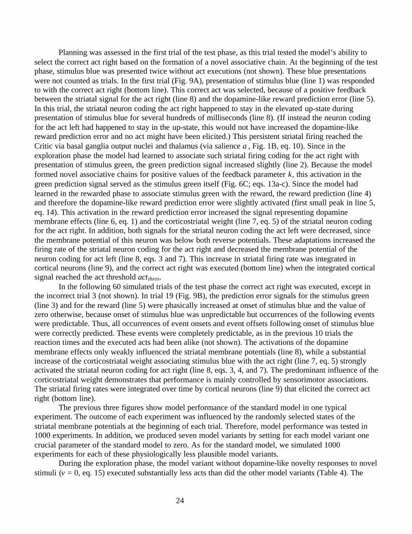
extinguished as a consequence of the Critic learning rule (lines 2 and 3, eq. 15). The green predictionsignal was slightly activated when the striatal neuron coding the act right was firing (line 4, line 5), assuch increased striatal firing had been followed by the corresponding act right in some, but not all,previous trials. Striatal activity influenced the green prediction signal via basal ganglia output nuclei,thalamus, and cortex (via salience α, Fig. 1B, eq. 10). Since act right had previously been followed bygreen presentations and an efference copy of the act signal reached the Critic, the green prediction signalwas fully activated when the act right was executed (bottom line), (Fig. 1B, eq. 13a). The green predictionpeaked at the correct value of three, as this value reflects the predicted future duration of stimulus green inunits of 100 msec. The striatal firing rates were integrated over time in cortical neurons, and the act rightwas executed (bottom line) when the cortical signal coding for act right reached the act threshold actthres(line 6).
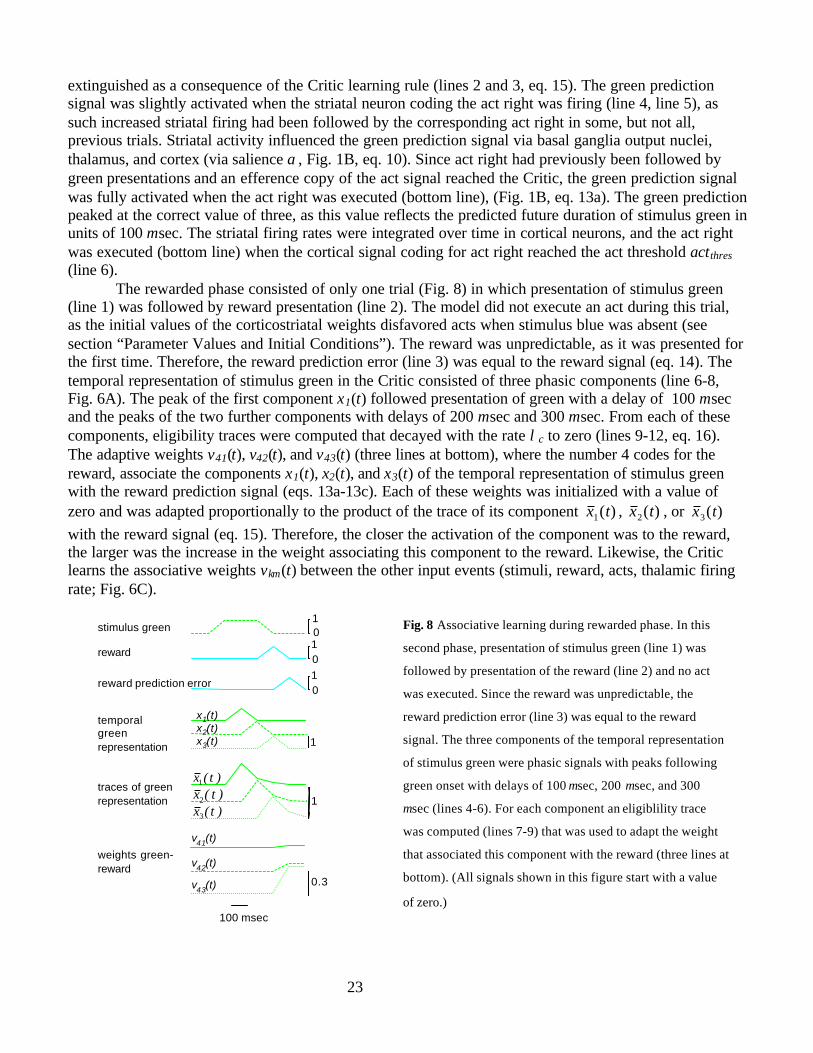
The rewarded phase consisted of only one trial (Fig. 8) in which presentation of stimulus green(line 1) was followed by reward presentation (line 2). The model did not execute an act during this trial,as the initial values of the corticostriatal weights disfavored acts when stimulus blue was absent (seesection “Parameter Values and Initial Conditions”). The reward was unpredictable, as it was presented forthe first time. Therefore, the reward prediction error (line 3) was equal to the reward signal (eq. 14). Thetemporal representation of stimulus green in the Critic consisted of three phasic components (line 6-8,Fig. 6A). The peak of the first component x1(t) followed presentation of green with a delay of 100 msecand the peaks of the two further components with delays of 200 msec and 300 msec. From each of thesecomponents, eligibility traces were computed that decayed with the rate λc to zero (lines 9-12, eq. 16).The adaptive weights v41(t), v42(t), and v43(t) (three lines at bottom), where the number 4 codes for thereward, associate the components x1(t), x2(t), and x3(t) of the temporal representation of stimulus greenwith the reward prediction signal (eqs. 13a-13c). Each of these weights was initialized with a value ofzero and was adapted proportionally to the product of the trace of its component )(1 tx , )(2 tx , or )(3 txwith the reward signal (eq. 15). Therefore, the closer the activation of the component was to the reward,the larger was the increase in the weight associating this component to the reward. Likewise, the Criticlearns the associative weights vkm(t) between the other input events (stimuli, reward, acts, thalamic firingrate; Fig. 6C).
Fig. 8 Associative learning during rewarded phase. In this
second phase, presentation of stimulus green (line 1) was
followed by presentation of the reward (line 2) and no act
was executed. Since the reward was unpredictable, the
reward prediction error (line 3) was equal to the reward
signal. The three components of the temporal representation
of stimulus green were phasic signals with peaks following
green onset with delays of 100 msec, 200 msec, and 300
msec (lines 4-6). For each component an eligiblility trace
was computed (lines 7-9) that was used to adapt the weight
that associated this component with the reward (three lines at
bottom). (All signals shown in this figure start with a value
of zero.)
stimulus green
reward prediction error
reward01
01
100 msec
01
weights green-reward
1
temporalgreenrepresentation
traces of greenrepresentation 1
0.3
x1(t)x2(t)x3(t)
)t(x1
)t(x2
)t(x3
v41(t)
v42(t)
v43(t)
24
Planning was assessed in the first trial of the test phase, as this trial tested the model’s ability toselect the correct act right based on the formation of a novel associative chain. At the beginning of the testphase, stimulus blue was presented twice without act executions (not shown). These blue presentationswere not counted as trials. In the first trial (Fig. 9A), presentation of stimulus blue (line 1) was respondedto with the correct act right (bottom line). This correct act was selected, because of a positive feedbackbetween the striatal signal for the act right (line 8) and the dopamine-like reward prediction error (line 5).In this trial, the striatal neuron coding the act right happened to stay in the elevated up-state duringpresentation of stimulus blue for several hundreds of milliseconds (line 8). (If instead the neuron codingfor the act left had happened to stay in the up-state, this would not have increased the dopamine-likereward prediction error and no act might have been elicited.) This persistent striatal firing reached theCritic via basal ganglia output nuclei and thalamus (via salience α, Fig. 1B, eq. 10). Since in theexploration phase the model had learned to associate such striatal firing coding for the act right withpresentation of stimulus green, the green prediction signal increased slightly (line 2). Because the modelformed novel associative chains for positive values of the feedback parameter κ, this activation in thegreen prediction signal served as the stimulus green itself (Fig. 6C; eqs. 13a-c). Since the model hadlearned in the rewarded phase to associate stimulus green with the reward, the reward prediction (line 4)and therefore the dopamine-like reward prediction error were slightly activated (first small peak in line 5,eq. 14). This activation in the reward prediction error increased the signal representing dopaminemembrane effects (line 6, eq. 1) and the corticostriatal weight (line 7, eq. 5) of the striatal neuron codingfor the act right. In addition, both signals for the striatal neuron coding the act left were decreased, sincethe membrane potential of this neuron was below both reverse potentials. These adaptations increased thefiring rate of the striatal neuron coding for the act right and decreased the membrane potential of theneuron coding for act left (line 8, eqs. 3 and 7). This increase in striatal firing rate was integrated incortical neurons (line 9), and the correct act right was executed (bottom line) when the integrated corticalsignal reached the act threshold actthres.
In the following 60 simulated trials of the test phase the correct act right was executed, except inthe incorrect trial 3 (not shown). In trial 19 (Fig. 9B), the prediction error signals for the stimulus green(line 3) and for the reward (line 5) were phasically increased at onset of stimulus blue and the value ofzero otherwise, because onset of stimulus blue was unpredictable but occurrences of the following eventswere predictable. Thus, all occurrences of event onsets and event offsets following onset of stimulus bluewere correctly predicted. These events were completely predictable, as in the previous 10 trials thereaction times and the executed acts had been alike (not shown). The activations of the dopaminemembrane effects only weakly influenced the striatal membrane potentials (line 8), while a substantialincrease of the corticostriatal weight associating stimulus blue with the act right (line 7, eq. 5) stronglyactivated the striatal neuron coding for act right (line 8, eqs. 3, 4, and 7). The predominant influence of thecorticostriatal weight demonstrates that performance is mainly controlled by sensorimotor associations.The striatal firing rates were integrated over time by cortical neurons (line 9) that elicited the correct actright (bottom line).
The previous three figures show model performance of the standard model in one typicalexperiment. The outcome of each experiment was influenced by the randomly selected states of thestriatal membrane potentials at the beginning of each trial. Therefore, model performance was tested in1000 experiments. In addition, we produced seven model variants by setting for each model variant onecrucial parameter of the standard model to zero. As for the standard model, we simulated 1000experiments for each of these physiologically less plausible model variants.
During the exploration phase, the model variant without dopamine-like novelty responses to novelstimuli (v = 0, eq. 15) executed substantially less acts than did the other model variants (Table 4). The
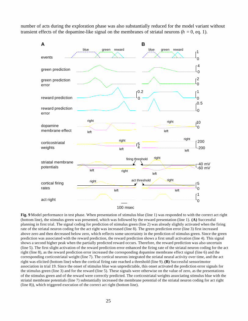
25
number of acts during the exploration phase was also substantially reduced for the model variant withouttransient effects of the dopamine-like signal on the membranes of striatal neurons (η = 0, eq. 1).
Fig. 9 Model performance in test phase. When presentation of stimulus blue (line 1) was responded to with the correct act right(bottom line), the stimulus green was presented, which was followed by the reward presentation (line 1). (A) Successfulplanning in first trial. The signal coding for prediction of stimulus green (line 2) was already slightly activated when the firingrate of the striatal neuron coding for the act right was increased (line 8). The green prediction error (line 3) first increasedabove zero and then decreased below zero, which reflects some uncertainty in the prediction of stimulus green. Since the greenprediction was associated with the reward prediction, the reward prediction shows a first small activation (line 4). This signalshows a second higher peak when the partially predicted reward occurs. Therefore, the reward prediction was also uncertain(line 5). The first slight activation of the reward prediction error enhanced the firing rate of the striatal neuron coding for the actright (line 8), as the reward prediction error increased the corresponding dopamine membrane effect signal (line 6) and thecorresponding corticostriatal weight (line 7). The cortical neurons integrated the striatal neural activity over time, and the actright was elicited (bottom line) when the cortical firing rate reached a threshold (line 9). (B) Successful sensorimotorassociation in trial 19. Since the onset of stimulus blue was unpredictable, this onset activated the prediction error signals forthe stimulus green (line 3) and for the reward (line 5). These signals were otherwise on the value of zero, as the presentationsof the stimulus green and of the reward were correctly predicted. The corticostriatal weights associating stimulus blue with thestriatal membrane potentials (line 7) substantially increased the membrane potential of the striatal neuron coding for act right(line 8)), which triggered execution of the correct act right (bottom line).
0
4green prediction
reward predictionerror
events
05cortical firing
rates
act threshold
0
1
reward prediction
100 msec
blue green reward blue green reward
1
00.5
0
act right
right
left
green predictionerror 0
2
1
0
right
left
-60 mV-40 mV
firing threshold
left right
striatal membranepotentials
right
left
dopaminemembrane effect
100
left
right right
left
corticostriatalweights -200
20045
right
left
right
left
0.2
0
A B
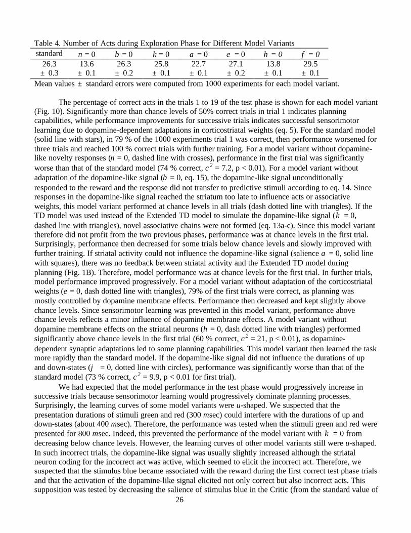
26
Table 4. Number of Acts during Exploration Phase for Different Model Variantsstandard ν = 0 β = 0 κ = 0 α = 0 ε = 0 η = 0 φ = 0
26.3± 0.3
13.6± 0.1
26.3± 0.2
25.8± 0.1
22.7± 0.1
27.1± 0.2
13.8± 0.1
29.5± 0.1
Mean values ± standard errors were computed from 1000 experiments for each model variant.
The percentage of correct acts in the trials 1 to 19 of the test phase is shown for each model variant(Fig. 10). Significantly more than chance levels of 50% correct trials in trial 1 indicates planningcapabilities, while performance improvements for successive trials indicates successful sensorimotorlearning due to dopamine-dependent adaptations in corticostriatal weights (eq. 5). For the standard model(solid line with stars), in 79 % of the 1000 experiments trial 1 was correct, then performance worsened forthree trials and reached 100 % correct trials with further training. For a model variant without dopamine-like novelty responses (ν = 0, dashed line with crosses), performance in the first trial was significantlyworse than that of the standard model (74 % correct, χ2 = 7.2, p < 0.01). For a model variant withoutadaptation of the dopamine-like signal (β = 0, eq. 15), the dopamine-like signal unconditionallyresponded to the reward and the response did not transfer to predictive stimuli according to eq. 14. Sinceresponses in the dopamine-like signal reached the striatum too late to influence acts or associativeweights, this model variant performed at chance levels in all trials (dash dotted line with triangles). If theTD model was used instead of the Extended TD model to simulate the dopamine-like signal (κ = 0,dashed line with triangles), novel associative chains were not formed (eq. 13a-c). Since this model varianttherefore did not profit from the two previous phases, performance was at chance levels in the first trial.Surprisingly, performance then decreased for some trials below chance levels and slowly improved withfurther training. If striatal activity could not influence the dopamine-like signal (salience α = 0, solid linewith squares), there was no feedback between striatal activity and the Extended TD model duringplanning (Fig. 1B). Therefore, model performance was at chance levels for the first trial. In further trials,model performance improved progressively. For a model variant without adaptation of the corticostriatalweights (ε = 0, dash dotted line with triangles), 79% of the first trials were correct, as planning wasmostly controlled by dopamine membrane effects. Performance then decreased and kept slightly abovechance levels. Since sensorimotor learning was prevented in this model variant, performance abovechance levels reflects a minor influence of dopamine membrane effects. A model variant withoutdopamine membrane effects on the striatal neurons (η = 0, dash dotted line with triangles) performedsignificantly above chance levels in the first trial (60 % correct, χ2 = 21, p < 0.01), as dopamine-dependent synaptic adaptations led to some planning capabilities. This model variant then learned the taskmore rapidly than the standard model. If the dopamine-like signal did not influence the durations of upand down-states (ϕ = 0, dotted line with circles), performance was significantly worse than that of thestandard model (73 % correct, χ2 = 9.9, p < 0.01 for first trial).
We had expected that the model performance in the test phase would progressively increase insuccessive trials because sensorimotor learning would progressively dominate planning processes.Surprisingly, the learning curves of some model variants were u-shaped. We suspected that thepresentation durations of stimuli green and red (300 msec) could interfere with the durations of up anddown-states (about 400 msec). Therefore, the performance was tested when the stimuli green and red werepresented for 800 msec. Indeed, this prevented the performance of the model variant with κ = 0 fromdecreasing below chance levels. However, the learning curves of other model variants still were u-shaped.In such incorrect trials, the dopamine-like signal was usually slightly increased although the striatalneuron coding for the incorrect act was active, which seemed to elicit the incorrect act. Therefore, wesuspected that the stimulus blue became associated with the reward during the first correct test phase trialsand that the activation of the dopamine-like signal elicited not only correct but also incorrect acts. Thissupposition was tested by decreasing the salience of stimulus blue in the Critic (from the standard value of
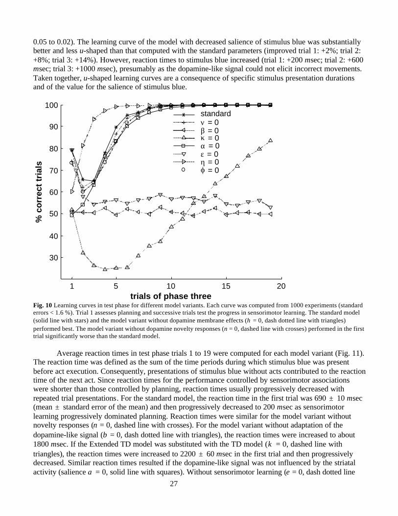
27
0.05 to 0.02). The learning curve of the model with decreased salience of stimulus blue was substantiallybetter and less u-shaped than that computed with the standard parameters (improved trial 1: +2%; trial 2:+8%; trial 3: +14%). However, reaction times to stimulus blue increased (trial 1: +200 msec; trial 2: +600msec; trial 3: +1000 msec), presumably as the dopamine-like signal could not elicit incorrect movements.Taken together, u-shaped learning curves are a consequence of specific stimulus presentation durationsand of the value for the salience of stimulus blue.
1 5 10 15 20
30
40
50
60
70
80
90
100
trials of phase three
% c
orr
ect
tria
ls
standard ν = 0 β = 0 κ = 0 α = 0 ε = 0η = 0 ϕ = 0
Fig. 10 Learning curves in test phase for different model variants. Each curve was computed from 1000 experiments (standarderrors < 1.6 %). Trial 1 assesses planning and successive trials test the progress in sensorimotor learning. The standard model(solid line with stars) and the model variant without dopamine membrane effects (η = 0, dash dotted line with triangles)performed best. The model variant without dopamine novelty responses (ν = 0, dashed line with crosses) performed in the firsttrial significantly worse than the standard model.
Average reaction times in test phase trials 1 to 19 were computed for each model variant (Fig. 11).The reaction time was defined as the sum of the time periods during which stimulus blue was presentbefore act execution. Consequently, presentations of stimulus blue without acts contributed to the reactiontime of the next act. Since reaction times for the performance controlled by sensorimotor associationswere shorter than those controlled by planning, reaction times usually progressively decreased withrepeated trial presentations. For the standard model, the reaction time in the first trial was 690 ± 10 msec(mean ± standard error of the mean) and then progressively decreased to 200 msec as sensorimotorlearning progressively dominated planning. Reaction times were similar for the model variant withoutnovelty responses (ν = 0, dashed line with crosses). For the model variant without adaptation of thedopamine-like signal (β = 0, dash dotted line with triangles), the reaction times were increased to about1800 msec. If the Extended TD model was substituted with the TD model (κ = 0, dashed line withtriangles), the reaction times were increased to 2200 ± 60 msec in the first trial and then progressivelydecreased. Similar reaction times resulted if the dopamine-like signal was not influenced by the striatalactivity (salience α = 0, solid line with squares). Without sensorimotor learning (ε = 0, dash dotted line
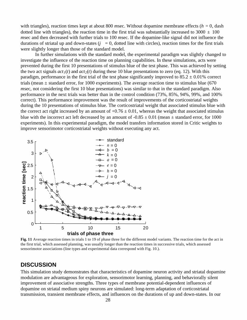
28
with triangles), reaction times kept at about 800 msec. Without dopamine membrane effects (η = 0, dashdotted line with triangles), the reaction time in the first trial was substantially increased to 3000 ± 100msec and then decreased with further trials to 100 msec. If the dopamine-like signal did not influence thedurations of striatal up and down-states (ϕ = 0, dotted line with circles), reaction times for the first trialswere slightly longer than those of the standard model.
In further simulations with the standard model, the experimental paradigm was slightly changed toinvestigate the influence of the reaction time on planning capabilities. In these simulations, acts wereprevented during the first 10 presentations of stimulus blue of the test phase. This was achieved by settingthe two act signals act1(t) and act2(t) during these 10 blue presentations to zero (eq. 12). With thisparadigm, performance in the first trial of the test phase significantly improved to 85.2 ± 0.01% correcttrials (mean ± standard error, for 1000 experiments). The average reaction time to stimulus blue (670msec, not considering the first 10 blue presentations) was similar to that in the standard paradigm. Alsoperformance in the next trials was better than in the control condition (73%, 85%, 94%, 99%, and 100%correct). This performance improvement was the result of improvements of the corticostriatal weightsduring the 10 presentations of stimulus blue. The corticostriatal weight that associated stimulus blue withthe correct act right increased by an amount of +0.76 ± 0.01, whereas the weight that associated stimulusblue with the incorrect act left decreased by an amount of -0.85 ± 0.01 (mean ± standard error, for 1000experiments). In this experimental paradigm, the model transfers information stored in Critic weights toimprove sensorimotor corticostriatal weights without executing any act.
1 5 10 15 200
0.5
1
1.5
2
2.5
3
3.5
trials of phase three
reac
tion
tim
e [s
ec]
standard ν = 0 β = 0 κ = 0 α = 0 ε = 0η = 0 ϕ = 0
Fig. 11 Average reaction times in trials 1 to 19 of phase three for the different model variants. The reaction time for the act inthe first trial, which assessed planning, was usually longer than the reaction times in successive trials, which assessedsensorimotor associations (line types and experimental data correspond with Fig. 10.).
DISCUSSIONThis simulation study demonstrates that characteristics of dopamine neuron activity and striatal dopaminemodulation are advantageous for exploration, sensorimotor learning, planning, and behaviorally silentimprovement of associative strengths. Three types of membrane potential-dependent influences ofdopamine on striatal medium spiny neurons are simulated: long-term adaptation of corticostriataltransmission, transient membrane effects, and influences on the durations of up and down-states. In our
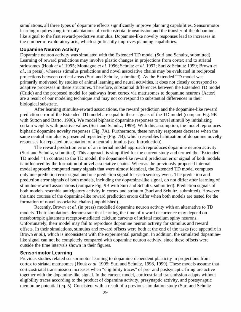
29
simulations, all three types of dopamine effects significantly improve planning capabilities. Sensorimotorlearning requires long-term adaptations of corticostriatal transmission and the transfer of the dopamine-like signal to the first reward-predictive stimulus. Dopamine-like novelty responses lead to increases inthe number of exploratory acts, which significantly improves planning capabilities.
Dopamine Neuron ActivityDopamine neuron activity was simulated with the Extended TD model (Suri and Schultz, submitted).Learning of reward predictions may involve plastic changes in projections from cortex and to striatalstriosomes (Houk et al. 1995; Montague et al. 1996; Schultz et al. 1997; Suri & Schultz 1999; Brown etal., in press), whereas stimulus predictions and novel associative chains may be evaluated in reciprocalprojections between cortical areas (Suri and Schultz, submitted). As the Extended TD model wasprimarily motivated by studies of animal learning and neural activities, it does not closely correspond toadaptive processes in these structures. Therefore, substantial differences between the Extended TD model(Critic) and the proposed model for pathways from cortex via matrisomes to dopamine neurons (Actor)are a result of our modeling technique and may not correspond to substantial differences in theirbiological substrate.
After learning stimulus-reward associations, the reward prediction and the dopamine-like rewardprediction error of the Extended TD model are equal to these signals of the TD model (compare Fig. 9Bwith Sutton and Barto, 1990). We model biphasic dopamine responses to novel stimuli by initializingcertain weights with positive values (Suri and Schultz, 1999). With this assumption, the model reproducesbiphasic dopamine novelty responses (Fig. 7A). Furthermore, these novelty responses decrease when thesame neutral stimulus is presented repeatedly (Fig. 7B), which resembles habituation of dopamine noveltyresponses for repeated presentation of a neutral stimulus (see Introduction).
The reward prediction error of an internal model approach reproduces dopamine neuron activity(Suri and Schultz, submitted). This approach is simplified for the current study and termed the “ExtendedTD model.” In contrast to the TD model, the dopamine-like reward prediction error signal of both modelsis influenced by the formation of novel associative chains. Whereas the previously proposed internalmodel approach computed many signals that were almost identical, the Extended TD model computesonly one prediction error signal and one prediction signal for each sensory event. The prediction andprediction error signals of both models, including the dopamine-like signal, do not differ after learning ofstimulus-reward associations (compare Fig. 9B with Suri and Schultz, submitted). Prediction signals ofboth models resemble anticipatory activity in cortex and striatum (Suri and Schultz, submitted). However,the time courses of the dopamine-like reward prediction errors differ when both models are tested for theformation of novel associative chains (unpublished).
Recently, Brown et al. (in press) modelled dopamine neuron activity with an alternative to TDmodels. Their simulations demonstrate that learning the time of reward occurrence may depend onmetabotropic glutamate receptor-mediated calcium currents of striatal medium spiny neurons.Unfortunately, their model may fail to reproduce dopamine neuron activity for stimulus and rewardoffsets. In their simulations, stimulus and reward offsets were both at the end of the tasks (see appendix inBrown et al.), which is inconsistent with the experimental paradigm. In addition, the simulated dopamine-like signal can not be completely compared with dopamine neuron activity, since these offsets wereoutside the time intervals shown in their figures.
Sensorimotor LearningPrevious studies related sensorimotor learning to dopamine-dependent plasticity in projections fromcortex to striatal matrisomes (Houk et al. 1995; Suri and Schultz, 1998, 1999). These models assume thatcorticostriatal transmission increases when “eligibility traces” of pre- and postsynaptic firing are activetogether with the dopamine-like signal. In the current model, corticostriatal transmission adapts withouteligibility traces according to the product of dopamine activity, presynaptic activity, and postsynapticmembrane potential (eq. 5). Consistent with a result of a previous simulation study (Suri and Schultz
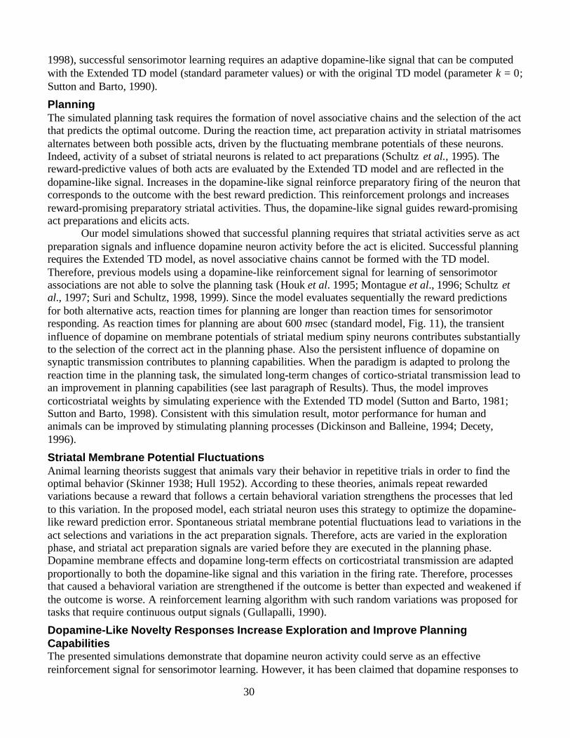
30
1998), successful sensorimotor learning requires an adaptive dopamine-like signal that can be computedwith the Extended TD model (standard parameter values) or with the original TD model (parameter κ = 0;Sutton and Barto, 1990).
PlanningThe simulated planning task requires the formation of novel associative chains and the selection of the actthat predicts the optimal outcome. During the reaction time, act preparation activity in striatal matrisomesalternates between both possible acts, driven by the fluctuating membrane potentials of these neurons.Indeed, activity of a subset of striatal neurons is related to act preparations (Schultz et al., 1995). Thereward-predictive values of both acts are evaluated by the Extended TD model and are reflected in thedopamine-like signal. Increases in the dopamine-like signal reinforce preparatory firing of the neuron thatcorresponds to the outcome with the best reward prediction. This reinforcement prolongs and increasesreward-promising preparatory striatal activities. Thus, the dopamine-like signal guides reward-promisingact preparations and elicits acts.
Our model simulations showed that successful planning requires that striatal activities serve as actpreparation signals and influence dopamine neuron activity before the act is elicited. Successful planningrequires the Extended TD model, as novel associative chains cannot be formed with the TD model.Therefore, previous models using a dopamine-like reinforcement signal for learning of sensorimotorassociations are not able to solve the planning task (Houk et al. 1995; Montague et al., 1996; Schultz etal., 1997; Suri and Schultz, 1998, 1999). Since the model evaluates sequentially the reward predictionsfor both alternative acts, reaction times for planning are longer than reaction times for sensorimotorresponding. As reaction times for planning are about 600 msec (standard model, Fig. 11), the transientinfluence of dopamine on membrane potentials of striatal medium spiny neurons contributes substantiallyto the selection of the correct act in the planning phase. Also the persistent influence of dopamine onsynaptic transmission contributes to planning capabilities. When the paradigm is adapted to prolong thereaction time in the planning task, the simulated long-term changes of cortico-striatal transmission lead toan improvement in planning capabilities (see last paragraph of Results). Thus, the model improvescorticostriatal weights by simulating experience with the Extended TD model (Sutton and Barto, 1981;Sutton and Barto, 1998). Consistent with this simulation result, motor performance for human andanimals can be improved by stimulating planning processes (Dickinson and Balleine, 1994; Decety,1996).
Striatal Membrane Potential FluctuationsAnimal learning theorists suggest that animals vary their behavior in repetitive trials in order to find theoptimal behavior (Skinner 1938; Hull 1952). According to these theories, animals repeat rewardedvariations because a reward that follows a certain behavioral variation strengthens the processes that ledto this variation. In the proposed model, each striatal neuron uses this strategy to optimize the dopamine-like reward prediction error. Spontaneous striatal membrane potential fluctuations lead to variations in theact selections and variations in the act preparation signals. Therefore, acts are varied in the explorationphase, and striatal act preparation signals are varied before they are executed in the planning phase.Dopamine membrane effects and dopamine long-term effects on corticostriatal transmission are adaptedproportionally to both the dopamine-like signal and this variation in the firing rate. Therefore, processesthat caused a behavioral variation are strengthened if the outcome is better than expected and weakened ifthe outcome is worse. A reinforcement learning algorithm with such random variations was proposed fortasks that require continuous output signals (Gullapalli, 1990).
Dopamine-Like Novelty Responses Increase Exploration and Improve PlanningCapabilitiesThe presented simulations demonstrate that dopamine neuron activity could serve as an effectivereinforcement signal for sensorimotor learning. However, it has been claimed that dopamine responses to
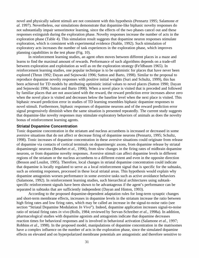
31
novel and physically salient stimuli are not consistent with this hypothesis (Pennartz 1995; Salamone etal. 1997). Nevertheless, our simulations demonstrate that dopamine-like biphasic novelty responses donot substantially impair sensorimotor learning, since the effects of the two phases cancel out and theseresponses extinguish during the exploration phase. Novelty responses increase the number of acts in theexploration phase (Table 4). This simulation result suggests that dopamine neuron responses stimulateexploration, which is consistent with experimental evidence (Stahle, 1992). Such stimulation ofexploratory acts increases the number of task experiences in the exploration phase, which improvesplanning capabilities in the test phase (Fig. 10).
In reinforcement learning studies, an agent often moves between different places in a maze andlearns to find the maximal amount of rewards. Performance of such algorithms depends on a trade-offbetween exploration and exploitation as well as on the exploration strategy (Fe'ldbaum 1965). Inreinforcement learning studies, one popular technique is to be optimistic for places that have never beenexplored (Thrun 1992; Dayan and Sejnowski 1996; Sutton and Barto, 1998). Similar to the proposal toreproduce dopamine novelty responses with positive initial weights (Suri and Schultz, 1999), this hasbeen achieved for TD models by attributing optimistic initial values to novel places (Sutton 1990; Dayanand Sejnowski 1996; Sutton and Barto 1998). When a novel place is visited that is preceded and followedby familiar places that are not associated with the reward, the reward prediction error increases above zerowhen the novel place is visited and decreases below the baseline level when the next place is visited. Thisbiphasic reward prediction error in studies of TD learning resembles biphasic dopamine responses tonovel stimuli. Furthermore, biphasic responses of dopamine neurons and of the reward prediction errorsignal progressively diminish when the same situation is presented repeatedly. The current study suggeststhat dopamine-like novelty responses may stimulate exploratory behaviors of animals as does the noveltybonus of reinforcement learning agents.
Striatal Dopamine ConcentrationTonic dopamine concentration in the striatum and nucleus accumbens is increased or decreased in someaversive situations that do not affect or decrease firing of dopamine neurons (Pennartz, 1995; Schultz,1998). Tonic increases of dopamine concentration in these aversive situations could originate from releaseof dopamine via contacts of cortical terminals on dopaminergic axons, from dopamine release by striataldopaminergic neurons (Betarbet et al., 1996), from slow changes in the firing rates of midbrain dopamineneurons, or from dopamine novelty responses. Aversive stimuli can affect dopamine levels in differentregions of the striatum or the nucleus accumbens to a different extent and even in the opposite direction(Besson and Louilot, 1995). Therefore, local changes in striatal dopamine concentration could indicatethat dopamine is locally regulated to serve as a local reinforcement signal that is specific for the subtasks,such as orienting responses, processed in these local striatal areas. This hypothesis would explain whydopamine antagonists worsen performance in some aversive tasks such as active avoidance behaviors(Salamone, 1992). In reinforcement learning studies, such hierarchical architectures using subtask-specific reinforcement signals have been shown to be advantageous if the agent’s performance can beseparated in subtasks that are sufficiently independent (Dayan and Hinton, 1993).
According to the proposed dopamine-dependent adaptation rules for long-term synaptic changesand short-term membrane effects, increases in dopamine levels in the striatum increase the ratio betweenhigh firing rates and low firing rates, which may be called an increase in the signal-to-noise ratio (seesection “Striatal Dopamine Modulation In Vivo”). Indeed, dopamine application increases signal-to-noiseratio of striatal firing rates in vivo (Rolls, 1984; reviewed by Servan-Schreiber et al., 1998a). In addition,pharmacological studies with dopamine agonists and antagonists indicate that dopamine decreasesreaction times for behavioral responses and is involved in behavioral activation (Salamone et al., 1997;Robbins et al., 1998). In the proposed model, manipulations of dopamine concentration in the matrisomeshave a complex influence on the number of acts in the exploration phase, since the simulated dopamineeffects on elevated and on hyperpolarized membrane potentials are antagonistic and therefore sensitive to
32
model parameters values (unpublished results). Moreover, sustained dopamine concentration increases donot serve as a reinforcement signal, since the direction of dopamine effects depends on the fluctuatingmembrane potential. The most reliable influence of sustained dopamine increases on the modelperformance is probably a focussing effect in some tasks due to an increase in the signal-to-noise ratio ofstriatal firing rates (Servan-Schreiber et al., 1998a,b).
CortexIn hippocampal slices, pulsative application of dopamine D1 agonists increases the number ofspontaneous bursts of CA1 pyramidal cells if the agonist application is contingent on spontaneous burstsbut not if it is applied noncontingently (Stein et al., 1993; Stein, 1997). This L-type calcium currentmechanism was seen as a cellular substrate of operant conditioning and closely resembles the simulatedeffects of dopamine on striatal medium spiny neurons. Dopamine membrane effects seem to stabilize theactivity patterns of cortical pyramidal neurons by suppressing weak and enhancing strong presynapticactivation (Durstewitz et al., 1999a,b). Thus, dopamine enhances cortical working memory activity(Sawaguchi and Goldman-Rakic, 1991; Durstewitz et al., 1999a,b). These mechanisms could contributeto dopamine-induced signal-to-noise ratio enhancement (reviewed by Servan-Schreiber et al., 1998a).Since dopamine effects in the cortex resemble those in the striatum, cortical dopamine modulation mayhave similar functions as those simulated in the current study for the striatum.
Saccadic Eye MovementsPlanning is a basic concept of modern control algorithms used in reinforcement learning and in
engineering sciences (Garcia et al. 1989; Sutton and Barto, 1998). Planning may not only be involved in“cognitive” tasks but also in “motor” tasks that cannot be solved with sensorimotor learning (Wolpert eta., 1995). Pure sensorimotor learning fails to explain behavior in tasks that require novel movements toreach a goal. For example, sensorimotor learning cannot explain learning of saccadic eye movements tofixate a behaviorally important object if the muscle commands vary due to varying start positions of thegaze. The proposed model suggests how such goal-directed movements could be generated. The proposedExtended TD model (Critic) learns prediction signals that precede sensory consequences of planned acts(Fig. 7B). Neural recording studies during saccadic eye movement tasks report direct evidence for thispostulated phenomenon. Sensory and sensorimotor neural activity in frontal eye fields (Goldberg & Bruce1990; Umeno & Goldberg 1997), superior colliculus (Walker et al. 1995), and lateral intraparietal area(Duhamel et al. 1992), striate and extrastriate cortex (Nakamura and Colby, 1999) anticipates the retinalconsequences of saccades about 100 msec before these saccades are elicited. The saccade preparation-dependent transformation of these receptive fields has been proposed to occur in frontal eye fields(Goldberg and Bruce, 1990) or in lateral intraparietal area (Dominey and Arbib, 1992). Instead ofassuming a nonadaptive task-specific mechanism for this transformation (Dominey and Arbib, 1992;Dominey, Arbib, and Joseph, 1995), the proposed model suggests that neurons in cortical areas learn toanticipate sensory consequences of intended saccades. Moreover, we suggest that this anticipatory activitycan select a reward-promising target via increases in dopamine neuron activity. This hypothesis wouldexplain the surprising fact that these largely retinotopically organized areas can select context-dependentsaccade targets for arbitrary start and target positions. This hypothesis is also consistent with the view thatdopamine attributes salience to novel and reward-related stimuli and thereby triggers the animal’s visualand internal attention to such targets (Pennartz 1995; Salamone et al. 1997; Redgrave et al. 1999).
AcknowledgementThis study was supported by a fellowship of the Swiss National Science Foundation (R.S.) and by aProgram Project grant from the Human Brain Project (M.A.).
33
REFERENCESAlexander GE, DeLong MR, Strick PL. Parallel organization of functionally segregated circuits linking
basal ganglia and cortex. Annu Rev Neurosci 1986;9:357-81Alexander GE, Crutcher MD. Functional architecture of basal ganglia circuits: neural substrates of
parallel processing. Trends Neurosci 1990 Jul;13(7):266-71Albin RL, Young AB, Penney JB. The functional anatomy of basal ganglia disorders. Trends Neurosci
1989 Oct;12(10):366-75Apicella, P., Scarnati, E., Ljungberg, T. and Schultz, W.: Neuronal activity in monkey striatum related to
the expectation of predictable environmental events. J. Neurophysiol. 68: 945-960, 1992Arbib, M.A. (1972). The metaphorical brain. New York: Wiley- Interscience.Balleine BW, Dickinson A Goal-directed instrumental action: contingency and incentive learning and
their cortical substrates. Neuropharmacology 1998 Apr-May;37(4-5):407-19Besson C, Louilot A Asymmetrical involvement of mesolimbic dopaminergic neurons in affective
perception. Neuroscience 1995 Oct;68(4):963-8Betarbet R, Turner R, Chockkan V, DeLong MR, Allers KA, Walters J, Levey AI, Greenamyre JT.
Dopaminergic neurons intrinsic to the primate striatum. J Neurosci 1997 Sep 1;17(17):6761-8Berns G. S. and Sejnowski T.J. (1996). How the basal ganglia make decisions. In: Neurobiology of
decision-making (Damasio A. R. et al., eds.), Springer Verlag Berlin Heidelberg.Blank T, Nijholt I, Teichert U, Kugler H, Behrsing H, Fienberg A, Greengard P, Spiess J The
phosphoprotein DARPP-32 mediates cAMP-dependent potentiation of striatal N-methyl-D-aspartateresponses. Proc Natl Acad Sci U S A 1997 Dec 23;94(26):14859-64
Brown, J., Bullock, D, and Grossberg, S. How the basal ganglia use parallel excitatory and inhibitorylearning pathways to selectively respond to unexpected rewarding cues. J Neuroscience, in press.
Calabresi P, Mercuri N, Stanzione P, Stefani A, Bernardi G. Intracellular studies on the dopamine-induced firing inhibition of neostriatal neurons in vitro: evidence for D1 receptor involvement.Neuroscience 1987 ; 20 (3): 757-71.
Calabresi P., Pisani A., Mercuri N. B. and Bernardi G. (1992) Long-term potentiation in the striatum isunmasked by removing the voltage-dependent magnesium block of NMDA receptor channels. Europ.J. Neurosci. 4, 929-935.
Calabresi P, Pisani A, Centonze D, Bernardi G Synaptic plasticity and physiological interactions betweendopamine and glutamate in the striatum. Neurosci Biobehav Rev 1997a Jul;21(4):519-23
Calabresi P, Saiardi A, Pisani A, Baik JH, Centonze D, Mercuri NB, Bernardi G, Borrelli E. Abnormalsynaptic plasticity in the striatum of mice lacking dopamine D2 receptors. J Neurosci 1997b Jun15;17(12):4536-44
Cepeda C, Buchwald NA, Levine MS. Neuromodulatory actions of dopamine in the neostriatum aredependent upon the excitatory amino acid receptor subtypes activated.Proc Natl Acad Sci U S A 1993;90 (20): 9576-80
Cepeda C, Chandler SH, Shumate LW, Levine MS Persistent Na+ conductance in medium-sizedneostriatal neurons: characterization using infrared videomicroscopy and whole cell patch-clamprecordings. J Neurophysiol 1995; 74 (3): 1343-8.
Cepeda C, Colwell CS, Itri JN, Chandler SH, Levine MS. Dopaminergic modulation of NMDA-inducedwhole cell currents in neostriatal neurons in slices: contribution of calcium conductances. JNeurophysiol 1998;.79.(1):.82-94.
Cepeda C, Levine MS. Dopamine and N-methyl-D-aspartate receptorinteractions in the neostriatum. DevNeurosci 1998; 20 (1): 1-18.
Cherubini E, Herrling PL, Lanfumey L, Stanzione P Excitatory amino acids in synaptic excitation of ratstriatal neurones in vitro. J Physiol (Lond) 1988; 400: 677-90.
34
Cohen JD, Servan-Schreiber D Context, cortex, and dopamine: a connectionist approach to behavior andbiology in schizophrenia. Psychol Rev 1992 Jan;99(1):45-77
Cooper DC, White FJ. Cocaine alters the bistable membrane potential in the nucleus accumbens: and invivo intracellular study in the mice. Soc Neurosci. Abstr. 1998; vol. 24.
Craik, K. (1943). The nature of explanation. Great Britain. Cambridge University Press.Dayan P. and Hinton G.E. 1993. Feudal reinforcement learning. In: Advances in Neural Information
Processing Systems 5 (S.J. Hanson and J.D. Cowan and C.L. Giles eds.), San Mateo, CA, MorganKaufmann: 271-278.
Dayan P and Sejnowski TJ (1996). Exploration bonuses and dual control. Machine Learning, 25, 5-22.Decety J. The neurophysiological basis of motor imagery. Behav Brain Res. 1996 May;77(1-2):45-52.Dickinson, A. (1980). Contemporary animal learning theory. Cambridge University press.Dickinson, A. and Balleine B (1994). Motivational control of goal-directed action. Animal Learning and
Behavior 22 (1): 1-18.Dominey PF, Arbib MA. A cortico-subcortical model for generation of spatially accurate sequential
saccades. Cereb Cortex 1992 Mar-Apr;2(2):153-75Dominey, P., Arbib, M., & Joseph, J.-P. (1995). A model of corticostriatal plasticity for learning
oculomotor associations and sequences. J. Cognitive Neurosci. 7 (3), 311-336.Duhamel, J.R., Colby, C.L., & Goldberg, M.E. (1992). The updating of the representation of visual space
in parietal cortex by intended eye movements. Science 255 (5040), 90-92.Durstewitz D, Kelc M, Gunturkun O. A neurocomputational theory of the dopaminergic modulation of
working memory functions. J Neurosci 1999a Apr 1;19(7):2807-22Durstewitz, D., Seamans, J. K., and Sejnowski, T. J. (1999b) Dopaminergic modulation of activity states
in the prefrontal cortex. Soc. Neurosci. Abstr., 25: 1216.Fel’dbaum, A. A. (1965). Optimal Control Systems. New York, NY: Academic Press.Fellous JM, Linster C Computational models of neuromodulation. Neural Comput 1998 May
15;10(4):771-805Fienberg AA, Hiroi N, Mermelstein PG, Song W, Snyder GL, Nishi A, Cheramy A, O'Callaghan JP,
Miller DB, Cole DG, Corbett R, Haile CN, Cooper DC, Onn SP, Grace AA, Ouimet CC, White FJ,Hyman SE, Surmeier DJ, Girault J, Nestler EJ, Greengard P. DARPP-32: regulator of the efficacy ofdopaminergic neurotransmission. Science 1998 Aug 7;281(5378):838-42 .
Gallistel CR (1990) The organization of learning. A Bradford Book, MIT press, Massachusetts.Garcia, C.E., Prett, D.M., & Morari, M. (1989). Model Predictive Control: Theory and Practice-a survey.
Automatica, 25, 335-348.Garcia-Munoz M, Young SJ, Groves PM. Presynaptic long-term changes in excitability of the
corticostriatal pathway. Neuroreport 1992 Apr;3(4):357-60Goldberg, M.E., & Bruce, C.J. (1990). Primate frontal eye fields. III. Maintenance of a spatially accurate
saccade signal. J. Neurophysiol., 64 (2), 489-508.Gonon F. Prolonged and extrasynaptic excitatory action of dopamine mediated by D1 receptors in the rat
striatum in vivo.J Neurosci 1997; 17 (15): 5972-8.Graybiel AM. Neurotransmitters and neuromodulators in the basal ganglia. Trends Neurosci 1990
Jul;13(7):244-54Gullapalli V. A stochastic reinforcement algorithm for learning real valued functions. Neural Networks, 3:
671-692 (1990)Hernandez-Lopez S, Bargas J, Reyes A, Galarraga E. Dopamine modulates the afterhyperpolarization in
neostriatal neurones. Neuroreport 1996; 7 (2): 454-6.Hernandez-Lopez S, Bargas J, Surmeier DJ, Reyes A, Galarraga E. D1 receptor activation enhances
evoked discharge in neostriatal medium spiny neurons by modulating an L-type Ca2+ conductance. JNeurosci 1997; 17 (9): 3334-42
35
Houk JC, Adams JL, Barto AG (1995) A model of how the basal ganglia generate and use neural signalsthat predict reinforcement. In: Models of Information Processing in the Basal Ganglia (Houk JC, DavisJL, Beiser DG eds), Massachusetts Institute of Technology: 215-232.
Hull, C. L. (1952). A behavioral system: An introduction to behavior theory concerning the individualorganism. New Haven: Yale University Press.
Jaeger D, Kita H, Wilson CJ Surround inhibition among projection neurons is weak or nonexistent in therat neostriatum. J Neurophysiol 1994 Nov;72(5):2555-8.
Katayama Y, Tsubokawa T, Moriyasu N Slow rhythmic activity of caudate neurons in the cat: statisticalanalysis of caudate neuronal spike trains. Exp Neurol 1980; 68(2):310-21
Kearns M and Singh S. “Bias-variance” error bounds for temporal difference updates. Submitted.Kita H. Glutamatergic and GABAergic postsynaptic responses of striatal spiny neurons to intrastriatal and
cortical stimulation recorded in slice preparations. Neuroscience 1996; 70 (4): 925-40.Kitai ST, Surmeier DJ. Cholinergic and dopaminergic modulation of potassiumconductances in
neostriatal neurons. Adv Neurol 1993; 60: 40-52.Lange KW, Robbins TW, Marsden CD, James M, Owen AM, Paul GM L-dopa withdrawal in Parkinson's
disease selectively impairs cognitive performance in tests sensitive to frontal lobe dysfunction.Psychopharmacology (Berl) 1992;107(2-3):394-404
Levine MS, Li Z, Cepeda C, Cromwell HC, Altemus KL. Neuromodulatory actions of dopamine onsynaptically-evoked neostriatal responses in slices. Synapse 1996; 24 (1): 65-78.
MacCorquodale K. and Meehl P.E. “Section 2: Edward C. Tolman”. pp. 177-266. In: Modern LerningTheory (Estes W.K. ed.), Appleton-Century-Crofts, New York, 1954.
Mackintosh, N.M. (1974). The psychology of animal learning. Academic Press, London.Montague PR, Dayan P, Sejnowski TJ A framework for mesencephalic dopamine systems based on
predictive Hebbian learning J Neurosci 1996 Mar 1;16(5):1936-47.Nakamura K. and Colby C.L. (1999). Updating of the visual representation in monkey striate and
extrastriate cortex during saccades. Soc. Neurosci. Abstr. 25 (1), 1163.Nicola SM, Malenka RC Modulation of synaptic transmission by dopamine and norepinephrine in ventral
but not dorsal striatum. J Neurophysiol 1998 Apr;79(4):1768-76Nisenbaum ES, Berger TW, Grace AA. Depression of glutamatergic and GABAergic synaptic responses
in striatal spiny neurons by stimulation of presynaptic GABAB receptors. Synapse 1993;14 (3): 221-42.
Nisenbaum ES, Xu ZC, Wilson CJ Contribution of a slowly inactivating potassium current to thetransition to firing of neostriatal spiny projection neurons. J Neurophysiol 1994 Mar;71(3):1174-89
Pacheco-Cano MT, Bargas J, Hernandez-Lopez S, Tapia D, Galarraga E. Inhibitory action of dopamineinvolves a subthreshold Cs(+)-sensitive conductance in neostriatal neurons. Exp Brain Res 1996; 110(2): 205-11
Pennartz CM The ascending neuromodulatory systems in learning by reinforcement: comparingcomputational conjectures with experimental findings. Brain Res Rev 1995 Nov;21(3):219-45
Piaget, J. (1954). The construction of reality in the child. New York: Basic books.Pineda JC, Galarraga E, Bargas J, Cristancho M, Aceves J Charybdotoxin and apamin sensitivity of the
calcium-dependent repolarization and the afterhyperpolarization in neostriatal neurons. J Neurophysiol1992 Jul;68(1):287-94
Redgrave P, Prescott TJ, Gurney K. Is the short-latency dopamine response too short to signal rewarderror? Trends Neurosci 1999 Apr;22(4):146-51
Robbins TW, Granon S, Muir JL, Durantou F, Harrison A, Everitt BJ. Neural systems underlying arousaland attention. Implications for drug abuse. Ann N Y Acad Sci 1998 Jun 21; 846:222-37.
Rolls ET, Thorpe SJ, Boytim M, Szabo I, Perrett DI. Responses of striatal neurons in the behavingmonkey. 3. Effects of iontophoretically applied dopamine on normal responsiveness. Neuroscience1984 Aug;12(4):1201-12
36
Rutherford A, Garcia-Munoz M, Arbuthnott GW An afterhyperpolarization recorded in striatal cells 'invitro': effect of dopamine administration. Exp Brain Res 1988;71(2):399-405
Salamone JD Complex motor and sensorimotor functions of striatal and accumbens dopamine:involvement in instrumental behavior processes. Psychopharmacology (Berl) 1992;107(2-3):160-74
Salamone JD, Cousins MS, Snyder BJ Behavioral functions of nucleus accumbens dopamine: empiricaland conceptual problems with the anhedonia hypothesis. Neurosci Biobehav Rev 1997 May;21(3):341-59
Sawaguchi T, Goldman-Rakic PS. D1 dopamine receptors in prefrontal cortex: involvement in workingmemory. Science 1991 Feb 22;251(4996):947-50
Schultz W. Predictive reward signal of dopamine neurons. J Neurophysiol. 1998 ;80(1):1-27.Schultz, W., Apicella, P., Romo, R. and Scarnati, E.: Context-dependent activity in primate striatum
reflecting past and future behavioral events. In: Models of Information processing in the basal ganglia(Eds. J.C. Houk, J.L. Davis and D.G. Beiser. MIT Press, Cambridge, MA, USA, pp. 11-28, 1995.
Schultz W, Dayan P, Montague PR A neural substrate of prediction and reward. Science 1997275(5306):1593-9
Servan-Schreiber D, Bruno RM, Carter CS, Cohen JD. Dopamine and the mechanisms of cognition: PartI. A neural network model predicting dopamine effects on selective attention. Biol Psychiatry 1998May 15; 43(10): 713-22
Servan-Schreiber D, Carter CS, Bruno RM, Cohen JD Dopamine and the mechanisms of cognition: PartII. D-amphetamine effects in human subjects performing a selective attention task. Biol Psychiatry1998 May 15; 43(10): 723-9
Skinner, B. F. (1938). The behavior of organisms: An experimental analysis. New York: D. AppletonCentury.
Smith DA and Bolam JP (1990) The neural network of the basal ganglia as revealed by the study ofsynaptic connections of identified neurons. TINS 13 (7):259-265.
Stahle L Do autoreceptors mediate dopamine agonist--induced yawning and suppression of exploration?A critical review. Psychopharmacology (Berl) 1992;106(1):1-13
Stern EA, Kincaid AE, Wilson CJ. Spontaneous subthreshold membrane potential fluctuations and actionpotential variability of rat corticostriatal and striatal neurons in vivo. J Neurophysiol 1997; 77(4) :1697-715.
Suri, R.E., and Arbib, M.A. Modeling sensorimotor learning in striatal projection neurons. Soc. Neurosci.Abstr. vol 24: p. 174. 1998.
Suri RE, Marmol JS, and Arbib MA. A documented online model of striatal dopamine modulation. 1999.In preparation at http://latte.usc.edu/~bmw.
Suri RE, Schultz W Learning of sequential movements by neural network model with dopamine-likereinforcement signal Exp Brain Res 1998 Aug;121(3):350-4
Suri RE, Schultz W A neural network model with dopamine-like reinforcement signal that learns a spatialdelayed response task. Neuroscience 1999;91(3):871-90
Suri and Schultz, submitted. Internal model reproduces anticipatory neural activity. Available athttp://www.cnl.salk.edu/~suri
Surmeier DJ, Bargas J, Hemmings HC Jr, Nairn AC, Greengard P Modulation of calcium currents by aD1 dopaminergic protein kinase/phosphatase cascade in rat neostriatal neurons. Neuron 1995Feb;14(2):385-97
Surmeier DJ, Kitai ST. D1 and D2 dopamine receptor modulation of sodium and potassium currents in ratneostriatal neurons. Prog Brain Res 1993; 99: 309-24.
Surmeier DJ, Eberwine J, Wilson CJ, Cao Y, Stefani A, Kitai ST. Dopamine receptor subtypes colocalizein rat striatonigral neurons.Proc Natl Acad Sci U S A 1992; 89 (21): 10178-82.
Sutton RS, Barto AG (1981) An adaptive network that constructs and uses an internal model of its world.Cognition and Brain Theory, 4(3): 217-246
37
Sutton, R.S., Barto, A.G. Time derivative models of Pavlovian reinforcement. In: Learning andcomputational neuroscience: Foundations of adaptive networks (eds Gabriel M. and Moore. J.) MITPress, Cambridge: 539-602, 1990.
Sutton, R.S., & Barto, A.G. (1998). Reinforcement Learning: An Introduction. MIT Press, BradfordBooks, Cambridge, MA. (Available: http://envy.cs.umass.edu /~rich/book/the-book.html)
Sutton RS Pinette B The learning of world models by connectionist networks. Proceedings of the seventhannual conference of the cognitive science society, Lawrence Erlbaum, Irvine, California, August1985: 54-64
Taylor AE, Saint-Cyr JA The neuropsychology of Parkinson's disease. Brain Cogn 1995 Aug;28(3):281-96
Thistlethwaite D., A critical review of latent learning and related experiments. Psychological Bulletin 48(2): 97-129, 1951.
Thrun, S. B. (1992). The role of exploration in learning control. In D.A. White and D.A. Sofge, eds,Handbook of Intelligent Control: Neural, fuzzy and adaptive approaches. New York, NY: VanNostrand Reinhold.
Umemiya M, Raymond LA Dopaminergic modulation of excitatory postsynaptic currents in ratneostriatal neurons. J Neurophysiol 1997 Sep;78(3):1248-55
Walker, M.F., Fitzgibbon, E.J., & Goldberg, M.E. (1995). Neurons in the monkey superior colliculuspredict the visual result of impending saccadic eye movements. J. Neurophysiol. 73 (5), 1988-2003.
Wallesch CW, Karnath HO, Papagno C, Zimmermann P, Deuschl G, Lucking CH. Parkinson's diseasepatient's behaviour in a covered maze learning task. Neuropsychologia 1990;28(8):839-49
Wickens JR, Begg AJ, Arbuthnott GW Dopamine reverses the depression of rat corticostriatal synapseswhich normally follows high-frequency stimulation of cortex in vitro. Neuroscience 1996 Jan;70(1):1-5
Wickens JR, Wilson CJ. Regulation of action-potential firing in spiny neurons of the rat neostriatum invivo.J Neurophysiol 1998; 79 (5): 2358-64
Williams GV, Millar J. Concentration-dependent actions of stimulated dopamine release on neuronalactivity in rat striatum. Neuroscience 1990; 39 (1):1-16.
Wilson CJ The generation of natural firing patterns in neostriatal neurons. Prog Brain Res 1993;99:277-97.
Wilson CJ. Dendritic morphology, inward rectification, and the functional properties of neostriatalneurons. In: Single Neuron Computation (McKenna T, Davis J, and Zornetzer SF eds.) AcademicPress, San Diego. 1992
Wilson CJ, Groves PM Spontaneous firing patterns of identified spiny neurons in the rat neostriatum.Brain Res 1981 Sep 7;220(1):67-80
Wilson CJ, Kawaguchi Y The origins of two-state spontaneous membrane potentialfluctuations ofneostriatal spiny neurons. J Neurosci 1996; 16 (7): 2397-410.
Wolpert DM, Ghahramani Z, Jordan MI An internal model for sensorimotor integration. Science 1995Sep 29;269(5232):1880-2.
Young, A.M., Ahier, R.G., Upton, R.L., Joseph, M.H., & Gray, J.A. (1998). Increased extracellulardopamine in the nucleus accumbens of the rat during associative learning of neutral stimuli.Neuroscience 83 (4), 1175-1183.