Embed Size (px)
Citation preview
JOURNAL OF BACTERIOLOGY, Feb. 1977, p. 668-677Copyright 0 1977 American Society for Microbiology
Vol. 129, No. 2Printed in U.S.A.
Mode of Action of Yeast Toxins: Energy Requirement forSaccharomyces cerevisiae Killer Toxin
NIGEL SKIPPER* AND HOWARD BUSSEYDepartment ofBiology, McGill University, Montreal, Quebec, Canada H3C 3G1
Received for publication 26 October 1976
The role of the energy status of the yeast cell in the sensitivity of cultures totwo yeast toxins was examined by using 42K release from cells as a measure oftoxin action. The Saccharomyces cerevisiae killer toxin bound to sensitive cellsin the presence ofdrugs that interfered with the generation or use ofenergy, butit was unable to efflux 42K from the cells under these conditions. In directcontrast, the Torulopsis glabrata pool efflux-stimulating toxin induced efflux ofthe yeast 42K pool was insensitive to the presence of energy poisons in cultures.The results indicate that an energized state, maintained at the expense ofadenosine 5'-triphosphate from either glycolytic or mitochondrial reactions, isrequired for the action of the killer toxin on the yeast cell.
The Saccharomyces cerevisiae killer toxinand the Torulopsis glabrata pool efflux-stimu-lating toxin (PEST) kill sensitive strains ofyeast by mechanisms that involve damage tothe plasma membrane. Both toxins are secretedinto the growth medium by producer strains,and when added to sensitive cultures, bothblock synthesis of macromolecules, dischargethe intracellular pools of adenosine 5'-triphos-phate and potassium, and increase the cultureturbidity. The toxin-induced alterations are de-layed and coordinate, appearing after about 40min at 22 to 240C (3-7).
In this paper we provide evidence that thetoxins are readily distinguished by the depend-ence of their action on the metabolic status ofthe sensitive cell; killer toxin action is blockedin energy-poisoned cultures, PEST action isnot. The nature of this energy requirement forthe killer toxin action and the similarity of therequirement to that known for the colicin K-Escherichia coli interaction (21) is discussed,particularly with regard to the mechanisms ofenergy-coupling in the yeast plasma mem-brane.
MATERIALS AND METHODSMaterials. 42KCI was purchased from New Eng-
land Nuclear Corp., Boston, Mass.; carbonyl cya-nide m-chlorophenyl hydrazone (CCCP), 2,4-dinitro-phenol (DNP), antimycin, and bovine serum albu-min were obtained from Sigma Chemical Co., St.Louis, Mo.; iodoacetic acid was from Eastman Or-ganic Chemicals Div., Eastman Kodak Co., Roches-ter, N.Y.; other chemicals were obtained fromFisher Scientific Co., Pittsburgh, Pa. Antimycin,DNP, and CCCP were used as solutions in ethanol;
the final ethanol concentration when these reagentswere used in cultures did not exceed 1%.
Strains, media, and culture conditions. Strainsof S. cerevisiae are given in Table 1; K19.10 was thetoxin-sensitive strain used in most experiments. T.glabrata ATCC 15126 was obtained from the Ameri-can Type Culture Collection, Rockville, Md. Forpreparation of toxins, T. glabrata or S. cerevisiaeK12 was grown in YEP medium containing 2% glu-cose (YEPD [8]). For experiments with toxins andinhibitors, sensitive cells were cultured at 22 to 24°Cin a medium containing either glucose or ethanol asthe predominant carbon and energy source. Thebasal medium (Na-YEP), based on the yeast mini-mal medium of Halvorson (12), contained yeast ex-tract (5 g); peptone (5 g); KCI (0.37 g); (NH4)2SO4 (4g); Na2HPO4 7H20 (13.4 g); succinic acid (5.8 g);CaCl2 * 2H20 (0.3 g); MgSO4- 7H20 (0.5 g); andFeSO4, MnSO4, ZnSO4, and CuSO4 (0.5 mg of each)in 1,000 ml. The Na-YEP was supplemented witheither 2% glucose (Na-YEPD) or 2% ethanol (Na-YEPE); the final pH was 4.7. Cultures of sensitivecells were inoculated at an absorbancy at 600 nm(Am6) of 0.8 and incubated to an A 00 of 1.2 to 1.3before use. Na-YEPE cultures were 50 ml in baffled250-ml flasks; they were inoculated from agar slantscontaining YEP and 4% glycerol and were incubatedwith vigorous shaking. For experiments measuring42K retention, Na-YEPE cultures were transferredto a roller drum as 2- to 3-ml portions. Na-YEPDcultures were inoculated from YEP slants contain-ing 2% glucose and were incubated throughout onthe roller drum. When required for plates, mediacontained 2% agar.
Culture turbidity. Turbidity was measured at 600nm in 1-cm path-length cuvettes in a Gilford 240spectrophotometer. An A6w of 1.0 was equivalent to1.4 x 107 colony-forming units (CFU) per ml and0.41 mg (dry weight) of cells per ml when strainK19. 10 was growing on glucose at 22 to 24°C;
668
on August 23, 2019 by guest
http://jb.asm.org/
Dow
nloaded from
ENERGY REQUIREMENT FOR YEAST KILLER TOXIN 669
ethanol-grown cultures at an A. of 1.0 contained1.7 x 107 CFU/ml and 0.31 mg (dry weight) of cells.
Viable counts and survival of treated cells. Cellsuspensions were diluted in Na-YEP and spread on
agar plates to obtain the viable count; survival isexpressed as the percentage of cells able to producecolonies at the end of an experiment, where 100% isthe CFU per milliliter immediately before additionof toxin or drug.
Preparation of toxins. PEST activity was par-
tially purified from culture supernatant fluid of T.glabrata by concentration on Amicon PM 30 mem-branes and precipitation with polyethylene glycol(6). The precipitates were washed twice with 12.5%polyethylene glycol in 0.1 M sodium acetate buffer(pH 4.7) (buffer), resuspended in buffer, and precipi-tated with 30% ethanol. After at least 2 h in the cold,the precipitate was collected at 27,000 x g for 30min, suspended in buffer, and frozen. The step withethanol was necessary to reduce the viable count ofthe PEST preparation to less than 103/ml. Killertoxin was prepared from cultures of S. cerevisiaeK12 by the same procedure used for PEST, withomission of ethanol precipitation. Table 2 summa-rizes a typical preparation for each toxin; in eachcase the final fraction was used in experiments withK19.10 cells. Killing units given in Table 2 were
measured with S. cerevisiae S14a as the sensitivestrain (4); protein was measured by the method of
TABLE 1. S. cerevisiae strains
Strain Genotype Source or refer-ence
K19 a trp5 leu1 M(k) J. M. SomersK19.10 a trp5 leu, M(o) J. M. SomersK12, ATCC a ade? 5 M(k) 6
28683S14a, ATCC a ade2.2 M(o) 628684
S14.96, a ade25 M(o) K. Al-AidroosATCC28685
Lowry et al. (24) with serum albumin as the stan-dard; and polysaccharide was measured by the pro-cedure of Badin et al. (1) with 1-glucose as thestandard.
Retention of the yeast potassium pool. The proce-dure for preparing suspensions of growing cellsloaded with 42K and for measuring the fraction oftheinitial cell-bound radioactivity subsequently re-
tained by cells was described previously (6). Thedata for 42K retention are expressed as the percent-age of 42K in the cells, where 100% is the cell-boundcounts per minute at zero time.
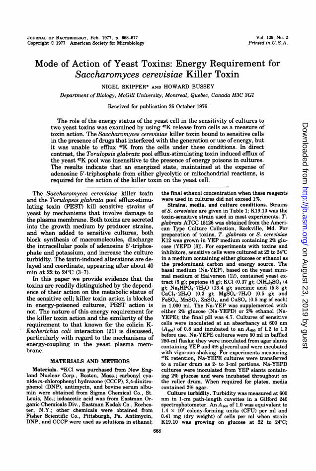
RESULTSPotassium release as a measure of toxin
action. Yeast toxins are routinely assayed byplating out treated cultures and calculating thesurvival; the method effectively measures bind-ing oftoxin to the cells but cannot give informa-tion on the course of events in the culture. Theprocedure that measures the relative stabilityofthe yeast 42K pool allows direct observation ofevents in the treated cell suspension. Activelygrowing cells took up 42K from the medium andmaintained an intracellular pool of the isotopeproportional to the cell density (Fig. 1A). Addi-tion of killer or PEST to loaded, sensitive, cellsresulted in almost complete release of radioac-tivity from the cells (Fig. 1B). The 42K pool inkiller-resistant (K12, K19, S14.96) and PEST-resistant (S14.96) strains was unaffected by theappropriate toxin, because it was lost to themedium at a low rate identical to that seen inuntreated cultures (Fig. 1B).
Effect of energy poisons on killer-inducedpotassium release. The relative survival ofsensitive cultures that had been treated withkiller toxin, when in either active growth orstationary phase, indicated that yeasts are
most susceptible to the toxin when in the expo-
TABLE 2. Partial purification of killer and PEST
Purification step KUa Protein Polysaccha- Sp act (KU/mg Purification Yield(mg) ride (mg) protein) (fold) (%)
KillerConcentrated culture 6.16 x 10¹⁰ 334.0 143 1.8 x 10⁸ 1 100supernatantb
Polyethylene-glycol 1.27 x 101° 5.8 37 2.19 x 10⁹ 12 21precipitate
PestConcentrated culture 2.56 x 10¹⁰ 356.0 160 7.19 x 10⁷ 1 100
supernatantePolyethylene-glycol 1.10 x 10'0 6.9 51 1.59 x 10⁹ 22 43
precipitateEthanol precipitate 7.56 x 10⁹ 4.7 32 1.61 x 10⁹ 22 29
a Killing units.b From 5 liters of S. cerevisiae K12, grown at 19°C to 450 Klett units (blue filter).c From 5 liters of T. glabrata, grown at 30°C to 550 Klett units.
VOL. 129, 1977
on August 23, 2019 by guest
http://jb.asm.org/
Dow
nloaded from
670 SKIPPER AND BUSSEY
nential phase; in addition, glycerol-grown cul-tures that bypassed six-carbon glycolysis wereless sensitive than glucose-grown cells (3). Thefollowing experiments were designed to exam-
ine the implication that sensitivity to killer
1 2A
40 °01
-30 .r-60-
20 _ 0
0.8LI
10 20-
0,7
00 60 120 210 0
60 120 180
Minutes Minutes
FIG. 1. Use ofthe yeast potassium pool to measuretoxin action. (A) Potassium loading. 42KC1 was
added to K19.10 cells growing on glucose, and theculture was sampled at intervals for turbidity (0)and for radioactivity in the cells (0). (B) Potassiumefflux in sensitive and resistant yeast. 42K-loadedcultures of strains K19.10, K19, K12, and S14.96 inNa-YEPD were harvested, washed, suspended ingrowth medium, and incubated at 22 to 4°C. At zero
time, K19.10 received either PEST to 150 pg ofpro-tein per ml (0), killer to 40 pg ofprotein per ml (0),or no additions (A); K12 and K19 received killer, 40pg ofprotein per ml (k), and S14.96 was treated witheither PEST, 100 pg ofprotein per ml (O), killer, 100pg ofprotein per ml (-), or no additions (A). Countsper minute remaining in cells was measured at inter-vals after zero time. The control (untreated cells)data for strains K12 and K19 were identical to thoseshown for strain K19.10 (A).
may be linked to energy metabolism.Glucose in appropriate concentration is
known to suppress mitochondrial function inyeast (15). When strain K19.10 was cultured inthe 2% glucose medium (Na-YEPD), it was sen-
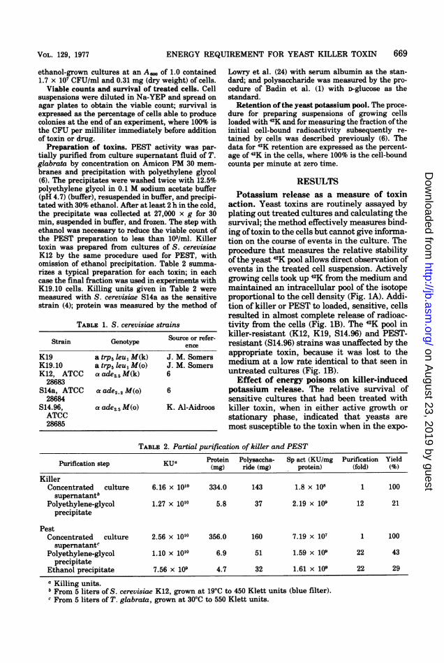
sitive to iodoacetate, an inhibitor of glyceralde-hyde 3-phosphate dehydrogenase, and to thereagents DNP and CCCP; the glucose-growncultures were not affected by cyanide or anti-mycin (Table 3). Killer-induced potassium ef-flux in glucose-grown cells was blocked by thedrugs that stopped growth; the inhibitors ofelectron transfer in the mitochondrion did notinfluence the effect of killer on the ion pool(Fig. 2). When grown on ethanol, K19.10 was
dependent on oxidative phosphorylation, sinceKCN or antimycin prevented growth (Table 3).Iodoacetate was also an effective inhibitor inthese cultures, presumably due to its inactiva-tion of yeast alcohol dehydrogenase (14). DNPor CCCP blocked the growth of the ethanol-grown cells (Table 3) as expected if these re-
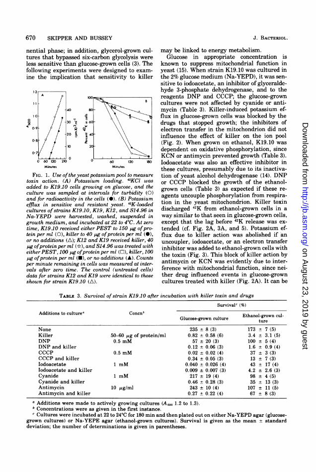
agents uncouple phosphorylation from respira-tion in the yeast mitochondrion. Killer toxindischarged 42K from ethanol-grown cells in a
way similar to that seen in glucose-grown cells,except that the lag before 42K release was ex-
tended (cf. Fig. 2A, 3A, and 5). Potassium ef-flux due to killer action was abolished if an
uncoupler, iodoacetate, or an electron transferinhibitor was added to ethanol-grown cells withthe toxin (Fig. 3). This block of killer action byantimycin or KCN was evidently due to inter-ference with mitochondrial function, since nei-ther drug influenced events in glucose-growncultures treated with killer (Fig. 2A). It can be
TABLE 3. Survival of strain K19.10 after incubation with killer toxin and drugsSurvivalc (%)
Additions to culturea ConcnbGlucose-grown culture ture
None 235 8 (3) 173 7 (5)Killer 50-60 ,tg of protein/ml 0.82 + 0.58 (6) 3.4 ± 3.1 (5)DNP 0.5 mM 57 20 (3) 100 5 (4)DNP and killer 0.12 ± 0.06 (3) 1.6 ± 0.9 (4)CCCP 0.5 mM 0.02 ± 0.02 (4) 37 ± 3 (3)CCCP and killer 0.34 ± 0.05 (3) 13 ± 7 (3)Iodoacetate 1 mM 0.040 ± 0.026 (4) 43 ± 17 (4)Iodoacetate and killer 0.009 ± 0.007 (3) 4.2 ± 2.6 (3)Cyanide 1 mM 217 ± 19 (4) 98 ± 4 (5)Cyanide and killer 0.46 ± 0.28 (3) 35 ± 13 (3)Antimycin 10 /Lg/ml 243 ± 10 (4) 107 ± 11 (5)Antimycin and killer 0.27 ± 0.22 (4) 67 ± 8 (3)
a Additions were made to actively growing cultures (A500 1.2 to 1.3)." Concentrations were as given in the first instance.c Cultures were incubated at 22 to 24'C for 180 min and then plated out on either Na-YEPD agar (glucose-
grown cultures) or Na-YEPE agar (ethanol-grown cultures). Survival is given as the mean ± standarddeviation; the number of determinations is given in parentheses.
J. BACTERIOL.
on August 23, 2019 by guest
http://jb.asm.org/
Dow
nloaded from
VOL. 129, 1977 ENERGY REQUIREMENT FOR YEAST KILLER TOXIN
0 A50 -
I I I .0
50
0
90' 18(
90 180
Minutes Minutes
FIG. 2. Potassium release in glucose-grown cells treated with killer toxin and drugs. 42K-labeled cultures ofK19.10 in Na-YEPD were incubated at zero time with killer, drug, killer and drug, or without additions, andthey were sampled at intervals for counts per minute remaining in cells. Concentrations: killer, 38 pg ofprotein per ml; antimycin, 10 pg/ml; cyanide, 1 mM; iodoacetate, 1 mM; DNP, 0.5 mM; CCCP, 0.5 mM. (A)Control (untreated cells) (A), plus killer (0), plus killer and cyanide (O), plus killer and antimycin (O),cyanide only (0), antimycin only (x). (B) Control (A), iodoacetate only (0), iodoacetate plus killer (O). (C)Control (A), DNP only (0), DNPplus killer (O). (D) Control (A), CCCP only (0), CCCP plus killer (O).
100 A
o0 8090 180
50
O 120
M inutes
FIG. 3. Potassium release in ethanol-grown cells treated with killer toxin and drugs. The experiments weredone as for Fig. 2, except that cells were loaded with 42K and incubated with additions in Na-YEPE. Drugconcentrations are given in the legend to Fig. 2; killer was added at 60 pg ofprotein per ml (A through C), and130 pg ofprotein per ml (D). (A) Control (A), plus killer (0), plus killer and antimycin (0), plus killer andcyanide (-), the data for antimycin or cyanide alone are given in the legend ofFig. 8. (B) Control (A), plusiodoacetate (0), plus iodoacetate and killer (-). (C) Control (A), plus DNP (0), plusDNP and killer (O). (D)Control (A), plus CCCP (0), plus CCCP and killer (O).
671
B
I I I
o.u_u50IF
4,
IC
C414
0 90
C
18(
B
14c
0
240
D
120
120
240
240
Minutes
50F
on August 23, 2019 by guest
http://jb.asm.org/
Dow
nloaded from
672 SKIPPER AND BUSSEY
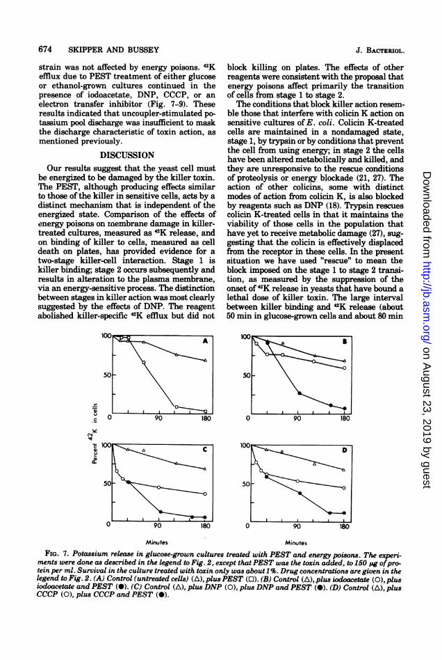
seen that the uncouplers themselves effluxed aconsiderable fraction of the yeast 42K pool (Fig.2C and D and 3C and D); however, the results ofexperiments with PEST-treated culturesshowed that such drug-stimulated ion releasewas not sufficient to mask the toxin-inducedrelease (see Fig. 7, 8, and 9). Since the PESTand the killer, if added alone to sensitive cul-tures, produced 42K efflux to a similar extent(Fig. 1B), it seemed that the uncouplers did infact abolish killer-induced ion release and didnot simply mask the killer action.DNP arrests the killing process at a stage
subsequent to killer binding. Table 3 summa-rizes the survival data for K19.10 cultures thathad been treated either with killer, with eachdrug, or with combinations of killer and drugs;the cells were grown on either glucose orethanol. When considered with the results forthe stability of the yeast potassium pool in cul-tures treated in the same way, the survivaldata suggested a distinction between two stagesin killer action: stage 1 is killer binding to cellsand is energy independent; stage 2 is the onsetof membrane alteration and is energy depend-ent. The clearest evidence comes from the cul-tures treated with DNP and killer toxin: incultures grown on either glucose or ethanol thecells did not show toxin-specific 42K release, yetthey died when plated out (Fig. 2C and 3C, andTable 3). These cells had evidently bound a
10(iA
80 -
60-
.c\C l 40
IV
aIL 20
lethal dose of killer in the presence ofDNP butwere arrested in this state until the reagentwas removed by dilution. Although not as eas-ily interpreted, the effects of the other drugs onkilling, under conditions where they blockedkiller-specific ion efflux, were not inconsistentwith a two-stage process for killer action. Anti-mycin or cyanide, although clearly having noeffect on either killer binding (measured asdeath on plates) or the onset of 42K efflux inglucose-grown cultures, reduced but did notabolish killing in respiring cells (Table 3, Na-YEPE cultures); either drug abolished 42K ef-flux in killer-treated, ethanol-grown cells (Fig.3A). CCCP and iodoacetate were themselveslethal to glucose-grown cultures to an extentthat masked any differential effects they mayhave had on killer binding and membrane al-teration (Table 3); in respiring cultures thesedrugs acted like antimycin or cyanide, affectingprimarily the onset of 42K efflux in killer-treated cells (Table 3, Fig. 3B and D).
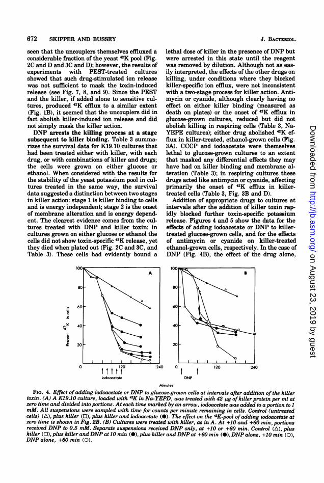
Addition of appropriate drugs to cultures atintervals after the addition of killer toxin rap-idly blocked further toxin-specific potassiumrelease. Figures 4 and 5 show the data for theeffects of adding iodoacetate or DNP to killer-treated glucose-grown cells, and for the effectsof antimycin or cyanide on killer-treatedethanol-grown cells, respectively. In the case ofDNP (Fig. 4B), the effect of the drug alone,
120 240 0t t ttt ttodoLcetate DNP
120 240
Minutes
FIG. 4. Effect ofadding iodoacetate or DNP to glucose-grown cells at intervals after addition of the killertoxin. (A) A K19.10 culture, loaded with "K in Na-YEPD, was treated with 42 ug of killer protein per ml atzero time and divided into portions. At each time marked by an arrow, iodoacetate was added to a portion to 1mM. All suspensions were sampled with time for counts per minute remaining in cells. Control (untreatedcells) (A), plus killer (0), plus killer and iodoacetate (-). The effect on the 42K-pool ofadding iodoacetate atzero time is shown in Fig. 2B. (B) Cultures were treated with killer, as in A. At +10 and +60 min, portionsreceived DNP to 0.5 mM. Separate suspensions received DNP only, at +10 or +60 min. Control (A), pluskiller (0), plus killer andDNP at 10 min (-), plus killer andDNP at +60 min (-), DNP alone, +10 min (0),DNP alone, +60 min (0).
J. BACTERIOL.
on August 23, 2019 by guest
http://jb.asm.org/
Dow
nloaded from
ENERGY REQUIREMENT FOR YEAST KILLER TOXIN 673
u 60 60
C4
20-20-
. . , , . I I , . .0 120 240 0 120 240
cyanide cntwnycinMinutes
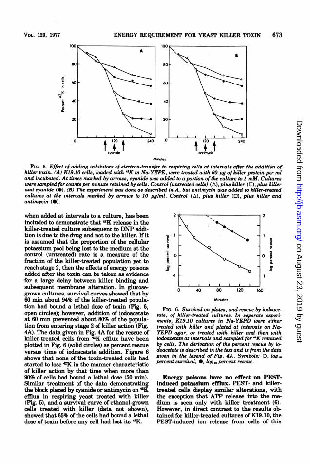
FIG. 5. Effect ofadding inhibitors of electron-transfer to respiring cells at intervals after the addition ofkiller toxin. (A) K1910 cells, loaded with 4K in Na-YEPE, were treated with 60 pg ofkiller protein per mland incubated. At times marked by arrows, cyanide was added to a portion of the culture to I mM. Cultureswere sampled for counts per minute retained by cells. Control (untreated cells) (A), plus killer (0), plus killerand cyanide (a). (B) The experiment was done as described in A, but antimycin was added to killer-treatedcultures at the intervals marked by arrows to 10 pg/mI. Control (A), plus killer (0), plus killer andantimycin (0).
when added at intervals to a culture, has beenincluded to demonstrate that 2K release in thekiller-treated culture subsequent to DNP addi-tion is due to the drug and not to the killer. If itis assumed that the proportion of the cellularpotassium pool being lost to the medium at thecontrol (untreated) rate is a measure of thefraction of the killer-treated population yet toreach stage 2, then the effects ofenergy poisonsadded after the toxin can be taken as evidencefor a large delay between killer binding andsubsequent membrane alteration. In glucose-grown cultures, survival curves showed that by60 min about 94% of the killer-treated popula-tion had bound a lethal dose of toxin (Fig. 6,open circles); however, addition of iodoacetateat 60 min prevented about 80% of the popula-tion from entering stage 2 of killer action (Fig.4A). The data given in Fig. 4A for the rescue ofkiller-treated cells from 2K efflux have beenplotted in Fig. 6 (solid circles) as percent rescueversus time of iodoacetate addition. Figure 6shows that none of the toxin-treated cells hadstarted to lose 42K in the manner characteristicof killer action by that time when more than90% of cells had bound a lethal dose (50 min).Similar treatment of the data demonstratingthe block placed by cyanide or antimycin on 2Kefflux in respiring yeast treated with killer(Fig. 5), and a survival curve of ethanol-growncells treated with killer (data not shown),showed that 65% of the cells had bound a lethaldose of toxin before any cell had lost its 2K.
Id
0 40 80 120 160
Minutes
FIG. 6. Survival on plates, and rescue by iodoace-tate, of killer-treated cultures. In separate experi-ments, K1910 cultures in Na-YEPD were eithertreated with killer and plated at intervals on Na-YEPD agar, or treated with killer and then withiodoacetate at intervals and sampled for 42K retainedby cells. The derivation of the percent rescue by io-doacetate is described in the text and is from the datagiven in the legend of Fig. 4A. Symbols: 0, log10percent survival; *, log,0 percent rescue.
Energy poisons have no effect on PEST-induced potassium efflux. PEST- and killer-treated cells display similar alterations, withthe exception that ATP release into the me-dium is seen only with killer treatment (6).However, in direct contrast to the results ob-tained for killer-treated cultures of K19.10, thePEST-induced ion release from cells of this
VOL. 129, 1977
on August 23, 2019 by guest
http://jb.asm.org/
Dow
nloaded from
674 SKIPPER AND BUSSEY
strain was not affected by energy poisons. 42Kefflux due to PEST treatment of either glucoseor ethanol-grown cultures continued in thepresence of iodoacetate, DNP, CCCP, or anelectron transfer inhibitor (Fig. 7-9). Theseresults indicated that uncoupler-stimulated po-tassium pool discharge was insufficient to maskthe discharge characteristic of toxin action, asmentioned previously.
DISCUSSIONOur results suggest that the yeast cell must
be energized to be damaged by the killer toxin.The PEST, although producing effects similarto those of the killer in sensitive cells, acts by adistinct mechanism that is independent of theenergized state. Comparison of the effects ofenergy poisons on rnembrane damage in killer-treated cultures, measured as 42K release, andon binding of killer to cells, measured as celldeath on plates, has provided evidence for atwo-stage killer-cell interaction. Stage 1 iskiller binding; stage 2 occurs subsequently andresults in alteration to the plasma membrane,via an energy-sensitive process. The distinctionbetween stages in killer action was most clearlysuggested by the effects of DNP. The reagentabolished killer-specific 42K efflux but did not
50
-n0
c
4,
-4,
90 180
block killing on plates. The effects of otherreagents were consistent with the proposal thatenergy poisons affect primarily the transitionof cells from stage 1 to stage 2.The conditions that block killer action resem-
ble those that interfere with colicin K action onsensitive cultures of E. coli. Colicin K-treatedcells are maintained in a nondamaged state,stage 1, by trypsin or by conditions that preventthe cell from using energy; in stage 2 the cellshave been altered metabolically and killed, andthey are unresponsive to the rescue conditionsof proteolysis or energy blockade (21, 27). Theaction of other colicins, some with distinctmodes of action from colicin K, is also blockedby reagents such as DNP (18). Trypsin rescuescolicin K-treated cells in that it maintains theviability of those cells in the population thathave yet to receive metabolic damage (27), sug-gesting that the colicin is effectively displacedfrom the receptor in these cells. In the presentsituation we have used "rescue" to mean theblock imposed on the stage 1 to stage 2 transi-tion, as measured by the suppression of theonset of42K release in yeasts that have bound alethal dose of killer toxin. The large intervalbetween killer binding and 42K release (about50 min in glucose-grown cells and about 80 min
Minutes Minutes
FIG. 7. Potassium release in glucose-grown cultures treated with PEST and energy poisons. The experi-ments were done as described in the legend to Fig. 2, except that PEST was the toxin added, to 150 pg ofpro-tein per ml. Survival in the culture treated with toxin only was about 1%. Drug concentrations are given in thelegend to Fig. 2. (A) Control (untreated cells) (A), plus PEST (0). (B) Control (A), plus iodoacetate (0), plusiodoacetate and PEST (-). (C) Control (A), plus DNP (0), plus DNP and PEST (O). (D) Control (A), plusCCCP (0), plus CCCP and PEST (O).
A
a0- I
J. BACTERIOL.
on August 23, 2019 by guest
http://jb.asm.org/
Dow
nloaded from
ENERGY REQUIREMENT FOR YEAST KILLER TOXIN
1Wo1
50
C
CN4
u
0 90 180
Minutes Minutes
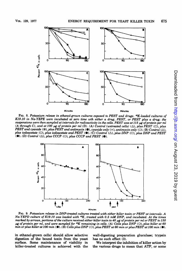
FIG. 8. Potassium release in ethanol-grown cultures exposed to PEST and drugs. 42K-loaded cultures ofK19.10 in Na-YEPE were incubated at zero time wth either a drug, PEST, or PEST plus a drug; thesuspensions were then sampled at intervals for radioactivity in the cells. PEST was at 115 pg ofprotein per ml(A through C), and at 100 pg ofprotein per ml (D). (A) Control (untreated cells) (A), plus PEST (0), plusPEST and cyanide (®), plus PEST and antimycin (m), cyanide only (x), antimycin only (0). (B) Control (A),plus iodoacetate (0), plus iodoacetate and PEST (a). (C) Control (A), plus DNP (0), plus DNP and PEST(X). (D) Control (A), plus CCCP (0), plus CCCP and PEST (A).
100 100IAB
80 80
-u 60 060
~40 40-
20-20-
0 , 120 240 0 , 120 240
Minuites
FIG. 9. Potassium release in DNP-treated cultures treated with either killer toxin or PEST at intervals. ANa-YEPD culture ofK19.10 was loaded with 42K, treated with 0.5 mM DNP, and incubated. At the timesmarked by arrows, portions ofthe culture received either killer toxin to 40 pg ofprotein per ml orPEST to 150pg ofprotein per ml, and were sampled for 42K remaining in cells. (A) Cells plus DNP (0), plus killer at 60min orplus killer at 100 min (-). (B) Cells plusDNP (0), plusPEST at 60 min orplusPEST at 100 min (0).
in ethanol-grown cells) should allow selectivedigestion of the bound toxin from the yeastsurface. Some maintenance of viability inkiller-treated cultures is achieved with the
wall-digesting preparation gluculase; trypsinhas no such effect (3).We interpret the inhibition of killer action by
the various drugs to mean that ATP, or some
A
VOL. 129, 1977 675
on August 23, 2019 by guest
http://jb.asm.org/
Dow
nloaded from
676 SKIPPER AND BUSSEY
form of energy derived from it, is required forthe maintenance of a state necessary for mem-brane damage to occur, a state analogous to theenergized plasma membrane in bacteria (13,31). By analogy to the situation in prokaryotes,it seems likely that a yeast plasma membraneadenosine triphosphatase (ATPase) couplesATP generated from glycolysis or from mito-chondrial reactions to membrane-linked work,perhaps via the proton-motive force in thechemiosmotic hypothesis of Mitchell (26). Thisenergy-coupling state is required for, and al-tered by, the action of the yeast killer toxin; itsmodification leads to the spectrum ofdamage tothe cell. A similar argument has been advo-cated to account for the effects ofcytochalasin Aon the yeast cell (22). The effects of iodoacetate,antimycin, and cyanide on killer action may beattributed to their blocking ATP synthesis, dueto inhibition of glyceraldehyde 3-phosphate de-hydrogenase and alcohol dehydrogenase (14,30), cytochrome c to b, electron transfer (11),and cytochrome c oxidase (23), respectively.The effect of iodoacetate reported here mayhave been nonspecific since we could not ar-
range a situation in which the alkylating re-
agent did not block the 42K efflux in killer-treated cells. lodoacetate is known to producegrossly altered membranes in yeast (25), albeitat concentrations higher than the 1 mM used inour experiments. ATP discharge due to uncou-
pling and to stimulation ofATPase in mitochon-dria (32) may account for the inhibition of killeraction by DNP or CCCP in the ethanol-growncultures, but the block of killer action by thesedrugs in glucose-grown cells is not easy to ex-
plain. Inhibition by DNP of energy-linked func-tions in organisms generating energy in a non-
respiratory mode is well known (13), and manyexamples have been reported in yeast growingeither anaerobically or in high glucose concen-
trations (2, 9, 28, 29, 33). It has been suggestedthat DNP dissipates glycolytic ATP in yeast bystimulating a promitochondrial ATPase (28).However, oligomycin or dicyclohexylcarbodi-imide, specific inhibitors of the mitochondrialATPase, do not block the effects of DNP on
energy-linked systems in fermenting yeast (10,19). An alternative view is that reagents suchas DNP and CCCP block any membrane-associ-ated and work-requiring systems by eliminat-ing proton gradients (13). CCCP is consideredto act by a similar mechanism to DNP (16, 17),and, in our experiments, the two drugs hadsimilar effects on both the stability of the yeast42K pool in untreated cultures and on the ionefflux in killer-treated cultures. These effects ofDNP and CCCP were not dependent on themode of energy generation in the cells, suggest-
ing that the reagents disrupted energy-linkedprocesses in either glycolyzing or respiringyeast by interacting with the plasma mem-brane.
ACKNOWLEDGMENTSThis research was supported by grants from the National
Research Council of Canada, the Quebec Department ofEducation, and the National Cancer Institute of Canada.
N.S. holds a postgraduate scholarship from the NationalResearch Council of Canada.We thank Donna Saville for assistance and R.G.E. Pal-
free for critical discussions of the manuscript.
LITERATURE CITED1. Badin, J., C. Jackson, and M. Schubert. 1953. Improved
method for determination of plasma polysaccharideswith tryptophan. Proc. Soc. Exp. Biol. Med. 84:288-291.
2. Borst-Pauwels, G. W. F. H., and S. Jager. 1969. Inhibi-tion of phosphate and arsenate uptake in yeast bymonoiodoacetate, fluoride, 2,4-dinitrophenol and ace-tate. Biochim. Biophys. Acta 172:399 406.
3. Bussey, H. 1972. Effects of yeast killer factor on sensi-tive cells. Nature (London) New Biol. 235:73-75.
4. Bussey, H. 1974. Yeast killer factor-induced turbiditychanges in cells and sphaeroplasts of a sensitivestrain. J. Gen. Microbiol. 82:171-179.
5. Bussey, H., and D. Sherman. 1973. Yeast killer factor:ATP leakage and coordinate inhibition of macromo-lecular synthesis in sensitive cells. Biochim. Biophys.Acta 298:868-875.
6. Bussey, H., and N. Skipper. 1975. Membrane-mediatedkilling of Saccharomyces cerevisiae by glycoproteinsfrom Torulopsis glabrata. J. Bacteriol 124:476-483.
7. Bussey, H., and N. Skipper. 1976. Killing of Torulopsisglabrata by Saccharomyces cerevisiae killer factor.Antimicrob. Agents Chemother. 9:352-356.
8. Bussey, H., D. Sherman, and J. M. Somers. 1973. Ac-tion of yeast killer factor. a resistant mutant withsensitive spheroplasts. J. Bacteriol. 113:1193-1197.
9. Chevallier, M. R., R. Jund, and F. Lacroute. 1975.Characterization of cytosine permeation in Saccharo-myces cerevisiae. J. Bacteriol. 122:629-641.
10. Galeotti, T., L. Kovac, and B. Hess. 1968. Interferenceof uncoupling agents with cellular energy-requiringprocesses in anaerobic conditions. Nature (London)218:194-196.
11. Hagihara, B., N. Sato, and T. Yamanaka. 1975. Type bcytochromes, p. 549-593. In P. D. Boyer (ed.), The en-zymes, vol. 11A, 3rd ed., Academic Press Inc., NewYork.
12. Halvorson, H. 0. 1958. Studies on protein and nucleicacid turnover in growing cultures of yeast. Biochim.Biophys. Acta 27:267-276.
13. Harold, F. M. 1972. Conservation and transformation ofenergy by bacterial membranes. Bacteriol. Rev.36:172-230.
14. Harris, I. 1964. Structure and catalytic activity of alco-hol dehydrogenases. Nature (London) 203:30-34.
15. Henson, C. P., C. N. Weber, and H. R. Mahler. 1968.Formation of yeast mitochondria. 1. Kinetics ofamino acid incorporation during derepression. Bio-chemistry 7:4431-4444.
16. Heytler, P. G. 1963. Uncoupling of oxidative phospho-rylation by carbonyl cyanide phenylhydrazones. 1.Some characteristics of m-Cl-CCP action on mito-chondria and chloroplasts. Biochemistry 2:357-361.
17. Hirose, S., N. Yaginuma, and Y. Inada. 1974. Disrup-tion of charge separation followed by that of the pro-ton gradient in the mitochondrial membrane byCCCP. J. Biochem. 76:213-216.
J. BACTERIOL.
on August 23, 2019 by guest
http://jb.asm.org/
Dow
nloaded from
ENERGY REQUIREMENT FOR YEAST KILLER TOXIN 677
18. Holland, I. B. 1975. Physiology of colicin action, p. 56-139. In A. H. Rose and D. W. Tempest (ed.), Advancesin microbiol physiology, vol. 12. Academic Press Inc.,London.
19. Huygen, P. L. M., and G. W. F. H. Borst-Pauwels.1972. The effect of N,N'-dicyclohexylcarbodiimide on
anaerobic and aerobic phosphate uptake by baker'syeast. Biochim. Biophys. Acta 283:234-238.
20. Jarrett, L., and R. W. Hendler. 1967. 2,4-dinitrophenoland azide as inhibitors ofprotein and ribonucleic acidsynthesis in anaerobic yeast. Biochemistry 6:1693-1703.
21. Jetten, A. M., and M. E. R. Jetten. 1975. Energy re-
quirement for the initiation of colicin action in Esche-richia coli. Biochim. Biophys. Acta 387:12-22.
22. Kuo, S. C., and J. 0. Lampen. 1975. Action of cytochal-asin A, a sulflhydryl-reactive agent, on sugar metabo-lism and membrane-bound adenosine triphosphataseof yeast. Biochim. Biophys. Acta 389:145-153.
23. Lardy, H. A., and S. M. Ferguson. 1969. Oxidativephosphorylation in mitochondria. Annu. Rev. Bio-chem. 38:99-134.
24. Lowry, 0. H., N. J. Rosebrough, A. L. Farr, and P. J.Randall. 1951. Protein measurement with the Folinphenol reagent. J. Biol. Chem. 193:265-275.
25. Maxwell, W. A., and E. Spoerl. 1972. lodoacetic acidinduced changes in Saccharomyces cerevisiae. Cyto-biology 5:309-312.
26. Mitchell, P. 1966. Chemiosmotic coupling in oxidativeand photosynthetic phosphorylation. Biol. Rev. Cam-bridge Philos. Soc. 41:445-502.
27. Plate, C. A., and S. E. Luria. 1972. Stages in colicinaction, as revealed by the action of trypsin. Proc.Natl. Acad. Sci. U.S.A. 69:2030-2034.
28. Ramos, E. H., L. C. de Bongioanni, M. L. Claisse, andA. 0. M. Stoppani. 1975. Energy requirement for theuptake of L-leucine by Saccharomyces cerevisiae.Biochim. Biophys. Acta 394:470-481.
29. Riemersma, J. C. 1968. Effects of sodium azide and 2,4-dinitrophenol on phosphorylation reactions and ionfluxes in Saccharomyces cerevisiae. Biochim. Bio-phys. Acta 153:80-87.
30. Segal, H. L., and P. D. Boyer. 1953. The role of sulfhy-dryl groups in the activity of D-glyceraldehyde 3-phosphate dehydrogenase. J. Biol. Chem. 204:265-281.
31. Simoni, R. D., and P. W. Postma. 1975. The energeticsof bacterial active transport. Annu. Rev. Biochem.44:523-554.
32. Slater, E. C. 1963. Uncouplers and inhibitors of oxida-tive phosphorylation, p. 503-516. In R. M. Hochsterand J. H. Quastel (ed.), Metabolic inhibitors, vol. 11.Academic Press Inc., New York.
33. Sumrada, R., M. Gorski, and T. Cooper. 1976. Ureatransport-defective strains of Saccharomyces cerevi-sile. J. Bacteriol. 125:1048-1056.
VOL. 129, 1977
on August 23, 2019 by guest
http://jb.asm.org/
Dow
nloaded from