Embed Size (px)
Citation preview

MNTP
Magnetoencephalography (MEG)
Erika J C Laing, MS
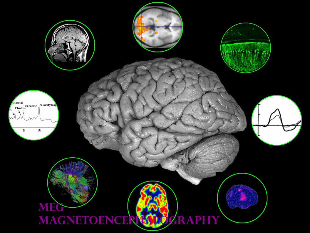
MEG
magnetoencephalography

A changing
electric field
generates a
magnetic field
M is for Magnetism
Right Hand Rule

A changing
electric field
generates a
magnetic field
M is for Magnetism

What does MEG measure?
Post-Synaptic Intracellular Current
Summed over 10000 or more neurons Organized Tangentially to the Scalp

Action potentials:
fields diminish too rapidly to sum
Pre-synaptic Post-synaptic
Postsynaptic currents:
fields diminish gradually
What does MEG measure?
Post-Synaptic Intracellular Current
Summed over 10000 or more neurons
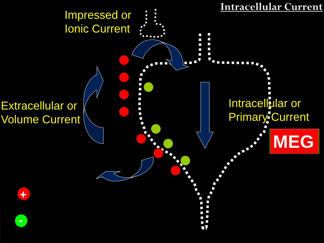
Impressed or
Ionic Current
Intracellular or
Primary Current Extracellular or
Volume Current
MEG
+
-
Intracellular Current

What does MEG measure?
Summed over 10000 or more neurons
~ 104-5
activated cells
Many neurons firing
in synchrony can
generate a magnetic
field observable at
the surface

9
MEG
MEG observes
currents
tangential to
the surface
best.
Magnetic fields from deep currents are
weaker at the surface
What does MEG measure?
Organized Tangentially to the Scalp

What does MEG measure?
Post-Synaptic Intracellular Current
Summed over 10000 or more neurons Organized Tangentially to the Scalp

EEG vs MEG
EEG = Electroencephalography
Post-Synaptic Extracellular Current
Summed over 10000 or more neurons Any orientation but signal is impeded by skull, skin, fat

EEG Vs MEG
12
Implications: EEG has lower spatial resolution
EEG can capture some signals MEG has poor sensitivity for
COMPLEMENTARY TOOLS

MEG and EEG Distributions

Noninvasive
2-3mm spatial
resolution
1ms temporal
resolution

Recording magnetic fields
SQUID Superconducting
QUantum
Interference
Device
SQUID works via
INDUCTION
to be superconductive,
must be cooled by
liquid helium

Recording magnetic fields
Induction
SQUID Superconducting
QUantum
Interference
Device

Recording magnetic fields
SQUID Superconducting
QUantum
Interference
Device

Types of sensors
Magnetometer - General magnetic fields
- Very sensitive
- Elekta, 4D
Planar Gradiometer - Focal magnetic fields
- Most sensitive to fields directly underneath it
- Elekta
Axial Gradiometer -Focal magnetic fields
- Most sensitive to fields directly underneath it
- CTF
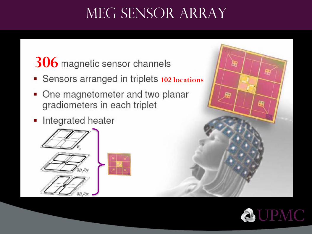
MEG Sensor array
306 102 locations
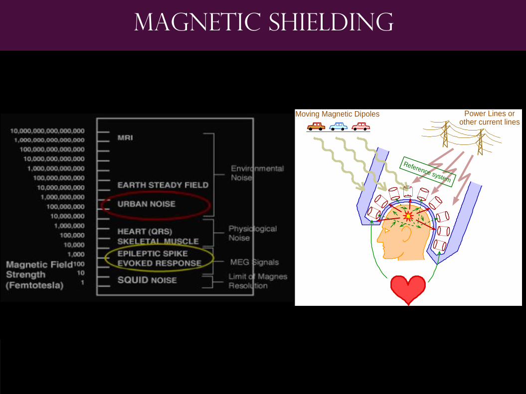
Magnetic shielding
Reference system
Moving Magnetic Dipoles Power Lines orother current lines

Physical shielding
3-ply µ-metal room
Magnetically Shielded Room (MSR)

The Machine
22
No Magnet
Quiet Machine makes no noise
Participant can sit or
lay down
Can record 128 EEG
simultaneously

Acquisition
Co-registration
Preprocessing
And Averaging
Localization
Monitoring Physiological
Artifacts
Defining the Head Shape
Recording the Head
Position
Data acquisition and analysis steps

Acquisition procedure
Subject Preparation
HPI Coils
(Head Position Indicator)
+ 4 HPI Coils, glued to head
+ A brief electrical pulse is sent to
the coils during acquisition. The
sensors record the pulses and
interpolates the head position
relative to the sensor helmet.

Acquisition procedure
Subject Preparation
EEG Electrodes
to collect artifacts
+ Vertical EOG
Above and below eyes – Eye Blinks
+ Horizontal EOG
Left and Right of eyes – Saccades
+ ECG
On chest – Heartbeat
+ EMG
On muscle of interest – Muscle Movement
+ don’t forget reference and ground

Acquisition procedure
Digitization
Digitize to make a 3D digital head shape file
+ 3 Fiducial Landmarks
Nasion, Left and Right Preauricular (ear)
+ 4 HPI Coils
+ Any additional EEG
+ A bunch of extra ones to get a good head
shape!

Acquisition procedure
Acquiring MEG Signals

Acquisition procedure
Acquiring MEG Signals
Earphones
Electrical
Stimulator
Presentation
Screen (moved to front!)
Also:
Button Pads
Button Gloves
Manual Tapper
Stimulus delivered
by E-Prime,
PsychToolBox, etc.

Collecting Data
29

Collecting Data
30

Collecting Data
31

Collecting Data
32

Acquisition
Co-registration
Preprocessing
And Averaging
Localization
Fitting Digitization
Points to Structural MRI
Data acquisition and analysis steps

Structural MRI

Fit the
Digitization
Points to
Structural MRI
Coregistration to MRI

Acquisition
Co-registration
Preprocessing
And Averaging
Localization
Filtering
Artifact Rejection
Selecting a time region
and baseline
Averaging by condition
Data acquisition and analysis steps
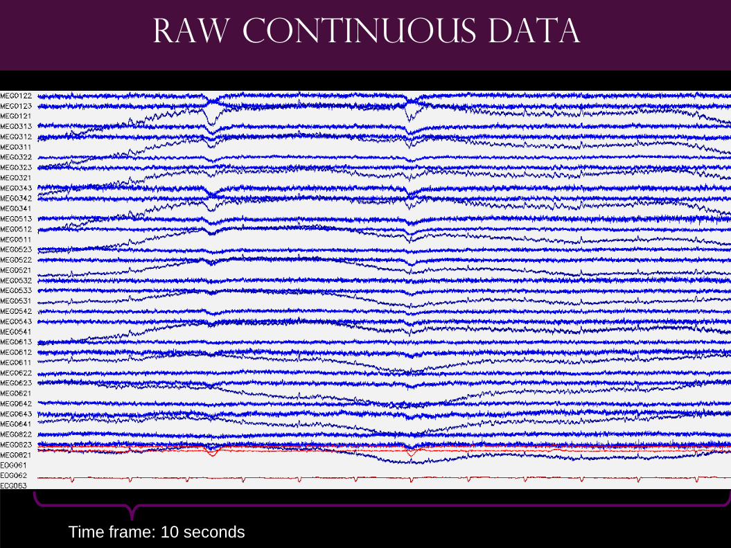
Time frame: 10 seconds
Raw continuous data

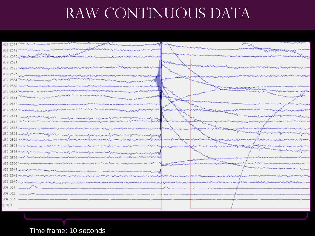
Time frame: 10 seconds
Raw continuous data
cardiac
drift
eye blink Buzz =
Line
noise
60Hz

Time frame: 10 seconds
Raw continuous data
Buzz =
Line
noise
60Hz

Neuronal Oscillations
Delta: 1-4Hz
Theta: 5-7Hz
Alpha: 8-12Hz
Beta: 15-29Hz
Gamma1: 30-59Hz
Gamma2: 60-90Hz
Cognition
Sleep
Neuronal
Injury

Time frame: 10 seconds
Raw continuous data

Time frame: 10 seconds
Filtered 1-40 Hz

Reference system
Moving Magnetic Dipoles Power Lines orother current lines
Sources of noise
saccade
swallowing
alpha
eye blink

Types of preprocessing
44
Reference system
Moving Magnetic Dipoles Power Lines orother current lines
Constant Sources of Noise:
SSP with Empty Room
Measurement
Patient artifact:
ICA or simply reject
Random external artifact:
MaxFilter (SSS)

Signal Space Projection (SSP)
45
Reference system
Moving Magnetic Dipoles Power Lines orother current lines Constant Sources of Noise:
SSP with Empty Room
Measurement Do a components analysis
of the signal when no
person is in the room.
Those signals are
presumed to regularly be
present.
Use Signal Space
Projection (SSP) to
remove those components
from the data.

Time frame: 10 seconds
Filtered 1-40 Hz

Time frame: 10 seconds
Filtered 1-40 Hz & SSP of Empty Room

Types of preprocessing
48
Reference system
Moving Magnetic Dipoles Power Lines orother current lines
Patient artifact:
ICA or simply reject

Artifact Removal: ICA
49
Isolates components based on independence
– Raw data from 306 channels transformed into component signals
– Visually inspect 306 components (or correlate with EOG/ECG channels)
– Compare with raw data
– Remove artifactual components
30 seconds of data
Blink?
Blink?
Cardiac?
Good?
Com
ponents
R
aw
Data
Chann
els

Time frame: 10 seconds
Filtered 1-40 Hz & SSP of Empty Room

Time frame: 10 seconds
Filt 1-40Hz; SSP of Empty Room; ICA components removed

Types of preprocessing
52
Reference system
Moving Magnetic Dipoles Power Lines orother current lines
Random external artifact:
MaxFilter (SSS)

Signal Space Separation (SSS)
53
MaxFilter, created by Elekta Calculate an INNER sphere
around the head but inside
the sensor helmet
Calculate an OUTER
sphere around the outside
of the sensors
Calculate estimates of
sources as if they were on
the surface of the INNER
and OUTER spheres
Remove those that are
stronger on the OUTER
sphere, more likely to be
noise
Time frame: 10 seconds
Raw continuous data

Time frame: 10 seconds
Maxfiltered data

Temporal extension
56
MaxST (or tSSS)
Look at data in 4 second
chunks
Correlate source signals
across chunks
Any source signals with
high correlations are
assumed to be artifacts

Maxfilter with temporal extension
57

Collecting Data
58

Baseline:
200ms
1000ms :Time-frame
1 Trial
x 200 =
1 sensor’s average
Averaging trials

1 Trial
200 Trials
Averaging trials

averages

Beep!
words

Acquisition
Co-registration
Preprocessing
And Averaging
Localization
WHERE IS THE
BRAIN ACTIVE?
Data acquisition and analysis steps

ELECTRIC! MAGNETIC!
From surface to localized
Maxwell’s Equations The Right Hand Rule

The problem
Forward
Problem
If we know this… We can calculate this!
Solvable!
? Inverse
Problem
The Problem?
We only know this….
Always an
estimate!

The estimation
The equivalent of triangulation
There are many kinds of localization strategies, here are two:
Dipole Fitting
Distributed Technique

Head models
Spherical
computationally
straightforward
unrealistic
Boundary
Element
computationally
difficult
realistic

1) Pick subset of sensors w/ peak 2) Pick Time Point; Observe Mag Field
4) Map to MRI 3) Measures of Quality
Goodness of Fit
% of activity explained by
forward solution based on
single dipole
Confidence Volume
volume within which you can be
95% confident that the dipole
exists
Equivalent
Current
Dipole
Technique

22ms 52ms 83ms
7 sensors
42 sensors
92 sensors
99.7% 99.2% 98.2%
84.6% 97.6% 85.8%
84.6% 97.6% 85.8%
Median Nerve
Dipole Fitting
Results
circle = size of
confidence volume

Equivalent Current Dipole
Best for a brain
response that does not
elicit a big network, or
for stimulus response
early in the time
course.
Heavily user
dependant

20 ms 44 ms 57 ms 88 ms 120 ms
Time course of SEF

20 ms 44 ms 57 ms 88 ms 120 ms
Time course of SEF
R L
L

Minimum Norm Estimate (MNE)
Realistic Head Model
Cortical surface
segmented into 5124
possible source
locations
1 location every 6.2 mm
(each 39 mm2)
* Can use spherical model
Estimate Current
Estimate a current
pattern from all sensors
to head model whose
forward solution
explains measured data
“Regularize”
Further constrain
estimate by suppressing
weak boundaries of a
source
Focuses
sources of
activity to
smaller
regions

Distributed
Minimum Norm
Estimate Dipole Fitting
Electrical Median Nerve Stimulation, 1Hz
SI: Post-Central
SII: Insula

Distributed Techniques
Great for brain responses
that elicit network activity,
like language
Much less user dependant
More automatable

Many Distributed Techniques
76

Distributed Methods
77
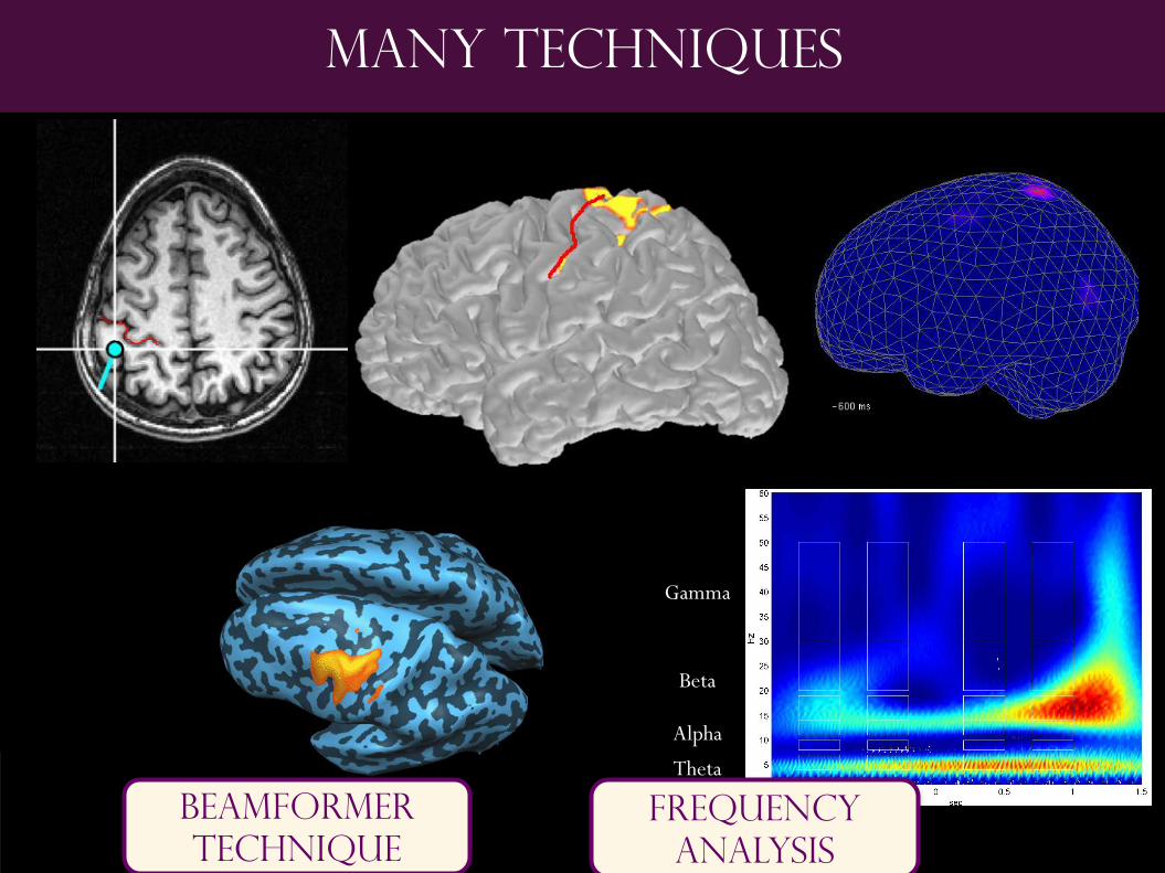
Many Techniques
MCE_2.mpeg
Beamformer
Technique
Theta
Alpha
Beta
Gamma
Frequency
analysis
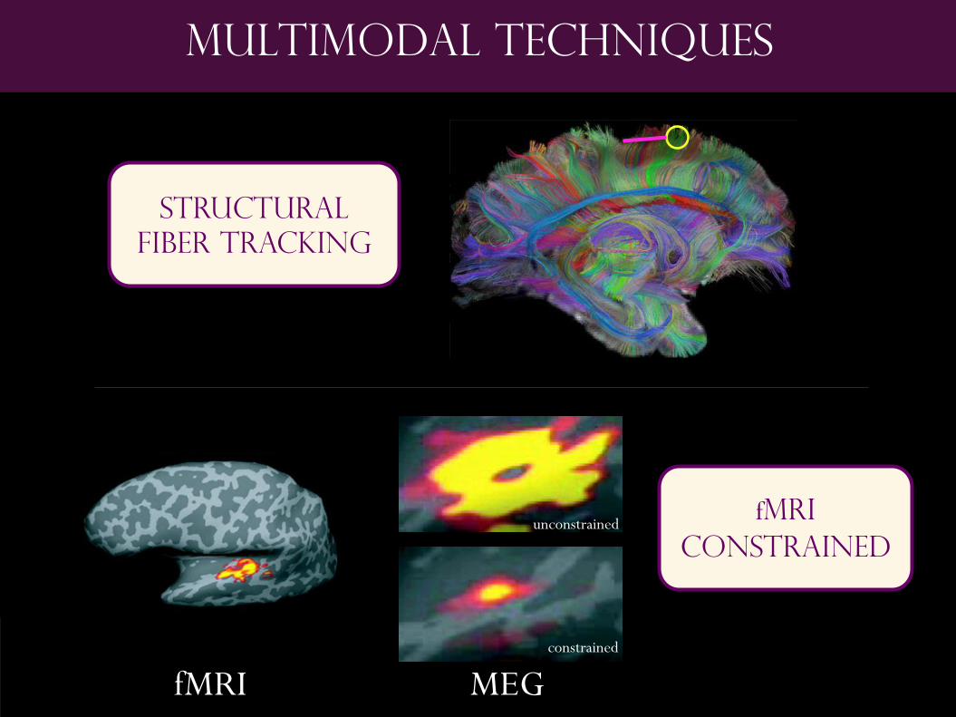
multimodal techniques
Structural
fiber tracking
fmri
constrained
fMRI MEG
unconstrained
constrained

Pre-surgical Mapping

Usefulness for surgical planning
Brain Mapping allows avoidance of critical areas
craniotomy

Usefulness for surgical planning
Brain Mapping allows avoidance of critical areas
Gamma Knife before after

Fetal MEG
30 weeks 36 weeks newborn
Latency of the peak
gets SHORTER

QUESTIONS?